A]H 1989; 2:655-658
Cellular Calcium Regulation in Hypertension
K. Hermsmeyer and P. Erne
In vascular m u s c l e cells, t w o distinct t y p e s o f functionally i m p o r t a n t c a l c i u m ( C a2 +) c h a n n e l s , called t r a n s i e n t (T) a n d s u s t a i n e d (L), are d i f f e r e n -tiated b y d i h y d r o p y r i d i n e c a l c i u m a n t a g o n i s t s (CaA). W e s t u d i e d t h e ratio of T / L C a2 + c h a n n e l s i n isolated, s p o n t a n e o u s l y c o n t r a c t i n g a z y g o u s v e n o u s cells of s p o n t a n e o u s l y h y p e r t e n s i v e rats ( S H R ) a n d Wistar-Kyoto rats ( W K Y ) b y q u a n t i t a t i n g C a2 + currents a n d i n t r a c e l l u l a r C a2 + r e l e a s e . W h i l e total t r a n s m e m b r a n o u s C a2 + c u r r e n t w a s not d i f f e r e n t between t h e t w o strains, t h e p r o p o r t i o n o f C a2 + currents carried b y L - t y p e c h a n n e l s w a s e n h a n c e d in vascular m u s c l e cells f r o m S H R . W e h a v e recently c o m p a r e d s u b c e l l u l a r d i s t r i b u t i o n of intracellular f r e e C a2 + c o n c e n t r a t i o n i n t h e s a m e cells, at rest a n d d u r i n g s t i m u l a t i o n , b y q u a n t i t a t i o n w i t h a digital p h o t o n - c o u n t i n g c a m e r a . F u r a - 2 fluorescence i n t e n s i t y s h o w e d t h a t C a2 + r e l e a s e w a s p r i n c i p a l l y f r o m s a r c o p l a s m i c r e t i c u l u m a n d t h a t cells f r o m S H R h a d h i g h e r l e v e l s of C a2 + u p o n c a l c i u m c h a n n e l s t i m u l a t i o n , e s p e c i a l l y at t h e cell p e r i p h e r y . T h e s e f i n d i n g s suggest f u n d a m e n t a l d i f f e r e n c e s i n S H R a n d W K Y v a s c u l a r m u s c l e cells i m p l i c a t i n g t h e i m p o r t a n c e o f c h a n g e s i n c a l c i u m c h a n n e l s , m o d u l a t i o n o f C a2 + r e l e a s e , a n d C a2 + u p t a k e i n S H R h y p e r t e n s i o n . A m J H y p e r t e n s 1989; 2 : 6 5 5 - 6 5 8 KEY WORDS: V a s c u l a r m u s c l e , c a l c i u m c h a n n e l s , c a l c i u m c o n c e n t r a t i o n , f u r a - 2 , c a l c i u m r e l e a s e , S H R , W K Y .
H
ypertension in its established phase is h e m o -dynamically characterized b y an increase in systemic vascular r e s i s t a n c e .1"3 Thisde-rangement is due to structural differences between normotensive and hypertensive subjects,4 but
also entails functional c o m p o n e n t s .5
As the force-generating bridging b e t w e e n actin and myosin depends on changes of intracellular calcium a c -tivity (calcium free-ion concentration) and h e n c e trans-membranous calcium m o v e m e n t s , modulations o f transmembranous calcium influx and intracellular
cal-From the Chiles Research Institute, Providence Medical Center and Departments of Medicine and Cell Biology, Oregon Health Science University, Portland, Oregon, and Department of Pharmacology and The Cardiovascular Center, University of Iowa, Iowa City, Iowa.
This research was supported by grants nos. HL38537 and HL38645 from the National Institutes of Health. P. E. was supported by the Swiss Foundation for Biological and Medical Fellowships.
Address correspondence and reprint requests to Kent Hermsmeyer, M.D., Chiles Research Institute, Providence Medical Center, 4805 NE Glisan Street, Portland, Oregon 97213. Present address for P. E., De-partments of Medicine and Research, University Hospital, 4031 Basel, Switzerland.
cium activity, ( C a2 +)i 7 h a v e a pivotal role in determining
vasoconstriction. Therefore, m e c h a n i s m s that lead to an increase in ( C a2 +) i through these processes cause
vaso-constriction.
Several lines of evidence suggest that abnormalities of calcium metabolism are involved in the pathogenesis o f established h u m a n hypertension. In particular, low serum concentrations of ionized C a2 + h a v e b e e n o b
-served in at least a fraction of patients with this dis-e a s dis-e .6 , 7 Furthermore, there is an e n h a n c e d
vasodila-t i o n8 , 9 and antihypertensive e f f e c t1 0 , 1 1 of calcium
antagonists in patients with essential hypertension. Studies using the platelet as a model of contractile systems indicate that alterations of C a2 + handling in
h u m a n hypertensives m a y occur at the cellular l e v e l .1 2 , 1 3
However, these studies do not determine whether simi-lar alterations are operative in vascusimi-lar muscle cells and, if so, w h e t h e r they are causally implicated or a conse-quence of elevated blood pressure.
To define vascular muscle mechanisms which are al-tered in an animal model of hypertension prior to the development of elevated blood pressure, studies were
656 HERMSMEYER AND ERNE AJH-AUGUST 1989-VOL 2, NO. 8
carried out on isolated azygous vein cells from three-day-old spontaneously hypertensive rats ( S H R ) a n d ge netically matched Wistar-Kyoto rats (WKY). T h e prepa ration a n d characterization of these cells from primary cultures were described in detail e l s e w h e r e .1 4 These
cells are spontaneously contracting and exhibit repeated contraction/relaxation cycles with a high shortening v e locity, high pharmacological sensitivity, and electro physiological i n t e g r i t y .1 5'1 6
M E T H O D S
Experiments were carried out on vascular muscle cells isolated from azygous veins of neonatal r a t s .1 7 1 8 W h o l e
cell recordings of C a2 + currents, isolated b y replacement
of N a+ a n d K+ in internal and external solutions allowed
separation of L a n d Τ types of current, where L are longer lasting (sustained) a n d Τ are transient.1 9
In cells from similarly prepared cultures, intracellular C a2 + w a s quantitated b y fura-2 fluorescence using the
three wavelength protocols explained in detail else w h e r e .2 0 High resolution (0.5 μτή) quantitation of C a2 +
was carried out with a two-stage microchannel plate camera (Hamamatsu V I M ) . Excitation of fluorescence at 3 4 0 , 3 6 0 , a n d 3 8 0 n m with emission at 5 1 0 n m (three wavelength analysis) provides point-by-point quantita tion of intracellular free C a2 + activity at each p i x e l .2 0
R E S U L T S
A l t e r e d C a l c i u m C u r r e n t s i n V a s c u l a r M u s c l e C e l l s o f S H R T h e rationale to study C a2 + currents is based on
the observation that alterations in C a2 + entry or exit
might well lead to changes in tension without interven tion b y other i o n s .2 1 Calcium which triggers
excitation-contraction coupling presumably enters vascular m u s cle cells through voltage-dependent C a2 + channels.
These precise C a2 + currents were recorded using
tight-seal pipettes b y patch-clamp t e c h n i q u e s .1 7'1 8'2 2 These
techniques allowed the identification a n d characteriza tion of t w o types of C a2 + channel currents. T h e low
threshold, transient channel (T) is activated with small depolarizations and inactivates rapidly. T h e high threshold, long-lasting channel (L, sustained) is acti vated at more positive m e m b r a n e potentials, inactivates slowly, a n d is sensitive to dihydropyridine calcium an tagonists.1 8 Based on these characteristics, it has been
suggested that the T-channel participates in the genera tion of C a2 +- d e p e n d e n t electrical spiking a n d spontane
ous activity in vascular muscle cells, whereas the L-channel is involved in the triggering a n d maintenance of vascular muscle contraction.
Peak amplitudes of T, L, a n d total (sum of Τ a n d L) C a2 + currents were measured in 3 0 cells from W K Y a n d
3 0 cells from S H R .1 9 While n o difference in total peak
amplitude of C a2 + current w a s observed in these cells
from neonatal rats, C a2 + current carried b y L (sustained)
channels contributed significantly more (62 ± 5 % ) to
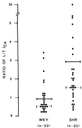
20 — y 5 — Λ 4 Η Ο Ο Ο I -< cc 3 Η 2 Η 1 Η ι . « . WKY (η=30) SHR (η=30)
Figure 1. Ratios of sustained (L) to transient (Γ) calcium current
were greater in SHR than WKY isolated vascular muscle cells measured under whole cell voltage-clamp conditions. Each dot represents one cell from which the ratios were measured in 30 SHR and 30 WKY, with means indicated by solid lines (significantly
different) and modes by dotted horizontal lines.19
total peak current in S H R than in W K Y (42 ± 7 % ) based on 6 0 experiments (P < 0.05). W h e n ratios (L/T) of C a2 +
current were calculated, the m e a n value for 3 0 W K Y was 0.9 a n d 2.9 for 3 0 S H R cells (Figure 1). These results demonstrate that even at the prehypertensive stage, L channels are more prominent in S H R than W K Y , sug gesting a genetic m e m b r a n e channel defect with the proper timing to b e a cause of increased C a2 + in vascular
muscle cells.
E l e v a t e d F r e e C a l c i u m C o n c e n t r a t i o n s at Subsarco-l e m m a Subsarco-l S i t e s T o study if these C a2 + currents asso
ciated with L channels are linked to altered intracellular C a2 + handling, the distribution of intracellular free C a2 +
activities, (Ca2 +)i, was measured in isolated vascular
muscle cells with fura-2. Incorporation of the fluores cent dye, quantitation of fluorescent intensities, and (Ca2 +X determination after correction for
inhomoge-neous dye distribution are described in detail else w h e r e .2 0-2 3
In a study quantitating the distribution of intracellular free C a2 + in neonatal vascular muscle cells from SHR
a n d W K Y ,2 4 w e have measured the average intracellular
dye concentration, a n d quantitated (Ca2 +)i of the whole
cytoplasm, as well as in defined regions. W e divided the cell into a peripheral boundary, 0.5 μτη distant from the
AJH-AUGUST 1989-VOL. 2, NO. 8 CELLULAR CALCIUM REGULATION IN HYPERTENSION 657
cell edges, and central areas, defined as all that remains. Quantitation of C a2 + was according to the following
equations which were applied to each of the 2 5 0 , 0 0 0 image elements
(Ca2 +X = Kd (cR - 1.36)/(3.48 - cR),
where Kd is the dissociation constant established b y cal
ibration procedures (239 n M at 3 7 ° C ) , R is the ratio of 510 n m fluorescence at 3 4 0 / 3 8 0 n m wavelength excita tion, and c corrects for the inhomogeneous dye distribu tion according to
c = 0.92 + 2 . 2 4 a e - °0 0 2 8 d F,
where F is the fura-2 fluorescence intensity count at 3 6 0 nm excitation wavelength, a corrects for day-to-day variability of the optical system as calibrated b y phos phor beads, and d adjusts for accumulation times. W e have performed these measurements in cells perfused with isotonic medium and following administration of 100 m M potassium and 1 μ Μ S D Z 2 0 2 - 7 9 1 ( + S ) , a ster eoselective dihydropyridine with calcium entry stimu lating properties.
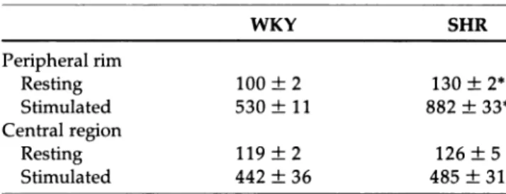
Intracellular fura-2 concentrations were similar in cells of both S H R and W K Y (62 and 85 μ Μ , respectively, not significantly different). ( C a2 +) i in central regions of
nonstimulated cells perfused with isotonic medium was comparable in both strains (WKY: 119 ν S H R : 1 2 6 n M ) . But ( C a2 +) j in the peripheral boundary was slightly ele
vated in S H R (WKY: 100 ν S H R : 130 n M , P < 0 . 0 5 ) . Upon stimulation of the cells by 100 m M potassium and SDZ 2 0 2 - 9 7 1 , the average myoplasmic C a2 + activity in
central regions of S H R tended to increase to a greater extent (WKY: 4 4 2 ν S H R : 4 8 5 n M ) accompanied by a more pronounced elevation of (Ca2"1^ in the peripheral
boundary (WKY: 5 3 9 ν S H R : 8 6 6 n M , Ρ< 0.05). These results point to the important role intracellular C a2 + re
lease from subsarcolemmal C a2 + pools m a y play in vas
cular muscle during the prehypertensive stage, suggest ing a defect of S H R C a2 + uptake or release mechanisms
in these cells.
D I S C U S S I O N / C O N C L U S I O N
These studies were directed towards the characteriza tion of cellular C a2 + handling abnormalities in S H R at a
prehypertensive age. T h e results demonstrate that b e fore development of elevated blood pressure in S H R , the sustained L C a2 + channel is more dominantly ex
pressed,1 9 and that C a2 + release or uptake mechanisms
from subsarcolemmal stores are already altered. T h e underlying factors leading to changes in the m e m b r a n e structure and intracellular organelles could well b e linked. For example, C a2 + entering through L channels
may have greater effects on the cytoplasmic sites which release intracellular C a2 +. Or, C a2 + entry through L
channels m a y have smaller effects on C a2 + uptake, as
suggested by Table 1. Whichever is true, these results
TABLE 1. C a2 + ACTIVITY (FREE ION
CONCENTRATION) IN AZYGOUS VEIN VASCULAR MUSCLE CELLS OF SHR ν WKY (nmol/L)
WKY SHR Peripheral rim Resting 100 ± 2 130 ± 2* Stimulated 530 ± 11 882 ± 33* Central region Resting 119 ± 2 126 ± 5 Stimulated 442 ± 36 485 ± 31
Resting condition was contractile cells between contractions in control
solution. Stimulated condition was cells in 100 mM K+ with 1 μΜ Ca2+
agonist (SDZ 202-791S). The peripheral rim is defined as the 0.5μm from the cell edge. Central region was all parts of the cell at least 1 μmfrom the cell edge, and actually includes the top and bottom peripheral edge contri butions to that fluorescence (ie, optical sectioning of fluorescence was not practical in these thin cells).
Values are means ± SEM; n = 3for WKY and 5 for SHR. *P<0.05.
underline the importance of C a2 + regulation of
sub-m e sub-m b r a n o u s C a2 + stores.
That total myoplasmic C a2 + activity in unstimulated
cells from both strains is comparable tallies with the finding that elevated ( C a2 +) i in platelets can only be
observed in older S H R with elevated blood p r e s s u r e .2 5
W h e t h e r a similar elevation of peripheral myoplasmic C a2 + activity in vascular cells from adult S H R (with ele
vated blood pressure) develops is u n k n o w n . However, it could be postulated that the e n h a n c e d responsiveness of S H R to stimuli giving rise to the observed C a2 + in
crease could lead to a profound resetting of C a2 + uptake
and extrusion mechanisms over time. T o further charac terize the responsible mechanisms, more studies of S H R are needed to investigate these processes in different ages and blood vessels.
R E F E R E N C E S
1. Cohn JN: Calcium, vascular smooth muscle, and calcium entry blockers in hypertension. Ann Intern Med 1983;98:806-809.
2. Bohr DF, Webb RC: Vascular smooth muscle function and its changes in hypertension. Am J Med 1984;77 (suppl 4A):3-16.
3. Hermsmeyer RK: Vascular muscle membrane cation mechanisms and total peripheral resistance. Hyperten sion 1987;10 (suppl I):20-22.
4. Folkow B: The hemodynamic consequences of adaptive structural changes of resistance vessels in hypertension. Clin Sci 1971;41:1-12.
5. Bolli P, Erne P, Hulthen UL, et al: Parallel reduction of calcium-influx-dependent vasoconstriction and platelet free calcium concentration with calcium entry and β-adrenoceptor blockade. J Cardiovasc Pharamcol 1984;6 (suppl 7 ) : 9 9 6 - 1 0 0 1 .
cal-658 HERMSMEYER AND ERNE AJH-AUGUST 1989-VOL 2, NO. 8
cium in patients with hypertension. Ν Engl J Med 1982;307:226-228.
7. Resnick LM, Laragh JH, Sealey JE, Alderman MA: Diva lent cations in essential hypertension. Relations between serum ionized calcium, magnesium and plasma renin activity Ν Engl J Med 1984;309:888-891.
8. Hulthen UL, Bolli P, Erne P, et al: Peripheral vasodilating effect of nitrendipine in man, in Scriabine A, Vanov S, Deck Κ (eds): Nitrendipine. Baltimore, Urban and Schwarzenberg, 1984, pp 4 6 3 - 4 6 8 .
9. Kiowski W, Bolli P, Erne P, et al: Mechanisms of action of calcium antagonists in hypertension. J Cardiovasc Phar macol 1987;10 (suppl 10):23-27.
10. Erne P, Bolli P, Bertel O, et al: Factors influencing the hypotensive effects of calcium antagonists. Hyperten sion 1983;5 (suppl II):97-102.
11. Erne P, Conen D, Kiowksi W, et al: Calcium antagonist induced vasodilation in peripheral, coronary and cere bral vasculature as important factors in the treatment of elderly hypertensives. Eur Heart J 1987;8 (suppl K):49-56.
12. Erne P, Bolli P, Burgisser E, Buhler FR: Correlation of platelet calcium with blood pressure: effect of antihyper tensive therapy. Ν Engl J Med 1984;310:1084-1088. 13. Resink TJ, Tkachuk VA, Erne P, Buhler FR: Platelet
membrane calmodulin-stimulated calcium-adenosine triphosphatase: altered activity in essential hyperten sion. Hypertension 1986;8:159-166.
14. Marvin WJ, Robinson RB, Hermsmeyer K: Correlation of function and morphology of neonatal rat and embryonic chick cultured cardiac and vascular muscle cells. Circ Res 1979;45:528-540.
15. Hermsmeyer K, Mason R: Norepinephrine sensitivity and desensitization of cultured single vascular muscle cells. Circ Res 1982;50:627-632.
16. Hermsmeyer K: Excitation of vascular muscle by norepi nephrine. Ann Biomed Eng 1983;11:567-577. 17. Sturek M, Hermsmeyer K: Calcium and sodium channels
in spontaneously contracting vascular muscle cells. Science 1986;233:475-478.
18. Bean BP, Sturek M, Puga A, Hermsmeyer K: Calcium channels in vascular muscle cells: modulation by dihy dropyridine drugs. Circ Res 1986;59:229-235. 19. Rusch N, Hermsmeyer K: Calcium currents are altered in
the vascular muscle cell membranes of spontaneously hypertensive rats. Circ Res 1988;63:997-1002. 20. Erne P, Hermsmeyer K: Intracellular C a2 + release in vas
cular muscle cells by caffeine, ryanodine, norepineph rine, and neuropeptide Y. J Cardiovasc Pharmacol
1988;12(suppl 5 ) : S 8 5 - S 9 1 .
21. Hermsmeyer K: Might nitrendipine enhance C a2 + trans
port in vascular muscle? in Merrill GF, Weiss HR (eds): Ca2 +-Entry Blockers, Adenosine and Neurohumors. Bal
timore, Urban and Schwartzenberg, 1983, pp 5 1 - 6 1 . 22. Hermsmeyer K, Rusch N: Felodipine actions on vascular
muscle C a2 + channels. J Cardiovasc Pharmacol 1987; 10
(suppl 1 ) 4 0 - 4 3 .
23. Erne P, Hermsmeyer K: Desensitization of norepineph rine includes refractoriness of calcium release in myo cardial cells. Biochem Biophys Res Commun 1988,151: 3 3 3 - 3 3 8 .
24. Erne P, Hermsmeyer K: Intracellular vascular muscle C a2 + modulation in SHR/WKY hypertension. Hyper
tension 1989;14 (in press).
25. Bruschi G, Bruschi ME, Caroppo M, et al: Cytoplasmic free C a2 + is increased in the platelets of spontaneously
hypertensive rats and essential hypertensive patients. Clin Sci 1985;66:179-184.