A/H 1996,9:13:-143
Hy ertension Does Not Affect Intracellular
Ca P
cium Uptake in Human Platelets
Marc Eberhnd,
Domirzique Eutf9uoz, md Paul
ErueThe relationship between the Ca’+ transport of platelet endoplasmic reticulum and hypertension was analyzed in 17 untreated patients exhibiting various degrees of hypertension. Each patient un-
derwent a 24-h recording of ambulatory blood
press~e. Platelets from patients wew permeabi- lized with saponin and the rate of ATP-driven thapsigargin-sensitive Ca2+ uptake det.::mined us- ing the fluorescent Ca’+ indicator flue-3. No rela- tionship behveen blood pressure (systolic, diastol- ic, day, night) and the rate of CaZC uptake into the
--
__----
sacmplasmic reticulum of platelets was found. A weak but insignificant correlation between Ca’+ uptake and the heart rate was noted. Therefore, the increase in cytosolic Ca2+ of platelets in hyperten- sion may not be dw to changes of the activity of Ca2+ uptake into the sacroplasmic reticulum. Am J Hypertens 1996;9:136-142
KEY WJRDS: Platelets, calcium transport, hypertension.
D
espite extensive research, the relationship of calcium and arterial hypertension re- mains unclear. intracellular Ca” is a major determinant of basal myogenic tone of vas- cular muscle. 1 herefore, an increase of cytosolic Ca” in arterial smooth muscle leads to an increase in pe-ripherai resistance characteristic for hypertension. The systemic hormonal signals associated with hyperten- sion influence endothelial cell, vascular muscle cells and blood cells. Eme et al’ have dwJmented an ele-
vation of platelet cytosolic frw 6” in hypertension and a close correlation of the CL’- levels in platelets and blood pressure in untreated h
Y+ pertensive pa- tients. Furthermore. both platelet Ca and vasocon-
striction were normalized by antihypertensive treat- ment,‘,2 indicating a close relationsh’ip between plate-
__-_- -_.-__-___
Receiwd March 6, 1995. Accepted June 3, 1995,
From the Department of Research, Kantonsspital, Base1 (ME), and the Diwsion of Cardinlogy. Kantonsspital, Luzrm (DE,PE), Switzw land.
Thts study was supported by the Swirr Nattonal Foundation (Vu 324Ml75.90 and 32439446.93~. the SEhweueriuhe Her.stiftunB, and the Langcardis Foundation.
Address conespondcnce and reprint requests to I’D D;. I’. Eme. Dwision of Cardiology. Kantonsspital, CH~JM Lurem 16, Switzcr- land.
0 1996 ly the Amoican [mmnl of Hylrrte~on, Ltd. Publishal by El& Scimce, Inc.
let Ca’+ dynamics and circulating hormonal factors. Cytosolic Ca2+ overload has been desctibed in a vari- ety of cells in both hypertensior?s and pressure- induced cardiac dysfunction.”
Whereas the relationship of platelet qtosolic Ca” and rasoconshiction has been clearly established, the mo!eccular basis of elevated Ca” levels remain, un- clear. The rapid rise in cytosolic Ca2’ observed upon exposure of platelets to humoral factors from hyper- tensives’,” indicates that systemic hormonal fac!ors in- fluence platelet cytosolic Ca’- and that the changes in platelet cytoso!ic Ca’+ are effected by a rapid mecha- nism, ie. Ca” influx across the plasma membrane or intracellular Ca” mobilization. The reversal of Ca” levels by means of Ca” antagonists has led to the hypothesis that Ca’* iflux is responsible for elevated platelet Ca”.‘,Y However, the enhanced hormone- induced elevation of platelet cytosolic Ca” is not cor- rected by antihypertensii e therapy,‘” demonstrating that the fundamental defect in Ca“ dvnamics has not been restored. .\loreover, witage-operated Ca’- chan- nels do not mediate agonist-evoked Ca” entry in platelets. ” E&ides voltage-operated Ca” channels. a variety of other mechanisms cw;tibutr to the Ca2’ dynamics of platelcts!‘~“: agonist-mediate.1 Ca” in-
138 EBEIWARD ET AL ASH-FEGRUARY 199GVOL. 5. NO. 2
Bux; l&and-g&d Ca’+ channels; Ca”-ATPare pumps present in both the plasma membrane and membrane of endoplasmic reticulum”,“; Na+/Ca*+ exchanger”; inositol 1,4,5+isphosph.&-induced Ca” rele.lse from intracellular Ca” stem: and a series of cytojolic and endoplasmic reticulum Ca’* binding proteins. Resink et allh have documented a reduction of the calmodu- linstimulated Ca’+-ATPaw activity and an increase in basal Ca”-ATPase activity in platelets from hyperten- sive compared to normal subjects, suggesting a reduc- tioq in plasma membrane Ca’+ exhusion activity and an activation of the sarcoplasmic reticulum Ca*+ pump in hypertension. Other studies also have desctibed a decrease in Ca”-ATPase activity in hypertension.“,” In the present study we have determined the rate of Ca” uptake in permeabilited platelets from 17 un- heated subjects with various degrees of hypertension in order to test if alteration of the sarcoplasmic retic- ulum Ca”-ATPase pump activity contributes to ele- vated platelet .$osolic Ca” in hypertension.
MATERIALS AND METHODS
Reagents Heparin, pentosan polysulfate, sapanin, and hexokinase were obtained from Sigma (St. Louis, MO). Rue-3 pentapotassium salt was from Molecular Probes (Eugene. OR). Flue-3 was stored in 50 pL ali- quots at a concentration of 1 mmol/L in dimethylsulf- oxide at -70°C. Ionomycin was from Calbiochem (San Diego, CA!, thapsigargin from LC Services Corpora- tion. Aminoethyl Biogel P-2 was purchased from Bio- Rad (Richmond, CA), and Sepharose CL-2B from Pharmacia (Uppsala, Sweden).
Buffer Solutions All experiments were performed in 20 mmol/L HEPES buffer containing 155 mmol/L KCI, 5 mmol/L NaCl, 2 mmol/L MgCl,, 2 mmol/L I&dithioetythritol, and 0.3 mmol/L phenylmethane- sulfonyl fluoride at pH 7.2, which was denoted buffer A. All solutions were depleted of Ca” by means of chromatography on EDTA-polyacrylamide as de- scribed previously.“,” Residual Ca” concentrations of solutions following treatment with EDTA- polyacrylamide were measured as described.20 Patients We analyzed platelets of 17 newly diag- nosed hypertensive patients who were referred ta the Division of Cardiology of the Kantonsspital Lucerne. None of the patients had received antihypertensive drugs before. Patients underwent a ltlead electcocar- diagram, a bicycle exercise test, and a 24-h recording of ambulatory blood pressure (HCR Blood Pressure, Disectru?ic, Burgdorf, Switzerland). Table 1 summa- rizes the clinical data and results of ambulatory 24-h blood pressure measuremenk.
preparation of Platelets Platelets were prepared from platelet-rich plasma by gel filtration*’ using Sepharose CL-2B (Pharmacia) and adjusted to 108
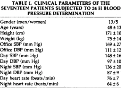
TABLE 1. CLINICAL PARAMETERS OF THE SE’IENTF.EN PATIENTS SUBJECIXD TO 24 H BLOOD
PRESSURE DETERMINATION Gender (men/women) Age (years) Height (cm) Weight (kg) Office SBP (mm Hg) Office DBP (mm Hg) Day SBP (mm Hg) Day DBP (mm Hg) Night SBP (mm Hg) Night DBP (mm Hg) Day heart rate (beats/min) Night heart rate (heats/min)
13/5 48+13 171flO 75+14 169f27 111f12 148f16 97flO 136f20 87f9 76f7 6426
cells/ml. Platelets were permeabiiized by addition of 20 kg saponin/l08 cells. Selective permeabilization was confirmed by measuring the release of flue-3 from cells loaded with flue-3 acetoxymethyl ester, and by monitoring the release of Ca” horn internal mem- branes in permeabilized cells following addition of ionomycin. Platelets were used within 6 h of blood withdrawal.
Measurement of Ca’+ Ca” concentrations were measured by means of the fluorescent Ca” indicator Buo-3. The indicator was added at a final concentra- tion of 1 bmol/L to 1 mL of a suspension of platelets (108 cells/ml) placed into a l-cm quartz cuvettc. The suspension was continuously stirred using a magnetic stirrer. Fluorescence was measured by means of a Per- kin-Elmer (Oakbrook, IL) 650-10s fluorimeter, inter- faced via an analog/digital converter (PC-28, Inshu- matic AG, Zurich, Switzerland) to a personal com- puter. Ruorescence of tluo-3 was excited at 505 nm and observed at 530 nm. ATP (1 mmol/L) and saponin (20 llg) were added to the cell suspension to activate Ca” uptake of intracellular Ca” stores. ATP at 1 mm~ol/L was found to saturate thapsigargin-sensitive Ca2’ uptake in platelets from many subjects of widely different degrees of hypertension (not shown).
Ca’+ concentrations were calculated frvn the fll;~ rescence signal using standard tikations of flue-3 with Ca2’. Binding of Ca” to flue-3 was expressed as signal versus concentration of added Ca”, and evalu&ed as describecLm From the dissociation constant (Kd) of the Ca’+-flu+3 complex, which was typically 900 nmol/L in buffer A. the fluorescence signal of Ca*+-free indi- cator (f,) and that of the Ca”-indicator complex (f,), the concentration of Ca” w.:s calcukted 3:. foIlor*-s: ICa2’l hn = Kd l Cf, - f,,/Cf,zc - f,, (1)
where f, represents the observed fluo-ce signal. K, was determined from tibations of flu-3 with Ca’+ (final concentrations between 0 and 5 mmol/L; eval-
A/H-fEBR11,4RY 1996-YOL 9. NO. 2 CALCIUM TRANSPORT IN HUMAN PLATELETS 139
uation according to Eberhard and Ernt?‘?. f, was de- termined after addition of EGTA (5 mmol/Lj, and tFc by extrapolation of the concentration of added Ca2’ to infinity. Figure 1 illustrates the calibration of fluores- cence. The Ca2’ flux due to the sarcoplasmir reticulum Ca”-ATl’ase pump, hence termed Cal’ uptake, was determined from the difference of the overall Ca’+ flux before and after addition of ?hapsigargin (200 nmol/ L), a specific inhibitor of the sarcoplasmic reticulum CaZ’-ATPase.22 In the presence of thapsigargi:l, a spontaneous Ca” flux out of the sarcoplasmic reticu- lum, the so-called leak, was observed and modelled by an exponential decay toward an equilibrium value (Figure IB):
[Ca”] = n * exp(-k . t) + [Ca*‘l,
(2)
where R and k are arbitrary consiants, and [Ca”],, the equilibrium concentration of Ca”. Thus, the rate of Ca” leak at a particular Ca” concentration is given byData Analysis Data evaluation and nonlinear least- squares fits were performed using standard proce- dures.2”2J Results are expressed as means + SD. I:ldi- vidual Ca” uptake and leak rate deterr%natiuns were averaged and SD ..x!*:s calculated if three or more individual values were measured. Linear least-squares resressions assuming uniform and unity SD values of the individual data points were used to analyze puta- tive relationships between binding data and clinical parameters. Percentile points 8’) were determined us- ing the two-tailed t distribution.
RESULTS
d[Ca”l/dt = -k l n. exp(-k * t) = -k *
uca2+1 - ICa2’iog)
The evaluatioil of fl:orescmce dat‘r and a typical Ca” transient used to datermine Ca” uptake rates, i.e. the (3) thapsigargin-sensitive Ca” flux, are shown in Figure
The rate of Ca” uptake is the difference bet ~veen the slop of the Ca” transient at a particular Ca’- concen- tration before addition o! thapsigargin (overal: Ca” flux, Figure I), and the rate of Ca” leak at that Cd’- concentration (equation 3).
140 EBERHARDEI’AL
1. At a given Ca” concentration similar Ca” uptake rates were obtained when thapsigargin was added at various degrees of store !oading (not shown). Since Ca” concentrations of suspensions of pemwabilized platelet were around and above 1 mmol/L, and since the Ca*’ uptake rate did not significantly depend on
A/H-FEHRUARY I!%-I’OL 9. NO. 2
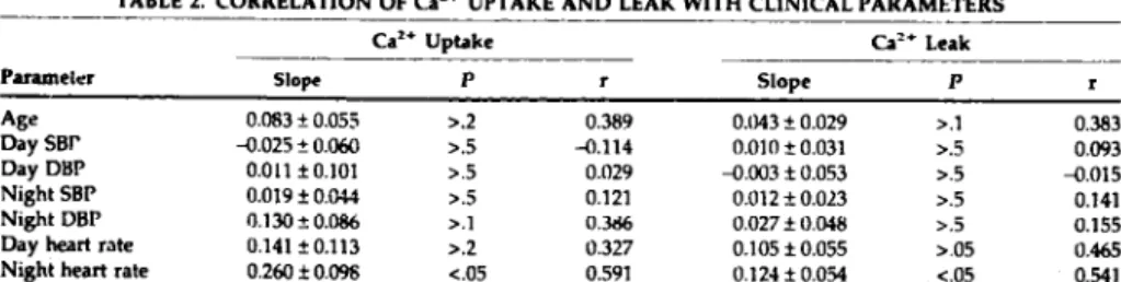
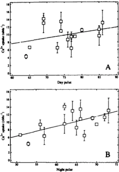
the concentration of Ca” in the range behveen 0.8 and 1.5 mmol/L (not shown), Ca’+ uptake rates were eval- uated at 1 mmol/L Ca” in the present study. To ex- amine if spontaneous Ca” leak of the Ca” stores is linked to hypertension, the Ca2+ leak (Ca2’ flux in the presence of thapsigargin) was also evaluated at 1 mmol/L Ca”. In two out of the 17 patients examined the CaZ’ concentration of the platelet suspension after permeabilization was too high to allow the determi- nation of Ca” uptake and leak rates at ! mmol/L Ca”. Figure 2 shows that Cai’ uptake into sarcoplasmic reticulum is not related to average daytime blood pressure. Similar results are obtained with otter blood pressure parameters (Table 1) and with Ca” leak from sarcoplasm~c rr:ix~lum (Tabie 2). A weak correlation between both Ca’+ uptak.e and Ca’+ leak and heart rate was observed (Figure 3, Table 2). although these cor- relations are flawed by the substantial error of the values of Ca” uptake and leak. Furthermore, no cor- relation was found behveen Ca” uptake and age (Ta- ble 2). To corroborate the relationship between Ca” fluxes and blood pressure we analyzed a group of blood donors consisting of four normotensive and ten hypertensive subjects who did not undergo 24-h blood pressure determination (Table 3). No significant cor- relation between flux parameters and blood pressure was detected within this group (not shown). In addi- tion, the differences in Ca” uptake and leak between the group of normotmsives and the two hypertensive groups were statistically not significant (Table 3).
DISCUSSION
There is a general agreement that plate!& cytosolic Ca” is increased in hypertension and that this process can be accomplished by rapid Ca’+-regulating pro- cesses.’ Both plasma membrane- and internal mem- brane-associated Ca’+ fluxes contribute to the ob- served changes in platelet cytosolic Ca”. Since the Ca’+-ATPase of sarcoplasmic reticulum is believed to represent the major contribution to Ca’+ remw;al from the cytoplasm,“,” xe have analyzed ATP-dependent
TABLE 2. CORRELATION OF Ca’+ UPTAKE AND LEAK WITH CLINICAL PARAMETERS
__-- .__~
Ca’* Uptake Ca’+ Leak -
PaIaUl&l Slope P r Sl0pe P r
Aw 0.063 A 0.055 >.2 0.389 0.043 f 0.029 >.I 0.383
Day SBr a.025 ? 0.060 >.5 -0.114 0.010 f 0.031 >.5 0.093
Day DBP 0.011 fO.lO1 >.5 0.029 a.003 t 0.053 >.5 a.015
Night SBP 0.019 i 0.044 ..5 0.121 0.012 + 0.023 >.5 0.141
Night DBP 0.130 A 0.086 >.l 0.3% 0.027 f 0.048 >.j 0.155
Day heart rate 0.141 f 0.113 >.2 0.327 0.105 f 0.055 >.05 0.465
Night heart rate 0.260 f 0.096 co5 0.591 0.124 * 0.054 co5 0.541 ~-
I5 kVprtnsla plimls wrd~plnx 2-h blood ptnsuw d&nGdion (TabIt I), for which Ca-‘* ~P!,I~I rind C$’ Imk ralur; uwc dclenind (UC ~UItsJ.
AIH-FEBRLIARY 199GV3L. 9, NO 2 CALCIUM TRANSPORT IN HUMAN PLATlXETS 141
Ca” transport into sarcopl3smic reticulum in platelets from a series of patients exhibiting various degrees of hypertension. None of the patients received antihy- pertensive therapy prior to analysis. Thapsigargin, a specific inhibitor of all isoforms of internal membrane GI~‘-ATP~s~‘~~~~, served to evaluate C&-A rPase transport activity and spontaneous Ca*+ leak of sarco- plasmic reticulum. No correlation of blood pressure with both CP” sptake and CL?’ leak and a weak but
insignificant correlation of Ca” uptake with heart rate were found. Ca” uptake and Ca” leak did not signif- icantly differ between hypertensive pntients and a normotensive control group.
An earlier study has documented a substantial in- crease of platelet calmodulin-insensifve Cd”-ATPase acti\ ity in ‘lypertension ” indicating a stimulation of internal membrane Ca”-ATPase in hypertension. The discrepancy to our results may result from the different selection of hypertensive patien!s, the xse of thapsig?rgin in the present study as opposed to cal- modulir. activation, and the determination oi Ca” transpo: t activities in permeabilized cells in the present .?xperiments rather than Ca’--dependent AT- Pase artisitv in membrane preparations used in the spldy of R&ink et a1.16 Takaya et al” found a decrease in maximal velocity and Michaelis constant of Ca”- ATPase in platelets from hypertensive patients. Since plasma membrane and internal membrane Ca”- ATPase activity was nor discriminated, the respective contributions of the two Ca”’ pumps could not ‘be evaluated in that shrdy. In agreement with our results, no correlation of platelet sarcoplasmic reticulum Cn’+- ATPase with blood pressure was documented, but a decrease of platelet plasma membrane Ca2+-ATPase activity in hypertension was found recently.‘” Consis- tent with the decrease in calmodulin-sensitive Ca’+- ATI’ase activity in hypertension,16 these studies indi- cate that removal of Ca” across the plasma membrane is responsible for the well established rise of cytosolic Ca” in hypertension.‘,’
Possible other mechanisms accounting for altered cytosolic Ca*’ in hypertension include increased ago- nist-induced 6” influx,‘O activation of voltage- operated Ca’+ channels,] altered inositol 1,4,5- trispho;fhate-induced Ca*’ efflux from internal Ca” stores, and a decreased activity of G+/Ca’+ exchangers present in the platelet plasma mem- brane.“,*’ Whereas the density of voltage-g&d Ca’+ channels in platelets is very low and plays only a mi- nor role in the Ca” balance of platelets,” enhanced agonist-induced 6” influx in platelets has been doc- umepted in patients with ischemic heart diseaw3 A
TABLE 3. COMPARISON OF Ca*+ UPTAKE AND LEAK BETWEEN HYPERTENSIVE PATIENTS ANV NORMOTENSIVE SUBJECTS
-
Normotensive Hypertensive Hypertensive’
142 EBERHARD ET AL A/H-FEBRUARY 1996-VOL. 9. NO. ?
decrease of platelet Na’/K’-ATPase activity in mild gestational hypertension has been suggested to con- tribute to increased cytosolic Ca” through the Na+/ Ca’+ exchanger.” However, Takaya et al” did not find altered platelet Na’/K*-ATPase activity in hyper- tension. Therefore, a decreased activity of the plasma membrane Ca’+-ATPase and increased agonist- induced Ca” influx are likely to contribute to the in- crease in plate& cytosolic Ca” in hypertension,
whereas ATPdriven Ca” uptake into internal Ca” stores remains rather unchanged.
In thzz present study a weak relationship between Ca” uptake into internal membrarzes and heart rate was noted (Figure 3). Clearly, more patients and a wider range of heart rates will be needed to decide if this correlation is significant. A link between heart rate, which is controlled by the sympathetic and para- sympathetic nervous system,w”.” and Ca2+ dynamics of the platelet wrcoplasmic reticulum could be pad- renergic receptors present in the heart and on plate- lets.Jz,33 Whereas hypertension is usually associated with an increase in sympathetic activity,” its role in the Ca” balance in platelets remains obscure, al- though Padrenergic receptors may affect Ca’+ pump- ing activity via phospholamban phosphorylation.”
REFERENCES 1. 2. 3. 4. 5. 6.
EM I’, Bolli P, Biirgisser E, Blihler FR: Correlation of platelet calcium with blood pressure: effect of antihy- pertensive therapy. N Engl J Med 1981;301:1084-IMU). LbUi P, Eme P, Hulthen LJL, et al: Parallel reduction cf calcium influx dependent vasoconstriction and platelet free calcium concentration with calcium entry and beta- adrenoceptor blockade. J Cardiovax Pharmacol 1984; 6:S99GS1001.
Rembold CM: Regulation of contraction and relaxation in arterial smooth murIo. Hypertension lW2;20:129- 137.
Brickman AS, Nyby MD, Van Hungen K, et al: Calci- tropic hormones, plabzlet calcium and blood pressure in essential hypertemion. Hypertensron 1990;16:515-
522.
Bruschi G, Bmschi ME, Caroppo M, et al: Cytoplasmic free ICa”l is increased in the platelets of spontaneously hyprtensive rats and essential hypertensive patients. Clin Sci 1985;68:179-184.
Morgan JP: Abnormal intracellular modulation of cal- cium as a mapr cause of cardiac contractile dysfunc- tion. N Engl J Med 1990;325:62M32.
Hollister AS, Menshikov M, Devyatova 0: Humoral factor, inha-platelet calcium, and hypertension. Am J Physiol 1992;5:131Sl34S.
Lindner A, Kenny M, Meacham AJ: Effects of a circu- kting facto1 in Patients with essential hypertension on intracellular free calcium in normal platelets. N Engl J
Med 1967;316:5U%513.
Ahn YS, Jy W, Harrington WJ, et al: Increased platelet calcium in thrombosis and related disorders and its correction by nifedipine. Thromb Res 1987;45:13>143.
10. Il. 12. 13. 14. 15. 16. 17. 18. 19. 20. 21. 22. 23. 24. 25. 20. 27. 28
Erne P, Resink T’J. Dolli P, et al: Free calcium rqonse to adrenaline in platelets of normal and hyprtensive (untreated and treated) subjects. J Hypertens 1984;2(suppI 3):159-161.
Rink TJ, Sage SD: Calcium signaling in human plate- lets. Annu Rev l’hysiol 1990;52:431-449.
Carafoh E: Intracellular calcium homeostasis. Annu Rev Biochem 1987;56:395-433.
Papp B, Enyedl A, Kovacs T, et al: Demostradon of two forms of calcium pumps by thapsigargin inhibition and radioimmunohlotting in platelet membrane vesicles. J Biol Chrm 1991;266:1?593-145%.
Dean WL: Structure, function and subcell&r iucnliza- tion of a human platelet CaZ*-ATPasu. Cell Calcium 1969;10:289-2Y7.
Valant PA, Adjei DN, Haynes VH: Rapid Ca”.. extruding via the Na*/Ca” exchanger of the human platelet. J Membr Biol 1992;13O:63472.
Resink TJ, Tkachuk VA, Emc I’, Biihler FR: Platelet membrane calmodulin-stimulated calcium-adenosine triphosphatase: altered activity in hypertension. Hy- Pertension l986;8:159-166.
Tnkaya J, Lasker N, Bamforth R, et al: Kinetics of Ca’+- ATPasc activation in platelet membranes of essential hypertensives and no¬ensives. Am J Physiol 1990;
25K98SC994.
Vezzoli G, Elli AA, Tripodi C, et al: Calcium ATPase in erythrocytes of spontaneously hypertensive rats of the Milan Grain. J Hypertens 1%5;5:64.5-648.
Eberhard M, Erne l? lnositol 1,4,5-trisphosphatc- induced calcium release in oermeabdized olatelcts is coupled to hydrolysis of in&itol 1,4,5-trisph’osphatc to inositol I,$-bisphosphatc. B&hem Biophys Res Com- mu” 1993;195:19-24.
Eberhard M, Erne P: Analysis of calcium binding to a-lactalbumin using a fluorescent calcium indicator. Eur J B&hem 1991;20231332-1338.
Erne I’, Mittelholzer E, Biirgisser E, et al: Measurement of receptor induced changes in intracellular free cal- cium in human plalelets. J Recept Res 1984;4:587-604. Lytton J, Westlin M, Hanley MR: Thapsigargin inhibits the sarcoplasmic or endoplasmic reticulum Ca”- ATI’ase family of Ca” pumps. J Biol Chcm 1991;266: 17c67-17071.
Press WH, Flannery BP, Tcukolsky SA, Vetterling WTz Numerical Recipes. Cambridge University Press, Cam- bridge, 1989.
Eberhard M: A set of programs for analysis of kinetic and cqudibrium data. CABIOS 1990;6:21~221. Wier WG: Cytoplasmic lCa**l m mammalian vcntrt- cldynamic control by celh~lar procesxs. Annu Rev Physiot 1990;52:467-48j.
Dean WL, Pop JE, Brier ME, Aronoff GR: Platelet cal- cium transport in hypertension. Hypertension 1994;U: 31-37.
Rengasamy A, Sourn S, Feinberg H: Platelet Ca2’ he meostasis: Na’Ca” exchange in plasma membrane vesicles. Thromb Haemost 1987;57:337-340. Kobayashi N, Okumura K, Hashimoto H, et al: In- .:rcased Ca” influx into platelets induced by thrombox-
AJH-FEBRUARY 19%-VOL. 9, NO. 2 CALCIUM TRANSPORT IN HUMAN PLATELETS 143
29.
30.
31.
ane A2 analog in patients with ischemic heart disease. Clin Cardiol 1989;12:456-460.
Romanini C, Tranquilli AL, Ceser N, et al: Modifica- tions induced by gestationai hypertension on platelet calcium transport. Exp Mel Pathol 1991;41:122-128. Rompelman 0: Rythms and analysis techniques, in
Stracker J, Westerhof N (ads): The Physin of Heart and Circulation. IOP Publishing Ltd., Bristol, England, 1993, pp 11&120.
Campbell DL, Rasmussen RL, Strauss HC: Electrophys iology of the sinus node, in Dangman KH, Miura IX teds): Electrophysiology and Pharmacology of the Heart. Marcel Dekker Inc., New York, 1991, pp S--108.
32.
33.
34.
35.
Brcxtde OE, Broedz A, Da”! A, et al: Receptor systems in t.ie non-failing human heart. Basic Res Cardiol 1992;87(suppl I):]-14.
Davi G, Now 5, Pinto A, et al: Platelet activation after adrcnergic shmulation in hypertensive patients: effect of acebutolol. Eur Heart J 1983:4:295-299.
Mancia G, Crassi G, Parati G, Daffonchio A: Evalilating sympathetic activity in human hypertension. J H)per- tens 1993;11(suppl 5):Sl>Sl9.
Luo W, Crupp IL, Haner J, ct al: Targe:ed ablation of the phospholamba” gene is awxiated with markcd,ly enhanced myocardial contractility and loss of I)-agomst stimulation. Circ Rn 1994;75:401-1C9.