Université de Sherbrooke
Involvement of Beta-arrestin 1 and Beta-arrestin 2 in store operated calcium entry
Par
Cynthia Sharmeen Département de Pharmacologie
Mémoire/Thèse présenté(e) à la Faculté de médecine et des sciences de la santé en vue de l’obtention du grade de maître ès sciences (M.Sc.) en pharmacologie
Sherbrooke, Québec, Canada [June, 2016]
Membres du jury d’évaluation Dr. Guylain Boulay Département de Pharmacologie
Dre. Christine Lavoie Département de Pharmacologie
Dr. Paul Pape
Département de physiologie
RÉSUMÉ
Implication de Beta-arrestin 1 et Beta-arrestin 2 dans l'entrée capacitative de calcium Par
Cynthia Sharmeen, Département de pharmacologie
Mémoire présenté à la Faculté de médecine et des sciences de la santé en vue de l’obtention du diplôme de maitre ès sciences (M.Sc.) en pharmacologie, Faculté de médecine et des
sciences de la santé, Université de Sherbrooke, Sherbrooke, Québec, Canada, J1H 5N4 La variation de la [Ca2+] intracellulaire participe à nombreux de processus biologiques. Les cellules eucaryotes expriment à la membrane plasmique une variété de canaux par lesquelles le calcium peut entrer. Dans les cellules non excitables, deux mécanismes principaux permettent l'entrée calcique; l'entrée capacitative de Ca2+ via Orai1 (SOCE) et l'entrée calcique activé par un récepteur (ROCE). Plusieurs protéines clés sont impliquées dans la régulation de ces voies d'entrée calcique, ainsi que dans l'homéostasie calcique. TRPC6 est un canal calcique impliquée dans l'entrée calcique dans les cellules à la suite d’une stimulation d’un récepteur hormonal. TRPC6 transloque à la membrane cellulaire et il y demeure jusqu'à ce que le stimulus soit retiré. Les mécanismes qui régulent le trafic et l'activation de TRPC6 sont cependant encore peu connus. Des découvertes récentes ont démontré qu'il y a un rôle potentiel de Rho kinase dans l'activité de TRPC6. Rho kinase est activée par la petite protéine G RhoA qui peut être activée par les protéines G hétérotrimériques Gα12 et Gα13. En plus de Gα12 et Gα13, les protéines de désensibilisation des GPCR β -arrestin 1 et / ou β-arrestin 2 peuvent aussi activer RhoA. Le but de notre étude est d'examiner la participation des protéines Gα12/13 et arrestin 1/ β-arrestin 2 dans l'activation de TRPC6 et de la protéine Orai1. Nous avons utilisé des ARN interférant (siRNA) spécifiques pour induire une réduction de l'expressionde Gα12/13 ou β-arrestin 1/β-β-arrestin 2. La conséquence sur l’entrée de Ca2+ dans les cellules a été ensuite déterminée par imagerie calcique en temps réel suite à une stimulation par la vasopressine (AVP), thapsigargin ou carbachol. Nous avons donc identifié que dans des cellules A7r5, une lignée cellulaire de musculaires lisses vasculaires où le canal TRPC6 exprimé de manière endogène, la diminution de l’expression des protéines Gα12 ou Gα13 ne semble pas modifier l’entrée Ca2+ induit par l’AVP par rapport aux cellules témoins. D'autre part, la diminution de l’expression β-arrestin 1 ou β-arrestin 2 dans des cellules HEK 293 ainsi que des cellules HEK 293 exprimant de façon stable TRPC6 (cellules T6.11) ont augmenté l’entrée de Ca2+ induite par thapsigargin, un activateur pharmacologique de SOCE. Des études de co-immunoprécipitation démontrent une interaction entre la β-arrestin 1 et STIM1, alors qu'aucune interaction n'a été observée entre les β-arrestin 1 et Orai1. Nous avons de plus montré à l'aide d'analyse en microscopie confocale que la diminution de l’expression β-arrestin 1 ou β-arrestin 2 n’influence pas la quantité d’Orai1 à la périphérie cellulaire. Cependant, des résultats préliminaires indiquent que la diminution de l’expression β-arrestin 1 ou β-arrestin 2 augmente la quantité de STIM1-YFP dans l'espace intracellulaire et diminue sa quantité à la périphérie cellulaire. En conclusion, nous avons montré que les β-arrestin 1 ou β-arrestin 2 sont impliquées dans l'entrée capacitative de Ca2+ (SOCE) et contrôlent la quantité de STIM1 dans le réticulum endoplasmique.
Mots clés: TRPC6, ROCE, SOCE, entrée calcique, signalisation cellulaire, imagerie calcique.
SUMMARY
Involvement of Beta-arrestin 1 and Beta-arrestin 2 in store operated calcium entry By
Cynthia Sharmeen, Department of pharmacology
Thesis presented to the Faculty of medicine and health sciences to obtain the degree Master of Science (M.Sc.) in pharmacology, Faculty of medicine and health sciences, Université
de Sherbrooke, Sherbrooke, Quebec, Canada, J1H 5N4
In an organism, intracellular [Ca2+] takes part in many biological processes. Eukaryotic cells express a variety of channels in the plasma membrane through which calcium can enter. In non-excitable cells, two main mechanisms allow calcium entry; the store-operated calcium entry via Orai1 (SOCE) and receptor-operated calcium entry (ROCE). Several key proteins are involved in the regulation of these calcium entry pathways as well as in calcium homeostasis. TRPC6 is a calcium channel implied in calcium entrance into the cells following hormonal stimulation and translocates to the plasma membrane. TRPC6 channel appear to the plasma membrane until the stimulus is present. Although, the mechanisms that regulate the trafficking and activation of TRPC6 are still little known. Recent findings have demonstrated that there is a potential role of Rho kinase in activity of TRPC6. Rho kinase is activated by the small G protein RhoA that itself can be activated by the heterotrimeric G proteins Gα12 and Gα13. In addition to Gα12 and Gα13 proteins, cytosolic GPCR desensitizing proteins β-arrestin 1 and/or β-arrestin 2 could also activate RhoA. The purpose of our study is to investigate the involvement of the proteins Gα12/13 and β-arrestin 1/β-arrestin 2 in the activation of TRPC6 and Orai1 protein. We used siRNA specific to Gα12/13 or β-arrestin 1/β-arrestin 2 to knockdown their endogenous expression. Then, calcium imaging in real time was performed in order to see the quantity of calcium entered into the cell following stimulation by vasopressin (AVP), thapsigargin, or carbachol. We hence identified that in A7r5 cell, vascular smooth muscle cell where TRPC6 channel expressed endogenously; reduced expression of Gα12 or Gα13 proteins does not seem to modify the AVP-induced Ca2+ entry compared to control cells. On the other hand, calcium imaging experiment in knocked down β-arrestin 1 or β-arrestin 2 in HEK 293 cells as well as HEK 293 cells stably transfected with TRPC6 (T6.11 cells) resulted in an increased thapsigargin-induced calcium entry. The co-immunoprecipitation studies demonstrate an interaction between β-arrestin 1 and STIM1, a calcium sensor in SOCE influx, while no interaction was observed between β-arrestin 1 and Orai1.We moreover showed by confocal microscopy that reduced expression of arrestin 1/ β-arrestin 2 does not influence the quantity of Orai1 at the cell periphery. Preliminary results showed that reduced expression of β-arrestin 1 or β-arrestin 2 increases the quantity of STIM1-YFP in the intracellular space and less it’s in peri-membrane space. In conclusion, we showed that β-arrestin 1 or β-arrestin 2 are involved in the store-operated calcium entry (SOCE) and control the quantity of STIM1 in the endoplasmic reticulum.
TABLE OF CONTENTS
RÉSUMÉ/ RÉSUMÉ ... iii
SUMMARY ... iv
LIST OF FIGURES ... vii
LIST OF TABLES ... viii
LIST OF ABBREVIATIONS ... ix
GqPCR ... ix
INTRODUCTION ... 1
1. Calcium: an important second messenger ... 1
2. Calcium homeostasis ... 2
2.1 Balancing excessive intracellular Ca2+ concentration ... 3
3. Calcium mobilisation ... 4
4. Voltage Independent Calcium Entry (VICC) ... 6
4.1 Store operated calcium entry (SOCE) ... 6
4.2. Receptor operated calcium entry (ROCE) ... 12
4.3 TRPC6: a potential therapeutic target ... 16
5. Research project ... 20
5.1 Problematic ... 20
5.2 Hypothesis ... 26
5.3 Objective of the study: ... 26
MATERIAL AND METHODS ... 27
6. Material ... 27
METHODS ... 29
7.1 Cell culture and Transfection ... 29
7.2 Protein Extractions ... 30
7.3 Determination of protein concentration ... 30
7.4 Separation of the proteins by electrophoresis on polyacrylamide gel and immunodetection of proteins ... 30 7.5 Co-immunoprecipitation ... 31 7.6 Confocal microscopy ... 32 7.7 Measurement of [Ca2+]i ... 32 7.8 Statistical Analysis ... 33 RESULTS ... 34
8.1 Importance of Gα12 and Gα13 in Ca2+ entry through ROCE ... 34
8.2 Knocking down β-arrestin 1 / β-arrestin 2 does not change in CCh induced calcium entry through ROC in HEK 293 cells stably expressingTRPC6 (T6.11) ... 36
8.3 Involvement of β-arrestin 1 / β-arrestin 2 protein in activation of SOCE and Orai1 in HEK 293T cells ... 42
8.4 β-arrestin 1 does not interact with Orai-Myc but interacts with STIM1 in HEK293T cells ... 47
8.5 Orai1 localization is not altered in β-arrestin 1 and β-arrestin 2 depleted cells ... 49
8.6 β-arrestin 1 and β-arrestin 2 depletion alter STIM1 intracellular distribution ... 50
9.1 Role of Rho in calcium signalling and Gα12 / Gα13 in Ca2+ entry through ROCE ... 52
9.2 Role of β-arrestin 1/ β-arrestin 2 in intracellular trafficking and involvement in calcium entry through ROCE ... 53
9.3 β-arrestin1/β-arrestin2 in activation of SOCE (Orai1) ... 54
9.4 Orai1 does not localize with β-arrestin 1/ β-arrestin 2 ... 55
9.5 β-arrestin 1 interacts with STIM1 in HEK293T cells ... 55
CONCLUSION ... 57
PERSPECTIVE ... 58
LIST OF FIGURES
Figure 1. Calcium homeostasis in smooth muscle ... 4
Figure 2. Calcium signalisation in non-excitable cell ... 5
Figure 3. Domain of STIM1 and STIM2 protein and STIM1 in SOC activation... 11
Figure 4 Structure of TRPC channels ... 15
Figure 5 Physiological activation mechanism of TRPC6 by AngII in native vascularmyocytes. ... 18
Figure 6. Regulation of Rho activity by Gα12/Gα13 or β-arrestin 1/ β-arrestin 2 ... 22
Figure 7 The expression of Gα12 and Gα13 is greatly diminished by small interference RNA in vascular myocytes, A7r5 ... 35
Figure 8 Calcium imaging in A7r5 cells treated with control Gα12 and Gα13 siRNA ... 36
Figure 9 The expression of β-arrestin 1 and β-arrestin 2 are reduced in siRNA treated T6.11 cells ... 39
Figure 10 Reduced level of expression of β-arrestin 1 and β-arrestin 2 in T6.11 cells increases Thapsigargin induced Ca2+ entry through SOCE and do not change in carbachol induced Ca2+ entry through ROCE ... 40
Figure 11 Reduced level of expression of β-arrestin 1 and β-arrestin 2 in T6.11 cell does not change the Ca2+ entry induced by 10 µM CCh compared to sicontrol. ... 41
Figure 12 Specificity of β-arrestin 1 and β-arrestin 2 knockdown in T6.11 cells. ... 42
Figure 13 The expression of β-arrestin 1 and β-arrestin 2 is greatly reduced by siRNA treatment of HEK293T cells ... 44
Figure 14 Reduced level of expression of β-arrestin 1 and β-arrestin 2 in HEK293T cells increase the Ca2+ entry induced by 1 µM Tg compared to sicontrol. ... 45
Figure 15 Calcium imaging in HEK293T cells depleted in β-arrestin 1, β-arrestin 2, Orai1 or Orai1 together with β-arrestin 1 or β-arrestin 2. ... 46
Figure 16 Specificity of β-arrestin 1 and β-arrestin 2 knockdown in HEK293T cells. ... 47
Figure 17 β-arrestin 1 interacts with STIM1 but not with Orai-Myc ... 48
Figure 18 Localization of Orai1 in β-arrestin 1 and β-arrestin 2 depleted HEK293 cells. ... 49
Figure 19 STIM1-YFP distribution in peri-membrane space is reduced in β-arrestin 1 and β-arrestin 2 KD cells. ... 51
Figure 20 Hypothesized model on regulation of β-arrestin 1 / β-arrestin 2 in SOCE activation. ... 56
LIST OF TABLES
Table 1: List of products used ... 27
LIST OF ABBREVIATIONS
ATP Adenosine triphosphate
AIF Apoptosis inducing factor AVP Arginine vasopressin
β2AR Beta 2 adrenargic receptor BSA Bovine serum albumin
CaSR Calcium sensing receptor CRAC Calcium release-activated calcium channel CCh Carbachol CAD CRAC activation domain CCE Capacitative Ca2+ entry
CC Coiled coil
CoIP Co-immunoprecipitation CPA Cyclopiazonic acid
DAG Diacylglycerol
EGTA Ethylene glycol tetraacetic acid ER Endoplasmic reticulum
FRET Fluorescence resonance energy transfer GAPs GTPase activating proteins
GDP Guanosine di-phosphate
GEF Guanosine exchange factor GPCR G protein-coupled receptor
GqPCR Gq protein-coupled receptor GRK G protein-coupled receptor kinase GTP Guanosine tri-phosphate
HBSS Hank’s buffered saline solution HRP Horseradish peroxidise
HEK 293 Human embryonic kidney cells
IgG Immunoglobulin G
IPAH Idiopathic pulmonary arterial hypertension IP Immunoprecipitation
IP3 Inositol 1, 4, 5-triphosphate IP3R IP3 receptor [Ca2+]i Intracellular Ca2+ concentration KO Knock out
KD Knock down
MAPK Mitogen activated protein kinase MEF Mouse embryonic fibroblast MLC Myosin light chain MOR μ opioid receptor
NCX Na+/ Ca2+ exchanger PFA Para-formaldehyde PTP Permeability transition pore
PLC Phospholipase C PIP2 Phosphatidylinositol 4, 5 bisphosphate
PIP3 Phosphatidylinositol 3,4,5-trisphosphate PI(3)K Phosphoinositide 3-kinase
PM Plasma membrane PMCA Plasma membrane calcium ATPase PKC Protein kinase C
PKG Protein kinase G PASMC Pulmonary arterial smooth muscle cell
RhoGEFs Rho-specific guanine nucleotide exchange factors ROCK Rho associated protein kinase
ROCs Receptor-operated Ca2+ channels RTK Receptor tyrosine kinase ROCE Receptor-operated calcium entry Rho Ras homologue
RIPA Radio-Immunoprecipitation Assay
SDS-PAGE Sodium dodecyl sulfate polyacrylamide gel electrophoresis SERCA Sarcoplasmic/endoplasmic reticulum calcium ATPase
SR Sarcoplasmic reticulum
SMC Smooth muscle cells STK Src family tyrosine kinase SOAR Stim orai activating region SOCs Store-operated Ca2+ channels SOCE Store operated calcium entry Tg Thapsigargin TRPC Transient receptor potential canonical VOCs Voltage-operated Ca2+ channels VICC Voltage independent calcium entry VSMC Vascular smooth muscle cell 7TMR 7 Trans membrane receptor
INTRODUCTION
1. Calcium: an important second messenger
It is widely known that calcium is an important second messenger carrying information at various time intervals to all processes important to cell life; ranging from birth through the development, function, and death of cells, tissues, through actions like muscle contraction, exocytosis, cell metabolism, transcription and proliferation (BERRIDGE MJ, 2000). The information that calcium carries is through binding to proteins followed by specific conformational changes and subsequently reaching the target. There are also membrane bound proteins that transport calcium across boundaries (CARAFOLI E, 2001). All the complex cellular processes are triggered through changes in intracellular calcium concentrations which are determined by the balance of activation and inactivation events (BERRIDGE MJ, 2003). Activation event is triggered by an increase in the intracellular calcium concentration. The high calcium concentration in the cell can be achieved from two different sources; one of which is the endoplasmic reticulum (ER), which is as an intracellular calcium reservoir. The second is the extracellular medium. The intracellular Ca2+ concentration ([Ca2+]i) in a resting cell is usually between 50 and 100 nM. Following stimulation by a hormone or desensitization, this concentration increases rapidly. The calcium concentrations of ER and the extracellular medium are approximately 1 to 2 mM, which is 10 000 to 20 000 times higher than that the concentration in the cytoplasm (CLAPHAM DE , 2007). So there is a large chemical gradient favouring calcium entry into the cytoplasm. On the other hand, inactivation event implicate in the removal of cytosolic Ca2+. The presence of pumps and exchangers as well as buffer actions altogether aid in the process of removal of calcium ions. Hence, there is always a physiological balancing of intracellular [Ca2+]i .
The plasma membrane (PM) contains several types of channels that permit the penetration of Ca2+ down its electrochemical gradient and maintain regulatory mechanisms of [Ca2+]i. These channels can be divided into three major groups: (1) channels in which the gating depends on “voltage” namely: VOCs, voltage-operated Ca2+ channels (Cav family), a
characteristic of “excitable cells,” that require depolarization of the PM for activation. (HESS P, 1984); (2) channels in which the gating depends on “ligand binding” (ROCs, receptor-operated Ca2+ channels): that generally involve by secondary messenger- linked G protein-coupled receptor (GPCR) activation; and (3) channels that are activated by the depletion of ER Ca2+ stores (SOCs, store-operated Ca2+ channels) through a mechanism known as capacitative calcium entry (CCE) (PUTNEY JW, 1986). It is important to mention that the, types of channels present (and/or predominant) vary with the cell type which could be either excitable or non-excitable (CARAFOLI E, 2001).
2. Calcium homeostasis
Too high concentration of [Ca2+]i into the cell can cause cell death through apoptosis (ORRENIUS S, 2003). These phenomenon urges in depth study on the level of intracellular [Ca2+]i in the cell as well as their regulation via different channels.
Calcium ions enter into the cells via SOC or ROC channels. They are stored in intracellular organelles; including Mitochondria and the ER. These organelles constantly accumulate Ca2+ ions and release them during certain cellular events. When there is an overloaded cytosolic Ca2+ upon various stimuli, Ca2+ accumulation takes place in mitochondria. Excessive accumulation of Ca2+ in mitochondria is a major cause of the opening of the permeability transition pore (PTP) in mitochondria (VALENTINA G, 2013). Activation of PTP located at the interphase of the inner and outer mitochondrial membranes causes the release of mitochondrial pro-apoptotic factors such as cytochrome C (CytC) and apoptosis inducing factor (AIF) in the cytoplasm, which in turn activate caspases (GALLUZZI L, 2012). In addition to mitochondria, ER is the major Ca2+ storage and Ca2+ depletion from ER can trigger apoptosis via the induction of SOCE (Store operated calcium entry) through STIM1-Orai1 pathway (MAGI S, 2016). Thus, any disturbances of ER Ca2+ homeostasis can lead to the induction of apoptosis.
On the other hand, an increase in cytosolic free Ca2+ due to the activation of calcium sensing receptor (CaSR) sets into motion a complex series of downstream signaling cascades to cause cell contraction, migration and proliferation. It is reported that, when
mRNA and protein expression of CaSR is upregulated, extracellular Ca2+ induced increase in cytosolic Ca2+ is enhanced (WAN J, 2013) (YAMAMURA A, 2012). It is also mentioned that the upregulation of TRPC6 (ROC) resulted in enhanced SOCE (YU Y1, 2004) (YU Y2, 2003) in pulmonary arterial smooth muscle cell (PASMC) from idiopathic pulmonary arterial hypertension (IPAH) patients in comparison to normal control PASMC. These findings indicate that, CaSR and TRPC6 functionally interact with each other to regulate cytosolic Ca2+ concentration in PASMC (HAIYANG T, 2016).
2.1 Balancing excessive intracellular Ca2+ concentration
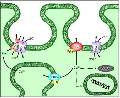
To avoid excessive accumulation of [Ca2+]i, which would be harmful for the cell, the exchanges of Ca2+ between the intracellular and extracellular medium or the ER occur through key proteins as plasma membrane calcium ATPase (PMCA), Na+/ Ca2+ exchanger (NCX) and sarcoplasmic/endoplasmic reticulum calcium ATPase (SERCA) (fig.1). The two main mechanisms that the cell uses to export Ca2+ to the extracellular medium are PMCA pump and the NCX. The PMCA pumps cytosolic Ca2+ into the extracellular medium by the hydrolysis of ATP. The PMCA pumps were discovered in red blood cell membranes by Schatzmann (SCHATZMANN HJ, 1966). NCX exchanger functions as an electrogenic gradient. The NCX have a low affinity for Ca2+ but they can lower quickly intracellular Ca2+ concentration in exchange for 3 Na+ ions into the cell (BERRIDGE MJ, 2012). NCX, an anti-porter membrane protein utilises the Na+ gradient provided by the Na, K-ATPase (NKA), to remove Ca2+ from cells (fig.1).
In mammals, SERCA is the primary transporter facilitating Ca2+ translocation into the sarcoplasmic reticulum (SR) (BERS DM, 1997). SERCA pumps the Ca2+ from the cytosol to the ER at the expense of ATP hydrolysis (POZZAN T, 1994). Two Ca2+ ions from the cytoplasm bind to SERCA pump. SERCA is then subsequently phosphorylated by ATP at Asp351. This high energy, phosphorylated form of SERCA translocates the Ca2+ ions into the SR lumen (Fig.1). The rate at which SERCA moves Ca2+ across the SR membrane can be controlled by the regulatory protein phospholamban (PLB/PLN). A decrease in the ER Ca2+ concentration resulting from SERCA pump inhibition is considered as a major cellular stress signal (ZHANG C, 2016). Thapsigargin (Tg) is a sesquiterpene lactone that can
induce calcium release and is by far the most widely used and efficient irreversible inhibitor of SERCA pump (WOOTTON LL, 2006). At low concentrations, Tg does not inhibit ATP-dependent Ca2+ pumps such as PMCA pumps. However, Tg is selective for different isoforms of SERCA. Indeed, the action of Tg is 60 times more potent on SERCA1 than on SERCA3 (WOOTTON LL, 2006) .
Figure 1. Calcium homeostasis in smooth muscle
The two families of Ca2+ transporter Ca-ATPase (PMCA) and Na/Ca exchanger (NCX) present in the plasma membrane of smooth muscle cells to efflux calcium. NCX is spatially coupled to the α2 isoform of the Na, K-ATPase (NKA α2) to directly regulate calcium transport. The PMCA is regulated by calmodulin (CaM). NKA α1 is a sodium-potassium antiporter. 2K+ enters into the cell in exchange for 3Na+ through NKA α1. NCX co-localised within caveolae and cholesterol-rich membrane domains with NKA α2 isoforms; PMCA with associated calmodulin (CaM) and NKAα1 isoforms; and SERCA with associated phospholamban (PLN) complexes. Taken from (FLOYD R, 2007)
3. Calcium mobilisation
Extracellular stimuli such as electrical, hormonal, or mechanical stimulation can produce calcium signals in various cell types. In eukaryotic cells, these stimulations cause calcium mobilization that can take place in two separate locations (Fig. 2): (1) Entry of calcium ion
across the PM or (2) its release from intracellular stores such as golgi, the endoplasmic and sarcoplasmic reticulum.
Figure 2. Calcium signalisation in non-excitable cell
Following stimulation of a GqPCR or RTK, phospholipase C β / γ is activated which allows hydrolysis of PIP2 into DAG and IP3. IP3 causes the release of Ca2+ from reserved ER by binding to its receptor channel IP3R. This release is counterbalanced by the action of SERCA pump. STIM1, the calcium concentration sensor of ER, activates ORAI1 channels when the ER reservoir is empty. It is believed that DAG can activate certain TRPC channels by PKC-independent mechanisms. Activation of these channels (TRPC6 and Orai1) allows calcium influx from the extracellular medium, and hence sustains signaling. The calcium release is triggered by binding of a wide variety hormones, neurotransmitters and secretagogues to their target receptors located at the plasma membrane. For example, in non-excitable cells, calcium release is induced following a stimulation of a G protein receptor (GPCR) or a receptor tyrosine kinase (RTK) that activate different types of phospholipase C (PLC), either β or γ. The PLC hydrolyze lipid precursor phosphatidylinositol-4, 5 bisphosphate (PIP2) into diacylglycerol (DAG) and inositol 1, 4, 5-triphosphate (IP3) (Figure 2). The PIP2 breakdown is marked by Ca2+ influx into the cell (PUTNEY JW, 2012). The most widely expressed intracellular Ca2+ channel, IP3 receptor (IP3R), releases Ca2+ from intracellular stores in response to binding of IP3 and Ca2+ (FOSKETT JK, 2007) (TAYLOR CW, 2010). IP3R plays an important role in regulating
intracellular [Ca2+]i levels in resting cells (PATTERSON RL, 2004), and the ER remains the major IP3-sensitive Ca2+ storage (IINO M, 1987). It is believed that DAG remains at the PM and activates protein kinase C (PKC) which can phosphorylate different substrates (LARGE WA, 2009). IP3Rs are believed to interact with many protein including a direct interaction between IP3Rs and TRPC (transient receptor potential canonical) channels is proposed to induce the opening of the TRPC channel (ZHANG Z, 2001) .
The next location in calcium mobilization is the calcium entry into the cell through several channels present on plasma membrane. Eukaryotic cells express a variety of calcium permeable channel at the plasma membrane, which can be classified into VOCs (L- and T-type calcium channels) and voltage independent calcium channels (SOCs, ROCs) (GUIBERT C, 2008). Voltage dependent calcium entry through VOC channels is mostly found in excitable cells (nerve and muscle cells) whereas in non-excitable cells, the main mechanisms allowing calcium to enter the cells are SOCE and receptor-operated calcium entry (ROCE). ROCs are believed to be generated through the breakdown of PIP2 by PLC upon receptor stimulation.
Our major interest in this research project is to study the entrance of Ca2+ in non-excitable cells through SOC and/ ROC channel.
4. Voltage Independent Calcium Entry (VICC)
4.1 Store operated calcium entry (SOCE)
Before the term SOCE was invented, Putney proposed the concept of capacitative Ca2+ entry (CCE) (PUTNEY JR., 1986). It was believed that Ca2+ enters the cytoplasm from outside the cell by traversing the ER (the “capacitor”). Later the term ‘‘capacitative Ca2+ entry’’ (CCE) was renamed to “store-operated calcium entry (SOCE)” which describe the phenomenon that a reduction in [Ca2+]i within the lumen of the Ca2+ storing organelles sends a signal to the PM Ca2+ channels. This signalling leads to the activation of SOC channels that causes calcium influx across the PM. The coupling phenomenon between ER
Ca2+ concentration and the activation of membrane Ca2+ entry channels results in refilling the Ca2+ storing organelles, such as ER (CASTEELS R, 1981). The SOC is activated for calcium entry only when there is ER calcium store depletion rather than the production of IP3 or agonist – receptor occupancy. Many experiments using a variety of drugs such as, Tg or cyclopiazonic acid (CPA) causes the depletion of internal Ca2+ stores. Both Tg and CPA mobilize Ca2+ within intact cells by inhibiting SERCA pump. Subsequently SOC is activated. SOCs are also activated by store depletion via as a consequence of IP3-induced Ca2+ release through IP3 receptors in the ER membrane (PAREKH AB, 2005).
Work involved in understanding how ER Ca2+ content is transmitted to the PM to regulate SOC channel opening pointed out involvement of a multi-domain, ER resident, Ca2+ binding protein named STIM1 (LIOU J , 2005).
4.1.1 STIM protein
STIM, which stand for stromal interaction molecules, acts as a calcium sensor for SOC influx. Scientific evidence suggests that STIM and Orai proteins might act as the Ca2+ sensor in the intracellular Ca2+ store and the putative calcium release-activated calcium channel (CRAC) at the PM, respectively (TAYLOR, 2006). SOC influx is the major mechanism for Ca2+ entry in many non-excitable cell types and STIM1 proteins have been identified as essential components in SOC influx along with Orai1. Orai1 is a calcium selective ion channel encoded by the Orai1 gene in humans (FESKE S, 2006). In addition to Orai1, mammalian cells express two additional homologous genes, Orai2 and Orai3. Thus STIM1-Orai1 is believed to provide precise local Ca2+ signals that involve multiple long-term cellular responses (ORITANI K, 1996). In that way, STIM proteins are responsible for the homeostatic control of cellular Ca2+ levels. STIM proteins are localized mainly at the ER and to a lesser extent to the PM.
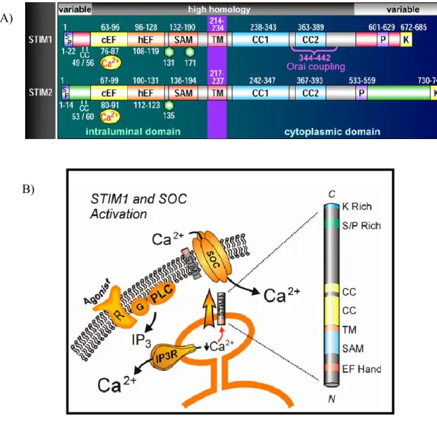
There are two different iso-forms of STIM protein that come from two distinct genes. STIM1 is a protein of 685 amino acids with a molecular weight between 80 and 90 kDa. STIM2 is a protein of 746 amino acids with a molecular weight between 105 and 115 kDa. STIM1 and STIM2 are highly homologous through 85% of their length, with the notable exception of high variability in their amino-terminal and carboxy-terminal segments. STIM proteins have a single trans membrane spanning segment with its NH2 terminus oriented toward the lumen of the ER (fig. 3). The N-terminus contain Ca2+ binding EF hand domain that predicts to lie within the ER lumen (ROOS J, 2005). The adjacent domain of EF-hand is the sterile motif (SAM) which is present near the predicted trans-membrane domain. The STIM proteins also contain protein–protein interaction domains, notably coiled-coiled domains (CC-1 and CC-2) in the cytoplasm near the predicted trans-membrane domain. The cytoplasmic C-terminal domain containing the CAD (CRAC activation domain also known as SOAR-STIM Orai activating region) that allows STIM proteins to bind and activate Orai1. Near the coiled-coil domains at C- terminal, there are a proline-rich domaine (P) and polybasic-rich domain (K) (fig. 3A). STIM1 and STIM2 are expressed ubiquitously throughout cell types in vertebrates (WILLIAMS RT, 2001). In most tissues STIM1 levels are higher than STIM2 levels, but an exception being brain and dendritic cells (BANDYOPADHYAY BC, 2011). At the cellular level, STIM1 is predominantly localized in the ER and in small proportion to the PM (MANJI SS, 2000). Until today, many scientific studies are still under progress to characterize exactly their localization, mode of activation and cellular function.
An important difference between the two isoforms of STIM (STIM1 and STIM2) is in their EF hand domain structures which bind Ca2+. The dissociation constant at equilibrium (Kd) of STIM1 (luminal EF-hand–SAM domain) for Ca2+ is about 200 μM whereas it is about 400 μM for STIM2. This Kd difference is presumably due to the differences in EF-hand domain structures and their affinity for Ca2+. When the luminal Ca2+ levels falls, Ca2+ dissociate from the canonical EF‑hand domain of STIM1 and activate Orai1 (fig. 3) (SOBOLOFF J , 2012).
The cytoplasmic C-terminal domain of STIM protein contains a small active fragment known as the STIM-Orai activating region (SOAR) or CRAC activation domain (CAD). SOAR or CAD is the minimal sequence necessary for interaction with and activation of Orai channels at the PM (PARK CY, 2009) (YUAN JP, 2009). Since STIM2 protein differs subtly from STIM1 in its N-terminal domain, thus there are differences in the rates of unfolding of the luminal N-terminal domain. As a result, there are distinct rates of activation of STIM1 and STIM2 in response to store depletion (SOBOLOFF J, 2012). The interaction and ability to gate Orai1 channels is lower for STIM2. Hence STIM2 responds slower than STIM1 and produces low but sustained Ca2+ entry via Orai1 channels (ZHOU Y, 2009). Experiment carried out by substitution of a single phenylalanine residue in the SOAR1 domain of STIM1 with a leucine residue found in SOAR2 domain of STIM2 prevents STIM1 to bind and activate both Orai channel, indicating that there is a physiologically important distinction between STIM1 and STIM2. STIM1 is a full Orai1-agonist. However leucine in STIM2 endows it as partial agonist, which is believed to be critical for limiting Orai1 activation by STIM2 (WANG X, 2014).
4.1.3 Mechanism of activation of STIM1 and Orai1 4.1.3.1 Sensing of low Ca2+ level in ER:
The decrease in [Ca2+]i in the ER is detected by STIMl protein. The EF-hand domain of STIM1 could function as Ca2+ sensor in the ER (ROOS J, 2005). Indeed, the luminal N- terminus of STIM1 includes two EF-hand motifs 1) a canonical cEF-hand (LIOU J, 2005) (ZHANG S, 2005) (13,25) and 2) a hEF-hand that does not bind with Ca2+ (fig. 3A) (STATHOPULOS PB, 2008). cEF and hEF hands associate tightly with the adjoining five-helix SAM domain. With Ca2+ bound to the dual EF-hand and SAM domain (EF-SAM), a tight, stable STIM1 EF-SAM configuration is formed. When Ca2+ dissociates, the EF-SAM domain unfolds and destabilizes (STATHOPULOS PB, 2008) and leads to rapid oligomerization of the EF-SAM regions. This results in the initial response of STIM1 to deplete ER Ca2+. Mutations in the EF-hand domain of STIM1 reduce its Ca2+ affinity and constitutively activate SOC entry (YEROMIN AV, 2006). The C-terminal cytosolic domain of Orai1 takes part in the interactions within a leucine-rich, acidic coiled-coil motif
of STIM1 protein as experimentally shown by FRET and patch-clamp studies (CALLOWAY N, 2010) (PARK CY, 2009).
4.1.3.2 Formation of puncta:
In physiologic conditions, STIM1 normally appears to be uniformly distributed on the membrane of the ER. On the other hand, Orai1 is a tetra spanning membrane protein localized at the PM and appears to be the functional store-operated channel moiety of STIM1 (PRAKRIYA M, 2006). Upon store emptying, it was observed that STIM1 undergoes a profound redistribution within the cell and leads to the appearance of densely labeled puncta, an aggregated region of ER STIM proteins (LIOU J, 2005). These regions are pre-existing ER-PM junctional domains (WU MM, 2006) within which accumulated STIM proteins capture and activate Orai channels. It requires two STIM1 monomers per Orai1 subunit to achieve optimal SOC channel activity. Interestingly, the puncta formation indicates that STIM1 puncta dissociate and return to an homogenous distribution on the ER when calcium enters the cells following SOCE activation (LIOU J , 2005).
4.1.3.3 Activation of SOCE:
These junctional ER-PM coupling domains are the functional sites of store-operated Ca2+ entry (MA HT, 2002) (BAUTISTA DM, 2004). Recently, it was found that an ER-resident protein, STIM-activating enhancer STIMATE (TMEM110) facilitates and enhances STIM1 puncta formation at ER-PM junctions (JING J, 2015).
Several scientific studies have shown that Orai1 at the PM interacts with STIMl on the ER, causing calcium entry into the cell. However, there are controversies in regard to the molecular mechanism and stoichiometry of the coupling process between these two proteins.
Figure 3. Domain of STIM1 and STIM2 protein and STIM1 in SOC activation
A) STIM protein domain structures. Comparison of STIM1 and STIM2 domain, including signal peptides (SP), a pair of highly conserved cysteines (CC), canonical cEF- and hEF-hands, SAMs, Asn-linked glycosylation sites (hexagons), TMs, coiled-coil regions (CC1 and CC2), proline-rich domains (P), and poly basic-rich domains (K). The minimal region of STIM1 known to be required for coupling to Orai1 is shown (positions 344–442). Taken from (DENG X, 2009)
B) The structure of STIM1 includes an EF hand and SAM domain NH2-terminal to a single trans-membrane (TM) domain; it is believed that, EF hand in particular may be involved in sensing ER Ca2+ levels, or in Ca2+ regulation at the plasma membrane. COOH-terminal to the TM domain is two coiled-coil (CC) domains, and a serine/proline-rich (S/P Rich) and lysine (K Rich) domain. Taken from (PAREKH AB, 2005).
A)
4.2. Receptor operated calcium entry (ROCE)
In non-excitable cells, the most studied pathway for calcium entry is SOC entry pathway or CRAC (Calcium Release Activated Calcium Channel) pathway. However, deletion of Orai or STIM proteins suggest that there are other pathways of calcium entry and other PM calcium channels that might be functionally involved (VIG M, 2008). The term receptor-operated channel (ROC) was simultaneously proposed, by Bolton (1979) and Van Breemen (1978) to define any plasmalemmal channel that is opened as a result of agonist binding to its receptor. ROC channels are thought to operate independently of any previous change in membrane potential. In smooth muscle cells (SMC), agonists may open ROC channels with significant permeability to Ca2+ ions as a result of second messengers produced following G alpha-protein activation of PLC (MCFADDEN I, 2002). In SMC and other cell types such as hepatocytes and platelets, ATP, ADP, acetylcholine, histamine, vasopressin, neurokinin A, and substance P have all been shown to activate receptor operated cation influx (ROSADO JA, 2000). ROCs have been described in both visceral (intestine, uterus) and vascular (aorta, ear artery, portal vein) smooth muscles and are activated by a large variety of agonists.
TRPC channels constitute a group of Ca2+ permeable cation channels that are activated in response to receptor-regulated PIP2 hydrolysis. PIP2 and DAG are antagonist to the TRPC binding site. Hence it is hypothesized that a simultaneous increase in DAG levels and decrease in PIP2 concentration is necessary for optimal activation of the channel (See section 6.1 for details). The TRPC subfamily consists of seven members (TRPC1–TRPC7). All of them are not responsible for ROC entry. It was found that, only TRPC3, 6, 7 are usually considered to be involved in ROC entry because they are sensitive to DAG and other components of GPCR signalling pathways (HOFMANN T, 1999). The mechanisms that regulate the activity of the TRPC are not well established and are the subject of several studies.
To distinguish between ROC and SOC channels: ROC channel corresponds to a Ca2+ permeable non-selective cation channel that is opened by an agonist but is not affected by
depleting intracellular Ca2+ stores whereas, SOC channel can be activated without any agonist acting on membrane receptors.
4.2.1 TRPC family
The first TRP gene was described in Drosophila (MONTELL C, 1989). Thus 28 TRP have been identified and grouped into seven families. These are grouped into following: TRPC ("C" for canonical), TRPV ("V" for vanilloid), TRPM ("M" for melastatin), TRPN, TRPA, TRPP ("P" for polycystic) and TRPML ("ML" for mucolipin). For TRPC, there are 4 sub-families. The Drosophila trp channel exhibits the greatest homology to the TRPCs. TRPs also contain sites for direct phosphorylation by serine/threonine and tyrosine kinases, although their role remains to be fully elucidated. The different TRPC families are expressed in mammalian cells.
4.2.2 Regulation and binding of TRPCs
As stated earlier, all channels of the TRPC family are activated by stimulation of receptors such as GPCRs and RTKs, that activate different isoforms of PLC (CLAPHAM DE, 2003). In general, members of the closely related subgroup TRPC3/6/7 can be activated by DAG, suggesting that DAG is the PLC-derived product mediating their physiological activation. In contrast, TRPC1, 4 and 5, which are also stimulated by receptor mediated PLC activation, are completely unresponsive to DAG (VENKATACHALAM K, 2002). However, the mechanism by which PLC-mediated TRPC stimulation remains controversial. Furthermore, there is evidence that TRPC1, TRPC4/5 and TRPC3/6/7 can function as ROC that are mostly insensitive to store depletion (LINTSCHINGER B, 2000); (TREBAK M, 2002 ) (PLANT TD, 2003).
4.2.3 Structural characteristic of TRPCs
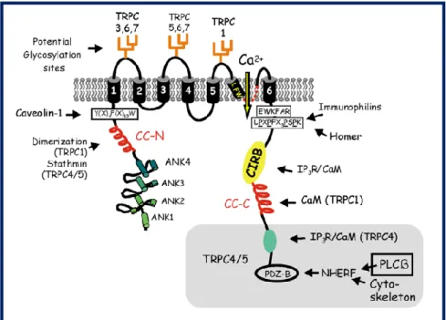
The common region, domain, motif of TRPC family includes the cytoplasmic N- and C-termini of TRPCs that are separated by six predicted trans-membrane domains (TM1–
TM6). The N-terminus of TRPC family includes three to four ankyrin repeats (ANK1–4), a predicted coiled coil region (CC) and a putative caveolin binding region(fig. 4) (ENGELKE M, 2002) (LEPAGE PK, 2007). A similar motif to Caveolin 1 (Y(x)4F(X)13W) is conserved in all members of the TRPC family located in the cytosolic N-terminus adjacent to the first transmembrane domain. The cytoplasmic C-terminus includes the TRP signature motif (EWKFAR) that is non-selectively permeable to cations, a highly conserved proline rich motif (LPXPFXXXPSPK), the CIRB (calmodulin/IP3 receptor binding) region which is a calmodulin binding region, and a predicted coiled-coil region (CC). There is a pore region (LFW, pore motif) between TM5 and TM6 of TRPC family that is presumed to take part in Ca2+ entry. The N- and C- termini are the sites where protein–protein interactions take place which are necessary for mediating channel trafficking, anchoring, localization, gating, and functional regulation (Fig. 4). However, there is uncertainty as to the exact assignment of the first two trans-membrane segment (VANNIER B, 1998) (DOHKE Y, 2004).
TRPC4 and TRPC5 contain a unique extended C-terminus containing a PDZ binding motif responsible for the interaction with the adaptor protein, NHERF, possibly linking the channels to PLCβ and the cytoskeleton (fig.4). The first three extracellular loops can be N-glycosylated. Different levels of constitutive activity within members of the TRPC family have been observed due to different patterns of N-glycosylation (VAZQUEZ G, 2004). The C-terminus proline-rich motif is presumed to interact with Homer (for TRPC1) and/or immunophilins. Moreover, there is another IP3/CaM domain at the C- terminus. The TRPC4–IP3R interaction is believe to inhibit by CaM in the presence of Ca2+ (BRAZER SC, 2003).
Figure 4 Structure of TRPC channels
The region shaded in gray at the extreme C- terminus is specific for TRPC 4 and 5. The C- terminus contains an additional binding region IP3/CaM specific to TRPC4. In CC-N terminus, the CC region of stathmin is presumed to responsible for the interaction. This interaction seemed to be specific for TRPC4 and TRPC5. A proline-rich motif (LPXPFXXXPSPK) is thought to be responsible for interaction with Homer (for TRPC1), LFW, amino acid motif conserved in the putative pore region of all TRPCs. Taken from (VAZQUEZ G, 2004).
4.2.4 Phosphorylation of TRPC channels
TRPCs contain sites for direct phosphorylation by serine / threonine and tyrosine kinases. TRPC3 is highly expressed in the brain, as well as in smooth and cardiac muscle cells (CLAPHAM DE, 2007) (HOFMANN T, 2000). TRPC3 are receptor operated, DAG-stimulated channel (HOFMANN T, 1999), which requires src mediated tyrosine phosphorylation for its availability for activation whereas Protein kinase G (PKG) was reported to have a role in negative regulation of human TRPC3 activity (KWAN HY, 2004). TRPC1 must be phosphorylated by PKCα in order to be active, which is needed in calcium entry (AHMMED GU, 2004). This phosphorylation is essential in the activation of SOCE dependent of TRPC1 by PIP2 in the portal vein of rabbit myocytes or in the myocytes of rabbit coronary artery (SALEH S, 2009). TRPC4 is phosphorylated on
tyrosine residues Y959 and Thr972 in its C-tail by tyrosine kinase of the Src family (STK), Fyn kinase (ODELL AF, 2005). The calcium entry is reduced in cells treated with inhibitors of tyrosine kinase of the Src family (STK), or in the cells expressing the mutants Y959 / 972F of TRPC4 (ODELL AF, 2005). PKC can also phosphorylate TRPC5 on its Thr972 site resulting in stimulation of muscarinic receptors upon optimal [Ca+]i levels, which desensitize the calcium channel (TRPC5). Inhibition of PKC by the specific inhibitor bisindolylmaleimide I (GF1), by inhibitor peptides, or by the expression of the TRPC5 T972A mutant, prevent or reduces TRPC5 desensitization (ZHU M, 2005). The TRPC6 interacts with Fyn that is independent of phosphorylation and is mediated by the SH2 domain of Fyn and the NH2-terminal cytoplasmic domain of TRPC6. Fyn seems to modulate the TRPC6 channel properties activated by DAG (HISATSUNE CY, 2004). According to Hisatsune et al. (HASSOCK SR, 2002) COS-7 cells overexpressing both Fyn and TRPC6 showed tyrosine phosphorylated TRPC6 following EGF treatment. Fyn is also able to phosphorylate TRPC6 in an in vitro kinase assay. Finally, Fyn and TRPC6 were shown to interact through the SH2 domain of Fyn and the channel N- terminal domain of TRPC6 in a manner independent of channel Phosphorylation. These findings contrast with those from Hassock et al. who observed no tyrosine phosphorylation of endogenous TRPC6 in human platelets. Hassock et al. reported that in platelets, endogenous TRPC6 and TRPC1 did not undergo tyrosine phosphorylation. The Phosphorylation of TRPC channel is important for TRPC channel activation and calcium entry.
4.3 TRPC6: a potential therapeutic target
Many pathophysiological situations like Focal segmental glomerulosclerosis (FSGS), idiopathic pulmonary arterial hypertension, and liver cancer are thought to arise from the imbalanced regulation of TRPC6 expression. Thus it is important to understand the mechanisms responsible for the regulation of TRPC6. FSGS is a channelopathy associated with TRPC6 (WINN MP, 2005) (REISER J, 2005). The symptoms of FSGS include proteinuria, hypertension, renal insufficiency, and eventual kidney failure. FSGS carries a missense mutation in the TRPC6 gene. In FSGS, TRPC6 mediated calcium signal is enhanced in response to agonists such as angiotensin II that promote kidney injury and proteinuria. The expression of TRPC6 has been reported to be higher in pulmonary artery
smooth muscle cells in Idiopathic pulmonary arterial hypertension (YU Y , 2004) (YU Y , 2009). An up-regulation of TRPC6 in hepatocytes has also been linked to liver cancer (BOUSTANY EL, 2008).
TRPC6, a member of the TRPC family, allows Ca2+ to enter into cells from the extracellular medium after stimulation by the PLC/ IP3 pathway. TRPC6 is known to be expressed in different types of cells such as vascular smooth muscle cells. The A7r5 cells are smooth muscle of rat thoracic aorta cell line use as a cellular model to study the function and activation of TRPC6 (JUNG S, 2002). It has been shown that the high expression of TRPC6 was essential for calcium entry induced by vasopressin in A7r5 cells (JUNG S, 2002). Since TRPC6 is highly expressed in vascular smooth muscle cells, many studies have investigated the potential involvement of TRPC6 in pulmonary hypertension.
4.3.1 Mechanism of activation of TRPC6 channel
To date, the complete mechanism of activation of TRPC6 is still unknown. Many hypothesis have been proposed for TRPC6 channel activation and thereby calcium entry into the cell.
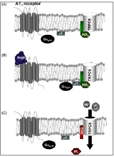
LARGE (LARGE WA, 2009) proposed a scheme for native TRPC6 activation in vascular myocytes (Fig. 5). According to his article, PIP2 is bound to TRPC6 in the resting state and GPCR stimulation of PLC cleaves PIP2 which simultaneously generates DAG and removes PIP2 from TRPC6. They proposed that DAG may be the ligand that causes channel opening and that DAG and PIP2 bind to the same site on calmodulin-binding domain within TRPC6 proteins. DAG and PIP2 have antagonist effects on TRPC6 activity and this model agrees with the hypothesis that a simultaneous increase in DAG levels and decrease in PIP2 concentrations is necessary for optimal activation of TRPC6 (HARDIE RC, 2003). Also, depletion of PIP2 using PI(3)K inhibitor, wortmannin and LY294002 only cause transient TRPC6 stimulation, indicating that simple removal of PIP2 from TRPC6 is not sufficient for TRPC6 channel (MONET M, 2013). The subsequent inhibition of PIP2 in continual presence of wortmannin constitutively generates DAG, which is necessary for channel
opening. It is therefore hypothesized that simultaneous PIP2 dissociation from TRPC6 and DAG association to this PIP2 site or similar site on TRPC6 could maximize TRPC6 activation and enhanced calcium entry. This also point out that DAG is necessary for TRPC6 channel opening (fig. 5).
Figure 5 Physiological activation mechanism of TRPC6 by AngII in native vascularmyocytes.
(A) At rest endogenous PIP2 is bound to TRPC6 proteins expressed at the plasma membrane. (B) Binding of Ang II to AT1 receptors activates G q/11 G-protein subunits which stimulate PLC activity and subsequent hydrolysis of PIP2 bound to TRPC6 proteins. (C) Optimal activation of TRPC6 channels occurs through both dissociation of PIP2 from the channel and association of DAG to the same or a similar site on the channel protein. Taken from (LARGE WA, 2009)
4.3.2 Trafficking of TRPC6 channel
Stimulation of cells regulates the PM expression of TRPC channels which enhanced Ca2+ influx. In HEK-293 cells stably expressing TRPC6, stimulation of muscarinic receptors triggers TRPC6 insertion into the plasma membrane, which is retained there as long as the stimulus is present (CAYOUETTE S, 2004). Our laboratory previously studied the trafficking of TRPC6 channels and showed that the rab GTPases (peripheral membrane protein) Rab9 and Rab11 regulate the intracellular trafficking of TRPC6 in Hela cells (CAYOUETTE S, 2010). Rab9 isknown to participate in the trafficking of proteins to the late endosomes and trans-golgi network (TGN) (LOMBARDI D, 1993). TRPC6 has been shown to interact and co-localize with Rab9 in vesicular structures (CAYOUETTE S, 2010). Over-expression of the dominant mutant of Rab9 resulted in an augmentation of TRPC6 levels at the PM and consequently, increased the Ca2+ influx upon carbachol (CCh) stimulation. Over-expression of Rab11, which is involved in recycling endosomes (REF), also increaseTRPC6 channel expression at the cell surface as well as Ca2+ entry (CAYOUETTE S, 2010). These results suggest that the intracellular trafficking of TRPC6 is regulated through the slow endosomal recycling pathway (CAYOUETTE S, 2010).
PI(3)K kinase localized in cytoplasm, phosphorylates PIP2 forming phosphatidylinositol (3,4,5)-trisphosphate (PIP3) (FOSTER FM, 2003). The inhibition of PI(3)K by treatment with PIK-93, LY294002 or Wortmannin reduces TRPC6 translocation to the PM and consequently Ca2+ entry in HEK293 cells stably expressing TRPC6 (T6.11 cells). On the other hand, phosphatase and tensin homolog (PTEN) opposes PI(3)K activity by dephosphorylating the 3-hydroxyl group of PIP3 to form PIP2. Knockdown of PTEN did not affect TRPC6 levels at the cell surface but increased the activity of the channel (MONET M, 2012). This demonstrate that PI(3)K and the phosphatase PTEN control the trafficking and activation of TRPC6 channels.
5. Research project
5.1 Problematic
Several studies have previously shown that the dysregulation of TRPC6 expression is associated with many pathophysiological conditions such as Focal segmental glomerulosclerosis, Idiopathic pulmonary arterial hypertension (WINN MP, 2005) (REISER J, 2005) (HEERINGA SF, 2009) (LEUNG PC, 2006) (SINGH I, 2007) (TAUSEEF M, 2012). Studies in our laboratory demonstrated that some TRPC6 translocates to the plasma membrane after hormonal stimulation (CAYOUETTE S & BOULAY G, 2007). However, the mechanisms involved in the intracellular trafficking and activation of TRPC6 are yet to be fully characterized. According to several previous studies using proteomic analysis (GOEL M, 2005) (BANDYOPADHYAY BC, 2005) many proteins have been identified as TRPC6 interacting partners including: Mixovirus resistance protein (MxA), Ras homolog gene A (RhoA), syntaxin, clathrin and dynamin (DE SOUZA, 2014).
Abnormal activation of the Rho/Rho-kinase pathway has been shown to play a role in diseases such as hypertension and bronchial asthma. Recent studies in our laboratory also found that there is a potential role of Rho kinase in TRPC6 activation (FRANCOEUR et al, unpublished data). These preliminary results showed that when the HEK-TRPC6 cells were treated with a Rho inhibitor, CT04 (Cytoskeleton Inc.), CCh induced calcium entry was increased by 50%. This increased calcium entry was also observed when ROCK (Rho kinase) inhibitors Y-27632 and HA-1077 were used. The Rho kinase can be activated by the small G protein RhoA. RhoA can be activated by the heterotrimeric G proteins Gα12 and Gα13 through the guanosine exchange factors (GEF) p115-RhoGEF, WIDTH or PDZ-RhoGEF (fig.6A). These results suggest a potential link between Rho kinase and TRPC6.
β-arrestin 1 and β-arrestin 2 have also been involve in the activation of RhoA (fig. 6B) (BARNES WG, 2005) (Ma X, 2012). It has been demonstrated that β-arrestin 2 act as a signal mediator in mitogen-activated protein kinase cascades. Angiotensin II type 1A
receptor mediated signaling is dependent on β-arrestin 2 and is mediated by a RhoA/ ROCK/MLCK-dependent pathway (CHRISTINA MG, 2010). β-arrestin 1 has also been shown to be required to activate the small GTPase RhoA leading to the re-organization of stress fibers (RhoA signals through ROCK) following the activation of the angiotensin II type 1A receptor (WILLIAM GB, 2004). This activation has been reported to be due to the association of β-arrestin 1 with the RhoGAP (a GTPase activating Protein), ARHGAP21. This interaction contributed to the inhibition of the RhoGAP activity of ARHGAP21, leading to higher RhoA activity (ANTHONY DF, 2011).
Given that our laboratory showed that RhoA could be involved in the regulation of TRPC6 (ROC), and that Gα12/Gα13 or β-arrestin 1/ β-arrestin 2 are known to be implicated in the activation of RhoA, we propose to verify if Gα12/Gα13 or β-arrestin 1/ β-arrestin 2 proteins are involved in calcium entry through ROC or SOC in non-excitable cells.
5.1.1 The Rho/Rho-kinase mediated pathway
The first Rho gene was cloned as a Ras homologue from Aplysia in 1985, soon followed by the three human homologues RhoA, RhoB, and RhoC (MADAULE P, 1985). These proteins are molecular switches which cycle between an inactive GDP-bound and an active GTP-bound state (Fig. 6). Under its active form, Rho interacts with several effectors such as Rho-kinase (ROCK), protein kinase N (PKN), rhophilin, rhotekin, citron, p140mDia (BISHOP AL, 2000). Rho-kinase, one of the best characterized Rho effectors, exists in two isoforms: ROKα (also called ROCK2) and p160ROCK (also known as ROKβ or ROCK1). Rho-kinases are serine/threonine protein kinases that contain an amino-terminal catalytic kinase domain, a central coiled-coil-domain that binds Rho-GTP, and a C-terminal pleckstrin homology domain which is split by a cystein-rich region (LEUNG T, 1995), (MATSUI T, 1996). One of the main substrates of Rho-kinase is myosin light chain phosphatase. Smooth-muscle myosin light-chain phosphatase (SMPP-1M) is a type I serine/threonine phosphatise. SMPP-1M can be inhibited indirectly by a phosphatase inhibitor CPI-17 (ETO M, 1995), (SOMLYO AP, 2000). CPI-17 is a phosphorylation dependent inhibitor protein of smooth muscle myosin phosphatase. In addition, CPI-17 independent phosphorylation of Rho-kinase can directly phosphorylate MLC20 (AMANO
M, 1996) and causes the smooth muscle contraction. The smooth muscle relaxation is opposed by the inhibition of SMPP-1M by CPI-17. Recently it has been shown that Rho might also cause phosphorylation of CPI-17 via its effectors protein kinase N (PKN) (HAMAGUCHI T, 2000) and Rho-kinase (KOYAMA M, 2000).
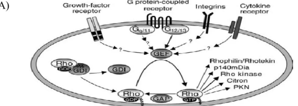
Figure 6 Regulation of Rho activity by Gα12/Gα13 or β-arrestin 1/ β-arrestin 2
A) Various classes of signaling receptors have been shown to activate Rho through the regulation guanine nucleotide exchange factor (GEF) proteins which catalyze the exchange of GDP for GTP on Rho GTPases. Two negative regulators of Rho activity, GTPase-activation proteins (GAP) and GDP-dissociation inhibitors (GDI) may also be regulated by receptor-mediated signals. Effector proteins of Rho are PKN (Protein kinase N); p140mDia (Mammalian homolog of Drosophila diaphanous); citron (citron-kinase) (WETTSCHURECK N, 2002).
B) β-arrestin-dependent signaling to RhoA or ERK1/2. β-arrestin 1 and Gαq/11 is involved in activation of Rho. Activation of RhoA leads to stress fiber formation via the downstream effector of RhoA, ROCK. Both arrestins and heterotrimeric G protein activation is required to activate Rho. Conversely, β-arrestin 2 signals to the MAPK component ERK1/2 independent of the heterotrimeric G protein pathway to ERK1/2 via PKC. Therefore, RhoA activation depends on heterotrimeric G protein and β-arrestin 1, however and ERK1/2 activation is independent on heterotrimeric G protein-independent and depends on β-arrestin 2.
A)
5.1.2 Regulation of Rho/Rho-kinase
The activity of Rho itself is regulated by three groups of proteins, which are found ubiquitously (Fig. 6): GTPase activating proteins (GAPs) which facilitate the inactivation of active GTP-bound Rho by increasing its intrinsic GTPase , GDP dissociation inhibitors (GDIs) which inhibit the dissociation of GDP and indirectly prevent Rho to translocate on the membranes (OLOFSSON B, 1999), and Rho-specific guanine nucleotide exchange factors (RhoGEFs) which facilitate the activation of Rho by promoting the dissociation of GDP and subsequent binding of GTP. Currently RhoGEF proteins are regarded as the main regulators of Rho activity.
The activity of Rho can also be regulated by numerous GPCR such as Gq/11, and it is well established that G-proteins of the Gα12 family, Gα12 and Gα13, which appear to be ubiquitously expressed, are involved in these GPCR activation of Rho (BUHL AM, 1995 ). A group of RhoGEFs have been identified to bind the activated α-subunit of Gα12/ Gα13, including p115RhoGEF/lsc, PDZ-RhoGEF, and LARG(fig.6). However, other receptors such as growth factor receptor, cytokine receptor, integrins are believed to take part in Rho activation through the activation of GEF. However, these receptors involvement in GEF activation is not confirmed yet (WETTSCHURECK N, 2002).
5.1.3 β-arrestin mediated signalling
β-arrestin 1 and β-arrestin 2 (non-visual arrestins, also known as arrestin 2 and arrestin 3), are ubiquitously expressed in most tissues and play an important role in regulating signal transduction by numerous GPCRs (ATTRAMADAL H, 1992; REITER E, 2006). Both of these arrestin isoform are 78% sequence homologous (ATTRAMADAL H, 1992). arrestin 1 and arrestin 2 accumulate in the cytoplasm following their overexpression. β-arrestin 2 has a leucine rich nuclear export sequence located on its C terminus, that is absent in β-arrestin 1. Hence, β-arrestin 1 but not β-arrestin 2, accumulates in the nucleus, and shows differences in nucleocytoplasmic shuttling (SCOTT MG, 2002). Both the β-arrestin 1 (418 amino acids) and β-β-arrestin 2 (410 amino acids) have nuclear localization sequences on their N termini. β-arrestin 1 and β-arrestin 2 can reciprocally regulate
signaling at certain receptors. For example, siRNA knockdown of β-arrestin 2 attenuates ERK signaling, whereas knockdown of β-arrestin 1 potentiates ERK signaling (AHN S, 2004). However, at other receptors such as the β2AR and the type 1 parathyroid hormone receptor (PTH1R), knockdown of either β-arrestin 1 or β-arrestin 2 decreases ERK signaling (SHENOY SK, 2006).
The classical role of β-arrestins includes their role in GPCR signaling and in signaling on endosome through MAPK activation. β-arrestins are belived to quench the GPCR signaling by sterically inhibiting the G protein interaction at the second and third intracellular loops of a GPCR (DEGRAFF JL, 2002) (MARION S, 2006). This steric hindrance uncouples GPCRs from the G protein signal transduction process (FERGUSON SS, 1996). Association of β-arrestins with the phosphorylated domains of the 7TMR trigger conformational changes in the GPCR at PM that allows β-arrestins to recruit clathrin and adaptor protein (AP-2). However, β-arrestins require C-terminal dephosphorylation for targeting internalized receptors to clathrin. This leading to the co-localization of the GPCR and β-arrestin in punctuated clathrin-coated pits at the cell surface or near. As a result, receptor desensitization via downstream kinases (MAPK) takes place (REITER E, 2012), (SHUKLA AK, 2011). Thus, another role of β-arrestins being as adaptor proteins that facilitate the activation of mitogen-activated protein kinase cascades (MAPK cascades). MAPK cascade relays signals from the PM to the nucleus. β-arrestins scaffold the MAPK signaling molecules (ERK), leading to phosphorylation and activation of extracellular signal mediated kinase (ERK 1/2) (DEWIRE, 2007). Ubiquitination of β-arrestin – GPCR complex prolongs the MAPK activity. Once internalized, the receptor continues for tubulovesicular early endosomes, where receptors are sorted to either recycling endosomes or multivesicular late endosomes. GPCRs in early endosome return to the plasma membrane. GPCRs in multivesicular late endosomes, traffic receptors to lysosomes for degradation (KANG D, 2014). In brief, the key roles of β-arrestins are to 1) inhibit the coupling of GPCR to the G-proteins and 2) recruit AP2 and clathrin to internalize the recptor in clathrin-coated vesicles, and 3) endosomal signaling (MAPK cascade).
β-arrestin 1 and β-arrestin 2 has also been involve in the activation of RhoA (WILLIAM GB, 2004) (CHRISTINA MG, 2010). It has been demonstrated that β-arrestin 2 act as a signal mediator in mitogen-activated protein kinase cascades. Angiotensin II type 1A receptor mediated signaling is dependent on β-arrestin 2 and is mediated by a RhoA/ ROCK/MLCK-dependent pathway (CHRISTINA MG, 2010). β-arrestin 1 has also been shown to be required to activate the small GTPase RhoA leading to the re-organization of stress fibers (RhoA signals through ROCK) following the activation of the angiotensin II type 1A receptor (WILLIAM GB, 2004). This activation has been reported to be due to the association of β-arrestin 1 with the RhoGAP (a GTPase activating Protein), ARHGAP21. This interaction contributed to the inhibition of the RhoGAP activity of ARHGAP21, leading to higher RhoA activity (ANTHONY DF, 2011). Moreover, β-arrestin 2 involve in translocation of p115RhoGEF, a regulator of RhoA to the PM from the cytosol. Hence, β-arrestins are believed to serve for diverse upstream signals to activate RhoA. For example, angiotensin II type 1A receptor-induced activation of RhoA require both Gq and β-arrestin 1(fig.xx) (BARNES WG, 2005). However, β-arrestin 2 signals to the MAPK component ERK1/2 independent of the heterotrimeric G protein pathway via PKC (fig.xx). Consequent activation of RhoA promotes the formation of focal adhesions and stress fibers (fig.xx). Overall, it seems that β-arrestins finetune RhoA activity through coordinated regulation of RhoA activators and inactivators.
5.2 Hypothesis
Given that RhoA has been shown to be activated by heterotrimeric G proteins Gα12 and Gα13 or by β-arrestin 1 / β-arrestin 2 proteins, and TRPC6 channel expression at the plasma membrane can be regulated by the RhoA/ROCK pathway (FRANCOEUR et al, unpublished data), we hypothesized that Gα12 / Gα13 and/or β-arrestin 1 / β-arrestin 2 are involved in RhoA activation in ROCE. Since RhoA can be activated by β-arrestins, we assume that β-arrestins can also modulate the SOCE pathway.
5.3 Objective of the study:
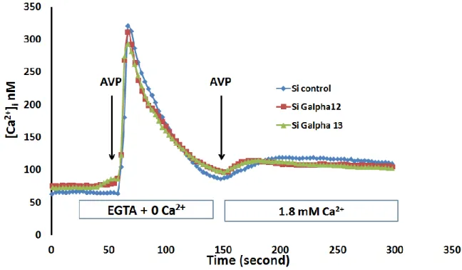
Objective 1: Determine if Gα12 and/or Gα13 protein influence the receptor operated Ca2+ entry through TRPC6 channel in vascular smooth muscle cells (A7r5)
The entrance of calcium via ROC was examined in vascular smooth muscle cells (A7r5) (expressing endogenous TRPC6) in which Gα12 or Gα13 were depleted using siRNA.
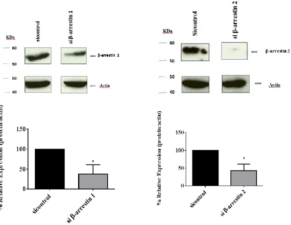
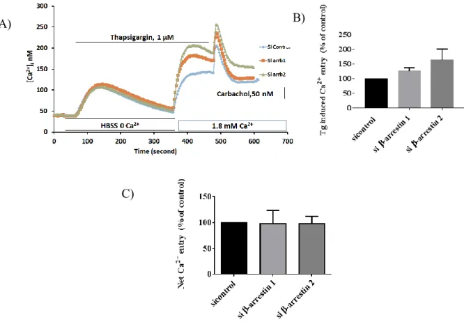
Objective 2: Determine if β-arrestin 1 and β-arrestin 2 influence the receptor operated (TRPC6) or store-operated Ca2+ entry (Orai1) in HEK 293T cells and HEK293 cells stably transfected with TRPC6 (T6.11 cells)
The entrance of calcium via SOCE was examined in HEK 293T cells and HEK293 cells stably transfected with TRPC6 (T6.11 cells) in which β-arrestin 1 or β-arrestin 2 were depleted using siRNA.
Objective 3: Determine if β-arrestin 1 and β-arrestin 2 interact with STIM1 or Orai1 protein to influence the store-operated Ca2+ entry (Orai1) in HEK 293T cells and HEK293 cells stably transfected with TRPC6 (T6.11 cells)
A co-immunoprecipitation assay was performed in HEK293T cells in which β-arrestin 1 was depleted using siRNA. Immunofluorescence assay was performed to examine the distribution of Orai1 or STIM1 protein in β-arrestin 1 and β-arrestin 2 depleted cells
MATERIAL AND METHODS
6. MaterialTable: List of products used
Products Manufacturer
Antibody Anti-Gα12 Santa cruz biotechnology, inc. Antibody Anti-Gα13 Santa cruz biotechnology, inc. Antibody Anti β-arrestin 1 AbCam (Cambridge, MA, USA)
Antibody Anti β-arrestin 2 Cell Signaling Technology (Boston, MA) Antibody anti-ST1M1 (Rabbit
polyclonal)
Millipore (Temecula, CA, USA) Antibody Anti-Actin Millipore (Temecula, CA, USA)
Anti-Myc Santa Cruz Biotechnology (Dallas, TX)
Arginin vasopressin (AVP) Calbiochem (San Diego, CA)
Bradford reagent BioRAD
BSA Sigma-Aldrich
Carbachol (CCh) Calbiochem (San Diego, CA)
DMEM Life Technologies Corporation
EGTA Sigma-Aldrich (St. Louis, MO, USA)
Fetal bovine serum (FBS) Invitrogen (Burlington, ON)
Fura-2 / AM Calbiochem (San Diego, CA)
G418 Invitrogen (Burlington, ON)
Hepes Invitrogen (Burlington, ON)
HRP conjugated sheep anti-mouse IgG
GE Healthcare UK Limited
HRP conjugated donkey anti-rabbit IgG
GE Healthcare UK Limited
L-glutamine Invitrogen (Burlington, ON)
Nitrocellulose membrane Perkin Elmer (Boston,MA)
Opti-MEM I Invitrogen (Burlington, ON)
Protein A-sepharose beads Santa Cruz Biotechnology (Santa Cruz, CA, USA)
Penicillin/streptomycin Invitrogen (Burlington, ON)
Para-formaldehyde (PFA) Electron Microscopy Science (Hatfield, PA)
SiRNA Control (ON-TARGET plus Control Pool)
Dharmacon, Inc. (Lafayette, CO, USA)
SiRNA Arrb1 Dharmacon, Inc. (Lafayette, CO, USA)
SiRNA Arrb2 Dharmacon, Inc. (Lafayette, CO, USA)
SiRNA Gα12 Dharmacon, Inc. (Lafayette, CO, USA)
SiRNA Gα13 Dharmacon, Inc. (Lafayette, CO, USA)
SiRNA Orai1 Dharmacon, Inc. (Lafayette, CO, USA)
Trypsin Invitrogen (Burlington, ON)
Triton X-100 Sigma-Aldrich
Tween 20 Fisher Chemical
Tris HCl Sigma-Aldrich
West Pico Chemiluminescent Reagent Thermo scientific (Rockford, IL)
Unless otherwise stated, all other reagents were from Laboratoire MAT (Quebec City, QC, Canada).