15 min
30 min
45 min
60 min
TAT
TAT-Ras
GAP
317-326Control
TAT
TAT-Ras
GAP
317-326Control
15 min
30 min
45 min
60 min
Trypsin-mediated detachment
Adhesion rate
B
480 min
480 min
C
TAT TAT-RasGAP317-326 ControlCell spreading 30 min after plating
Figure S1
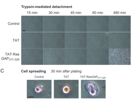
Figure S1. Kinetics of TAT-RasGAP317-326-mediated adhesiveness increase.
A. U2OS cells were subjected to an adhesion assay for 30 minutes in presence of the indicated concentrations of TAT and TAT-RasGAP317-326 (the statistical significance of the observed
differences was assessed by one-way ANOVA).
B. Representative images of the adhesion assay performed in Figure 1E. Scale bar: 100 µm. C. Higher magnification of cells 30 minutes after plating. Scale bar: 40 µm.
A
0 200 400 500 300 0 5 10 15 20 Peptide concentration [µM] TAT TAT-RasGAP317-326Number of cells per mm
2 100
*
*
*
*
: TAT vs. TAT-RasGAP317-3260 75 50 100 M21 M21L % adherent cells
Control w/o trypsin Control with trypsin
TAT-RasGAP317-326 with trypsin
TAT with trypsin
25 0.93 0.32 +/ -0.44 0.32 +/ -0.15 0.02 +/ -0.41 0.27 +/ -integrin-αV +
-B
integrin-β1 1.13.105 integrin-α3 4.45.104 integrin-αV 2.72.104 integrin-β5 3.00.104 integrin-α2 1.34.104 integrin-α6 7.38.103 integrin-α5 2.97.103 integrin-β2 < 5.102 integrin-α9 < 5.102 < 5.102 integrin-β4 < 5.102 integrin-α4 integrin-β3 N/A integrin-β6-8 integrin-α1 integrin-α7-11 N/A N/A N/A Copies/cell ProteinD
48h 24h 48h 72h -48h siGFP si-Integrin-β1 130 130 kDa HeLa 0.11 0.11 +/ -0.10 0.20 +/ -0.24 0.26 +/ -0.16 0.10 +/ -0 75 50 100 25 % adherent cellssiGFP siIntegrin-β1HeLa
Integrin-β1 RasGAP
C
A
α1β1 α2β1 α3β1 α4β1 α5β1 α6β1 α7β1 α8β1 α9β1 α10β1 α11β1 αVβ1 αXβ2 αMβ2 αLβ2 αDβ2 αVβ3 α6β4 αVβ5 αVβ6 αEβ7 αVβ8 α4β7 β2 β1 β3 β4 β5 β6 β7 β8 α2β3 α integrin subunits 48h- 48h 24h 48h 72h siGFP siCD44 130 90 kDa CD44 RasGAP HeLa 1.3 0.6 +/ -5.0 1.9 +/ -1.7 2.3 +/ -3.0 1.1 +/ -0 75 50 100 25 % adherent cellssiGFP siCD44HeLa
E
F
0 200 400 600PBS Collagen Fibronectin Laminin
1.5 +/ 3.3 -0.0 +/ 0.0 -0.0 +/ 0.0 -0.0 +/ 0.0 -4.5 +/ 4.1 -0.6 +/ 0.6 -0.0 +/ 0.0 -0.0 +/ 0.0 -0.3 +/ 0.6 -0.0 +/ 0.0
-Number of cells per mm
2
Control w/o trypsin
Control with trypsin TAT-RasGAPTAT with trypsin317-326 with trypsin
PBS Collagen Fibronectin Laminin
TAT TAT-Ras
GAP317-326
Control w/o trypsin Control
With trypsin
Figure S4
Figure S4. Integrins, CD44 and caspases are not required for TAT-RasGAP317-326-mediated adhesion increase. A. The twenty-four known integrin heterodimers are represented (8).
B. Wild-type (M21) or αV-integrin-null (M21L) melanoma cells were treated 8 hours with 20 µM TAT, 20 µM TAT-RasGAP317-326 or left untreated. The cells
were then subjected to a trypsin-mediated detachment assay.
C. HeLa cells were transfected with siRNAs targeted against GFP (siGFP) or β1-integrin (siIntegrin-β1). Forty-eight hours later, the cells were incubated 8 hours with 20 µM TAT, 20 µM TAT-RasGAP317-326 or left untreated and were subjected to a trypsin-mediated detachment assay. The upper panel depicts the
loss of β1-integrin expression after siRNA transfection.
D. Expression levels of the indicated integrin family members in U2OS cells [data taken from (9)].
E. CD44 was silenced in HeLa cells with siRNAs. Forty-eight hours later, the cells were incubated 8 hours with 20 µM TAT, 20 µM TAT-RasGAP317-326 or left
untreated. The cells were then subjected to a trypsin-mediated detachment assay. The upper panel depicts the loss of CD44 expression after siRNA transfection.
F. U2OS cells were grown overnight on bacteriological Petri dishes that had been coated beforehand with collagen, fibronectin or laminin. The cells were then treated or not with 20 µM TAT or 20 µM TAT-RasGAP317-326 for 8 hours. A trypsin-based detachment assay was finally performed. Representative