HAL Id: tel-02918071
https://tel.archives-ouvertes.fr/tel-02918071
Submitted on 20 Aug 2020
HAL is a multi-disciplinary open access
archive for the deposit and dissemination of sci-entific research documents, whether they are pub-lished or not. The documents may come from teaching and research institutions in France or abroad, or from public or private research centers.
L’archive ouverte pluridisciplinaire HAL, est destinée au dépôt et à la diffusion de documents scientifiques de niveau recherche, publiés ou non, émanant des établissements d’enseignement et de recherche français ou étrangers, des laboratoires publics ou privés.
Analyse de la composition et de la fonction de la
machinerie basale de transcription au cours du
développement et de la différenciation
Paul Louis Bernard Bardot
To cite this version:
Paul Louis Bernard Bardot. Analyse de la composition et de la fonction de la machinerie basale de transcription au cours du développement et de la différenciation. Génomique, Transcriptomique et Protéomique [q-bio.GN]. Université de Strasbourg, 2018. Français. �NNT : 2018STRAJ034�. �tel-02918071�
UNIVERSITÉ DE STRASBOURG
ÉCOLE DOCTORALE 414 - Sciences de la Vie et de la Santé
IGBMC - CNRS UMR 7104 - Inserm U 1258 - Université de Strasbourg
THÈSE
présentée par :Paul BARDOT
soutenue le : 28 juin 2018
pour obtenir le grade de : Docteur de l’université de Strasbourg Discipline/ Spécialité : Aspects moléculaires et cellulaires de la biologie
Analyse de la composition et de la fonction
de la machinerie basale de transcription au
cours du développement et de la
différenciation
THÈSE dirigée par :
M. TORA Làzlò Directeur de recherche, IGBMC, Université de
Strasbourg, France
RAPPORTEURS :
Mme. DOSTATNI Nathalie Directeur de recherche, Institut Curie, Paris 6, France
M. TIMMERS Marc Professeur, German Cancer Consortium (DKTK)
Partner Site Freiburg, German Cancer Research Center (DKFZ), Allemagne
AUTRES MEMBRES DU JURY :
M. WEBER Michaël Directeur de recherche, Biotechnologie et signalisation
A
CKNOWLEDGEMENTSLa thèse est faite de haut et de bas qui nécessite de la volonté et surtout un soutien de nombreuses personnes. Je tiens ici à présenter les remerciements à toutes celles et tous ceux qui m’ont aidé, à leur échelle, à réussir ce défi.
First of all, I would like to thanks Nathalie Dostatni, Andreas Mayer, Marc Timmers and Michaël Weber for accepting and taking time to judge my thesis. I would like also to thank my mid-thesis committee for their kind guidance and precious advice: Gérard Gradwohl, Gabor Papai and Marc Timmers.
Cette thèse a pu avoir lieu grâce à la confiance que m’a accordée Làszlò, afin de continuer ce projet, et qui a toujours été bienveillant à mon égard. Je ne puis oublier Stéphane, qui m’a permis d’étudier la biologie du développement sous un angle nouveau et qui a fait preuve d’une grande disponibilité et d’une grande patience. Merci de m’avoir transmis ta rigueur et ton regard critique, que je n’aurai de cesse de continuer à approfondir par la suite. Je vous remercie également tous les deux, ainsi que Vincent, d’avoir pris le temps de relire mon manuscrit.
A l’IGBMC, j’ai pu profiter des services des différentes plateformes et je remercie ici, Betty, Amélie, Marion et toute l’équipe de culture cellulaire pour la préparation des milieux, pour leur sérieux et leur disponibilité. A l’animalerie, je remercie Sylvie et Martine qui ont toujours été d’une grande aide. Je remercie aussi Catherine Birck pour le service de filtration sur gel. A la cytométrie en flux, j’ai pu compter sur Claudine et Muriel. Les personnes de l’administration ont également été très précieuses, merci à Francine, Annick, France, Sandra et Mathilde.
Au sein du labo Tora, je souhaite remercier chacun de ses membres, présents et passés avec qui j’ai passé de bons moments. Plus que des collègues, ce sont des amitiés qui se sont forgées à travers le temps. Changwei et Sascha mes plus proches voisins de paillasse avec qui on pouvait discuter de tout. Eli, toujours bienveillante et d’une grand complicité, cela a été un plaisir de discuter avec toi, notamment à propos de culture. Tiago, Pooja, Ivanka, Gizem, Nikolaos, Federica, Véronique, Farah, Kenny, Vincent, Matthieu, Fang, Michael, Didier, Emma, David vous avez tous été des personnes très agréables avec qui j’ai apprécié passer du temps au laboratoire et en-dehors. Je n’oublierai pas la chance que j’ai eu d’évoluer dans un
3 environnement aussi agréable. Une pensée pour David Umlauf qui m’a supervisé lors de mon stage dans ce même laboratoire en master 1.
L’aventure a commencé au sein du laboratoire d’Olivier Pourquié, et je remercie tous les membres que j’ai connu à cette époque pour les bons moments passés. Notamment Olivier, Caroline et Goncalo avec qui c’est toujours un plaisir de se retrouver autour d’un verre.
Je n’oublie pas aussi tous ceux qui m’ont donné goût à la recherche. Marc Leborgne qui m’a ouvert les portes se son laboratoire à Lyon pour ma toute première expérience de recherche. Par la suite, je remercie Rémi Mounier et Bénédicte Chazaud de m’avoir accueilli dans leur laboratoire à l’institut Cochin où j’ai vraiment pris goût à la recherche.
Merci à tous mes collègues doctorants du SPB, de l’Addal, du Réseau BIOTechno pour ces moments partagés dans des projets associatifs, qui m’ont permis aussi de diversifier mes compétences et de rencontrer des personnes agréables, dynamiques et curieuses.
La thèse aura réellement été une course d’endurance au sens figuré, qualité que j’ai pu également développer au sens strict grâce à tous mes amis du club cycliste de l’UC Vendenheim Serge, Etienne, Loïc, Cindy, Yann, Gaël, Stéphane et tous les autres. Vous m’avez appris à ne jamais lâcher et à me dépasser, je n’en ressors que plus fort après ces quatre ans de partage d’une passion commune autour du vélo.
Je me rappellerai aussi tous les bons moments passés au cours de ma thèse avec mes amis du lycée, qui me connaissent bien : Adrien C., Adrien S., Alexis, Angéline, Benji, Mickaël, Rémi, Pauline
Depuis le début de mes études, mes parents m’ont toujours fait confiance, sans me pousser vers une voie quelconque, et m’ont soutenu dans tous les choix que j’ai pu faire. Ils savent plus que n’importe qui, combien ce diplôme est important à mes yeux. Merci de m’avoir permis d’étudier, de m’avoir aidé et d’être toujours présent quand il le faut. Merci également à mes frères et sœur pour les retrouvailles en famille qui ont contribué à mon équilibre personnel. Enfin, plus personnellement, je tiens à remercier Vanessa qui m’a toujours soutenu et supporté en plus de m’avoir permis de vivre pleinement épanoui.
4
A
BSTRACTDuring development, tightly regulated gene expression programs control cell fate and patterning. In eukaryotes, transcription initiation requires the assembly of the preinitiation complex at promoters. TFIID is the first general transcription factor to bind the promoter, and is essential for RNA Polymerase II recruitment. TFIID is an evolutionary conserved multi-subunit complex composed of TBP and 13 TAFs in metazoans. However, TFIID composition was shown to be variable. In HeLa cells, at least two types of TFIID complexes co-exist, and in addition several paralogs of TBP and TAFs exist. Most of the data concerning the composition of TFIID comes mainly from C. elegans, Drosophila and human cells for metazoans. However, little is known about the exact composition of TFIID in vivo in metazoans and its biological role. The first goal of my thesis was to characterize the precise composition of TAF10-containing complexes in metazoans, TFIID and SAGA, in the embryo and in different cellular contexts. TFIID and SAGA composition was analyzed in thymocytes, mES cells and in the whole embryo at E9.5 by immunoprecipitation followed by mass spectrometry. All the TFIID and SAGA subunits were detected with some differences depending on the cellular context. TFIID sub-complexes could also be detected in the nucleus of mES cells. I showed that TAF10 is essential for the assembly of TFIID and SAGA complexes. In order to determine the role of TAF10-containing complexes during development, Taf10 was conditionally deleted in the embryonic mesoderm derivatives at E7.5. It resulted in an efficient TAF10 protein loss in the presomitic and lateral plate mesoderm from E8.5. The conditional deletion of Taf10 resulted in a growth arrest at E9.5 and initial paraxial mesoderm differentiation was not prevented in the absence of TAF10 whereas lateral plate differentiation was altered. At E9.5, steady state mRNA levels were unchanged in the presomitic mesoderm, with only a minor subset of genes dysregulated. Our data suggest that the canonical TFIID and SAGA are dispensable for early paraxial mesoderm development, arguing against their proposed generic role in transcription. To better understand their role in transcription, newly-transcribed mRNA have been analyzed in mES cells after the inducible deletion of Taf10. The results obtained here indicate that mRNA synthesis is strongly impaired for most of the genes tested and suggest a global role for TFIID and SAGA in transcription initiation in mammals. It was observed that the steady-state mRNA levels were not altered to the same extent than mRNA synthesis indicating that there might be a compensation. Ongoing genome-wide analyses will provide a more comprehensive view of the transcription dependency on TFIID and SAGA.
5
R
ESUME EN FRANÇAISIntroduction
La transcription est une étape essentielle à la mise en place des programmes d’expression génique qui contrôlent les processus de développement. Chez les eucaryotes, la transcription des pré-ARNm est catalysée par l’ARN polymérase II (Pol II) associée à une machinerie basale. D’autres complexes tels que le Médiateur, les enzymes de remodelage de la chromatine et les facteurs de transcription sont impliqués dans la coordination de l’activation de la transcription. Chez les eucaryotes, un des complexes capable de modifier la chromatine est le complexe SAGA (Spt-Ada-Gcn5 acétyltransferase). L’initiation de la transcription au promoteur est contrôlée par l’assemblage séquentiel et ordonné des facteurs généraux de transcription en un complexe de pré-initiation (PIC). Parmi eux, TFIID (facteur IID) intervient dans la reconnaissance des séquences caractéristiques du promoteur. Chez la levure, TFIID a été montré comme étant nécessaire pour la transcription par l’ARN Pol II de la quasi-totalité du génome.
TFIID est un complexe multi-protéique composé de TBP (TATA-binding protein) et de 13 TAFs (TBP-associated factors) chez les métazoaires et de 14 Tafs chez la levure, et qui sont conservées à travers l’évolution. Le complexe TFIID a été initialement caractérisé chez plusieurs espèces dont la drosophile, la levure et les cellules humaines. Un grand nombre des sous-unités TAFs se caractérisent par la présence d’un domaine HFD (Histone fold domain) permettant leur dimérisation. Cette caractéristique en fait des protéines structurales importantes pour l’assemblage de TFIID. D’autres sous-unités paralogues de TBP et de certains TAFs ont également été découvertes et identifiées chez les métazoaires : TRF1/TLF1 (TBP related
factor/TBP like factor 1), TRF2, TRF3/TBPL2, TAF4 et TAF4b, TAF7 et TAF7L et TAF9 et
TAF9b. L’existence de machineries basales de transcription spécifiques de certains tissus a ainsi été démontrée dans les cellules germinales et dans les adipocytes. Par ailleurs, dans les cellules humaines cancéreuses HeLa, il a été montré qu’au moins deux types de complexes TFIID coexistent, avec ou sans TAF10. Toutes ces données montrent que la composition de la machinerie basale de transcription peut varier selon le contexte cellulaire. Le modèle structural de TFIID obtenu d’après la surexpression de ses sous-unités dans les cellules consiste en un cœur structural composé de TAF4, TAF5, TAF6, TAF9, TAF12 complété par
TAF2-TAF8-6 TAF10, le complexe « huit-TAFs ». Enfin, le complexe « holo-TFIID » est formé par l’incorporation finale de TBP, TAF1, TAF3, TAF7, TAF11 et TAF13.
Le complexe SAGA partage certains TAFs avec TFIID comme TAF9, TAF10, TAF12 ainsi que les paralogues de TAF5 et TAF6 (au moins chez l’humain et la souris): TAF5L et TAF6L respectivement. Ces TAFs appartiennent au cœur structural de SAGA, qui est donc très similaire au cœur structural de TFIID. SAGA présente une organisation modulaire, à savoir le cœur structural, le module de dé-ubiquitinylation (permettant la dé-ubiquitinylation de l’histone H2B1), le module d’acétylation des histones (permettant l’acétylation de la lysine 9 de l’histone H3) et interagit avec TBP. SAGA partage aussi certaines de ses sous-unités avec d’autres complexes transcriptionnels. Récemment, SAGA a également été montré comme étant requis pour la transcription de la quasi-totalité du génome chez la levure. TFIID et SAGA sont donc deux complexes clés pour la transcription catalysée par l’ARN Pol II.
La plupart des TAFs ont un rôle dans l’assemblage des complexes TFIID et de SAGA, et ne possèdent pas d’activité enzymatique connue, à l’exception de TAF1 pour qui des activités controversées d’acétyltransférase, de kinase et d’ubiquitinylation ont été décrites. Chez les métazoaires, TFIID est important pour permettre l’activation de la transcription in vitro, et interagit physiquement avec des co-activateurs et des facteurs de transcription. Cela dit, chez les métazoaires, la fonction de TFIID in vivo est moins bien connue. La mutation nulle des sous-unités Tbp, Taf7, Taf8 et Taf10 chez la souris est létale, liée à un défaut d’implantation du blastocyste. Il a été montré que la transcription par l’ARN Pol I et l’ARN Pol III dans les blastocystes Tbp-/- était affectée mais pas la transcription catalysée par l’ARN Pol II. TAF10,
une sous-unité ubiquitaire, est une protéine d’architecture nécessaire à l’assemblage de TFIID. Bien qu’ubiquitaire, TAF10 présente cependant un phénotype différent en fonction du contexte cellulaire et du stade de développement. Il a été montré que TAF10 est nécessaire à l’embryogenèse précoce chez la souris. En culture, les cellules de la masse interne
Taf10-/-
meurent massivement par apoptose alors que les cellules du trophectoderme (les
trophoblastes) Taf10-/- sont viables. Cependant, l’endoréplication et la transcription par l’ARN Pol II sont bloquées dans les trophoblastes Taf10-/-. TAF10 joue donc un rôle important lors de la transcription selon le contexte cellulaire. Par ailleurs, il a aussi été montré que la transcription se poursuit chez l’adulte en l’absence de TAF10 dans les cellules de l’épiderme et du foie. La
7 sous-unité TAF7 a également été montrée comme étant différemment requise lors de la maturation des cellules de thymocytes ainsi que pour la transcription en fonction du type cellulaire étudié. L’ensemble de ces résultats suggèrent donc une variabilité́ de la composition de la machinerie basale de transcription, selon le contexte cellulaire et le stade de développement.
La variabilité de la composition de TFIID ainsi que sa fonction in vivo reste mal connue. Afin de mieux comprendre le rôle de la machinerie basale de transcription, nous avons choisi d’utiliser le développement embryonnaire comme paradigme. En effet, au cours du développement, la cellule œuf est à l’origine des différents types cellulaires, aux fonctions très diverses et spécialisées, de l’organisme à partir d’une information génétique unique. Pour cela, le contrôle de l’expression des gènes joue un rôle essentiel. De plus, les programmes transcriptionnels contrôlent les processus cellulaires à l’œuvre dans l’embryon, telles que la prolifération et la différenciation des cellules ou la morphogenèse. Afin d’étudier la machinerie basale de transcription pendant le développement, nous avons choisi ici le mésoderme présomitique (PSM).
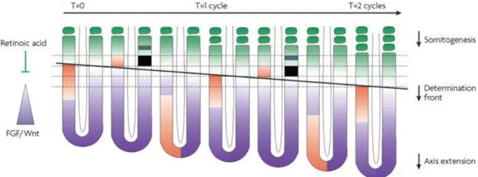
Le PSM est le siège d’un processus particulier dans l’embryon : la segmentation. Chez les vertébrés et la plupart des insectes, la segmentation est un processus séquentiel. Le PSM dérive du mésoderme paraxial qui forme deux bandes de tissu flanquant le tube neural. La segmentation du PSM au cours de l’embryogenèse, aussi appelée somitogenèse, est responsable de la formation des somites, précurseurs du squelette axial et des muscles du tronc et des membres. Chez la souris, une paire de somites est ajoutée toutes les deux heures à l’extrémité antérieure du PSM. La formation d’un nombre défini de segments de taille précise, spécifique à chaque espèce, est le résultat d’un contrôle spatio-temporel de l’expression des gènes. En effet, une horloge moléculaire et un front de détermination contrôlent la spécification de nouveaux segments. Le front de détermination est formé à l’intersection des gradients postéro-antérieurs de Wnt/FGF et d’un contre-gradient d’acide rétinoïque. Les forts niveaux de FGF maintiennent les cellules les plus postérieures à l’état de progéniteurs tandis qu’un contre-gradient d’acide rétinoïque induit la différenciation. Lorsqu’un groupe de cellules traversent le front de détermination, correspondant à un niveau seuil de gradient, ce groupe de cellules devient compétent à répondre au signal de l’horloge permettant la spécification d’un nouveau segment. L’horloge moléculaire est caractérisée par l’expression cyclique des gènes
8 des voies de signalisation Notch, Wnt et FGF. Certains de ces gènes codent des répresseurs qui permettent la mise en place de boucles de rétro-inhibition, à l’origine des oscillations moléculaires de l’expression des gènes. Ainsi, le temps nécessaire à la transcription des gènes impliqués dans des boucles de rétro-inhibition est un paramètre crucial à l’établissement de ces oscillations. Par conséquent, les étapes d’épissage et d’export nucléaire des ARNm suivant la transcription, ainsi que la demi-vie des ARNm constituent les paramètres de ces oscillations. En revanche, la transcription seule n’est pas un facteur limitant dans l’établissement des oscillations. L’expression dynamique des gènes liée à une transcription cyclique et l’expression classique des gènes dans le PSM, sous l’influence de cascades de signalisation, en font un paradigme intéressant pour l’étude de la transcription au cours du développement.
Objectifs
La composition de TFIID est variable et a été principalement décrite chez la levure, la drosophile et les cellules humaines mais peu de choses sont connues quant à la fonction de ce complexe et de sa variabilité in vivo chez les métazoaires. En utilisant la sous-unité TAF10, partagée entre TFIID et SAGA, comme point de départ pour l’étude de la machinerie basale de transcription, les buts de ma thèse de doctorat étaient:
(1) De caractériser dans l’embryon sauvage la composition, qui n’a pas encore été déterminée de façon précise, de la machinerie basale de transcription, notamment TFIID et SAGA;
(2) De développer et de valider des approches alternatives permettant l’utilisation d’approches biochimiques classiques afin d’approfondir la caractérisation de TFIID et de SAGA dans différents contextes cellulaires;
(3) D’étudier le rôle de TFIID et SAGA au cours de la somitogenèse dans l’embryon à 9,5 j.p.c;
(4) D’analyser la contribution de TFIID et SAGA à la transcription catalysée par l’ARN Pol II dans un modèle mammifère.
Résultats
Afin d’analyser les complexes TFIID et SAGA chez la souris, par immuno-précipitation couplée à la chromatographie liquide avec spectrométrie de masse en tandem, j’ai tout d’abord optimisé les conditions expérimentales. Pour cela j’ai testé l’efficacité de plusieurs anticorps,
9 pour la plupart initialement dirigés contre les épitopes de sous-unités humaines, à partir d’extraits cellulaires totaux de thymocytes. Les anticorps anti-TAF7 et anti-TBP ont été sélectionnés pour immuno-précipiter le complexe TFIID. Les anticorps TRRAP et anti-SUPT3 ont été sélectionnés pour immuno-précipiter le complexe SAGA. La quantité de matériel a également été réduite de 4 mg à 0.7 mg afin de pouvoir réaliser ces expériences dans l’embryon de souris à 9,5 j.p.c (jours post coitum).
L’analyse à partir d’extraits cellulaires totaux d’embryons entiers à 9,5 j.p.c a révélé la présence du complexe TFIID canonique. Des résultats similaires ont été obtenus dans les thymocytes et dans les cellules souches embryonnaires murines (mES). Ces résultats clarifient la composition de TFIID dans l’embryon et indiquent qu’il s’agit du complexe canonique. De plus, pour la première fois dans l’embryon, la composition du complexe SAGA a été décrite. Toutes les sous-unités du complexe décrites dans la littérature ont été détectées en proportions différentes pour certaines entre les cellules de thymocytes, de mES et l’embryon. Ces données montrent que la composition de TFIID et SAGA est globalement conservée quel que soit le contexte cellulaire étudié ici avec quelques différences selon le type cellulaire.
La quantité de matériel à partir d’embryons étant limitée pour des approches biochimiques plus approfondies, les cellules mES ont été utilisées pour préparer des extraits nucléaires. L’analyse préliminaire par chromatographie d’exclusion à partir de ces échantillons a permis de détecter le complexe holo-TFIID mais aussi au moins deux autres sous-complexes à des poids moléculaires inférieurs à 1 MDa. Parmi les anticorps testés par western-blot, un premier sous-complexe contenant au moins TAF5, TAF8, TAF10 et TBP et le second de 670 kDa contenant au moins TAF5 et TBP ont été détectés. Ces résultats confirment la présence de sous-complexes de TFIID dont la composition reste à déterminer avec précision.
La délétion inductible du gène codant la sous-unité TAF10 dans l’embryon entier à 7,5 j.p.c et dans les cellules mES a permis la déplétion complète de la protéine dans l’embryon à 9,5 j.p.c et quasi totale dans les cellules mES deux et cinq jours plus tard. Les expériences d’immuno-précipitation contre TAF7 et TBP à partir d’extraits cellulaires totaux ont montré que les sous-unités de TFIID associées étaient faiblement détectées indiquant que TAF10 est requis pour l’assemblage complet de TFIID. Ces résultats ont été confirmés par la chromatographie d’exclusion à partir d’extraits nucléaires de cellules mES mutantes pour
sous-10 unités tandis que les deux autres sous-complexes étaient toujours présents. Cela indique qu’en l’absence de TAF10, peu de complexes holo-TFIID sont retrouvés et que majoritairement il s’agit de complexes TFIID partiellement assemblés. Toutefois, il a été noté que l’assemblage de TFIID était plus fortement affecté dans l’embryon que dans les cellules mES en l’absence de TAF10, soulignant des différences liées au contexte cellulaire.
TAF10 a également été montré comme étant requis pour l’assemblage complet du complexe SAGA dans l’embryon et les cellules mES. Comme pour TFIID, l’assemblage du complexe était plus fortement affecté dans l’embryon que dans les cellules mES. Toutefois, malgré ce défaut d’assemblage, les niveaux d’acétylation de l’histone H3 sur la lysine 9 ainsi que les niveaux d’ubiquitine de l’histone H2B1 n’étaient pas affectés en l’absence de TAF10, suggérant que le complexe SAGA reste fonctionnel.
Afin d’étudier le rôle de TFIID et SAGA au cours du développement, nous avons choisi de déléter Taf10 dans le mésoderme paraxial où la transcription est dynamique avec certains gènes qui ont une expression cyclique. L’analyse de la délétion du gène codant la sous-unité TAF10 a été réalisée. La délétion de Taf10 ubiquitaire inductible ou conditionnelle dans les dérivés du mésoderme à partir de 7,5 j.p.c, entraine la disparition totale de la protéine et provoque un sévère ralentissement de la croissance embryonnaire entre 9,5 j.p.c et 10,5 j.p.c. La délétion conditionnelle de Taf10 dans les dérivés du mésoderme n’affecte pas la formation et la différenciation précoce des somites à 9,5 j.p.c. Toutefois, la délétion de Taf10 n’est pas viable à long terme, puisqu’elle conduit à la mort de l’embryon vers 10,5 j.p.c, probablement en raison de la dégénérescence du placenta et de l’allantoïde chez les mutants
T-Cre ;Taf10flox/flox. L’analyse transcriptomique du PSM entre les embryons sauvages et mutants
a montré que l’expression de la plupart des gènes n’est pas perturbée en l’absence de TAF10. Cependant, certains gènes dont Cdkn1a (p21) et Cdkn1c (p57) codant des inhibiteurs du cycle cellulaire, sont surexprimés chez les mutants, suggérant que l’arrêt de la croissance des embryons mutants est lié au blocage du cycle cellulaire et donc de la prolifération cellulaire. Ces résultats mettent en évidence que l’expression de la plupart des gènes est normale en l’absence de TAF10, alors que des travaux précédents ont montré que TAF10 est nécessaire à la transcription dans l’embryon. Toutefois, la transcription reste fonctionnelle seulement pendant un intervalle de temps précis en l’absence de TAF10 et les embryons mutants meurent à partir de 10,5 j.p.c. Cela suggère donc que la transcription peut dans certaines conditions
11 fonctionner avec une machinerie basale alternative sans TAF10 dans l’embryon. Cette fenêtre de temps représente donc une opportunité pour étudier la machinerie qui permet la transcription dans l’embryon en l’absence de TAF10. Un autre fait marquant est l’effet différent produit par la délétion de Taf10 selon les tissus. En effet, le mésoderme de la plaque latérale (LPM), qui est également ciblé par la délétion conditionnelle de Taf10, présente une mort cellulaire importante à 9,5 j.p.c alors que le mésoderme paraxial est normal. Cela suggère une variabilité de la machinerie de transcription en fonction du contexte comme cela a été démontré dans les cellules du trophectoderme et les cellules de la masse interne. L’ensemble des résultats confirment que la transcription dans l’embryon est particulière, avec des spécificités propres à certains tissus.
L’induction de la délétion de Taf10 dans les cellules mES provoque un fort ralentissement de leur croissance trois jours après, récapitulant le phénotype observé dans l’embryon. Toutefois, aucune mort cellulaire massive n’a été détectée, suggérant une diminution de leur prolifération. L’analyse de la prolifération par incorporation d’EdU n’a pas permis de révéler une forte réduction de la prolifération trois jours après l’induction de la délétion, mais il n’est pas exclu qu’elle le soit par la suite, quatre ou cinq jours après l’induction de la délétion. Ces résultats indiquent que les cellules mES se comportent différemment des cellules de la masse interne, qui sont incapables de survivre dans les blastocystes Taf10-/- commecela a été rapporté dans la littérature et permettent donc de servir comme un bon modèle d’étude pour l’analyse du rôle de TFIID et SAGA dans la transcription. Les cellules de tératocarcinome murines F9, en revanche, sont quasiment incapables de se maintenir et de proliférer en culture en l’absence de TAF10 confirmant le phénotype décrit dans la littérature et soulignant ainsi l’importance du contexte cellulaire dans les différences de phénotype obtenues avec la délétion de Taf10.
De façon surprenante, nous avons montré que TAF10 n’est pas indispensable pour l’expression globale des gènes dans le PSM, à l’exception de certains gènes. La délétion de
Taf10 dans les cellules mES impactent différemment les niveaux d’ARN totaux des gènes testés
par RT-qPCR, avec l’augmentation des niveaux d’ARN totaux pour Cdkn1a et Cdkn1c notamment, qui récapitulent ce qui a été observé dans le PSM. De même, Gas5 et Taf1d, dont les niveaux d’ARN totaux sont diminués dans le PSM sont également diminués dans les cellules mES mutantes. Il a été montré chez la levure qu’une diminution du taux de la synthèse des
12 ARNm peut être compensée par une diminution du taux de la dégradation des ARN, normalisant ainsi les niveaux des ARN totaux. Par conséquent, les niveaux d’ARN totaux peuvent ne pas toujours refléter l’état de la transcription. Afin d’analyser directement la transcription, j’ai adapté la technique de marquage métabolique des ARN nouvellement synthétisés aux cellules mES. Cette technique repose sur le marquage pendant un temps court, ici 10 minutes, des ARNm en cours de synthèse grâce à l’incorporation de l’analogue de l’uridine, le 4-thiouridine (4sU). Les ARN marqués sont ensuite biotinylés et purifiés avec des billes de streptavidine magnétiques. L’analyse par RT-qPCR d’un panel de gènes a montré que la transcription est fortement affectée pour la quasi-totalité des gènes, avec une réduction d’au moins 50 % du niveau des ARNm nouvellement synthétisés. TAF10 est donc requis à la transcription au moins pour ces gènes, et il apparait qu’un phénomène important de compensation a lieu dans les cellules mES. En effet, plus que la normalisation des niveaux d’ARN totaux, il y a une surcompensation liée à une probable plus forte diminution du taux de dégradation des ARN par rapport à leur taux de synthèse. Une stabilisation accentuée des transcrits résultant en une accumulation plus forte des transcrits pourrait expliquer que les niveaux d’ARN totaux de certains gènes comme Cdkn1a et Cdkn1c sont fortement augmentés. Dans le but d’étudier l’état de la transcription de façon globale au niveau de tout le génome, j’ai tenté dans un premier temps d’utiliser la technique Transient Transcriptome
sequencing. Cette technique repose sur le marquage métabolique avec le 4sU des ARN
nouvellement synthétisés avec une étape préliminaire de fragmentation des ARN. Cette étape permet de ne purifier uniquement les fragments d’ARNm marqués au 4sU et donc de s’affranchir du biais lié aux fragments d’ARNm déjà synthétisés avant le début du marquage et qui ne correspondent pas à de la transcription naissante. Malgré plusieurs tentatives, le manque de reproductibilité du profil des ARNm fragmentés associé à la faible efficacité de purification des ARNm nouvellement synthétisés, n’ont pas rendu possible leur séquençage. C’est pourquoi, la technique de 4sU-sequencing des ARNm purifiés après leur marquage par le 4sU a été utilisée. Les résultats étant en cours d’analyse, je ne dispose pas lors de la rédaction de ce manuscrit d’informations concernant l’état global de la transcription dans les cellules mES après la délétion de Taf10.
13 Au cours de ma thèse, j’ai pu clarifier la composition des complexes TFIID et SAGA dans l’embryon ainsi que les différences de composition en fonction du contexte cellulaire. J’ai également montré que la sous-unité TAF10 est requise pour l’assemblage complet des complexes TFIID et SAGA. De plus, j’ai montré que le rôle général de TFIID et SAGA peut être nuancé dans l’embryon. Enfin, j’apporte ici des éléments indiquant que l’initiation de la transcription est sévèrement affectée en l’absence de TAF10, suggérant que le rôle global de TFIID et SAGA dans la transcription pourrait être conservé dans les cellules de mammifères.
Publication:
P. Bardot, S. D. Vincent, M. Fournier, A. Hubaud, M. Joint, L. Tora, and O. Pourquié, “The TAF10-containing TFIID and SAGA transcriptional complexes are dispensable for early somitogenesis in the mouse embryo.,” Development, vol. 144, no. 20, pp. 3808–3818, Oct. 2017.
Communications:
Bardot P, Platania A, Pourquié O, Tora L and Vincent S. Characterization and functional analyses of the basal transcription machinery during development and cellular differentiation. Communication sous forme d’affiche. Tri-regional stem cell & developmental biology meeting. 9 Décembre 2016, IGBMC, Illkirch.
Paul Bardot, Olivier Pourquié, László Tora, Stéphane D. Vincent. Analysis of the TFIID and SAGA transcriptional complexes composition and function during development. Communication orale. Journées du campus d’Illkirch. 28-29 Mars 2017, ESBS, Illkirch.
Paul Bardot, Olivier Pourquié, László Tora, Stéphane D. Vincent. Analysis of the TFIID and SAGA transcriptional complexes composition and function during development. Présentation sous forme d’affiche. Keystone symposia, « Gene Control in Development and Disease (X6) ». 23-28 Mars 2018, Whistler.
14
T
ABLE OF CONTENTS Acknowledgements ... 2 Abstract ... 4 Résumé en français ... 5 List of figures ... 19 List of tables ... 21 List of annexes ... 22 List of abbreviations ... 23INTRODUCTION ... 3
I. Chromatin organization of the genome ... 5
1. Chromatin organization... 5
2. Epigenetic modifications... 8
a. DNA methylation ... 8
b. Post-translational modifications of histone proteins ... 8
II. Mechanisms of eukaryotic RNA Pol II transcription initiation ... 10
1. Core promoter ... 10
2. Pre-Initiation Complex assembly at the promoter ... 11
a. RNA Polymerase II : a multi-subunit complex ... 11
b. The General Transcription Factors ... 12
c. Structural aspects of the PIC assembly model ... 17
3. Promoter proximal pausing ... 17
4. Transcription reinitiation... 18
5. Control of transcription activation ... 18
a. Distal cis-regulatory elements ... 18
b. Transcription factors ... 19
c. Co-activators ... 19
i. Mediator ... 19
ii. Chromatin remodeling complexes ... 20
15
1. Discovery of TBP ... 21
2. TBP-Associated-Factors form a large TFIID multi-subunit complex ... 21
3. Distinct TFIID complexes exist ... 24
4. Architecture of TFIID and structural model for the assembly ... 25
5. TAFs are transcriptional co-activators ... 28
6. Enzymatic activities of TAFs ... 29
IV. The SAGA co-activator complex, a TAF-containing complex ... 30
1. Identification and characterization of the SAGA complex and SAGA-related complexes in yeast 30 2. The SAGA complex is conserved in metazoan... 32
a. Conservation and divergence of the SAGA subunits ... 32
i. PCAF complex ... 32
ii. STAGA ... 32
iii. TFTC ... 33
b. ATAC ... 33
3. Modular organization of SAGA ... 34
a. Core structural module ... 36
b. SPT module ... 36
c. Tra1/TRRAP ... 36
d. DUB module ... 37
e. HAT module ... 38
V. TFIID and SAGA are required for transcription of nearly all genes in S. cerevisiae ... 38
1. TFIID and SAGA control the expression of a large fraction of the genome ... 38
2. Classification of genes as TFIID-dominated or SAGA-dominated ... 39
3. New approaches reveal a global role for TFIID and SAGA ... 40
a. TFIID ... 40
b. SAGA ... 41
VI. TFIID and SAGA roles during embryonic development in metazoans ... 43
1. TFIID and SAGA subunit expression pattern during development ... 44
a. TAF paralogs ... 44
b. TBP paralogs ... 44
c. TFIID composition is variable ... 45
d. GCN5 and PCAF display different expression pattern ... 46
2. TFIID role during development ... 46
16
b. Some TFIID subunits are differentially required during development ... 47
i. TAF10 and TAF7 are differentially required depending on the cellular context ... 47
ii. TAF10 is differentially required depending on the developmental stage ... 47
iii. TAF10 is required for initial gene activation ... 48
3. SAGA role in development ... 48
a. HAT module ... 48
b. DUB module ... 50
c. SUPT20 ... 50
VII. A new paradigm to study the role of transcriptional complexes ... 51
1. Vertebrate segmentation ... 51
2. The clock and wavefront model ... 52
a. The segmentation clock defines the pace ... 53
b. The wavefront determines the new somites ... 53
3. Gene oscillatory expression in the PSM ... 54
a. Negative feedback loops generates expression oscillation ... 54
b. Mathematical modeling of gene oscillation ... 55
c. Determination of the parameters controlling gene oscillation ... 56
i. Transcription elongation is not a critical parameter for gene oscillation ... 56
ii. Role of splicing ... 57
iii. Role of transcript stability ... 58
4. The role of co-activators in somitogenesis... 59
VIII. Goals of the thesis project ... 60
MATERIAL & METHODS ... 62
1. Mouse lines ... 63
2. Cell culture ... 63
a. Mouse F9 embryonal carcinoma cells ... 63
b. Mouse embryonic stem cells ... 63
c. T-cell leukemia cell line T29 ... 64
3. Cell count and cell death assay ... 64
a. Cell count and viability assay ... 64
b. Apoptosis assay ... 64
4. Proliferation assay ... 64
5. Cellular extracts ... 65
17
b. Whole Cell Extracts ... 65
i. Large scale whole cell extract preparation ... 65
ii. Small scale whole cell extract preparation ... 66
c. Nuclear extracts ... 66
6. Bradford protein assay ... 67
7. Immuno-precipitation... 67
8. Western-blot ... 68
9. Antibodies ... 68
10. Mass spectrometry ... 69
11. Gel filtration ... 69
12. 4sU metabolic labeling of newly-synthesized mRNA and purification ... 70
a. 4sU labeling ... 70
b. Total RNA extraction and isolation ... 70
c. Purification of newly-synthesized mRNA ... 70
13. RNA fragmentation ... 73
14. RT-qPCR... 73
15. Gene primers ... 73
RESULTS ... 75
IX. Publication: “The TAF10-containing TFIID and SAGA transcriptional complexes are dispensable for early somitogenesis in the mouse embryo” - (Bardot et al. 2017) ... 76
X. Biochemical characterization of TAF10-containing complexes ... 108
1. Technical optimization of immuno-precipitation for the TAF10-containing complexes .. 108
a. Antibody validation ... 108
b. Starting material reduction ... 110
c. Processing and analysis of the proteomics data ... 111
2. TFIID and SAGA characterization in different cellular contexts ... 112
3. Phenotype characterization ... 114
a. Experimental workflow ... 114
b. Cellular growth, viability and cell death analyses ... 117
c. Cellular proliferation analysis... 117
4. TAF10 is required for TFIID and SAGA full assembly in pluripotent cells ... 118
a. Residual TAF10 protein detected by mass-spectrometry ... 118
b. TAF10 is required for TFIID full assembly ... 120
18
d. TAF10 is required for SAGA full assembly ... 125
XI. Analysis of the transcriptional function of TFIID and SAGA ... 127
1. Gene expression analysis of steady-state mRNA levels in mES cells ... 127
2. Newly-transcribed mRNA analysis ... 130
a. Technical validation ... 130
b. Newly-transcribed mRNA is globally affected in the absence of TAF10 ... 131
c. Genome-wide analysis of nascent transcription ... 133
GENERAL DISCUSSION & PERSPECTIVES ... 136
XII. Composition of TFIID and SAGA during development ... 137
1. No alternative TFIID complexes are detected ... 137
2. Characterization of TFIID sub-complexes in mES cells ... 137
3. SAGA composition ... 138
XIII. The architectural role of TAF10 in TFIID and SAGA assembly ... 138
1. TAF10 is required for TAF8 stability ... 138
2. TAF10 is required for TFIID and SAGA full assembly ... 139
3. Residual TAF10 protein is detected ... 140
XIV. Role of TFIID and SAGA in vivo ... 141
1. TAF10 is essential and required for cellular viability and cellular growth ... 141
2. TAF10 is differentially required between PSM and LPM tissues ... 142
3. Limb bud formation but not vertebrate segmentation is affected at E9.5 in T-Cre; Taf10 mutants ... 143
XV. Role of TFIID and SAGA in mammalian transcription ... 145
1. TAF10 is required for transcription of many genes with some notable exceptions ... 145
2. Hypothetical model for gene activation by TFIID and transcription maintenance ... 146
3. Determining the respective contribution of TFIID and SAGA ... 147
4. Steady-state gene expression can be sustained with altered TFIID and SAGA complexes147 5. Potential mechanisms of compensation of mRNA decay in response to a decrease in mRNA synthesis ... 148
CONCLUSIONS ... 150
BIBLIOGRAPHY ... 152
19
L
IST OF FIGURESFigure 1: Resolution of the three eukaryotic RNA Polymerases Figure 2: Chromatin organization of the genome
Figure 3: Histone Fold Domain interactions
Figure 4: RNA Pol II core promoter elements diversity
Figure 5: Schematic representation of the preinitiation complex multi-step assembly model at the promoter for RNA Pol II recruitment
Figure 6: Schematic representation of the role of Mediator in transcription initiation Figure 7: Schematic representation of the TFIID subunits
Figure 8: Schematic representation of the TFIID model of assembly
Figure 9: Schematic representation of the modular organization of SAGA in relation with the other transcriptional complexes
Figure 10: Schematic model of the general role of TFIID, SAGA and Mediator complexes in
Saccharomyces cerevisiae
Figure 11: Schematic representation of the presomitic mesoderm and the signaling gradients Figure 12: Vertebrate segmentation of the PSM
Figure 13: Schematic representation of negative feedback loops for gene oscillation Figure 14: Workflow of 4sU metabolic labeling of newly-synthesized mRNAs
Figure 15: TAF10-containing complexes characterization in different cellular contexts Figure 16: TAF10 is required for normal mES cell growth
Figure 17: TAF10 is required for mouse teratocarcinoma F9 cells growth
Figure 18: Distribution of TAF10 peptides detected by mass spectrometry in immuno-precipitation experiments
20 Figure 19: TFIID assembly defect in mES cells at day 3 after Taf10 deletion
Figure 20: TFIID assembly defect in mES cells at day 5 after Taf10 deletion Figure 21: TFIID assembly defect in mES cells at day 3 after Taf10 deletion Figure 22: SAGA enzymatic activities are not affected after Taf10 deletion Figure 23: SAGA assembly defect in mES cells at day 5 after Taf10 deletion Figure 24: Steady-state mRNA levels analyses by RT-qPCR
Figure 25: Newly-synthesized mRNA levels analyses by RT-qPCR Figure 26: Comparison of 4sU-seq and TT-seq methods
Figure 27: RNA fragmentation optimization profiles Figure 28: 4sU-labeled mRNA fragmentation profiles
21
L
IST OF TABLESTable 1: Overview of the histone post-translational modifications
Table 2: Unified nomenclature for the TFIID subunits including orthologs and paralogs Table 3: Composition of orthologous SAGA complexes
Table 4: Validation for immuno-precipitations of antibodies raised against TFIID and SAGA subunits
22
L
IST OF ANNEXESAnnexe I: R code
Annexe II: Antibody validation for TAF1, TAF2, TAF3 and TAF4 immuno-precipitations Annexe III: Antibody validation for TAF7 and TAF8 immuno-precipitations
Annexe IV: Antibody validation for TBP immuno-precipitation Annexe V: Antibody validation for TAF10 immuno-precipitation
Annexe VI: Antibody validation for GCN5, ATXN7L3, and SUPT20H immuno-precipitations Annexe VII: Antibody validation for SUPT3H immuno-precipitation
Annexe VIII: Antibody validation for TRRAP immuno-precipitation Annexe IX: TAF10 is required for normal mES cell growth
Annexe X: 4OHT treatment does not affect controls mES cellular growth and viability Annexe XI: Cellular growth is not affected by doxycycline in F9 wild-type cells Annexe XII: Nouveau Chapître de la Thèse
23
L
IST OF ABBREVIATIONS4OHT: 4-hydroxy tamoxifen 4sU: 4-thiouridine
4TU: 4-tiouracile Ac: acetylation
AF: Activation function containing region ARC: Activator Recruited Factor
ATAC: Ada2a containing complex ATP: Adenosine Tri Phosphate BrdU: 5-Bromo-2-deoxyuridine BRE: TFIIB Recognition Element CC: Close Complex
CDK: Cyclin Dependent Kinase cDTA: comparative dynamic transcriptome analysis
CHD: Chromodomain Helicase DNA-binding
ChEC-seq: Chromatin Endogenous Cleavage sequencing
ChIP-seq: Chromatin Immuno-precipitation Sequencing
Cre: Cre recombinase
CTD: Carboxyl Terminal Domain
DNA: Deoxyribo Nucleic Acid DPE: Downstream Promoter Element DRIP: VDR-interacting proteins DSIF: DRB sensitivity-inducing factor DTT: dithiothreitol
DUB: De Ubiquitinylation EC: Elongation Complex
EDTA: Ethylene Diamine Tetra Acetic Acid
EdU: 5-ethynyl-2-deoxyuridine EM: Electron Microscopy EtOH: Ethanol
FGF: Fibroblast Growth Factor GTF: General Transcription Factor HAT: Histone Acetyle Transferase HCl: Chloridric acidHDAC: Histone Deacetylase
HFD: Histone Fold Domain Inr: Initatior
IP: Immuno-precipitation
3 LFNG:
Beta-1,3-N-acetylglucosaminyltransferase lunatic fringe
LIF: Leukemia Inhibitory Factor LPM : Lateral Plate Mesoderm Me: Methylation
mES: mouse embryonic stem cell MgCl2: Magnesium Chloride miRNA: micro RNA
MTE: Motif Ten Element
MudPIT: Multidimensional Protein Identification Technology
NaCl: Sodium Chloride
NELF: Negative Elongation Factor NFR: Nucleosome Free Region NLS: Nuclear Localizing Sequence
NSAF: Normalized Spectral Abundance Factor
OC: Open Complex
PBS: Phosphate Buffer Saline PIC: Pre Initiation Complexe PM: Paraxial Mesoderm
PSMx: Peptide Spectrum Match
PSM: Pre Somitic Mesoderm
RARE: Retinoic Acid Response Element Rcf: Relative centrifugal force
RNA Pol: Ribo Nucleic Acid Polymerase RNA: Ribo Nucleic Acid
RPB: RNA Pol II subunit
SAGA: Spt Ada Gcn5 Acetyltransferase SLIK: SAGA Like complex
SPT: Suppressor of Ty
SWI/SNF: Switch/Sucrose Non Fermentable
TAF: TATA-Binding Protein Associated Factors
TBP: TATA-Binding Protein TBPL: TBP Like Factor TCT: TcT-E element
TFII: Transcription Factor II TFTC: TBP Free TAF complex TLF: TBP Like Factor
TRAP: Thyroid Hormone Receptor Associated Proteins
TRF: TBP Related Factor TSS: Transcription Start Site
4 UAS: Upstream Activator Sequence
UTR: UnTranslated Region
WHHERE: Wdr5, HDAC1, HDAC2, RERE/ATROPHIN2
3
4 In 1959, Weiss and Gladstone described an RNA polymerase activity from rat liver nuclei (Weiss et al. 1959). Transcription was defined as the conversion of DNA into RNA, and thus represents the first step of genome expression. Chromatographic analyses from purified nuclei from sea urchin and rat liver, and the transcription inhibition mediated by α-amanitin, brought the first evidences of the existence of three RNA Polymerases (RNA Pol) that catalyze transcription in the nucleus (figure 1): RNA Pol I, RNA Pol II and RNA Pol III (except plants that have five RNA Polymerases, reviewed in (Duda 1976)) (Kedinger et al. 1970; Roeder et al. 1969). While RNA Pol I transcribes large ribosomal RNAs, RNA Pol II transcribes protein coding genes, messenger RNAs, and also non-coding RNAs, and RNA Pol III transcribes small RNAs (tRNA, 5S RNA).
Figure 1: Resolution of the three eukaryotic RNA Polymerases. Activity measurement (units/µg protein) based on the incorporation of Uridine Mono Phosphate (UMP) into RNA/10min/ml of the fractions eluted by chromatography obtained from soluble enzyme preparation from sea urchin nuclei gastrula (52h development) (Roeder and Rutter 1969).
Transcription is a multi-step process composed of: (1) initiation with the recruitment of the RNA Pol II to the promoter, (2) elongation with productive mRNA synthesis and (3) termination that corresponds to the release of RNA Pol from DNA. Furthermore, transcription
5 represents a critical step for gene regulation, and plays major roles in the development of an organism by controlling many cellular processes as well as generating the different cell types.
The focus of my thesis concerns the general mechanisms of RNA Pol II transcription initiation. Firstly, I will describe how DNA is packaged in the eukaryotic nuclei and its implication for transcription. Then, I will detail the molecular mechanisms that govern RNA Pol II transcription with a special emphasis on the general transcription factor TFIID and the co-activator SAGA. Particularly, I will detail the importance of the components of the transcriptional apparatus in the control of gene expression in vivo and in a developmental context. Finally, I will present the new paradigm that I used to study RNA Pol II transcription in my thesis.
I. Chromatin organization of the genome
1. Chromatin organization
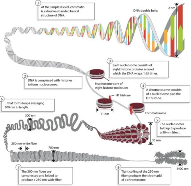
Genetic information in eukaryotes is packaged into the nucleus of every cell. In the nucleus, DNA adopts a chromatin structure (figure 2), where it is wrapped around proteins called histones. Histone octamers form nucleosomes, the basic organization unit of chromatin. Each octamer contains two copies of histones: H2A, H2B, H3 and H4. Histone proteins form heterodimer through their Histone Fold Domain (HFD), which consists in three α-helices (α1, α2, and α3) connected by short loops L1 and L2 (figure 3) [reviewed in (Arents et al. 1991)]. Nucleosomes are separated between each other by a DNA linker. In addition, the histone H1 binds the nucleosome and allows a higher order of chromatin organization into 30 nm fibers and chromosomal 300 nm fibers. Chromatin is described as differentially condensed. Firstly, heterochromatin represents the highly condensed form and contains both non-coding gene regions (constitutive heterochromatin) and genes that are not expressed (facultative heterochromatin) (Bannister et al. 2011). Secondly, euchromatin is a decondensed chromatin that contain expressed genes. Chromatin organization is very dynamic and can change from one cell type to another, especially during development. Chromatin organization is of high importance as it impacts gene expression by modulating the accessibility of the transcription machinery on DNA. As a consequence, coding regions must be free of nucleosomes prior gene activation. The initial view of the chromatin organization has evolved, since the description of
6 at least five chromatin domains in Drosophila (Filion et al. 2010) and a more complex spatial organization of the genome with chromosome territories, A/B compartments, topologically associating domains, and chromatin loops [reviewed in (Serizay et al. 2018)].
Figure 2: Chromatin organization of the genome. DNA is packaged in the nucleus under chromatin where it is wrapped around histone proteins. Multiple organisation levels structure the chromatin. Adapted from Pierce, Benjamin. Genetics: A Conceptual Approach, 2nd ed.
7 Figure 3: Histone Fold Domain interactions. (a) Scheme of the fours histone proteins with their HFD, helices correspond top rectangles and the black line represents the DNA interactions. Structure of (b) H2A-H2B dimer and (C) H3-H4 dimer (Dutnall and Ramakrishnan 1997).
8 2. Epigenetic modifications
Genome accessibility can be modulated by epigenetic modifications, that is to say, inherited modifications that do not alter the DNA sequence per se and that participate to the regulation of gene expression.
a. DNA methylation
First of all, DNA can be directly methylated through the covalent transfer of a methyl group to the C-5 position of the cytosine ring, mostly at CpG dinucleotides [reviewed in (Li et al. 2014)]. Interestingly, DNA methylation of cytosine is not found at the same level in all species such as in Drosophila, where it was shown to be much lower (Boffelli et al. 2014), and nematodes for instance. Furthermore, it was shown that DNA can be also methylated on N(6)-adenine, as shown in several organisms (Greer et al. 2015; Liu et al. 2016; Wu et al. 2016; Zhang et al. 2015). In mammals, DNA methylation represents an epigenetic “lock” of gene expression. The methylated sequences include satellite DNAs, repetitive elements, non-repetitive intergenic DNA, and exons of genes. However, there are CpGs that remain unmethylated, and are found in CpG islands. Methylation is thought to play several roles in gene silencing such as X chromosome inactivation, for gene dosage compensation. Methylation of DNA controls the accessibility of transcription factors and can also be bound by specific factors that further recruit co-repressor complexes [reviewed in (Li et al. 2014)].
b. Post-translational modifications of histone proteins
Amino-terminal tails of histones can also be modified and thus affect the nucleosome structure [reviewed in (Bannister et al. 2011)]. Several post-translational modifications of histones have been reported so far, and are listed in (Table 1) [reviewed in (Tessarz et al. 2014)]. Histone PTMs can affect directly chromatin compaction by modifying the chemical properties of their interactions with DNA. For instance, acetylation removes the positive charge from the histone tails rendering them neutral resulting in a more relaxed chromatin structure. PTMs are regulated by writer proteins that add PTMs on histones, while erasers remove them. PTMs are “read” by specific protein factors capable to recognize specific PTM or a combination of them, through specific domains [reviewed in (Lalonde et al. 2014)]. The recruitment of such proteins can also lead to chromatin modification and regulate transcription. Histone PTMs have
9 been proposed to constitute an additional biological “code” for regulating gene expression and epigenetic inheritance [reviewed in (Jenuwein et al. 2001)].
Table 1: Overview of the histone post-translational modifications. Adapted from (Tessarz and Kouzarides 2014).
Post-translational modification
Histone (residue) Proposed function
Methylation H1 (Arginine, Lysine, Tyrosine)
H2A (Arginine, Lysine, Glutamine, Tyrosine) H2B (Arginine, Lysine) H3 (Arginine, Lysine) H4 (Arginine, Lysine) Chromatin compaction, rDNA transcription, Transcription, Acetylation H1 (Lysine) H2A (Lysine) H2B (Lysine) H3 (Lysine) H4 (Lysine) Chromatin compaction, DNA repair, DNA replication, Transcription
Phosphorylation H1 (Lysine, Tyrosine) H2B (Serine)
H3 (Threonine, Tyrosine) H4 (Serine, Tyrosine)
Chromatin compaction, DNA repair, Transcription
Formylation H1 (Lysine) H2B (Lysine) H3 (Lysine) H4 (Lysine)
10
Oxidation H4 (Lysine)
Crotonylation H2A (Lysine) H2B (Lysine) Transcription Hydroxylation H1 (Lysine) H2A (Tyrosine) H2B (Lysine, Tyrosine) H4 (Tyrosine)
Ubiquitinylation H2A (Lysine) H2B (Lysine) H3 (Lysine)
Transcription
Succinylation H2A (Lysine) H2B (Lysine) H3 (Lysine) H4 (Lysine)
Citrullination H1 (Arginine)
Propionylation H4 (Lysine)
II. Mechanisms of eukaryotic RNA Pol II transcription initiation
1. Core promoterEukaryotic genes consist in a promoter region depleted of nucleosomes, and so-called Nucleosome Free Region (NFR), where transcription starts, defined as the Transcription Start Site (TSS), and a gene body that contains the open reading frame. The core promoter is defined as the minimal DNA region bound by the transcription machinery sufficient for basal transcription [reviewed in (Kadonaga 2012)]. It contains sequences (figure 4) that are bound by specific proteins that recruit RNA Pol II to the promoter.
11 Figure 4: RNA Pol II core promoter elements diversity. The core promoter can display numerous elements, alone or in combination (Vo ngoc et al. 2017).
The TATA-box was the first core promoter element to be identified (Gannon et al. 1979) and is located 25-35 base pairs before the TSS but is not present in all metazoan genes (Jin et al. 2006; Kimura et al. 2006; Yang et al. 2007). Actually, only a minority of genes, about 20% in yeast, contain a TATA-box (Basehoar et al. 2004). So, higher eukaryotes genes harbor a combination of several different sequence elements such as for example the Initiator (Inr), the TFIIB Recognition Element (BRE), the Downstream Promoter Element (DPE), the Motif Ten Element (MTE) (figure 4) and others [reviewed in (Kadonaga 2012; Vo ngoc et al. 2017)].
2. Pre-Initiation Complex assembly at the promoter
a. RNA Polymerase II : a multi-subunit complex
RNA Pol II enzyme is responsible for directing the synthesis of mRNA and has been shown to be conserved from yeast to human [reviewed in (Young 1991)]. This enzyme has a mass >0.5 MDa and chromatographic analyses revealed that it consists in a multi-subunit complex composed of 12 polypeptides (Bartholomew et al. 1986; Edwards et al. 1991) [reviewed in (Young 1991)]. The subunits RPB5, 6, 8 and 10 are shared by all three RNA Polymerases while RNA Pol II is characterized by the presence of RPB4, 7, 9 and RPB1 which harbors the Carboxyl Terminal Domain (CTD) (Carles et al. 1991; Hampsey 1998; Wild et al.
12 2012; Woychik et al. 1990). The resolution of its structure revealed four structural domains: the core, the clamp, the shelf, and the jaw lobe (Cramer et al. 2001).
The CTD of RBP1 consists in tandemly repeated heptapeptides and contains the consensus sequence Tyr-Ser-Pro-Thr-Ser-Pro-Ser (YSPTSPS). The number of repeated hexapeptides depends on the species and range from 26-27 times in Saccharomyces up to 52 times in mouse [reviewed in (Young 1991)]. The CTD can be post-translationally modified by phosphorylation and also glycosylation (Kelly et al. 1993) [reviewed in (Young 1991)]. The CTD is under an unphosphorylated state when RNA Pol II is recruited to the Pre Initiation Complex (PIC), while serine-5 phosphorylation is associated with transcription initiation and serine-2 phosphorylation with elongation (Bartholomew et al. 1986; Cadena et al. 1987; Chesnut et al. 1992; Laybourn et al. 1990).
b. The General Transcription Factors
Transcription initiation is an orchestrated process that requires the assembly of the Pre-Initiation Complex (PIC) that recruits the RNA Pol II enzyme to initiate mRNA synthesis at the promoter in NFRs delimited by an upstream -1 and a downstream +1 nucleosome (Jiang et al. 2009). In vitro transcription within cellular-free systems showed that RNA Pol II is not sufficient to direct accurate transcription from a DNA template, and that additional factors are required: the General Transcription Factors (GTFs) (Luse et al. 1980; Weil et al. 1979) [reviewed in (Thomas et al. 2006)]. Those factors have been identified from chromatography analyses of crude cell extracts and used for incubation with purified RNA Pol II. Initially, four nuclear factors have been identified for accurate RNA Pol II transcription initiation from four enzymatically active fractions (A, B, C and D) followed by the characterization of additional GTF from the sub-fractionation of the C active fraction (Samuels et al. 1982; Weil et al. 1979). Those RNA Pol II GTFs were named according their fraction elution, as Transcription Factor II (TFII). They include TFIIA, TFIIB, TFIID, TFIIE, TFIIF and TFIIH (Flores et al. 1989, 1992; Ge et al. 1996; Sawadogo et al. 1985). The GTFs are evolutionary conserved large multi-subunit complexes, except TFIIB which is a single polypeptide chain.
13 Figure 5: Schematic representation of the preinitiation complex multi-step assembly model at the promoter for RNA Pol II recruitment. According to the canonical model of PIC assembly, TFIID binds DNA first, notably through TBP which binds the TATA-box and bend DNA. TBP-DNA is stabilized by TFIIB and TFIIA. RNA Pol II is brought to this complex by TFIIF followed by TFIIE and TFIIH, which will melt DNA, initiating the transcription bubble, before productive elongation by RNA Pol II (Sainsbury, Bernecky, and Cramer 2015).
14 In the canonical model of PIC assembly (figure 5) described by Steve Buratowski and colleagues (Buratowski et al. 1989), the GTFs are assembled to the promoter hierarchically. TFIID comes first and recognizes the core promoter sequences. TBP binds the TATA-box, and distorts DNA (Starr et al. 1995), and other TFIID subunits, can also recognize additional elements. TFIID is stabilized on the promoter with TFIIA and TFIIB. RNA Pol II is brought to the PIC with TFIIF. The PIC assembly is finalized with the recruitment of TFIIE and TFIIH. TFIIH is responsible for the transition from the PIC to the open complex through DNA melting.
Nevertheless, an alternative model based from the observation that RNA Pol II purified together with a set of GTFs, TFIID and TFIIA excepted, and other co-activators led to the holoenzyme model [reviewed in (Thomas et al. 2006)]. According to this model, TFIID and TFIIA come first followed by the binding of a pre-assembled RNA Pol II complex with GTFs, remodelers and co-activators. Evidences exist for both assembly pathways but they are still debated in the community [reviewed in (Thomas et al. 2006)].
TFIID
TFIID is the first GTF to be recruited to the promoter and nucleates the PIC assembly. TFIID contains TBP and 13, in metazoan, or 14, in yeast, TBP-associated factors (TAFs). TBP binds TATA-box element but other TAFs are also capable of binding additional core promoter elements. Since TFIID is one of the topic of the thesis, it will be described in more details thereafter.
TFIIA
TFIIA is essential in yeast, where it is composed of two polypeptides encoded by TOA1 and TOA2 (Ranish et al. 1991, 1992) whereas in metazoan it is composed of three polypeptides: the α, β and γ subunits (DeJong et al. 1993; Yokomori et al. 1993a). However, the α and β proteins are encoded by a single gene and are cleaved post-translationally or not depending on the cell-type [reviewed in (Høiby et al. 2007)]. TFIIA has been identified as a component of the basal transcription machinery, and was purified as an interacting partner of TFIID (DeJong et al. 1993; Ranish et al. 1991, 1992; Reinberg et al. 1987; Yokomori et al. 1993a). So, it has been proposed that TFIIA stabilizes TBP binding to DNA (Yokomori et al. 1994; Weideman et al. 1997) and controls TBP/ TFIID dimerization, thus accelerating the binding of TBP to DNA (Coleman et al. 1999). The stimulatory effect of TFIIA on transcription comes from its
anti-15 repressor activity of inhibitors including Mot1/TAF-172, NC2/Dr1, topoisomerase I, and TAF1 (Auble et al. 1993; Chicca et al. 1998; Inostroza et al. 1992; Kokubo et al. 1998; Merino et al. 1993). It has been shown that TFIIA acts also as a coactivator by physically interacting with several factors (Kobayashi et al. 1998; Kraemer et al. 2001; Ozer et al. 1994; Yokomori et al. 1993a) and is thus required for activation of several genes (Kobayashi et al. 1995; Lieberman 1994; Lieberman et al. 1997; Stargell et al. 1995, 2000).
TFIIB
TFIIB is a single polypeptide (Ha et al. 1991; Maldonado et al. 1990; Malik et al. 1993) that was shown to bind the TFIIA-TFIID complex (Maldonado et al. 1990). TFIIB contains three domains: the amino-terminal zinc ribbon domain (the B-ribbon) that contacts the RNA Pol II, the finger domain inserted in the RNA Pol II active center and the carboxy-terminal domain (the B-core, comprising two cyclin folds) interacting with RNA Pol II and TBP (Barberis et al. 1993; Buratowski et al. 1993; Malik et al. 1993). TFIIB can also recognize upstream and downstream elements of the TATA-box of the AdE4 promoter: the BREu and BREd elements (Lagrange et al. 1998; Qureshi et al. 1998).
TFIIF
TFIIF was initially found as an RNA Pol II interacting partner (Sopta et al. 1985) and is formed by the hetero-dimerisation of its two subunits RAP30 and RAP74 proteins (Burton et al. 1988). In vitro studies showed that transcription initiation can occur to a certain extent without TFIIE and TFIIH but not without TFIIF, illustrating the critical role of TFIIF (Pan et al. 1994). TFIIF plays a role in PIC formation at several levels. TFIIF was shown to facilitate RNA Pol II recruitment to the TFIIB and D complex and stabilizing the PIC (Flores et al. 1991). It has been also described that TFIIF can induce a topological conformation that stabilizes a TBP-TFIIB-pol II-TFIIF- promoter DNA complex (Hou et al. 2000). Moreover, by interacting directly with TFIIE (Maxon et al. 1994) it mediates the recruitment of both TFIIE and TFIIH [reviewed in (Orphanides et al. 1996)]. In yeast, TFIIF has also been shown to control the start site selection (Ghazy et al. 2004), for which TFIIB is also involved (Fairley et al. 2002). Not only TFIID plays multiple roles in transcription initiation, but it also facilitates and enhances the transition between initiation and elongation (Cheng et al. 2007a; Cojocaru et al. 2008; Renner et al. 2001; Schweikhard et al. 2014; Újvári et al. 2011).
16 TFIIE
TFIIE is recruited with TFIIH to the PIC and is responsible together with TFIIH for promoter melting (Holstege et al. 1996). It is a hetero tetramer composed of two subunits, α and β (Ohkuma et al. 1994; Peterson et al. 1991; Sumimoto et al. 1991). TFIIE binds to TFIIH, TFIIB, promoter DNA, RNA Pol II and help recruiting TFIIH (Flores et al. 1989; Forget et al. 2004; Maxon et al. 1994; Watanabe et al. 2003). Furthermore, TFIIE has been shown to stimulate the ATPase, CTD kinase and DNA helicase activities of TFIIH which helps the formation of an initiation-competent Pol II complex (Ohkuma et al. 1994, 1995; Serizawa et al. 1994)
TFIIH
The last factor to be recruited to the PIC TFIIH has been discovered as an indispensable factor for transcription initiation in vitro, and was purified from rat liver and HeLa cells and was originally called general transcription factor-δ or BTF2 (Conaway et al. 1989; Gerard et al. 1991). Interestingly, TFIIH has been also shown to be required for RNA Pol I transcription [reviewed in (Compe et al. 2016)]. TFIIH is a multi-subunit complex that consists of 10 subunits [reviewed in (Compe et al. 2016)]. The complex is organized into two sub-complexes: the core complex and the cyclin-dependent kinase (CDK)-activating kinase (CAK) complex. The core complex contains xeroderma pigmentosum group B complementing protein (XPB), p62, p52, p44, p34 and p8 and the CAK complex contains CDK7, cyclin H and MAT1 [reviewed in (Compe et al. 2016)]. TFIIH displays three ATP-dependent activities with the subunits XPD (catalyzing a 3' --> 5' DNA helicase activity), XPB (catalyzing a 5' --> 3' DNA helicase activity) and CDK7 (catalyzing a kinase activity). From the reconstitution of TFIIH, the role of the XPB DNA helicase was elucidated (Tirode et al. 1999). During transcription initiation, XPB catalyzes the formation of the open complex in a ATP-dependent manner before the synthesis of the first phosphodiester bond of nascent transcripts (Tirode et al. 1999). Recently, it was shown that only the ATPase activity of XPB is required for transcription initiation suggesting that no helicase activity is required for transcription initiation (Alekseev et al. 2017). Moreover, TFIIH controls the elongation efficiency by preventing the premature arrest of RNA Pol II activity at promoter-proximal sites (Dvir et al. 1997; Moreland et al. 1999; Yan et al. 1999). The CDK7 subunit phosphorylates the RNA Pol II CTD at serine 5 and 7, and thus plays a role in coupling transcription and RNA processing [reviewed in (Compe et al. 2016)]. In addition,