HAL Id: tel-03139891
https://tel.archives-ouvertes.fr/tel-03139891
Submitted on 12 Feb 2021HAL is a multi-disciplinary open access archive for the deposit and dissemination of sci-entific research documents, whether they are pub-lished or not. The documents may come from teaching and research institutions in France or abroad, or from public or private research centers.
L’archive ouverte pluridisciplinaire HAL, est destinée au dépôt et à la diffusion de documents scientifiques de niveau recherche, publiés ou non, émanant des établissements d’enseignement et de recherche français ou étrangers, des laboratoires publics ou privés.
The role of tumor necrosis factor receptor family
(TNFRF) members in regulatory T cell biology
Martina Lubrano Di Ricco
To cite this version:
Martina Lubrano Di Ricco. The role of tumor necrosis factor receptor family (TNFRF) members in regulatory T cell biology. Immunology. Sorbonne Université, 2019. English. �NNT : 2019SORUS222�. �tel-03139891�
1
Universite Pierre et Marie Curie
Ecole doctorale 394 « Physiologie, Physiopathologie et Thérapeutique »
Centre d’Immunologie et des Maladies Infectieuses de Paris
Equipe « Biologie des lymphocytes T régulateurs et Thérapie »
The se de doctorat d’Immunologie
The role of Tumor Necrosis Factor
Receptor Family (TNFRF) members in
regulatory T cell biology
Pre sente e par
Martina Lubrano di Ricco
Soutenue publiquement le Lundi 25 Mars 2019 devant un jury compose de:
Professeur Guy GOROCHOV
Présidente
Professeur José COHEN
Rapporteur
Docteur Silvia PICONESE
Rapporteur
Docteur Véronique BAUD
Examinateur
Docteur Yenkel GRINBERG-BLEYER
Examinateur
2
To my parents, Luca and Ilaria,
To my sibling, Lolli and Zack
And to my love, Dodo.
4
INDEX
ACKNOWLEDGMENTS ... 8 ABBREVIATIONS ... 12 PREFACE ... 15 INTRODUCTION ... 191. The immune tolerance ... 19
1.1 Central tolerance ... 19
1.2 Peripheral tolerance ... 21
2. Foxp3+CD4+Regulatory T cells ... 24
2.1 Discovery ... 24
2.2 Phenotypic markers ... 24
2.3 Features of Treg stability and activation ... 28
2.4 Treg development ... 31
2.4.1 Development of tTreg ... 32
2.4.2 Development of pTreg ... 35
2.5 Treg stability and plasticity ... 36
2.5.1 Treg stability ... 37
2.5.2 Class control ... 39
2.6 Role of tTreg and pTreg ... 40
2.6.1 Autoimmunity ... 40
2.6.2 Allergies and asthma ... 40
2.6.3 Infections ... 41
2.6.4 Pregnancy ... 41
2.6.5 Tumors ... 41
2.6.6 tTreg and pTreg ... 41
2.7 Impact of inflammatory cytokines on Treg ... 42
2.8 Mechanisms of suppression ... 44
2.8.1 Suppression by inhibitory cytokines ... 45
2.8.2 Suppression by cytolysis... 45
2.8.3 Suppression by metabolic disruption ... 46
2.8.4 Suppression by targeting dendritic cells ... 46
5
3.1 The discovery of the TNFSF ... 49
3.2 Structure ... 50
3.3 Signaling ... 51
3.4 General features on the roles of TNFRSF members ... 54
3.4.1 TNFRSF members and Tconv ... 54
3.4.2 TNFRSF members and Treg ... 56
3.4.3 TNFRSF members and disease ... 56
3.5 TNFR ... 58
3.5.1 Expression and main features ... 58
3.5.2 Role in AD ... 59
3.5.3 Role in cancer ... 59
3.5.4 Role in Tconv ... 60
3.5.5 Role in Treg ... 60
3.6 GITR ... 61
3.6.1 Expresion and main features ... 61
3.6.2 Role in AD ... 61
3.6.3 Role in cancer ... 62
3.6.4 Role in Tconv ... 62
3.6.5 Role in Treg ... 63
3.7 4-1BB ... 64
3.7.1 Expression and main features ... 64
3.7.2 Role in AD ... 64
3.7.3 Role in cancer ... 65
3.7.4 Role in Tconv ... 66
3.7.5 Role in Treg ... 66
3.8 OX40 ... 67
3.8.1 Expression and main features ... 67
3.8.2 Role in AD ... 67
3.8.3 Role in cancer ... 68
3.8.4 Role in Tconv ... 68
3.8.5 Role in Treg ... 69
3.9 DR3 ... 70
3.9.1 Expression and main features ... 70
6 3.9.3 Role in Tconv ... 70 3.9.4 Role in Treg ... 71 3.10 Other TNFRSF members ... 72 3.10.1 CD30 ... 72 3.10.2 HVEM... 72 3.10.3 CD40 ... 73 3.10.4 CD27 ... 73 3.10.5 DR5 ... 74 AIM OF WORK ... 76 PAPER ... 79 ADDITIONAL RESULTS ... 122 DISCUSSION ... 124
1. The importance of Treg and TNFRs ... 124
2. Role of TNFRs members in Treg biology ... 125
3. Role of TNFRs in iTreg ... 130
BIBLIOGRAPHY ... 133
ANNEX 1 ... 147
8
ACKNOWLEDGMENTS
This PhD was and will be one of the most important experience of my life. It made me
grow as scientist but also as a person. Moreover, I had the opportunity to discover a new
country and a new city that I ended up to love. There have been happy and exciting
moments, rapidly followed by desolation and sadness. I would have never gone through
all, without the support and comprehension of many people around me and I would like
to take this chance to acknowledge them.
First I would like to thank Prof. José Cohen and Dr. Silvia Piconese to have accepted the
role of “Rapporteur” and also for their helpful advice on my manuscript. I equally thank
Prof. Guy Gorochov, Dr. Véronique Baud and Dr. Yenkel Grinberg-Bleyer to be part of my
jury. The thesis defense is the best occasion to conclude this journey and confront with
experts in the field.
I also thank Dr. Benoit Salomon for supervising me during these 3 years. We have had
some technical issues, starting this PhD project, that helped me to boost my
determination and willingness to complete my thesis, thanks also to your daily support. I
have strengthened my background in immunology thanks to your admirable knowledge
and passion. I thank you also for the humanity and personality that you showed during all
my thesis.
I would like to give a big hug and thank to Emilie because of her fundamental support
during my thesis. Not only you formed me scientifically, when I joined the lab, but you
were also a great friend. This PhD would not have been the same without your help and
support. I will never forget our chats and all the moments we spent together in and
outside the lab. Thanks for showing me Paris, France and French traditions. Thanks for
introducing me to your friends and Tristan, we had such great times all together.
I also thank all Benoit’s lab: Jordane and Salomé, who helped me several times in my big
experiments with many mice, but also Pukar, Nicolas and Maryam. I also thank previous
members, Romain and Simon, for helping me to settle in the lab and for their friendship.
A big thank to Sylvie because she shared with me her big experience, permitting me to
9
advance with my project. I also acknowledge Gilles because of his help with the analysis
of big data. Especially, thanks to all the lab members because you taught me French
during all the thesis!
I would like to thank Dr. Christophe Combadiére because he greeted me warmly in the
center since the beginning. I also thank all the secretary staff of the CIMI, Maryse and
Djamila because of their help to ensure a smooth progression of my thesis. Thanks to all
the membes of the CIMI that cooperated and helped me during my experiments.
I equally thank Flora, Christelle, Doriane, Olivier, Bocar and Maria because of their expert
help to take care of all the mice.
I thank the EU commission because of the scholarships assigned to establish the
ENLIGHT-TEN consortium. I kindly thanks Rebecca Ludwig because she organized excellently all the
meetings and courses. I thank as well all the PIs and ESRs because of their support and
advice during all the program.
I also thank all the staff at the platform at the ICM for the RNA-sequencing as well as
Genomatix and Sanger Institute because they hosted me to learn analysis of big data.
I thank again Dr. Véronique Baud and Davi for their expertise in the biochemical analysis.
Moving to family and friends, I want to thanks my bestfriend, soulmate and sister, Lolli.
Thanks for moving here with me, we discovered together this city and I will never forget
all the experiences we have done together. I could not have done all this without you, we
will be always together.
A big thank to my parents (and zack) that supported me every days and were close to me
in every occasion. You gave me the strength to keep going during all these years boosting
my determination.
During my PhD, I also had the chance to meet an old classmate that now has become my
lovely partner. You supported me since we met and helped me whenever possible.
Thanks for accepting this long distance relationship, I cannot wait to move to live
together.
10
I want to thanks Matte because even from Berlin, he was always available for me and
came to visit me often, you have always been a great friend and support. I also thank
Mary because of the crazy moments we spent together when you came to visit me in
Paris as well as in Italy.
Lastly, I want to thank all the rest of my family, cousins, aunts, uncles and grandparents
and all the other friends, that anyhow supported me and came to visit me during my
thesis. I have really appreciated your love.
12
ABBREVIATIONS
AIRE
AutoImmune Regulator
AP-1
Activator Protein 1
APCs
Antigen Presenting cells
CD
Cluster of differentiation
CNS
Conserved Non-coding sequence
CTLA-4
Cytotoxic T-Lymphocytes-associated Antigen 4
DCs
Dendritic cells
EAE
Experimental Autoimmune Encephalomyelitis
EMSA
Electrophoretic Mobility Shift Assay
Foxp3
Forkhead box P3
GFP
Green Fluorescent Protein
GITR
Glucocorticoid-Induced TNF Receptor
HAT
Histone Acetyl Transferase
ICOS
Inducible T-cell COStimulator
IDO
Indoleamine 2 3-dioxygenase
Ig
Immunoglobuline
IKK
IkB kinases
IL
InterLeukine
IFN
Interferon
IPEX
Immune dysregulation Polyendocrinopathy Enteropathy X-linked
KO
knock-Out
LAG-3
Lymphocyte Activation Gene-3
LN
Lymph Nodes
MFI
Mean Fluorescence Intensity
MHC
Major Histocompatibility Complex
NFAT
Nuclear Factor of Activated T cells
NF-kB
Nuclear Factor-kB
NK
Natural killer cells
NKT
Natural killer T cells
13
Nrp1
Neuropiline 1
PBS
Phosphate Buffered Saline
PD-1
Programmed Death 1
PKB
Protein Kinase B (or Akt)
STAT
Signal Transducer and Activator of Transcription
TCR
T cell receptor
Tconv
Conventional T cells
Teffs/Tconv Effector/Conventional T cells
Tfh
Follicular T helper cells
TGF-
Transforming Growth Factor
Th
T helper cells
TNF
Tumor Necrosis Factor
TNFR
TNF Receptor
iTreg
In vitro induced regulatory T cells
pTreg
Peripheral regulatory T cells
tTreg
Thymic regulatory T cells
15
PREFACE
Very often I try to give a definition of “autoimmunity”, but I still find it hard. I imagine
autoimmunity as a mosaic, as suggested by Shoenfeld and Isenberg in 1989. A mosaic is a
“picture or pattern produced by arranging together small pieces of stone, glass, etc.”. If
you move any of each piece you might have a different picture. Autoimmunity could be
compared to a mosaic to explain two major features that render autoimmune diseases so
complicated. First, although different mechanisms are involved in driving autoimmunity,
there are shared factors among several autoimmune diseases. Second, this can explain
why there is a high heterogeneity among people classified with the “same autoimmune
disease”.
The interest in autoimmune diseases has grown recently because of the rising incidence
of many of them which is still unexplained. Several hypotheses have been proposed: the
too hygienic lifestyle which does not challenge enough the immune system, or the
western diet with a low fiber content.
But the truth is that the immune system will remain a problem as long as variation and
natural selection remain the laws of nature. Indeed, more than a century ago Darwin and
Wallace taught us that life depends upon adaptation of an organism to its ever-changing
environment. And the immune system needs to distinguish component of the
environment that promote survival from those that threaten it.
The tools used by the human immune system for recognition have been refined,
specialized and expanded and many have been reshaped from invertebrate predecessors.
The way of doing research and interpreting the immune system have been changed
during the years. For example, historically “self” was used to describe an entity fixed
during early development and responsible for imposing and maintaining natural immune
tolerance in the primary lymphoid tissues. However, several new findings rendered the
self and non-self distinction more difficult: should we consider the microbiota self? And
products derived from apoptotic cells?
Among various mechanisms for establishing and sustaining immunological self-tolerance
and immune homeostasis, T-cell-mediated suppression of immune responses toward self
16
and non-self antigens has always attracted enormous interest.
The idea of the existence of suppressor cells, now regulatory T cells (Treg), came in the
early 70s. Gershon and Kondo found that T cells not only augmented but also dampened
immune responses and that this down-regulation was mediated by T cells different from
the helper cells. However, in the following years the interest for these cells collapsed due
to technical issues such as absence of markers to distinguish suppressor cells from the
others, ambiguity in the molecular basis of suppression and difficulty in preparing
antigen-specific suppressor T-cell clones. But also immunologists failed to proof a direct
connection between immunological diseases and anomaly of suppressor cells.
Therefore, for some years it was thought that the suppressor activity was due to T cells
that start to produce immunosuppressive cytokines such as IL-10.
Then, in the 90s, many researchers decided to focus more on studying self-tolerance and
how it can be inhibited and this approach led to the finding that the immune system
naturally harbors T cells and thymocytes with suppressive activity, later called Treg.
The immune system is composed of several tissues and cells and it is important that each
component works perfectly to avoid unbalanced responses.
The Treg are key component of this system because they are involved in keeping the
tolerance against self-antigens and therefore their dysfunction can lead to the
development of autoimmunity. These cells also need to switch off the responses of the
immune system once the threat is eliminated to avoid pathologic inflammation. On the
opposite, Treg are a barrier for cancer immunotherapy because they block the specific
response against tumor cells. Therefore, it is highly important to enrich our awareness of
these cells to be able to manipulate them to control different immunopathologies and to
develop promising therapies.
In addition, in the last years the majority of the research on autoimmune diseases has
been based on the reductionist principle of isolating a single variable within complex
background. This idea is now challenged and now scientists have started to study the
immune system as a complex network in which changes in one parameter can alter the
whole system.
With this new way of studying biology in general, the wet lab work is not sufficient
anymore if we want to enlarge our knowledge and be able to connect a biological finding
17
with a clear explanation.
Indeed, one of the major revolutions is the use of Next Generation Sequencing which
dramatically changed the way of doing research. It permits us to take a shot of what is
going on in even a single cell in a particular condition. It can generate high amount of data
which then brings to the problem of how to interpret them.
Being passionate of immunology and eager to learn and use new bioinformatics tools to
arrive where the lab research is not enough, I decided to start this thesis project.
Nowadays, there are new techniques, such as the –omics, that could help to fasten the
research driving to new findings. With this work I hope I have contributed, at least in a
small part, to the missing information regarding the immune system in general and the
regulatory T cells in particular.
19
INTRODUCTION
1. The immune tolerance
Tolerance refers to the prevention of an immune response against a particular antigen.
Normally, the immune system is tolerant against self-antigens to avoid to attack body’s
own cells and tissues, therefore there is a sort of balance between self-antigen-driven
tolerance and pathogen-driven immunity. A shift toward the extremes such as lack of
response (immunodeficiency) or an excessive response (autoimmunity or allergy) results
in pathophysiological conditions [1].
The thymus is the site where the central tolerance of T cells takes place, which is one of
the most important mechanisms that lead to the elimination of autoreactive T cells.
However, some autoreactive cells might escape the central checkpoints and circulate in
the tissues. This is the reason why there is also a peripheral tolerance that protects us.
1.1
Central tolerance
Thymus is the site of central tolerance, which occurs throughout life even if this organ
undergoes significant atrophy during aging. T cell precursors are produced in the bone
marrow, and then they migrate to the thymus to complete their development becoming
CD4
+or CD8
+T cells and acquiring the expression of the T cell receptor (TCR) which
permits the selection of T cells. During postnatal life, in humans, around 10-100
hematopoietic precursors enter the thymus per day. Then there are several rounds of
division resulting in the generation of about 5x10
7T cells daily.
Central tolerance consists in positive selection of lymphocytes that express a functional
TCR and then negative selection based on the affinity of TCR-MHC (Major
Histocompatibility Complex) binding and together they cause the death of around
90%-95% of thymocytes [1]. This process ensures that only T cells with a functional TCR and
that does not recognize self-antigen with high or intermediate avidity, are released in the
circulation.
The TCR comes in two different versions,
or
, each chain of the dimer having one
variable domain and one constant. T cells are able to produce hypervariable TCRs thanks
to the recombination of variable (V) and joining (J) gene segments from the pools of
20
and
TCR genes and V, diversity (D) and J gene segments from the pool of
and
TCR
genes. There are genes that encode the enzymes RAG-1 and RAG-2 which mediate these
processes. Splicing inaccuracies and insertion of nucleotides around the V(D)J junctions
further increase diversity. This permits to have cells that can recognize a broad spectrum
of foreign antigens but some of them also recognize self-molecules.
Initially, bone-marrow derived progenitors enter the thymus as double negative (DN)
thymocytes, defined by the choice of CD4 or CD8 expression (Figure 1).
Figure 1 – Schematic view of the different steps occurring during central tolerance in the thymus From: Passos, G.A., D.A. Mendes-da-Cruz, and E.H. Oliveira. Front Immunol, 2015. 6: p. 352
Thymocyte development and interactions with the microenvironment. Thymocytes migrate and interact with microenvironmental components; these interactions are responsible for selective processes. Medullary thymic epithelial cells (mTECs) express AIRE, to present all peripheral antigens to thymocytes.
In humans, they stay in the cortex around 2 weeks undergoing approximately 20
divisions.
Then, the TCR
-chain rearrangement permits the survival of only DN thymocytes with a
functionally rearranged chain (positive selection). At this step, they become double
positive (DP) thymocytes, defined by the co-expression of CD4 and CD8.
Following, there is the lineage commitment because they become single positive (SP)
thymocytes expressing either CD4 or CD8.
Now, thymocytes translocate to the medulla, in a CCR7-dependent way [2], where they
reside for 4-5 days before receiving the “exit permit” [3].
21
subsequent negative selection during which thymocytes are selected based on the affinity
of the binding TCR-MHC. Indeed, both rearrangements are random which gives the
possibility to recognize a wide array of antigens, including self-molecules. That is why the
thymocytes are subjected to the positive selection, where only cells with a TCR that is
able to bind to MHC molecules survive, and negative selection where cells that bind MHC
with a strong affinity are eliminated. In this way if the affinity is too low, the TCR might
not be functional and cells die within a few days; however, if it is too high, they might be
potential autoreactive cells and they are eliminated by clonal deletion. Thymocytes that
bind the MHC with low to intermediate affinity complete this development and enter the
periphery as mature, naïve T cells.
One issue regarding the central tolerance was how T cells can be tolerized in the thymus
for antigens which are not expressed there. Later, it was shown that thymic medullary
epithelial cells (mTECs) express a wide array of antigens representing essentially all
organs, including genes expressed in a spatially-restricted fashion and developmentally
and temporally regulated genes [1]. It was indeed discovered a gene called AutoImmune
REgulator (AIRE), expressed in mTECs, that functions as a transcription factor to drive the
expression of peripheral tissues antigens in those cells [4]. The negative selection is
operated by both mTECs but also dendritic cells (DCs) since mTECs are poor antigen
presenting cells for obiquitous antigens and very few mTECs express a given antigen. DC
may be resident in the thymus (CD11c
hiand pDC) or coming from the periphery (CD11b
+),
and they can cooperate with mTECs acquiring antigens from them [5], or alternatively,
they can transport peripheral antigens, in a CCR9-dependent way, into the thymus,
contributing to immune tolerance [6].
Some of T cells, that have a high affinity TCR, that survive negative selection differentiate
into CD4
+Foxp3
+Treg that prevent autoimmunity and plays an important role in
modulating inflammatory responses [7].
1.2
Peripheral tolerance
Although central tolerance is a very efficient mechanism, it cannot eliminate all
self-reactive lymphocytes. For this reason, there is the peripheral tolerance, which is
important when lymphocytes encounter self-antigens for the first time, such as food
22
antigens, developmental antigens and antigens displayed during chronic infections [8]. In
fact, there is a clear association between autoimmunity and defects in genes involved in
peripheral tolerance confirming its importance. It is possible to divide it into two
categories: one that act directly on T cells (T-cell intrinsic) and the other which is
mediated by different types of suppressive cells, and among these, Treg are the most
important (T-cell extrinsic).
For the T-cell intrinsic part, it is possible to identify 4 mechanisms [9]:
Ignorance: when T cells cannot detect the antigen and this can happen either
because it is unaccessible, like in immune-privileged sites (such as eyes and brain),
or because the amount of antigen is very low and not enough to mount a
response.
Anergy: which means that T cells are functionally inactivated because of the lack
of all the necessary signals. This happens because T cells to be activated need an
antigen-specific signal (signal 1) but also a costimulatory one (signal 2), mediated
mainly by CD28 [10] and when the latter is missing it can impair T cell activation.
But the lack of costimulatory signals is not the only mechanism, indeed the
expression of some inhibitory molecules, such as Cytotoxic
T-Lymphocytes-associated Antigen 4 (CTLA4) and Programmed Death-1 (PD-1), can induce anergy
in T cells. CTLA4 can compete with CD28 for the binding of CD80 and CD86
expressed on Antigens Presenting Cells (APCs) [11], [12] while PD-1 binds its ligand
and inhibits T cell proliferation and function [13].
Phenotype skewing: the cytokine environment present during the encounter with
the antigen highly influences T cell phenotype. Indeed, there are several subsets
of CD4
+T cells, with different phenotype and functions, that can be induced with a
different cytokine milieu. A common example is the shift versus T-helper 2 cells
which downregulates autoimmunity in some mouse models [14].
Apoptosis: this is perhaps the most important mechanism and involves the
receptor Fas expressed on T cell and its ligand (FasL) present on APCs [15].
As mentioned above, Treg are the most important mediators of T-cell extrinsic
suppression. There are some Foxp3
-CD4
+T cells, namely T
23
when naïve CD4
+T cells are cultivated with IL-10 and suppress T cells by producing IL-10
[16], and T
h3 cells which suppress by producing Transforming Growth Factor-
(TGF-
But the ones that seem the most important in peripheral tolerance are the
Foxp3
+CD4
+T cells which can be divided in tTreg or nTreg (thymic or natural Treg) or
pTreg (peripheric Treg) based on their generation. The nTreg are generated from SP CD4
+T cells which recognize the MHC with high affinity and require also CD28/CD80/CD86
signal for their induction while the pTreg derive from naïve T cells. They keep tolerance by
several ways, mainly acting by inhibiting other cells[9].
24
2. Foxp3
+CD4
+Regulatory T cells
Foxp3
+CD4
+T cells are the best characterized population of regulatory cells and, as
already mentioned, there are several types of these cells based on their development.
They are indispensable for the maintenance of self-tolerance and immune homeostasis.
And indeed their defects cause autoimmune diseases, immunopathologies and allergies
[18].
Their discovery was one of the major findings in immunology and helped to acquire more
knowledge regarding the immune system and autoimmunity; it also opened the door to
cell therapy based on Treg expansion ex vivo and their transfer into patients to treat
autoimmune diseases and inhibit graft-versus-host disease after bone marrow
transplantation [19].
2.1
Discovery
The first evidences for the existence of Foxp3
+CD4
+T cells came in 1969 when a neonatal
thymeoctomy at day 3 after birth resulted in autoimmune damage of various organs; and
this condition was reverted with the transplantation of thymus 7 days later [20].
One year later, it was shown that cells derived from the thymus can suppress immune
responses [21].
Then, in the following years, it was shown that adult thymeoctomy causes an
autoimmune disease called thyroiditis [22] which can be reverted with the injection of
CD4
+T cells from syngeneic mice [23]. These results showed that not only there are
pathogenic autoreactive T cells but also T cells that suppress autoimmunity. The key
problem was to identify a marker to enrich these regulatory cells.
2.2
Phenotypic markers
The growing interest for the possibility to identify markers to define Treg pushed
researchers to conduct several experiments. One of the most important was to deplete a
CD4
+T cell suspension of a population of CD5
highcells and to transfer the rest to
T-cell-deficient athymic nude mice; the result was the development of autoimmunity in several
organs [24]. Reconstitution of depleted CD4
+T cell subpopulation inhibited
autoimmunity. Indeed, the first identified marker of Treg was CD5 or Lyt-1.
25
More efforts to search for something more specific led to the identification of CD25 which
is the
chain of the IL-2 receptor. Transfer of T cells depleted of CD25
+T cell causes
autoimmunity in athymic nude mice, while a cotransfer of CD25
+CD4
+T cell inhibits the
development of autoimmunity [25].
Indeed IL-2 is a key cytokine for Treg development, survival and function and mice lacking
IL-2 develop T cell-mediated fatal lymphoproliferative/inflammatory disease with
autoimmune features [26].
Mice deficient in CD25 or CD122 (the
chain of the IL-2 receptor) succumb to a similar
disease.
In humans, CD25 deficiency is indistinguishable from IPEX (immune dyregulation,
polyendocrinopathy, enteropathy, X-linked syndrome), an autoimmune disease
characterized by defective Treg [27]. Evidences suggest that the syndrome is due to
deficiency or dysfunction of Foxp3
+Treg for 3 reasons: the number of these cells is
reduced in mice lacking either CD25 or IL-2 [28], T cell-specific deficiency of Signal
Transducer and Activator of Transcription (STAT) 5a and b (which mediate signaling from
IL-2 receptor) abrogates the development of Foxp3
+Treg causing autoimmunity [29] and
lastly, high dose of neutralizing anti-IL-2 antibody to normal neonatal mice reduces the
number of Foxp3
+CD25
+CD4
+T cells for a limited period [30].
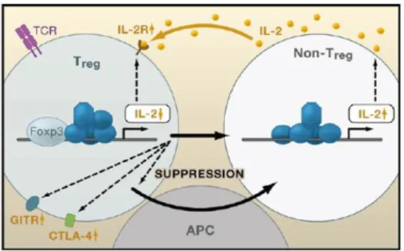
It is possible to conclude that IL-2 has a pivotal role in immune homeostasis since it is
produced by activated non-regulatory T cells (Treg are unable to produce it), and it
contributes to differentiation, maintenance, expansion and activation of Treg which in
turn limits the expansion of non-regulatory T cells (Figure 2). Disruption of this feedback
loop promotes the development of autoimmunity.
Figure 2 – Key role of IL-2 in immune homeostasis
26
IL-2 signaling in Treg, non-Treg and APCs. Foxp3 represses IL-2 transcription in Treg, making them highly dependent on exogenous IL-2 (produced by non-Treg). Foxp3 regulates, as well, the expression of Treg-associated molecules as CD25, GITR and CTLA4.
But it was only in the early 2000s that was discovered the main marker of Treg: Forkhead
box P3 (Foxp3). It is a member of head/winged-helix family of transcription factors and is
a
master
regulator
gene
of
Treg
development
and
function.
It was identified as a defective gene in Scurfy mice, which exhibit an X-linked recessive
disease that causes death one month after birth. This mice show hyperactivation of CD4
+T cell and overproduction of proinflammatory cytokines [31]. Furthermore, mutations of
the human FOXP3 gene causes the human counterpart of Scurfy which is called IPEX [32].
Given the similarities of the diseases caused by Foxp3 mutations among the species, it
was hypothesized that it must have a key role in Treg development and function [33],
[34], [35].
To confirm this, it was shown that ectopic transduction of Foxp3 gene in CD25
-CD4
+T cell
can convert them to CD25
+CD4
+Treg-like cells that were suppressive and could control
autoimmunity [33]. Moreover, Foxp3-deficient mice and Scurfy mice have few
CD25
+CD4
+Treg, and inoculation of CD25
+CD4
+T cell prevents systemic inflammation in
Scurfy mice [35].
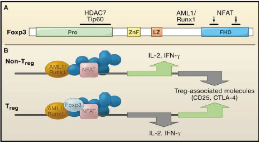
Foxp3 interacts with several transcription factors: Nuclear Factor of Activated T cells
(NFAT), Runt-related transcription factor 1 (AML1/Runx1), the histone acetyl transferase
(HATs)/histone deacetyl transferase(HDAC), Nuclear Factor-kB (NF-kB) and many others
(Figure 3A).
Figure 3 – Control of Treg by Foxp3
27
In A, the structure of Foxp3 locus. Black bars are binding sites for other transcription factors or chromatin-remodelling enzymes. In B, NFAT and AMLA/Runx1 regulate genes encoding molecules in Treg and non-Treg, depending on the presence of Foxp3.
Upon activation, NFAT forms a complex with Activator Protein 1 (AP-1) and NF-kB and
promotes the expression of IL-2, Il4, Ctla4, and other genes in conventional T cells
contributing to their differentiation in effector T cells [36]. In these cells, AML1/Runx1
binds to the IL-2 promoter facilitating the assembly of transcriptional activation
complexes driving IL-2 production. In contrast, in Treg, AML1/Runx1 binds physically to
Foxp3 and disruption of this binding impairs Foxp3-dependent suppression of IL-2
production.
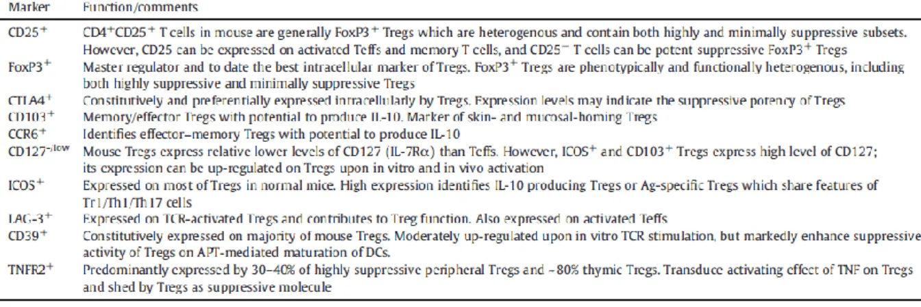
There are other markers of Treg related to their function that are differentially expressed
compared to Tconv, in both humans and mice. I will list the mains [37], which will be
further explained in the “function” chapter of Treg:
CTLA-4: which downregulates T cell activation by competing with CD28 for B7
binding
CD103: which is expressed mainly by mouse Treg and identify IL-10 producing Treg
and is a marker of skin and mucosal retention of Treg
CCR6: which is a marker of Th17 cells
CD127: Treg have low expression of this marker compared with conventional T
cells
ICOS (Inducible T-cell COStimulator): which is expressed by the majority of mouse
Treg and by half of human Treg, namely whose that produce IL-10 and TGF-
,
while ICOS
-cells produce only TGF-
LAG-3 (Lymphocyte Activation Gene-3), a ligand of MHC II, which is expressed on
activated cells
CD39: which characterize functional Treg and contribute to their suppression
consuming extracellular ATP
TNFR2: which binds TNF and is expressed at very low level on conventional T cells
Here there are two tables resuming, in a more extensive way, the main markers of mouse
and human Treg (Table 1).
28 Table 1 – Markers of mouse and human Treg
From: Chen, X. and J.J. Oppenheim. Int Immunopharmacol, 2011. 11(10): p. 1489-96.
2.3
Features of Treg stability and activation
Recently, researchers have put a lot of efforts to couple NGS techniques to Treg to
unravel their core signature meaning the major genes characterizing their phenotype.
To this regard, Benoist et al. [38] compared the transcriptome of Treg and conventional T
cells (Tconv) from mice and humans at the steady state using single cell RNA-sequencing.
They saw two separated clusters for Treg and Tconv, as expected. However, surprisingly,
they saw some so-called “furtive Treg” with a transcriptome clustering with the Tconv
(26% in mice and 55% in humans). Foxp3 was detected in those cells at similar levels to
29
those of other Treg and canonical Treg-signature genes were also overexpressed in these
cells compared to surrounding Tconv. However, some of these key
genes were present at
lower levels compared to other Treg, suggesting that they could be “weaker” Treg with a
poorly suppressive phenotype.
In addition, they also saw Tconv and Treg clustering together in three separated minor
clusters. One of these contains cells expressing genes associated with residence in B cell
follicles (T follicular helper and regulatory cells) and the others contain cells that
upregulate a set of genes associated with early response to TCR engagement. These
observations mean that even if Treg are generally distinguishable from Tconv, the two
populations were imbricated at different levels with an important degree of overlap and
that TCR signaling may drive similar programs in both Treg and Tconv.
To go deeper with the analysis, they systematically compared Treg with closest Tconv and
identified a small set of genes (IL-2ra, IL-2rb, Ikzf2, Ctla4, Capg, Tnfrsf4, Tnfrsf18, Izumo1r,
Chchd10,
Gpr83
and
Foxp3)
overexpressed
by
Treg
in
all
clusters.
Moreover, they also pointed out that in the big cluster of Treg it is possible to identify six
clusters, half of which contain resting and the other half activated Treg according to Sell
(CD62L) and Ccr7 expression. Among the activated ones, there were Treg expressing
genes of early cell activation thus evoked TCR-mediated activation, while others
characterized by a diverse set of genes.
These data describe well Treg identity which is not so separated from Tconv but also their
heterogeneity suggesting that in inflammatory conditions or in different tissues we might
find Treg expressing different sets of genes.
There are several papers supporting this heterogeneity investigating the transcriptomes
of intratumoral Treg.
One of this [39] compares the transcriptome of Th1, Th17 and Treg infiltrating colorectal
or non-small-cell lung cancers to the same subsets from normal tissues. It was noticed
that tumor infiltrating Treg cluster together in both tumors suggesting common features
in
these
cells
dictated
probably
by
the
tumor
microenvironment.
They up-regulated, also at protein level, several immune checkpoints (GITR, OX40, TIGIT,
LAG-3, TIM-3) and their ligands (OX40L, Galectin-9, CD70). In general, they showed an
enrichment in genes related to lymphocyte activation and with increased suppressive
30
activity (OX40, CTLA4 and GITR). It was also noticed that tumor infiltrating Treg express
high amounts of 4-1BB, a costimlulatory molecule that seems to mark antigen-activated
Treg.
Moreover, Treg display the most pronounced differences in gene expression among CD4+
T
cell
subsets
infiltrating
normal
and
tumor
tissues.
This suggests that tumor microenvironment influences specific gene expression in Treg
and this correlates with the evidence that Treg from different tissues are instructed by
environmental factors to display different gene-expression profiles.
Another work [40]compared Treg from untreated human breast carcinomas, normal
mammary gland and peripheral blood and showed that tumor infiltrating Treg are very
similar to tissue-resident Treg but very different from Treg from peripheral blood. Thus,
residence in a non-lymphoid tissue, regardless of whether it is healthy or with oncogenic
transformation, serve as a major determinant of gene-expression characteristics. Once
again tumor Treg were enriched in genes for cytokines signaling, defense response,
inflammatory response and lymphocyte activation compared to cells from peripheral
blood. Of these genes, a subset was more highly expressed by Treg in tumor compared to
tissue-resident cells, including EBI3, OX40 and IL1R2.
Lastly, it was investigated the impact of Helios in Treg identity. This transcription factor
might be dispensable for Treg activity at the steady state but seems to be essential during
inflammation to maintain a stable phenotype and potentiate suppressive function.
Indeed, Helios deficiency in Treg leads to enhanced antitumor immunity that reflects the
induction of an unstable phenotype and conversion of Treg into Tconv within the tumor
microenvironment [41].
It was shown that Helios-deficient intratumoral Treg displayed increased levels of some
receptors associated with final Treg differentiation as Icos, Tnfrsf18, Tnfrsf9, Klrg1 and
Ilr1. But they also up-regulated factors involved in Th lineage determination and cytokine
production. In general Helios-deficient Treg resemble the so-called “tissue Treg”.
Moreover, the expression levels of GITR and OX40 have been positively correlated with
the strength of TCR signaling during Treg development and in mature Treg. Increased
expression of those receptors promotes Treg maturation. Since GITR is highly
up-31
regulated in Helios-deficient Treg this might implicate that the TCR repertoire in those
cells is skewed toward increased self-reactivity [41].
Interestingly, recently it was demonstrated an important role of the TNFSF in the
generation of effector Treg (eTreg) from central Treg (cTreg) [42]. Indeed, Treg generated
in the thymus recirculate through secondary lymphoid organs as cTreg. In this state they
already have regulatory functions, however they do not express the full repertoire of
molecules associated with their suppressive capacity. This means they undergo further
differentiation in the periphery to acquire a fully suppress phenotype, like dowregulation
of CD62L, upregulation of Blimp-1, ICOS, TIGIT, CD73, PD-1 and IL-10 production.
Maturation and functional specialization of Treg are associated with their propensity to
localize to non-lymphoid tissues, and indeed the majority of Treg in those tissues are
eTreg. IRF4 is required for this differentiation and its deletion in Treg impairs their ability
to suppress inflammation [43]. IRF4 can be activated by TCR signaling which is required
for Treg functional maturation in the periphery. However, Treg survive and suppress
without TCR, suggesting that there should be additional signals promoting Treg survival
and function [44]. In fact, other signals can be driven through TNFR family members,
which are highly expressed on Treg. It was demonstrated that they activate RelA which
has a critical role in development and maintenance of eTreg. Deficiency of RelA
compromised the fitness of eTreg in lymphoid and non-lymphoid tissues [42] (See also
Annex 1).
All these data underlie the main genes involved in Treg biology but also highlight the
heterogeneity of these cells based on their development, residency, activation status and
impact of the surrounding environment. One of the major driver of Treg diversity is surely
the site of their generation, which will be discussed in the next paragraph.
2.4
Treg development
As already mentioned, it is possible to distinguish 2 major types of Foxp3
+CD4
+T cells
based on the site of development: thymic Treg (tTreg) generated in the thymus and
peripheral Treg (pTreg) for peripherally generated cells. Then, there is a third class, called
induced Treg (iTreg) for in vitro generated cells. They are generated starting from naïve
32
CD4
+T cells (defined by Foxp3
-CD44
lowCD62L
hi) isolated and cultured with
anti-CD3/anti-CD28 in the presence of IL-2 and TGF-
2.4.1 Development of tTreg
Foxp3
+thymocytes are detectable from a late DP stage to SP stage, constituting around
5% of CD4
+thymocytes and less than 1% of CD8
+thymocytes (Figure 4A).
Figure 4 – Thymic development of nTreg
From: Sakaguchi, S., et al. Cell, 2008. 133(5): p. 775-87
In A, the percentage of Foxp3+ cells in thymocytes subpopulation. In B, Development of Foxp3+ Treg in the thymus involves interaction with stromal cells. Foxp3- thymocytes turn on a Treg differentiation program when they receive signals produced by the interaction between their TCRs and MHC/self-peptide complexes on stromal cells. The signal involves also accessory molecules and stromal cell-derived humoral factors.
Evidences suggest that tTreg may have a TCR with higher affinity for thymic
MHC/self-peptide ligands than those of other T cells and that those self-reactive cells are selected
as tTreg precursors. This was confirmed in double-transgenic mice expressing a
transgene-encoded peptide in thymic stromal cells at high level and in this case T cells
expressing the TCR specific for the peptide differentiate into Treg [7],[45].
In contrast, Treg fail to develop when these mice express either low-affinity transgenic
TCR or a high concentration of the peptide, presumably due to insufficient positive
selection or strong negative selection, respectively. The self-reactivity of Treg could be a
consequence of positive selection of highly self-reactive T cells or a resistance to negative
selection [46].
In addition to the affinity of the TCR/MHC interaction, the intensity of the binding
between accessory molecules contributes to tTreg generation. Deficiency of CD28, CD40,
CD11a/CD18, or CD80 and CD86 results in a reduction of tTreg [47].
At a cellular level, both mTEC and DCs in the thymus contribute to tTreg generation.
Interestingly, Aire-deficient mice develop an autoimmune disease similar to the one
33
produced by Treg depletion. A study suggests that Aire
+mTECs expressing tissue antigens
induce the development of Treg specific to recognize tissue specific antigens [48].
An interesting feature is that it seems that Foxp3 is not required for this initial cell fate
determination in the thymus [49],[50]. In mice where the Foxp3 enconding region was
substitute with a reporter GFP protein, GFP-positive thymocytes, which do not express
Foxp3, have a Treg phenotype although they are not suppressive in vitro. This suggests
that the interaction between thymocytes and stromal cells turns on a program of
transcriptional regulation (with or) upstream Foxp3; and once it is on, Foxp3 stabilizes
and sustains Treg phenotype and confers suppression activity (Figure 4B).
Recent genome-wide analysis showed different patterns of DNA methylation or histone
modification between Tconv and Treg.
For example, Treg-specific DNA hypomethylation (associated to open chromatin and
therefore accessibility to transcription factors) occurs in a limited number of loci, half of
which are located in genes important for Treg phenotype and functions [51].
Furthermore, some changes in DNA methylation are highly stable, such as the
demethylation of the conserved noncoding sequence 2 (CNS2) in the intron 1 of Foxp3
which is bound by Foxp-3, Gata-3 and STAT5 in Treg and enhances the expression of
Foxp3 [52],[53],[54]. In contrast, DNA methylation of Ilra varies with the activation status
of Treg.
Treg-specific epigenetic pattern appears in developing Treg, already in the SP stage, and
progresses from the thymus to the periphery. Treg-specific DNA hypomethylation and
Foxp3 expression have distinct roles in tTreg development. For example, Foxp3 represses
some molecules important for Treg functions (IL-2 and IFN-
), whereas Treg-specific
epigenetic changes do not [51].
Therefore it seems that there is a cooperation between Foxp3 and epigenetic
modifications, supported by the fact that Treg-specific DNA hypomethylated regions in
the genome are different from Foxp3-binding sites [55].
Moreover, chromatin accessibility of Foxp3-binding sites is similar between Treg and
Tconv [56], suggesting that Foxp3-dependent regulation is independent of epigenetic
changes.
34
and stable phenotype. TCR stimulations is important for both events, but while Foxp3
expression seems to depend on the intensity of TCR stimulation, the epigenetic changes
depend more on the duration of TCR stimulation [51]. Therefore, only thymocytes that
have acquired the Treg-specific epigenetic changes become poised to express Foxp3, and
if they do express Foxp3 they will become mature Treg. While thymocytes that have
acquired the Treg-specific epigenetic pattern, but do not express Foxp3, can potentially
become Treg, Foxp3-expressing thymocytes withouth the correct epigenome lose Foxp3
and fail to differentiate into Treg [57] (Figure 5).
Figure 5 – A model for nTreg development in the thymus
From: Ohkura, N., Y. Kitagawa, and S. Sakaguchi. 2013. 38(3): p. 414-23.
TCR rearrangement generates diverse TCRs that recognize with different intensities and durations self-ligands (shown as gradients). Foxp3 expression is driven by high intensity TCR stimulation, while DNA hypomethylation is induced by high duration of TCR stimulation. When the signal is high and persistent, stable Treg develop. Foxp3+ cells without the right epigenome are unstable Treg, while T cells with the right Treg-type epigenome without Foxp3 expression are potential Treg. Too strong or too weak intensity of TCR stimulation leads to apoptosis (negative selection) and death by neglect (positive selection).
It is possible to conclude that thymic Treg development is a two-step process. An initial
signal driven by TCR and CD28 leads to the expression of IL-2R and chromatin remodeling.
A second, TCR-independent step, occurs when Treg progenitors respond to IL-2
stimulation, driving the expression of Foxp3.
However, there are addition signals that play a role in thymic Treg development and since
a hallmark of Treg progenitors is the high expression of TNFRSF members, their role was
investigated. GITR expression precedes the induction of Foxp3, therefore it might support
35
the conversion into mature Treg. In addition to GITR, it was found the expression of OX40
and TNFR2 on Treg progenitors. Moreover, mTECs express the corresponding ligands
GITRL, OX40L and TNF. It was also shown that thymic progenitors that receive the
strongest TCR signaling during development have the highest GITR, OX40, TNFR2
expression. The expression of these receptors seems to be driven by TAK1, downstream
of the TCR and CD28 signaling.
How the TNFRSF members promote the development of Treg is not clear. One possibility
could be that they enhance sensitivity to IL-2, allowing Treg to respond to lower
concentration of IL-2. An alternative mechanism could be the induction of antiapoptotic
proteins. Of course it might also be a cooperation of different mechanisms [58].
2.4.2 Development of pTreg
Some of the early evidences of peripheral conversion of naïve conventional CD4
+cells into
Treg originated from adoptive transfer experiments in which polyclonal CD25
-CD4
+naïve
T cells were injected into lymphopenic mice or mice containing a monoclonal T cell
repertoire devoid of tTreg. In this condition, part of donor population became
CD25
+CTLA-4
+GITR
+and acquired Foxp3 expression and suppressive activity [59],[60].
The conditions for the development of pTreg vary a lot, for instance they appear in
mesenteric LN during induction of oral tolerance [61], they continuously differentiate in
the lamina propria of the gut in response to microbiota and food antigens [62], but they
are also generated in chronically inflamed tissues [63] and transplanted tissues [64].
The requirements necessary for pTreg development are the following: TCR stimulation,
IL-2 and TGF-
. TGF-
induces Foxp3 transcription cooperating for the binding of NFAT and
STAT5 on the Foxp3 gene [65]. In vivo, neutralization of TGF-
impairs [66] oral tolerance
and inhibits the differentiation of antigen-specific pTreg. In contrast, the role of IL-2 in the
development of pTreg is not very clear. IL-2 is required in cultures of naïve CD4
+T cells,
stimulated with anti-CD3 and TGF-
, for the TGF-
-mediated Foxp3 transcription and
suppressive activity of iTreg [67]. IL-2 signaling activates STAT5 which binds to the Foxp3
gene and may cooperate for Foxp3 induction [29].
As for tTreg development, there also other signals that impact on pTreg generation. Some
researchers have showed that TNFRSF members might actually inhibit the differentiation
36
of pTreg. Zhang et al., starting from observations that TNF inhibits iTreg differentiation in
vitro, decided to investigate its impact in vivo. They saw, in a model of EAE, that
neutralization of TNF ameliorates the disease increasing the levels of Treg but not their
function. And they showed that the increased percentage of Treg is due to the increased
number of iTreg while nTreg were not affected. Moreover, depletion of iTreg abolished
the effect of the anti-TNF treatment. The proposed mechanism involves the binding of
TNF to the TNFR2 which inhibits Foxp3 transcription through inhibition of TGF-
-induced
Smad3 phosphorylation, decreasing its binding to the Foxp3 promoter during iTreg
generation. The inhibition of Smad3 phosphorylation is mediated by Akt, directly
activated by the TNF signaling [68].
Given the negative role of TNFR2 in iTreg and pTreg generation, other receptors of the
family have been investigated. It was recently showed that OX40 inhibits iTreg
differentiation in vitro and this mechanism was studied. Gene array analysis of activated
CD4
+T cells with and without OX40 costimulation revealed the upregulation of Batf3 and
Batf following OX40 triggering. BATF3 and BATF are members of the AP-1 family
transcription factors and in the Foxp3 locus there are five putative AP-1 binding sites and
indeed one way of inhibition of iTreg generation through OX40 involves repression of
Foxp3 expression by BATF3 and BATF. However, this is not the only way for OX40 to
inhibit iTreg because in the absence of BATF3 and BATF it activates AKT-mTOR pathway
that suppresses Foxp3 expression[69].
(See fig.1 in “Additional Results” for further details of the role of TNFRs in iTregs).
2.5
Treg stability and plasticity
The molecular and cellular bases of Treg stability remain a key issue of Treg research. To
use Treg in clinical for immunosuppression, it is important to know exactly how they
develop
in
terms
of
Foxp3
expression
and
epigenetic
changes.
What does Treg stability mean, first of all? It could be defined by the maintenance of all
the following characteristics: Foxp3 expression, suppressive activity and lack of effector
activity. Stable Foxp3 expression is due mainly to specific epigenetic modifications. As
already mentioned, the CNS2 at the 5’UTR of Foxp3 must be hypomethilated in Treg
allowing transcription [70]; and this pattern is maintained mainly by IL-2.
37
Many studies, in the beginning, have suggested that Treg development leads to a
terminally differentiated population [71], suggesting Treg are a highly stable lineage.
However, others have suggested that Treg can lose Foxp3 expression and develop a
pro-inflammatory, memory-like phenotype in certain disease states [72].
Indeed tTreg have been shown to convert to different helper T cells such as Th1, Th17 or
Tfh cells [73]. Under lymphopenic conditions, many Treg were found to lose Foxp3 and
start to produce IL-2 and IFN-
.
The topic is still controversial in part because of different strains of mouse used, then
because of different methods used to sort Treg and lastly, it is also possible that T cells
transiently upregulate Foxp3 during development leading to a “false labeling” of cells
[75].
2.5.1 Treg stability
Most reports have suggested that these unstable Foxp3
+cells constitute a minor fraction
under normal conditions. Thus, there must be mechanisms that prevent the pathogenic
conversion of Treg.
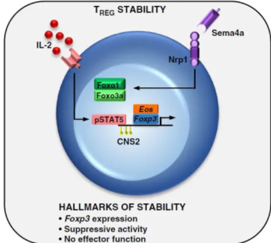
Once Foxp3 is up-regulated, its expression must be kept to assure Treg function and
stability. One important molecule is STAT5 (which is downstream of the IL-2 signaling),
which binds the Foxp3 promoter, stabilizing the locus [29].
But also Foxo1 and Foxo3a are involved because they prevent Treg from taking effector
functions [76]; indeed in the absence of Foxo1, Treg development is diminished and they
are unable to suppress in vitro leading to autoimmunity in vivo [77]. Stable Treg have
reduced Akt expression, leading to enhanced Foxo1 and Foxo3 in the nucleus [78] (Figure
6).
38 Figure 6 – Maintenance of Treg stability in vivo
From: Overacre, A.E. and D.A. Vignali. Curr Opin Immunol, 2016. 39: p. 39-43.
IL-2 signaling leads to phosphorylation of STAT5 that contributes to Foxp3 expression. Moreover, Nrp1 increases the suppressive activity of Treg. All these are hallmark of stability of Treg.
Eos, a zinc-finger transcription factor, was identified as a key factor for Treg stability [79].
Overexpression of Eos avoids Treg reprogramming even in destabilizing environments.
Another important pathway is mediated by Neuropilin-1 (Nrp1):Semaphorin-4a (Sem4)
axis. Most of Treg express high levels of Nrp1 and it increases function and stability of
Treg [80]. Indeed, Nrp1-deficient Treg lose stability and suppressive capacity in tumor
microenvironment and blocking antibodies to Nrp1 or Sem4 were found to limit tumor
growth. This pathway seems to activate Phosphatase and Tensin Homolog (PTEN), which
in turn limits Protein Kinase B (PKB/Akt) activity, leading to increase nuclear translocation
of Foxo1 and Foxo2, stabilizing Foxp3 expression.
As already said, the hypomethylation of CNS2 is also critical, not for Foxp3 induction but
maintenance. Indeed in vitro generated Treg with TGF-
and IL-2 does not present this
pattern and are indeed more unstable; while pTreg generated in vivo present the Treg
specific demethylated region (TSDR) [70].
In addition to the transcriptional and epigenetic mechanisms controlling Treg stability,
there are also MicroRNAs (miRNA). In model of Dicer-deficiency, Treg are unstable and
ineffective suppressor, producing effector cytokines and losing Treg-associated proteins
[81]. MiR-155 is important for maintaining Treg fitness downregulating the STAT signaling
inhibitor SOCS1, augmenting responsiveness to IL-2 [82].
39