HAL Id: tel-03001290
https://tel.archives-ouvertes.fr/tel-03001290
Submitted on 12 Nov 2020HAL is a multi-disciplinary open access archive for the deposit and dissemination of sci-entific research documents, whether they are pub-lished or not. The documents may come from teaching and research institutions in France or abroad, or from public or private research centers.
L’archive ouverte pluridisciplinaire HAL, est destinée au dépôt et à la diffusion de documents scientifiques de niveau recherche, publiés ou non, émanant des établissements d’enseignement et de recherche français ou étrangers, des laboratoires publics ou privés.
Omics of human plasma lipoproteins : role in
cardiometabolic diseases
Emil Zakiev
To cite this version:
Emil Zakiev. Omics of human plasma lipoproteins : role in cardiometabolic diseases. Human health and pathology. Sorbonne Université, 2019. English. �NNT : 2019SORUS436�. �tel-03001290�
Sorbonne Université
Ecole Doctorale Physiologie, Physiopathologie et Thérapeutique (ED394)
INSERM-UMR-S 1166/Equipe 4
Omics of human plasma lipoproteins:
Role in cardiometabolic diseases
Par Emile ZAKIEV
Thèse de Doctorat de Physiopathologie
Dirigée par Dr. Anatol KONTUSH
Présentée et soutenue publiquement le 28 Juin 2019
Devant un jury composé de :
Dr. Philippe LESNIK Directeur de Recherche INSERM Président du
Jury Sorbonne Université
Dr. Anne NEGRE-SALVAYRE Directeur de Recherche CNRS Rapporteur
Université Toulouse III Prof. Dominique
BONNEFONT-ROUSSELOT
PU-PH de l’Hôpital Pitié Salpêtrière
Rapporteur Université Paris Descartes - Paris 5
Dr. Mikael CROYAL Ingénieur d'études Examinateur
Université de Nantes
Dr. Boris HANSEL MCU-PH de l’Hôpital Bichat - Claude Bernard Examinateur Université Paris Diderot – USPC
Dr. Anatol KONTUSH Directeur de Recherche INSERM Directeur de
thèse Sorbonne Université
TABLE OF CONTENTS
ACKNOWLEDGMENTS ... 1 LIST OF ABBREVIATIONS ... 5 RESUME ... 8 ABSTRACT ... 11 INTRODUCTION ... 14 1. Introduction to lipoproteins ... 14 1.1 LDL ... 16 1.2 HDL ... 161.3 Other lipoprotein classes ... 17
2. Epidemiology of lipoproteins ... 18
2.1 LDL ... 18
2.2 HDL ... 19
2.3 Other lipoprotein classes ... 20
3. HDL ... 20 3.1 Structure ... 20 3.2 Composition ... 22 3.2.1 Classic techniques ... 22 3.2.2 Omics approaches ... 24 3.2.2.1 Proteome ... 25 3.2.2.2 Lipidome ... 27 3.2.2.3 Glycome ... 31 3.3 Heterogeneity ... 31 3.4 Metabolism ... 35 3.4.1 Biosynthesis ... 35 3.4.2 Remodelling ... 36 3.4.3 Catabolism ... 36
3.5 Biological activities and structure-function relationships ... 37
3.6 Dysfunctional HDL ... 41
3.6.1 Cardiometabolic diseases and atherosclerosis ... 41
3.6.2 Altered composition and impaired biological function ... 43
3.6.3 Altered metabolism ... 45
3.7 Therapeutic targeting of abnormal HDL metabolism ... 45
3.7.1 CETP inhibition ... 45
3.7.3 LPL activation ... 48
3.7.4 Nicotinic acid ... 49
3.7.5. Other agents ... 50
4. LDL ... 51
4.1 Structure and composition ... 51
4.1.1 Proteome ... 51 4.1.2 Lipidome ... 52 4.1.3 Glycome ... 52 4.2 Metabolism ... 52 4.2.1 Biosynthesis ... 52 4.2.2 Catabolism ... 54
4.3 Altered composition and metabolism in cardiometabolic diseases ... 54
4.4 Therapeutic targeting of abnormal LDL metabolism ... 56
WORKING HYPOTHESIS AND OBJECTIVE ... 59
METHODS ... 61
1. Subjects ... 61
2. Blood samples ... 62
3. Isolation of lipoproteins ... 62
4. Chemical analysis of lipoproteins ... 64
5. Heterogeneity and mean size of HDL ... 64
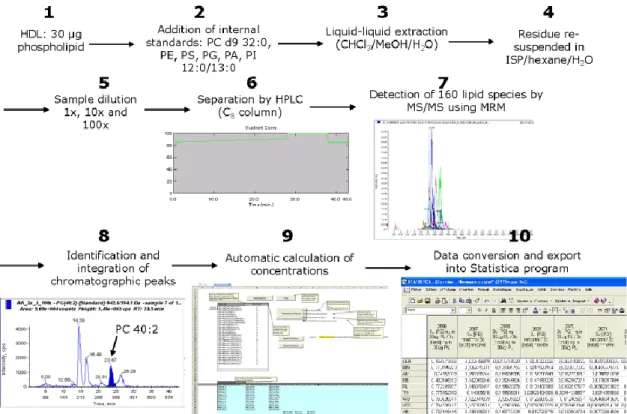
6. Lipidomic analysis ... 65 6.1 Lipid standards ... 66 6.2 Lipid extraction ... 67 6.3 LC/MS analysis ... 68 6.4 Quantification ... 68 6.5 Sphingosine-1-phosphate ... 69 7. Glycomic analysis ... 70 8. Functional assays ... 71 8.1 Cholesterol efflux ... 72 8.2 Antioxidative activity of HDL ... 72 8.3 Anti-apoptotic activity of HDL ... 74
8.4 Cellular cholesterol accumulation induced by LDL ... 74
9. Statistical analysis ... 75
RESULTS ... 79
1.1 Lipidome ... 79
1.2 Function ... 83
2. HDL lipidome and functionality in cardiometabolic diseases ... 83
2.1 Familial apoA-I deficiency ... 83
2.1.1. Abundances of individual lipid species. ... 85
2.1.2. PCA ... 92
2.1.3. Heatmaps ... 92
2.1.4. Bubble plots ... 92
2.1.5. Structural plots ... 96
2.1.6. Network analysis ... 100
2.1.7. Metabolic pathways affected by apoA-I deficiency ... 105
2.1.8. Lipid-related genes affected by apoA-I deficiency ... 105
2.2 ST-elevated myocardial infarction ... 108
2.2.1. Characteristics of subjects and overall abundances of lipid classes and species ... 108
2.2.2. PCA ... 116
2.2.3. Heatmaps ... 116
2.2.4. Bubble plots ... 116
2.2.5. Structural plots ... 117
2.2.6. Network maps ... 123
2.2.7. Metabolic pathways affected by STEMI ... 126
3. HDL and LDL glycome in normolipidemic controls ... 127
3.1. Glycomic profiling of LDL and HDL ... 127
3.2. Role of glycome in biological activities of lipoproteins ... 135
3.2.1. Cholesterol efflux capacity of HDL ... 135
3.2.2. Cellular cholesteryl ester accumulation induced by LDL ... 136
3.2.3. Heterogeneity, mean size and LCAT activity of HDL ... 137
3.2.4. Relationships between the glycome and biological properties of LDL & HDL... 138
DISCUSSION AND CONCLUSIONS ... 142
1. Lipidome of normolipidemic HDL particles ... 142
2. Lipidome of HDL particles in CMD ... 145
2.1 Lipidome of HDL particles isolated from apoA-I–deficient patients ... 145
2.2. Lipidome of HDL particles isolated from STEMI patients ... 150
3. Glycome of normolipidemic HDL and LDL ... 154
4. General conclusions ... 158
ACKNOWLEDGMENTS
My PhD project has been developed at the UMRS 1166 Research Unit of the French National Institute for Health and Medical Research (INSERM) and the University of Sorbonne (Doctoral School ED394 “Physiology, physiopathology and therapeutic”). Here I would like to acknowledge people who have supported me during this process.
First, I would like to express my sincere gratitude towards my supervisor, Dr. Anatol Kontush, a persistent and rigorous scientist with an unrivaled expertise in his field. His optimism, practicality and wisdom saved the day for me for too many times to count. Without his constant support I would certainly have not been able to finish this project. He taught me all the intricacies of being a scientist in a modern world – a knowledge not to be underestimated. I am profoundly grateful for his kindness for giving me the opportunity to perform my PhD in his team.
I would also like to thank Dr. Isabelle Guillas-Baudouin, who is an honored professional in her field, humble, energetic and appealing. Isabelle was always there when I needed help, be it a theoretical or a practical question, even if she was extremely busy with her own research or teaching. Her mere presence was comforting; during my project her advices were often stalemate-breaking.
I would also like to thank Dr. Maryam Darabi-Amin, with whom we became good friends, and who also helped me with my project considerably. She was always helpful in my project even though, being a postdoc at the time, she had a lot of other important things to do.
During these years, I also befriended another PhD student, now Dr. Feng Ma, and I thank him for his constant moral support and good company. I will remember our giggles during lunch breaks and those good times spent together during the free time of scientific conferences.
I owe Mr. Vasily Sukhorukov and Ms. Emilie Tubeuf a debt of gratitude for teaching me how to perform the experiments the first time when I came to the lab, it was a really nice memory of their careful guidance. Dr. Eric Frisdal and Dr. Wilfried Le Goff were always there to teach me the lab techniques as well, and I am sincerely grateful to them too.
I would like to thank Dr. Marie Lhomme and Dr. Fabiana Rached for helping me to perform the measurements in the lipidomics. I am also grateful for substantial support from Professors Raul Santos and Carlos Serrano from the University of Sao Paolo, Professor Gordan Lauc and Dr. Maja Pučić Baković and Dr. Ivan Gudelj from the University of Zagreb, and Professor Patrice Therond from the Hospital of Le Kremlin-Bicetre. Without their input, my thesis would not have happened.
Many thanks to all the members of Teams 4 and 5 of UMRS 1166. I would like to thank all the scientists in the team, especially Dr. Philippe Lesnik, Dr. Maryse Guerin, Dr. Philippe Couvert, Dr. Thierry Huby, Dr. Emmanuel Gautier and Ms. Martine Moreau for their help and constructive criticism in the development of my project.
I would equally like to express my gratitude to the members of my thesis committee, Dr. Philippe Lesnik, Dr. Anne Negre-Salvayre, Prof. Dominique Bonnefont-Rousselot, Dr. Boris
Hansel and Dr. Mikael Croyal, whose helpful participation was essential for this project to become fruitful.
I send my special thanks to Mme Severine Lowinski, Dr. Isabelle Cremer and Dr. Catherine Monnot of the Doctoral School ED394 for their constant support and care. Also big thanks to the secretary staff of our department, especially to Mme Natalie Abiola, Mme Olga Nobel, Mme Roberte Le-Galleu, Mme Angelique Riquelme, and Mme Martine Duquesne for their constant work on making our work possible.
Furthermore, I would like to acknowledge the financial support which was provided by the Ministry of Foreign Affairs of the French Republic (Vernadski scholarship) and support provided by the Institute for Atherosclerosis Research, Skolkovo, and its director, Prof. Alexander Orekhov. Thanks to these means of support I was able to do my project in France. Our team also gratefully acknowledges continuous financial support from French National Institute for Health and Medical Research (INSERM).
I would like to conclude with thanking one special person for her continuous support and endless love. I am deeply grateful to her for enduring my whims. She would always make me believe in myself when I felt like the world had me down on my knees and support me in every crucial decision in my life. “Behind every great man, there is a great woman”, they say. While I certainly have not achieved grandiosity by any means, I would not be even at this level without the Great Woman of My Life - Adele.
to my dear sister Galiya, to my kind cousins Renat and Nailya, to my cool uncle Ildar, and to my awesome grandparents Mirfatyh and Elena for their unconditional love and support in good and hard times during my sometimes-stormy PhD process.
LIST OF ABBREVIATIONS
A
A1AT α1-antitrypsin
AAPH Azo-initiator 2,2′-azo-bis-(2-amidinopropane) hydrochloride
ABCA1 ATP binding cassette transporter A1
ABCG1 ATP binding cassette transporter G1
ACN Acetonitrile
ACS Acute Coronary Syndrome
AMI Acute myocardial infarction
ApoA Apolipoprotein A ApoB Apolipoprotein B ApoC Apolipoprotein C ApoD Apolipoprotein D ApoE Apolipoprotein E ApoF Apolipoprotein F ApoJ Apolipoprotein J ApoL Apolipoprotein L ApoM Apolipoprotein M B
BET Bromodomain and extra-terminal domain
C
CAD Coronary artery disease
CCHS Copenhagen City Heart Study
CDS Phosphatidate cytidiltransferase
CE Cholesteryl ester
Cer Ceramide
CETP Cholesteryl ester transfer protein
CM Chylomicron
CMD Cardiometabolic disease
CHD Coronary heart disease
CVD Cardiovascular disease
D E
EDTA Ethylenediaminetetraacetic acid
EL Endothelial lipase
eNOS Endothelial nitric oxide synthase
ER Endoplasmic reticulum
F
FA Formic acid
FAID Familial apoA-I deficiency
FC Free cholesterol
FDR False discovery rate
FH Familial hypercholesterolemia
G
GC Gas chromatography
GLTP Glycolipid Transfer Protein
GP Glycerophospholipid
H
HDL High-density lipoprotein
HDL-C High-density lipoprotein-cholesterol
HILIC-SPE hydrophilic interaction liquid chromatography solid-phase extraction
HILIC-UHPLC-FLD
Hydrophilic-interaction ultra-high-performance liquid chromatography with fluorescence detection
HPLC High-performance liquid chromatography
HL Hepatic lipase
hsCRP High-sensitivity C-reactive protein
HUVEC Human umbilical vein endothelial cells
I
ICAM-1 Intracellular adhesion molecule-1
IDL Intermediate-density lipoprotein
L
LA Linoleic acid
lbLDL Large buoyant LDL
LC Liquid chromatography
LC-MS Liquid chromatography – mass spectrometry
LC/MS/MS Liquid chromatography–tandem MS
LC-ESI-MS/MS Liquid chromatography–electrospray ionization–tandem MS
LCAT Lecithin-cholesterol acyltransferase
LDL Low-density lipoprotein
LDL-C Low-density lipoprotein-cholesterol
LDLR Low-density lipoprotein receptor
Lp(a) Lipoprotein(a)
LpA-I apoA-I-containing particle
LpA-I:A-II apoA-I- and apoA-II-containing particle
LPC Lysophosphatidylcholine
LPL Lipoprotein lipase
LpPLA2 Lipoprotein-associated phospholipase A2
LPS Lipopolysaccharide
LRP1 LPLR-related protein-1
LXR Liver X receptor
M
MALDI-TOF-MS Matrix-assisted laser desorption ionization time-of-flight MS
MI Myocardial infarction
miRNAs, miR MicroRNAs
MPO Myeloperoxidase
MS Mass-spectrometry
N
NO Nitric oxide
NMR Nuclear magnetic resonance
PA Phosphatidic acid
PAF-AH Platelet-activating factor-acetyl hydrolase
PBS Phosphate-buffered saline
PC Phosphatidylcholine
PCA Principal component analysis
PCSK7/9 Proprotein convertase subtilisin/kexin type 7/9
PE Phosphatidylethanolamine
PG Phosphatidylglycerol
PGI2 Prostacyclin
PI Phosphatidylinositol
PI3K Phosphatidylinositol 3-kinase
PL Phospholipid
PLOOH Phospholipid hydroperoxide
PLA2G1B Group IB Phospholipase 2
PLTP Phospholipid transfer protein
PNGase F Peptide:N-glycosidase F
PON1 Paraoxonase 1
PPARα Peroxisome proliferator-activated receptor
PS Phosphatidylserine
R
RCT Reverse cholesterol transport
rHDL Reconstituted HDL
RXR Retinoid X receptor
S
S1P Sphingosine-1-phosphate
SAA Serum amyloid A
sdLDL Small dense LDL
SM Sphingomyelin
SL Sphingolipid
SNPs Single-nucleotide polymorphisms
sPLA2 Secretory phospholipase A2
SR-B1 Scavenger receptor class B type 1
STEMI ST-segment elevation myocardial infarction
T T2D Type 2 diabetes TC Total cholesterol TG Triglyceride TGRL Triglyceride-rich lipoprotein TOF Time-of-flight U UC Ultracentrifugation V
VLDL Very low-density lipoprotein
RESUME
Omics vise à la caractérisation et à la quantification collectives de pools de molécules
biologiques qui traduisent notre compréhension de la structure et de la fonction d'un organisme ou d'une partie de celui-ci. Les lipoprotéines plasmatiques humaines jouent un rôle important dans le développement de la résistance à l'insuline, de la dyslipidémie, de l'hypertension, de l'adiposité centrale et de maladie cardiovasculaire (MCV), le tout collectivement appelé maladie cardiométabolique (MCM). En effet, on sait que la diminution des taux de cholestérol
lipoprotéine de haute densité (LHD) et l’augmentation des taux de cholestérol lipoprotéine de basse densité (LBD) sont associées à un risque accru de MCM. En dépit des succès enregistrés dans le développement de médicaments normalisant les taux de cholestérol-LBD (C-LBD), les MCM représentent toujours la majorité de morbidité et des décès dans les pays développés. Nos nouvelles tentatives de réduction du risque de MCM ne réussissent pas toujours, reflétant parmi d'autres notre méconnaissance de biologie des lipoprotéines qui inclut, comme éléments clés, la fonction biologique et les caractéristiques de composition des particules de lipoprotéine lequel les rendent distinctement indispensables pour les conditions métaboliques saines. Notre
compréhension du protéome des LHD et des LBD a beaucoup progressé, alors que leur lipidome et leur glycome n’ont guère retenu l’attention de la communauté scientifique ces dernières années. Ce travail porte sur la lipidomique et la glycomique des LHD et LBD plasmatiques chez l'humain en ce qui concerne leurs fonctions biologiques chez les sujets sains et les patients atteints de MCM, comprenant déficit familial en apoA-I (DFAI) - dyslipidémie déterminée génétiquement entraînant un risque cardiovasculaire (RV) élevé, et infarctus du myocarde avec élévation du segment ST (IMEST), une présentation aiguë des MCV.
Dans la section Introduction du manuscrit, connaissances existantes sur la structure, la fonction et le métabolisme des LBD et des LHD est discuté, tout en abordant brièvement d'autres classes de lipoprotéines, avec un accent particulier sur les approches omiques. La section Méthodes décrit comment le recherche a été accompli, décrivant les méthodes de recrutement de patients, de prélèvements sanguins, d’analyses chimiques des lipoprotéines, de dosages fonctionnels, de détermination par chromatographie en phase liquide-spectrométrie de masse en tandem de la composition en lipoprotéines et d’aboutir aux méthodes statistiques utilisées. La section suivante Résultats décrit les résultats sur le lipidome des lipoprotéines chez les sujets normolipidémiques, le lipidome des lipoprotéines dans le MCM et le glycome des lipoprotéines chez les sujets normolipidémiques. Le lipidome de lipoprotéines, isolé de sujets normolipidémiques et MCM, a été caractérisé et quantifié en environ 160 espèces lipidiques individuelles, appartenant à 9 sous-classes lipidiques: la phosphatidylcholine (PC), la sphingomyéline (SM), la
lysophosphatidylcholine (LPC), la phosphatidyléthanolamine (PE), le céramide (Cer), la phosphatidylsérine (PS), l’acide phosphatidique (PA) et le phosphatidylglycérol (PG). Le lipidome normolipidémique a révélé une forte hétérogénéité parmi les principales sous-populations de HDL (2b, 2a, 3a, 3b et 3c), de petites denses HDL3b et HDL3c présentant un potentiel plus élevé en termes de propriétés anti-athérogènes et enrichies en LPC, PI, PS et PA. En MCM, des perturbations du lipidome des espèces affectées par les LHD de toutes les classes de lipides. Notamment, les LHD DFAI manquaient de nombreuses espèces lipidiques
polyinsaturées, telles que PC 34:2 et PC 40:8, tout en possédant des quantités accrues d'espèces saturées telles que PC 32:0 et PC 34:0, alors que IMEST LHD possédait des quantités réduites d'espèces lipidiques polyinsaturées, notamment PC 36:2 et PC 38:5, tout en possédant des quantités accrues d'espèces pro-inflammatoires lysophospholipidiques saturées, telles que LPC 16:0, LPC 18:0 et PA 32:0. Des altérations du lipidome dans le MCM ont été associées à des
voies de fonctionnement incorrect du métabolisme de l'acide linoléique et des glycérophospholipides.
Quand les glycomes des LHD et LBD normolipidémiques ont été caractérisés, plus de 17 pics chromatographiques distincts ont été identifiés, présentant une diversité élevée. Structurellement, la composition comprenait des fractions du complexe, du mannose élevé aux hybrides
N-glycanes. Les fonctions biologiques des HDL, à savoir la capacité d'efflux de cholestérol cellulaire, les activités antioxydantes et anti-apoptotiques, se sont avérées diminuées dans le MCM comparativement aux témoins sains. Les lipidomes et les glycomes ont montré des associations avec les fonctions biologiques des lipoprotéines, établissant ainsi des relations structure-fonction distinctes pour les deux MCM étudiés. Le manuscrit est fini par une discussion des résultats et des conclusions générales, au cours desquelles on suppose les applications
potentielles des résultats obtenus. Nous proposons notamment un concept de normalisation de la fonction LHD en les exposant à des perfusions de certains lipides qui ont montré des associations avec la fonctionnalité LHD dans nos tests.
Ces études ont été publiées dans le Journal of Clinical Lipidology [1] et Biochimica et Biophysica Acta - Molecular and Cell Biology of Lipids [2].
ABSTRACT
Omics aims at the collective characterization and quantification of pools of biological molecules that translate into our understanding of the structure and function of an organism and/or its components. Human plasma lipoproteins play important roles in the development of insulin resistance, dyslipidemia, hypertension, central adiposity and cardiovascular disease (CVD), all collectively referred to as cardiometabolic disease (CMD). Indeed, decreased levels of high-density lipoprotein (HDL)-cholesterol (HDL-C) and increased levels of low-high-density lipoprotein (LDL)-cholesterol (LDL-C) are known to be associated with an increased risk of CMD. Despite successes in the development of drugs normalizing LDL-C levels, CMD still account for the majority of morbidity and mortality in developed countries. Our further attempts in decreasing CMD risk do not always succeed reflecting among others our poor grasp of lipoprotein biology which includes, as key elements, biological function and compositional features of lipoprotein particles that make them distinctly indispensable for healthy metabolic conditions. Major advances have been made in our understanding of the proteome of HDL and LDL, while their lipidome and glycome has not enjoyed much attention of scientific community. This thesis focuses on lipidomics and glycomics of human plasma HDL and LDL in respect to their biological functions in healthy subjects and in patients with CMD, including familial apoA-I deficiency (FAID), a genetically determined dyslipidemia resulting in elevated cardiovascular (CV) risk, and ST-elevated myocardial infarction (STEMI), an acute presentation of CVD.
In the Introduction section of the manuscript, existing knowledge about LDL and HDL structure, function and metabolism is discussed, while briefly touching upon other lipoprotein classes, with a particular emphasis on the omics methods. The Methods section describes how this research was accomplished, detailing approaches to patient recruitment, blood sampling, chemical analysis
of lipoproteins, functional assays, liquid chromatography-tandem mass-spectrometry determination of lipoprotein composition and statistical methods employed. The following Results section describes findings on the lipidome of HDL in normolipidemic subjects, lipidome of HDL in CMD, and glycome of LDL and HDL in normolipidemic subjects. Lipidome of HDL, isolated from normolipidemic and CMD subjects, was characterized and quantified into ~160 individual lipid species belonging to 9 lipid subclasses, including phosphatidylcholine (PC), sphingomyelin (SM), lysophosphatidylcholine (LPC), phosphatidylinositol (PI),
phosphatidylethanolamine (PE), ceramide (Cer), phosphatidylserine (PS), phosphatidic acid (PA), and phosphatidylglycerol (PG). Normolipidemic lipidome revealed high heterogeneity among major HDL subpopulations (2b, 2a, 3a, 3b and 3c), with small, dense HDL3b and HDL3c showing enhanced anti-atherogenic properties and being enriched in LPC, PI, PS, and PA relative to large, light HDL. In CMD, perturbations of the lipidome of HDL affected species from all lipid classes. Notably, FAID HDLs lacked numerous polyunsaturated lipid species, including PC 34:2 and PC 40:8, while possessing increased amounts of saturated species, including PC 32:0 and PC 34:0, while STEMI HDL possessed decreased amounts of polyunsaturated lipid species,
including PC 36:2 and PC 38:5, while possessing increased amounts of saturated lysophospholipid pro-inflammatory species, such as LPC 16:0, LPC 18:0 and PA 32:0.
Alterations of the HDL lipidome in CMD were associated with improperly functioning pathways of linoleic acid and glycerophospholipid metabolism.
When glycomes of normolipidemic HDL and LDL were characterized, more than 17 distinct moieties were identified and quantified, revealing high compositional diversity. Structurally, lipoprotein glycans varied from complex, high-mannose to hybrid N-glycans. Biological functions of HDL, namely cellular cholesterol efflux capacity, antioxidative and anti-apoptotic
activities, were found to be diminished in CMD as compared to healthy normolipidemic controls. Both lipidome and glycome revealed multiple associations with biological functions of
lipoproteins, thereby establishing distinct structure-function relationships for the two CMD studied. The manuscript is completed with discussion of the results and general conclusions, suggesting potential applications of the results obtained. Notably, we propose a concept of normalizing HDL function through intravenous infusions of certain lipids that showed associations with HDL functionality in our in analysis.
These studies were published in Journal of Clinical Lipidology [1] and Biochimica et Biophysica Acta - Molecular and Cell Biology of Lipids [2].
INTRODUCTION
1. Introduction to lipoproteins
Lipids provide structural, signaling (steroid hormones), and energetic functions (bile acids, nutritional fatty acids). The structural function of lipids involves formation and sustaining of membranes, and this is important in separation of water-soluble and water-insoluble media of the human organism. Since our blood is water-based, lipids need to be compartmentalized in order to be transported through such polar milieu. Here is where lipoproteins come into play: they form micellar structures, engulfing lipids into ready-transportable “packages” with special apolipoproteins being attached to them, serving as ever-vigilant “courier”.
These packages have hydrophobic lipids, including triglyceride (TG) and cholesterol esters (CE), as their major “cargo”. The package is wrapped around with a monolayer of phospholipids (PL) and free cholesterol (FC). Proteins, primarily apolipoproteins and enzymes, serve as an all-in-one transportation company courier: they “pack” the lipids inside, ensuring their safety, and then “knock on the door” of a cell, delivering the lipid cargo to the cell, or from the cell.
Lipoproteins are distinguished by their structural, biological, chemical and physical properties. Differences in physical properties allows separating lipoproteins according to their density, charge and size. In the order of increasing density, major lipoproteins in human plasma are chylomicron (CM), very low-density lipoprotein (VLDL), intermediate-density lipoprotein (IDL), low-density lipoprotein (LDL) and high-density lipoprotein (HDL).
Table 1. Density, size, molecular weight and apolipoprotein contents of different human plasma lipoproteins [3, 4] Lipoproteins Density (g/ml) Diameter (nm) Molecular mass (Da) Major apolipoproteins CM 0.93 75-1200 50-1000×106 B48, E, C, A-Ⅰ VLDL 0.93-1.006 30-800 10-80×106 B100, E, C IDL 1.006-1.019 27-35 5-10×106 B100, E LDL 1.019-1.063 18-27 2.3×10⁶ B100 HDL2 1.063-1.125 9-12 360×103 A-Ⅰ, A-Ⅱ, C HDL3 1.125-1.210 7-9 175×103 A-Ⅰ, A-Ⅱ, C
Pre-β HDL 1.210-1.240 <7 (discoidal) 28-70×103 A-Ⅰ
Lipoprotein(a) (Lp(a)) forms additional lipoprotein subpopulation which consists of an LDL-like particle and the specific apolipoprotein (a) [5]. Density, size and major apolipoproteins of these lipoproteins are listed in Table 1 and Figure 1. LDL and HDL particles play key roles in the development of cardiometabolic disease (CMD), which is a term encompassing insulin
resistance, dyslipidemia, hypertension, central adiposity and cardiovascular disease (CVD) [6]; therefore, we initially focus on these two lipoproteins.
Figure 1. Density and diameter of human plasma lipoproteins[7]
1.1 LDL
LDL particles possess a hydrophobic core predominantly comprised of CE with a minor amount of TG, packaged into PL-FC coat with a single apoB100 molecule and some other minor proteins embedded into it. LDL particles display size of 18-27 nm and density of 1.019-1.063 g/ml. LDL perform their lipid transporting actions with a various degree of lipid capacity [8] and they deliver lipid to cells via receptor-mediated endocytosis [9, 10].
1.2 HDL
HDL are lipoproteins with a diameter of 7-12 nm and density of 1.063-1.21 g/ml. As LDL, HDL carry CE, TG and other lipids in its hydrophobic core, but the core/surface ratio is much smaller in HDL. The coating is made of a monolayer of PL and FC, with apolipoproteins embedded into it.
1.12 1.16
The most abundant of these apolipoproteins are apolipoprotein A-I (apoA-I) and apolipoprotein A-II (apoA-II), which are crucial to HDL metabolism [11, 12].
HDL pool in the circulation is highly heterogeneous and can be separated according to size and density using ultrancentrifugation, gel electrophoresis, and FPLC or on the basis of apolipoprotein composition into three classes: apoA-I-only HDL (LpA-I), apoA-I- and apoA-II-containing HDL (LpA-I:A-II), or apolipoprotein E (apoE)-containing HDL [13]. HDL possess multiple antiatherogenic properties which primarily include cellular cholesterol efflux capacity together with antioxidative, anti-inflammatory, antithrombotic, anti-infectious and vasodilatory activities.
1.3 Other lipoprotein classes
CMs are secreted by the intestine after a meal. These are large and light (75-1200 nm, mean density of 0.93 g/ml) spherical particles [14, 15], predominantly consisting of TG with minor amounts of CE, covered by a monolayer of PL and FC, with proteins residing in the monolayer [16-20]. ApoB48 is the main structural protein of CM. CM levels in the circulation surge shortly after a fatty meal and rapidly decrease in normolipidemic individuals [21, 22]. CMs are formed in the endoplasmic reticulum (ER) of enterocytes and are released into the circulation via the lymphatic system [23, 24]. Elevated CM production is associated with dyslipidemia and increased CVD risk in metabolic disorders, such as in Type 2 diabetes (T2D).
VLDLs are large (30-800nm, 0.93-1.006 g/ml) particles produced by the liver from TG, FC, and apolipoproteins and let off into the circulation [25]. Unlike CM, VLDL transports endogenous liver-derived lipids to peripheral tissues, making it the key transport vehicle for endogenous lipids. Apolipoprotein B100 (apoB100) is the main structural protein of VLDL which ensures its proper assembly and secretion [26]. Newly produced VLDL particles containapoB100, apolipoprotein C (apoC)-Ⅰ, apoE, PL, FC, CE, and TG. During maturation in the circulation, they acquire apoC-II
and apoE from HDL. VLDL is converted in the circulation to IDL and LDL and is a metabolic precursor of LDL [27]. Both IDLs and LDL are recognized by receptors in the liver and endocytosed by the hepatocytes. Once cholesterol level inside IDLs exceeds the level of TG, IDL finally becomes LDL.
Term “TG-rich lipoproteins (TGRL)” encompasses lipoproteins that display a high content of TG, such as CM and VLDL, secreted by the intestine and the liver, respectively. TG in TGRL is hydrolyzed by lipoprotein lipase (LPL), producing free fatty acids and glycerol. Similarly to LDL, remnants of CM and VLDL produced by lipolysis may enter the arterial wall and provoke formation of atherosclerotic lesions [28-30].
2. Epidemiology of lipoproteins
2.1 LDL
Our interest to LDL is primarily derived from the fact that elevated LDL-cholesterol (LDL-C) levels are associated with an increased risk of coronary heart disease (CHD), as shown in multiple large-scale epidemiological studies. Framingham Heart Study, for example, explicitly specified LDL-C as an independent risk factor for CHD after multivariate adjustments [31]. According to this study, circulating LDL-C levels of higher than 130 mg/dl are associated with an increased risk of CHD, reaching values of approximately +70% for the relative likelihood of the development of CHD in patients with LDL-C levels higher than 160 mg/dl. The Atherosclerosis Risk in Communities Study estimated increase in relative risk of CHD as approximately 40% for every 38.8 mg/dl (1 mmol/l) increase from the median values of LDL-C of 88 mg/dl and 95 mg/dl for women and men, respectively [32]. A meta-analysis revealed that single nucleotide polymorphisms (SNPs) of genes associated with increased LDL-C levels were implicated in increased risk of coronary artery disease (CAD) [33]. These genes were coding for the apoB and
apoE protein families, proprotein convertase subtilisin/kexin type 9 (PCSK9) and LDL receptor (LDLR). Other epidemiological studies produced similar results [34-37]. On the other hand, the opposite is also true: naturally occurring SNPs that reduce circulating LDL-C levels are
associated with decreased risk of CHD [38]. The risk decrease is stronger than in patients with statin treatment started later in life reaching similar reduction in LDL-C levels, implying
cumulative effect of the exposure to high levels of LDL-C [39, 40]. All these observations made LDL-C to be colloquially known as “bad cholesterol”.
2.2 HDL
CHD patients typically display reduced circulating levels of HDL-cholesterol (HDL-C) as
compared to healthy controls [41, 42]. In the 60s [43] and 70s [44], low concentrations of HDL-C were implicated in the poor clearance of excessive plasma cholesterol and, as a corollary, were proposed to represent the main cause of CHD development. Combined with the observation of 2-3 % decrease of CHD risk for each additional 1 mg/dl increase of HDL-C [45, 46], and
additionally reinforced by a multitude of large-scale epidemiological studies, reporting an inverse relationship between levels of HDL-C and the risk of CHD [47-52], HDL-C earned itself a moniker “good cholesterol”.
A whole plethora of factors is known to affect HDL-C levels: race [53-55], gender (with women displaying higher levels than men) [56, 57], conditions with compromised metabolism, including familial dyslipidemias [58], T2D [59, 60] or obesity [57].
There is however no consensus on the exact role of HDL subclasses in CVD [61, 62]. It has been reported that small, dense particles, such as HDL3c, are more efficient in performing their biological roles, than large, light HDL2 [63], while at the same time levels of small dense HDL
particles are increased in patients with CHD and levels of large, light HDL are decreased [62], adding to the controversy.
2.3 Other lipoprotein classes
Role of TGRL particles in the atherosclerosis is not particularly straightforward. Indeed,
atherosclerosis and CVD do not preferentially affect individuals with extremely high plasma TG, while those with high LDL-C levels develop premature atherosclerosis [64, 65]. This observation be explained by large TGRLs not being able to penetrate the arterial wall to lay a harmful lipid deposition there [66, 67]. On the other hand, moderately elevated TG levels most probably
feature TGRL particles small enough to enter the arterial wall, yet big enough to cause all sorts of atherosclerotic mayhem there [68-70]. Copenhagen City Heart Study (CCHS), Women’s Health Study and Emerging Risk Factors Collaboration consistently associated increased risks of
myocardial infarction (MI), CHD, stroke and all-cause mortality with raised levels of TG [71-74]. Furthermore, increased fasting triglycerides were associated with elevated risk of ischaemic stroke. Other studies demonstrated that the concentration of non-HDL-cholesterol rather than raised plasma TG was associated with CHD [75-77].
3. HDL
3.1 Structure
ApoA-I is a major component of HDL, accounting for approximately 70% of HDL protein mass [78]. Depending on the presence or absence of a lipid cargo in an HDL particle and this cargo’s composition, apoA-I can occur in multiple forms which include lipid-free apoA-I, a “belt” of discoid HDL (also known as pre-β HDL) [79], or a structural component of spherical HDL particles [78].
Figure 2. X-ray crystal structure of lipid-free apoA-I [78]
The lipid-free apoA-I most probably does not exist in plasma as the protein avidly interacts, trace amounts of lipids. Resulting lipid-poor apoA-I accounts for only 5–10% of total plasma apoA-I yet is believed to possess a crucial role in the HDL metabolism. ApoA-I is a 243-amino acid, 28-kDa single polypeptide organized into eight α-helical segments of 22 amino acids and two 11-mer repeats, leaving only 44 N-terminal amino acids not pertaining to any tertiary structure (
Figure 2). Another element in the process of HDL maturation (see section 3.4.2) is represented by discoidal HDL which contains small amounts of amphipatic lipids, such as PL and FC, alongside the apoA-I itself. The apoA-I in the discoidal HDL particles is thought to form a double-belt around the amphipatic lipid bilayer (Figure 3A). Double-belt in this model is formed by two apoA-I molecules wrapped beltwise around a small patch of bilayer containing 160 lipid molecules with each monomer having a ringlike C-terminal domain, with a curved, planar amphipathic alpha helix with an average of 3.67 residues per turn, and with the hydrophobic surface curved toward the lipids [80]. The next and last incarnation of the HDL lifecycle involves
spherical HDL (Figure 3A). Compared to discoidal HDL, spherical HDL additionally contains hydrophobic lipid core formed by TG and CE. In this configuration apoA-I becomes embedded into the surface lipid layer of the particle, while keeping contact with the lipid core [78].
Arrangement of the three apoA-I chains in the spherical HDL particle was proposed by Silva et al. [81] in which the three apoA-I chains are paired with each other for half of their length to form a symmetric construct made of three folded rings coined the Trefoil model [82].
3.2 Composition
Figure 3. Different means of HDL subpopulation classification: A) by maturation step B) by protein content C) by size D) by charge [83, 84]
3.2.1 Classic techniques
As mentioned above, HDL particles possess a surface coat and a lipid core. The surface coat consists of apolipoproteins (approximately 50-55% of total HDL mass), unesterified cholesterol (4%) and PL (20-30%). The core consists primarily of CE (14-18%) and TG (3-6%). PL
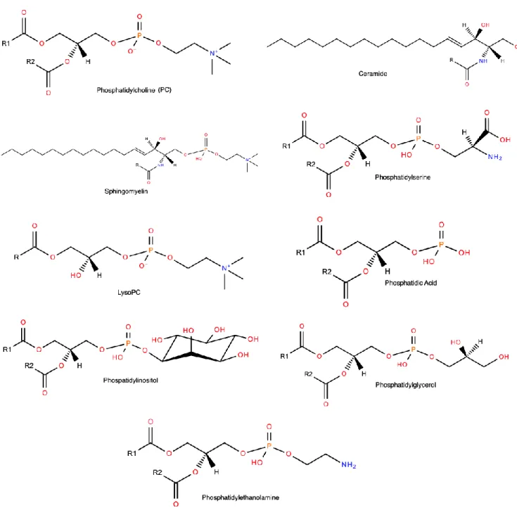
lysophosphatidylcholine (LPC), phosphatidylinositol (PI), phosphatidylethanolamine (PE), ceramide (Cer), phosphatidylserine (PS), phosphatidic acid (PA), and phosphatidylglycerol (PG) [85] (Figure 4).
Generally, before the arrival of proteomic techniques all HDL proteins identified by classic techniques were attributed to one of the three categories which involved apolipoproteins, lipid transfer proteins, and enzymes. Apolipoproteins included apoA-I, apoA-II, apoA-IV I, II, C-III and apoE, with apoA-I being the most prominent of them in terms of the abundance (70% of the protein mass of HDL), while apoA-Ⅱ was found to account for about 20% of total HDL protein, which rendered it a second-major protein component of HDL [86]. Lipid transfer proteins included phospholipid transfer protein (PLTP) and cholesteryl ester transfer protein (CETP). Enzymes of HDL encompassed lecithin:cholesterol acyltransferase (LCAT),
paraoxonase-1 (PON1), PON3 and platelet-activating factor-acetyl hydrolase (PAF-AH). Among these proteins identified by classic biochemical assays, many play important biological roles: for example, LCAT converts FC into CE, while PAF-AH and PON1 exert plausible antioxidative activities [87-91].
In order to successfully realize the qualitative and quantitative analysis of lipid components of HDL in the past, many analytical methods had to be employed, including thin-layer
chromatography (TLC), gas chromatography (GC), liquid chromatography (LC), enzyme-linked immunosorbent assays (ELISA) and nuclear magnetic resonance (NMR) which allowed
characterizing major lipid classes [92]. However, analysis of molecular lipid species was either impossible or highly laborious using these techniques.
Lest we forget about the glycan part of the HDL. Indeed, it has been long known that in addition to lipids and proteins, lipoproteins contain carbohydrates as a minor component (e.g. 3.3 wt% in HDL) [93]. However, without qualitative and quantitative analysis of HDL carbohydrates was not feasible using traditional methodology (see section 3.2.2.3 Glycome).
Omics refers to a field of study in biology ending in -omics, such as genomics - study of genes and their function, proteomics - study of proteins, lipidomics - study of cellular lipids or glycomics - study of cellular carbohydrates.
3.2.2.1 Proteome
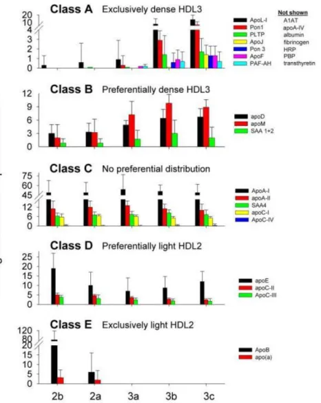
Advances in omics approaches allowed for a detailed molecular characterization of individual proteins present in HDL particles. Before the widespread arrival of liquid chromatography-mass spectrometry (LC-MS), the proteins had to be purified and subsequently exposed to Edman degradation – a slow and painful process, requiring the protein of interest to be broken into fragments of 50 amino acid long, with each such fragment taking up more than full three days of constant work from an automatic Edman sequencer to be analyzed [94]. On the other hand, liquid chromatography–electrospray ionization–tandem MS (LC-ESI-MS/MS), can detect more than 100 different proteins in HDL during a single run (Figure 5) [90]. Some of these proteins are cross-validated by classical methods yet revealing many previously undetected minor proteins representing less than 5% of HDL protein mass. Multiple complement-regulatory proteins and a diverse array of distinct serpins with serine-type endopeptidase inhibitor activity, as well as acute-phase response proteins were thereby detected, supporting the idea that HDL is of central importance in inflammation and immune response [90, 95]. More importantly, proteomics allowed not only to find minor yet crucial proteins in HDL, but also permitted a reliable quantification of their abundances across heterogeneous HDL subpopulations of HDL2 and HDL3 (see Figure 5; more details in section 3.3 Heterogeneity) in both normolipidemic subjects and subjects with disorders of lipid metabolism and CAD. Example of such proteins,
differentially presented in the control vs CAD subjects, may include acute-phase-response proteins, C4A/C4B, C9, and vitronectin [90]. Indeed, HDL from CAD patients was selectively
enriched in proteins that play critical roles in macrophage biology, lipid metabolism, and the inflammatory response (Figure 6) [90].
Figure 5. Abundance patterns of representative proteins across major HDL subpopulations. Class A — limited to dense HDL3, those listed showed the same pattern (not shown). Class B -
prefer dense HDL3 but are in all subfractions. Class C - evenly distributed. Class D — prefer light HDL2 but are in all subfractions. Class E — limited to light HDL2. Error bars =1 S.D
Figure 6. Relative abundance of proteins isolated from HDL3 of control and CAD subjects. HDL3 was isolated from plasma of 6 control subjects and 7 subjects with established CAD. The
relative abundance of proteins in the CAD subjects versus the control subjects was assessed by the peptide index. *P < 0.05 by Student’s t test (peptide number) or Fisher’s exact test (subject
number) [90].
3.2.2.2 Lipidome
Thanks to mass spectrometry, which bests the classic techniques in that it combines high sensitivity and specificity, high throughput and high accuracy [92], we can delve into lipidomics with a relative ease. Before the arrival of the omics technologies, analysis of individual lipid components of lipoproteins was probably even more cumbersome than the assessment of the composition of
their protein moieties. Indeed, lipids are an extremely diverse group of compounds consisting of tri-, di- and monoacylglycercols, free fatty acids, PL, sterols, carotenoids and vitamins A and D among others [96] and require a plethora of analytical techniques to be analyzed properly.
Although lipidomics is still considered to be in its early stages of development [92], lipidomic analysis already made significant progress in the determination of the lipoprotein composition. PC, the principal plasma phospholipid that accounts for 32-35 mol % of total lipids in HDL, is a structural lipid, consistent with its even distribution across HDL subpopulations (Figure 7) [97]. LPC is another important phospholipid subclass in HDL (1.4-8.1 mol % of total lipids) derived from regulated degradation of PC by phospholipases, including LCAT. PE is moderately abundant in HDL (0.7-0.9 mol % of total lipids). Plasmalogens are minor phospholipids which contain a vinyl ether linked fatty acid essential for their antioxidative properties [98]. PI, PS, PG, PA and cardiolipin are negatively charged minor (0.8 mol % of total lipids) PL present in HDL which may impact its net surface charge and modulate lipoprotein interactions with lipases, extracellular matrix and other protein components [97].
SM, a structural lipid which enhances surface rigidity, is the major HDL sphingolipid (SL) (5.6-6.6 mol % of total lipids), which largely originates from TGRL [98]. Cer is a minor (<0.1 mol % of total lipids) sphingolipid intermediate implicated in cell signalling, apoptosis, inflammatory responses, mitochondrial function and insulin sensitivity. Both SM and Cer are enriched in large, light relative to small, dense HDL [97]. Among lysosphingolipids, sphingosine-1-phosphate (S1P) is particularly interesting, reflecting its key role in vascular biology [99]. S1P is associated preferentially with small, dense HDL particles [98], consistent with their elevated content of apoM [95], a carrier for S1P [100].
Unesterified (free) sterols are located in the surface monolayer of HDL particles and regulate its fluidity. HDL sterols are dominated by cholesterol, reflecting the pivotal role of lipoproteins in
the cholesterol transport through the body. CE are largely (up to 80%) produced in HDL and form its lipid core [101]. Most of HDL CE is accounted for by cholesteryl linoleate. HDL-associated TG are dominated by species containing oleic, palmitic and linoleic acid moieties [101]. Minor HDL lipids include diacylglycerides, monoacylglycerides and free fatty acids.
Figure 7. Differential representation of the major glycerosphingolipid classes in five major HDL subpopulations [97].
Akin to the discoveries of the newfangled proteomics, lipidomics readily reveals differences between HDL subpopulations, as well as between HDL obtained from normolipidemic controls and from metabolically compromised patients. An example of the first would be the report of Camont and colleagues documenting differences between major HDL subpopulations in normolipidemic patients [97]. As it can be seen in Figure 7, differences between the five major
HDL subpopulations (HDL2b, 2a, 3a, 3b and 3c) were observed in the contents of PC, SM, LPC, PI, PE, PS, Cer, PA and PG. Concentrations of PS, PC, LPC and PA generally increased in parallel with increase in hydrated density from HDL2b to HDL3c. Indeed, small, dense HDL were enriched in PC, PS, and PA relative to large, light HDL (P=0.01, P=0.003, and P<0.001 for trend, respectively). HDL3c was enriched in LPC (+48%; P=0.05) and PS (17-fold; P<0.01) relative to HDL2b. Similarly, PE, PI, and PG tended to concentrate in small, dense HDL; these trends did not, however, attain significance. Proportion of SM and Cer decreased progressively in parallel with HDL density from 20% and 0.19% of total PL+SL in HDL2b to 14% and 0.11% in HDL3c. Another report details lipid classes of HDL that are most affected by ST-elevated
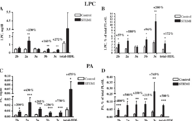
myocardial infarction (STEMI) [102], and which include LPC and PA (Figure 8). Many if not all of these lipids are biologically active and play either signalling role (LPC and PA), or are
believed to be involved in anti-apoptotic and anti-inflammatory features of HDL (PS, Cer and PA) [103-109]. However, all reports known to date do not provide a detailed representation of the contents of molecular lipid species, although more than 200 species of lipids are known to be present in the lipidome of HDL [97, 110].
Figure 8. Levels (A, C) and content (B, D) of LPC (A, B) and PA (C, D) in HDL subpopulations and in total HDL, expressed as mg/dl (A, C) and % of total PL + SL (B, D), in STEMI patients
and control subjects; *p < 0.05 vs. controls [102].
3.2.2.3 Glycome
HDLs are highly sialylated particles in which most of the glycans contain one or two sialic acid residues [111]. Several HDL proteins, including LCAT, CETP, apoCs, fetuin A and
-1-antitripsin, are glycosylated, while the glycosylation of apoA-I remains controversial [112-114]. Remarkably, carbohydrate residues are specifically associated with distinct HDL proteins, such as apoC-III which contains O-linked glycans [115]. Glycans, attached not to proteins, but rather to lipids directly with a covalent bond (glycolipids), contain sialic acids as well [116].
HDL particles are heterogeneous in size, charge, protein content or by biological activities they exert.
Ultracentrifugation (~200 000 g for 48 h) allows HDL particles to be subfractionated by density into light, large, lipid-rich HDL2 particles with a density of 1.063-1.125 g/ml and small, dense, protein-rich HDL3 particles with a density of 1.125-1.21g/ml [13].HDL2 and HDL3 are two major subpopulations of spherical HDL. HDL2 particles contain PL, FC, CE and TG at
approximately 25, 6, 18 and 9 wt %, respectively, while HDL3 contains PL, FC, CE and TG at approximately 23, 3, 14, and 7 wt %, respectively [117]. Further fractionation of HDL2 and HDL3 into five increasing-density subpopulations of HDL2b, HDL2a, HDL3a, HDL3b, and HDL3c can be achieved by density gradient ultracentrifugation (Figure 3C) [13, 118].
It is also possible to subfractionate HDL particles by their charge and size (Figure 3D). Namely, 2D gel electrophoresis separates plasma HDL particles into: (a) very small discoidal precursor HDL of pre-β mobility (pre-β-1 HDL, diameter about 5.6 nm), which contains apoA-I and PL; (b) very small discoidal HDL of α mobility (α-4 HDL, diameter about 7.4 nm), which contains apoA-I, PL, and free cholesterol; (c) small spherical HDL of α mobility (α-3 HDL, diameter about 8.0 nm), which contains apoA-I, apoA-II, phospholipid, free cholesterol, cholesteryl ester, and triglyceride; (d) medium-sized spherical HDL of α mobility (α-2 HDL, diameter about 9.2 nm), which contains the same constituents as α 3 HDL; and (e) large spherical HDL of α mobility (α-1 HDL), which contains the same constituents as α-3 and α-2 HDL, except for the near
Figure 9. The apoA-I–containing HDL subpopulation profiles of a CHD patient (A) and a healthy individual (control) (B), with a schematic diagram of all of the apoA-I-containing HDL particles
shown on the right (C). Below panel (A) is a plot of a densitometric scan across the α-migrating HDL-particle region indicating the presence of 4 α-migrating HDL particles ranging in mean particle diameter from very large α-1 HDL (11.0-nm diameter) to very small pre-β-1 HDL (5.6-nm diameter). In the schematic diagram α-migrating apoA-I-containing particles in the α-2 region (9.2-nm diameter) and in the α-3 region (8.1-nm diameter) contain both I and
apoA-II (more heavily shaded), whereas all other particles containing apoA-I, including small α-4 HDL (7.4-nm diameter), do not contain appreciable amounts of apoA-II (less heavily shaded). The asterisk marks the serum albumin or α front. Based on their composition, very small pre-β-1
HDL and small α-4 HDL are discoidal particles that do not contain cholesteryl ester or triglyceride, whereas medium, large, and very large α-3, -2, and -1 HDL are spherical and contain cholesteryl ester and triglyceride in their cores. CHD patients in general in the untreated
state tend to have significant decreases in the levels of apoA-I in very large and large α-migrating HDL and modest increases in apoA-I in very small pre-β-1 HDL and small α-4 HDL.
Plasma HDL can also be subfractionated and classified on the basis of their apolipoprotein composition into particles containing apoA-I (LpA-I) and those containing apoA-I and apoA-II (LpA-I:A-II) (Figure 3B). LpA-I and LpA-I:A-II are the major HDL subpopulations accounting for approximately 35% and 65% of plasma apoA-I, respectively. The cardioprotective roles of LpA-I and LpA-I:A-II are however controversial. A general conclusion resulting from these studies is that increased HDL-C concentrations involving an increase in the large HDL subclasses containing both LpA-I and LpA-I:A-II is associated with decreased CHD risk, whereas the
reduction in lipid-poor LpA-I and pre-β HDL is associated with increased CHD risk [13].
Finally, NMR can quantify three distinct HDL subclasses differing in size, notably large (8.8-13.0 nm), medium (8.2-8.8 nm) and small (7.3-8.2 nm) HDL [13]
HDL heterogeneity is also observed at the level of the proteome, lipidome and glycome. For example, proteins, exclusively present in HDL3, but not present in HDL2, include apoL-I, apoF, PON1/2, PLTP, apoJ, PON3, α1-antitrypsin (A1AT), apoA-IV, albumin, fibrinogen, Hrp, PBP and transthyretin [95]. In a similar fashion, PS is predominantly associated with the more
biologically active HDL3 particle pool and thus, its HDL content is positively correlated with all metrics of HDL functionality [97]. In contrast, SM and Cer are preferentially found in large, light HDL [97]. Potential heterogeneity of the HDL glycome however remains undeterminate.
3.4 Metabolism
Figure 10. HDL particle remodelling and metabolism [120]
3.4.1 Biosynthesis
ApoA-I is initially secreted by the liver and the intestine as either a mere apoA-I protein or as a lipid-poor apoA-I:PL complex, which is then exposed to the cholesterol and phospholipid transfer activity of the cellular ABCA1 (ATP binding cassette transporter A1) transporter and facilitates cholesterol efflux, resulting in the formation of pre-β HDL [13, 78]. Hepatically secreted apoA-II associates with LpA-I during this step to form LpA-I:A-II. The role of apoA-II in HDL
metabolism has not been definitively established, although apoA-II has been reported to delay HDL remodeling and to reduce cholesterol uptake by hepatic scavenger receptor class B type I (SR-B1) [13]. Mice models also suggest that apoE and apoC-Ⅲ might also ensure biosynthesis of mature HDL particles from lipid-free apolipoproteins [121]. When LPL hydrolyses TG from TGRL, a by-product of small HDL precursors can be generated from the redundant surface components of TGRLs (CMs and VLDL).
3.4.2 Remodelling
Discoidal pre-β HDL particles are subjected to LCAT, which forms CE from FC, and mature into a full-fledged spherical HDL which constitutes a majority of plasma HDL pool [78]. Plasma CETP primarily transfers CE from mature spherical HDL to apoB-containing particles, particularly VLDL, in exchange for TG [122]. As a result of CETP activity, HDL becomes depleted of CE and enriched in TG, which accelerates HDL catabolism. PLTP is involved in the interconversion of HDL2 and HDL3 and that conversion generates small nascent HDL [123].
Both surface and core lipids of HDL are extensively modified by lipases. Hepatic lipase (HL) breaks down primarily tri- and diglycerides but also phospholipids of HDL, resulting in the reduction of HDL particle size. Endothelial lipase (EL) primarily hydrolyses HDL phospholipids but also triglycerides. Lastly, further remodeling of HDL occurs through particle fusion or PLTP-mediated transfer of surface remnants such as PL from LPL-hydrolized CMs of VLDL, eventually producing mature HDL [123]. Mature spherical HDL can further grow via effluxing cellular cholesterol and phospholipid through ATP-binding cassette transporter G1 (ABCG1) [124]. Selective uptake of HDL-C via SR-B1 includes apoA-Ⅰ-dependent binding of HDL to SR-B1, transfer of HDL-CE to the cell membrane and intracellular hydrolysis of CE by cholesterol esterase to FC, with a release of small cholesterol-depleted HDL particles [125].
3.4.3 Catabolism
In healthy individuals, LpA-I is catabolized at a faster rate than LpA-I:A-II. The major sites of catabolism of the protein components of LpA-I and LpA-I:A-II are the liver and kidney, and the majority of HDL cholesterol is transported to the liver. Kinetic models incorporating the different rates of catabolism of LpA-I and LpA-I:A-II have been developed. The differential rates of metabolism of LpA-I and LpA-I:A-II have been proposed to be related to the decreased ability of apoA-I-containing lipoproteins to reassociate with HDL particles following cholesteryl ester
delivery to the liver via the SR-B1 receptors. This process includes apoA-Ⅰ-dependent binding of HDL to SR-B1, transfer of HDL-CE to the cell membrane and intracellular hydrolysis of CE by cholesterol esterase to FC, with a release of small cholesterol-depleted HDL particles [125]. Intracellular FC is subsequently excreted into the bile or converted to bile acids for excretion [11, 125].
CETP also plays a role in the catabolism of HDL: CETP mediates the transfer of CE from HDL to apoB-containing lipoproteins (VLDL and LDL) in exchange for TG from the latter, thereby forming CE-depleted and TG-enriched HDL. CE-enriched TGRL transport CE to the liver and bind to the LDL receptor for subsequent particle uptake [125, 126].TG-enriched HDLs are hydrolyzed by HL and EL to reduce particle size and release apoA-Ⅰ, which interacts with ABCA1 in the next lipidation cycle [11, 127, 128].
HDL particles can be removed from the circulation by holoparticle HDL receptors, such as cubulin present in kidney proximal tubules and the ectopic -chain of ATP synthase at the surface of hepatocytes [129, 130].
3.5 Biological activities and structure-function relationships
Beneficial effects of HDL are not limited to their influence on lipid metabolism. HDL equally possess cellular cholesterol efflux, antioxidative, anti-inflammatory, anti-apoptotic,
Figure 11. Biological activities of HDL [132]
Since several decades, the major biological role of HDL is thought to involve the removal of excess cholesterol from peripheral tissues to the liver for subsequent biliary excretion [133, 134]. Lipid-loaded macrophages that acquired cellular debris and modified or aggregated lipoproteins and are unable to metabolize nor to get rid of them, form foam cells. These foam cells are always found in the atherosclerotic lesions, implying their key role in atherogenesis. Cholesterol efflux from macrophages can be mediated through (a) one-directional, ATP-dependent pathways involving ABCA1 and ABCG1 transporters; (b) a bi-directional, ATP-independent pathway mediated by SR-B1; and (c) passive diffusion according to the gradient of cholesterol concentration [135, 136]. Mutations in ABCA1 lead to severe HDL deficits, manifesting as Tangier disease or other forms of familial hypoalphalipoproteinemia [137]. Highlighting the role of ABCA1 in cholesterol metabolism is the observation that any type of modulation (over- or under-representation) of this
transporter impacts the capacity of apoA-Ⅰ to perform cholesterol efflux [138]. Nuclear liver X receptors (LXRs) type α and β and retinoid X receptor (RXR) regulate the expression of ABCA1 [139]. LXRs expression is modulated by cellular concentrations of oxysterols, a product of enzymatic modification of cholesterol. Therefore, LXR agonists and oxysterols can modulate the transcription of ABCA1 in macrophages, thereby enhancing cholesterol efflux to lipid-poor HDL particles. However, it is challenging to isolate lipid-poor apoA-Ⅰ from the bloodstream, because after being secreted, it rapidly matures into spherical HDLs. Such mature, spherical HDLs can also perform cellular cholesterol efflux through ABCG1 transporter pathway [140, 141]. SR-B1 mediates another pathway of cholesterol efflux from microphages to mature HDL [142], and in knockout mice models was shown to be atheroprotective [143, 144].
ApoA-I and apoA-II are the primary acceptors in the cholesterol efflux process, while apoA-IV, apoC-I, apoE and apoM can efficiently accept cellular cholesterol, too [122, 145-148]. Enzymes also exert major functions in cellular cholesterol efflux and reverse cholesterol transport (RCT). Thus, LCAT participates in the transfer of cholesterol from peripheral cells to the liver, ensuring formation of CE which is subsequently taken up by the liver via SR-B1. HDL lipids are also relevant in its cholesterol efflux capacity; thus, HDL containing liquid-crystal unsaturated PLs is a more efficient cholesterol acceptors than HDL containing gel-state saturated PLs [149, 150]. ApoA-I and S1P of HDL can inhibit expression of pro-inflammatory adhesion molecules in the cells, thereby diminishing inflammation [151]. These adhesion molecules include vascular cell adhesion molecule-1, endothelial-leukocyte adhesion molecule 1 and intracellular adhesion molecule-1 [152, 153]. HDL promotes proliferation of endothelial cell and attenuates apoptosis of the endothelial cells [12, 121, 153], inhibits monocyte activation, inhibits secretion of pro-inflammatory cytokines and chemokines and decreases activation of neutrophils in the arterial wall [124, 154].
Furthermore, HDL can inhibit one-electron and two-electron oxidation of LDL, two most common types of LDL oxidation to occur in vivo [155, 156]. The mechanisms of such actions are well understood: one-electron oxidants generate phospholipid hydroperoxides, which are sequestered by HDL and reduced by the redox-active methionine residues of apoA-I into redox-inactive PL hydroxides and methionine sulphoxides [157, 158]. Two-electron oxidation of LDL can be intercepted by direct oxidant scavenging of HDL [159]. ApoA-I, apoA-II, apoA-IV, apoE, apoJ, apoM as well as PON1, PAF-AH and LCAT exert antioxidative activities [120, 131, 160-167]. As mentioned in section 3.3 Heterogeneity, HDL subpopulations are not equally potent in their key biological functions, antioxidative function being one them: small, dense HDL are more potent in their antioxidative activity than large, light HDL [168].
ABCG1-mediated efflux can serve as a mediator in the removal of oxidized lipids from the cells to HDL, with the latter exerting cytoprotective function. Oxidized lipids activate apoptotic caspases and may compromise the integrity of mitochondria [151]. HDL stimulates release of nitric oxide (NO) mediated by endothelial nitric oxide synthase (eNOS), and production of prostacyclin (PGI2) in endothelial cells to maintain normal function of vascular endothelium [169]. Endothelial NO beneficially regulates inflammatory response of endothelium, platelet activity and proliferation of vascular smooth muscle cells [133].Studies show that HDL promotes expression and activity of eNOS, which increases production of NO [170-173]. Activation of eNOS mediated by HDL includes HDL binding to the endothelial SR-B1, which triggers cell signaling pathways of phosphatidylinositol 3-kinase (PI3K)/Akt and the phosphorylation of eNOS [172, 173]. Moreover, NO production mediated by eNOS can be promoted by HDL-associated S1P via S1P1/S1P3 receptors [173, 174]. The eNOS activation can also be mediated by HDL-associated PON1 [170]. HDL possesses anti-coagulation activity, namely it inhibits activation of platelets and
to possess strong ability to quench infection by eliminating lipopolysaccharides (LPS), should they infiltrate the bloodstream. For example, apoA-I-overexpressing mice subjected to systemic inflammation by LPS show reduced CD14 expression on monocytes as compared to their wild-type counterparts [177].
Content-wise, there is no clear unanimity about the components with the most significant role. Abundances of specific lipids, such as sphingosine-1-phosphate (S1P) [178], or groups of lipids, such as PS [97, 179, 180] may modulate HDL’s efficiency to reduce risk of CHD. Recent studies demonstrated that miRNAs, associated with CVD, can be exported or released by cells and circulate with the blood in a remarkably stable form, namely bound to apolipoproteins of HDL, and modulated in the CVD [181, 182], but this assumption was not confirmed by others [183]. Finally, the activity of myeloperoxidase (MPO), and PON1 - both HDL-associated enzymes, were also found to be associated with CVD [184-186]
Measurements of HDL biological performance could have potentially presented a viable
alternative to the largely obsolete HDL-C metric, if they themselves were free of controversies: in one study cellular cholesterol efflux capacity of HDL was negatively correlated with the
development of CHD [187], while another showed that enhanced cholesterol efflux was associated with elevated CVD risk [188].
3.6 Dysfunctional HDL
3.6.1 Cardiometabolic diseases and atherosclerosis
Metabolic conditions associated with elevated CHD risk, such as dyslipidemia, insulin resistance, obesity and inflammation, are collectively called cardiometabolic diseases (CMD). Alterations in the composition, structure, and metabolism of HDL particles under such conditions lead to the loss
of their atheroprotective functions, rendering them “dysfunctional” [189]. In addition, CMD are frequently characterized by low plasma HDL-C.
Several genetically determined conditions feature low HDL-C, accelerated CHD and
dysfunctional HDL. Thus, homozygous or compound heterozygous apoA-I deficiency is a rare condition which results in complete absence of apoA-I from plasma accompanied by a marked decrease in HDL-C and increased risk of premature CVD [190]. Subjects with heterozygous forms of apoA-I deficiency feature plasma HDL-C and apoA-I levels that are about 50% of normal. HDL biogenesis is disrupted in apoA-I deficiency as a result of abnormal HDL
production and deficient LCAT activation by apoA-I. Remarkably, some rare variants of apoA-I, including apoA-I Milano and apoA-I Paris, are paradoxically associated with low HDL-C levels and reduced risk of CVD paralleled by greater longevity [191].
Most known ABCA1 mutations result in Tangier disease associated with the deficiency in cellular cholesterol efflux to lipid-free and lipid-poor apolipoproteins [192]. Low HDL-C is a common characteristic of ABCA1 deficiency, frequently resulting in elevated CV risk [192]. Naturally occurring mutations in LCAT are another common cause of low plasma HDL-C
(Figure) [193]. Familial LCAT deficiency results from the complete loss of LCAT activity, while fish-eye disease is associated with a change in the substrate specificity of LCAT that becomes inactive towards HDL, while retaining its CE-generating activity towards apoB-containing lipoproteins. The latter property can be important in accelerating atherosclerosis in fish-eye disease relative to familial LCAT deficiency [194]. In these diseases, plasma HDL-C and apoA-I levels are reduced, while plasma FC is elevated.
Genetic defects of LPL may lead to hypertriglyceridemia and low HDL-C [195]. Thus, familial LPL deficiency is a rare disorder characterised by severe hypertriglyceridemia and marked
![Figure 1. Density and diameter of human plasma lipoproteins[7]](https://thumb-eu.123doks.com/thumbv2/123doknet/14516187.721848/21.918.206.708.110.535/figure-density-diameter-human-plasma-lipoproteins.webp)
![Figure 3. Different means of HDL subpopulation classification: A) by maturation step B) by protein content C) by size D) by charge [83, 84]](https://thumb-eu.123doks.com/thumbv2/123doknet/14516187.721848/27.918.302.616.411.797/figure-different-subpopulation-classification-maturation-protein-content-charge.webp)

![Figure 7. Differential representation of the major glycerosphingolipid classes in five major HDL subpopulations [97]](https://thumb-eu.123doks.com/thumbv2/123doknet/14516187.721848/34.918.145.786.275.769/figure-differential-representation-major-glycerosphingolipid-classes-major-subpopulations.webp)
![Figure 10. HDL particle remodelling and metabolism [120]](https://thumb-eu.123doks.com/thumbv2/123doknet/14516187.721848/40.918.251.666.152.507/figure-hdl-particle-remodelling-metabolism.webp)
![Figure 11. Biological activities of HDL [132]](https://thumb-eu.123doks.com/thumbv2/123doknet/14516187.721848/43.918.263.683.111.578/figure-biological-activities-of-hdl.webp)