ABSTRACT. In the treatment of osteoporosis, the tolerance of oral bisphosphonates is often low. The high potency of ibandronate allows iv bolus injections that can be repeated every 2 to 3 months. However, the best dose and time inter-val of the treatment with iv ibandronate is still debated. Efficacy of 2-mg ibandronate injected every 3 months was tested in men with osteo-porosis over 2 yr, in a prospective, open study. Fourteen men with primary osteoporosis, mean age 57±12 yr (range: 40-73), received 2-mg iban-dronate iv every 3 months over 2 yr. All got 1 g/day calcium and 880 UI/day vitamin D for 2 yr. Bone mineral density (BMD) increased after 2 yr by 6.7±1.5% (mean change±SEM) at lumbar spine (p<0.001), by 3.2±08% at trochanter
(p<0.001) and by 1.4±1.1% at femoral neck (ns). Serum`-crosslaps and osteocalcin decreased sig-nificantly by 30-45 and 30%, respectively, during the 2 yr of treatment. Serum calcium increased from the lower to the middle tertile of the nor-mal range during the 2 yr of the study. The ob-served decrease of bone remodelling and the in-crease of BMD are of the same magnitude as those described with oral bisphosphonates. The increase of plasma calcium confirms the positive effect of the supplementation with calcium and vitamin D. These results suggest that 3 months are a good interval between two doses of iv iban-dronate, when 2 mg are given.
(J. Endocrinol. Invest. 26: 728-732, 2003)
©2003, Editrice Kurtis
INTRODUCTION
Osteoporosis is less common in men than in wom-en, but approximately 25 to 30% of all hip fractures occur in men, and many men have vertebral defor-mities (1, 2). The causes of osteoporosis in men in-clude excess of glucocorticoids, hypogonadism, and a variety of other systemic conditions, medica-tions and lifestyle factors; in about 50% there is no obvious cause. Only few therapies have been eval-uated in men. Testosterone therapy has limited ef-ficacy in men with hypogonadism (3). Alendronate significantly increased spine and hip bone mineral density (BMD), and decreased the incidence of ver-tebral fractures (4). However, bisphosphonates cause gastrointestinal side effects in many patients (5), and must be taken under strict conditions, which affects compliance. These limitations led to the
in-vestigation of intermittent iv administration of aminobisphosphonate, which showed an increase in BMD of the spine and femoral neck after 3 monthly infusions of pamidronate and ibandronate (6, 7). Ibandronate [1-hydroxy-3-(methylpenty-lamino)-propylidene bisphosphonates] is a potent aminobisphosphonate with proven efficacy in the treatment of malignant hypercalcemia (8) and of post-menopausal osteoporosis (9). For the treat-ment of osteoporosis, iv ibandronate, 2 mg every 3 months as bolus injection, seems to be effective (7). The goal of the present open pilot study was to investigate the effect of 2 mg of ibandronate, ad-ministered every 3 months over 2 yr, on BMD and on biochemical markers of bone metabolism in men with primary osteoporosis.
MATERIALS AND METHODS
Study subjects
The patients (age 40 to 80 yr) were referred to the out-patient clinic for osteoporosis at the University Hospital of Lausanne, Switzerland. This pilot study was open and monocentric. The in-clusion criteria were the presence of one osteoporosis fracture (vertebra, femoral neck or wrist) and a BMD at the lumbar spine of at least 1 SD below the Z-score, or at least 1.5 SD below the Z-score without any fracture. Men with secondary causes of
os-Key-words: Bisphosphonate, bone mineral density, ibandronate, osteo-porosis in men.
Correspondence: Olivier Lamy, MD, Department of Internal Medicine, CHUV, BH10 CH-1011 Lausanne, Switzerland.
E-mail: [email protected] Accepted March 14, 2003.
Intravenous ibandronate in men with osteoporosis: An open
pilot study over 2 years
O. Lamy, L. Sandini , I. Pache, S. Fatio, J. Burnand and P. Burckhardt
teoporosis were ineligible, as were those with other bone dis-eases, vitamin D deficiency, renal disease (indicated by a serum creatinine concentration >160 mol/l), known endocrinopathy af-fecting bone metabolism (hypogonadism, hyperparathyroidism, hyperthyroidism, etc.), medication affecting bone metabolism (corticosteroids or cyclosporine during the preceding 6 months; calcitonin, fluor or bisphosphonates during the preceding 12 months), a history of cancer other than basal-cell carcinoma of the skin, or a severe disability disease with a life expectancy of less than 2 yr. All patients had given their written informed con-sent prior to enrolment. The study was approved by the local Ethics Committee.
Treatment
All patients included in this study received 2-mg ibandronate every 3 months as a bolus injection at 0, 3, 6, 9, 12, 15, 18 and 21 months. All got oral supplementation with 1000-mg calcium and 880-UI vitamin D every day during the 2 yr of treatment. Baseline and follow-up study
In the pre-treatment period, a complete history was obtained, and each patient underwent a physical examination, electro-cardiography, chest radiography, and serum parameters [os-teocalcin, C-telopeptides, PTH, 25 (OH) vitamin D and 1,25 (OH) vitamin D, complete blood cell count, electrolytes, crea-tinine, albumine, liver-function tests]. The patients included re-turned for visits, biochemical measurements and treatment at 0, 3, 6, 9, 12, 15, 18, 21 and 24 months. The BMD of the lum-bar spine (anteroposterior view), femoral neck, trochanter and distal forearm was measured by dual-energy X-ray absorp-tiometry (Hologic QDR 2000) at baseline and at 6, 12, 18 and 24 months. Quality control of the BMD measurements, includ-ing teachinclud-ing of relevant technicians and phantom assessments, was performed before and throughout the study by checking accuracy and reproducibility on a daily basis. The precision of our Hologic QDR 2000 equipment was 1.3% at the lumbar spine, 1.8% at the femoral neck and 1.9% at the trochanter (7). The precision was evaluated by repeated measurement in 10 subjects (4 measurements per subject) according to Gluer et al. (10). The long-term stability was monitored during the study by measuring an object of constant mass (European spine phan-tom) on daily basis. The device showed a high stability during all the study with an in vitro long-term reproducibility between 0.37 and 0.47% in BMD per year. All scans were reviewed by a blinded expert, and reanalyzed upon request. BMD is ex-pressed in grams of hydroxyapatite per cm2. Posteroanterior
and lateral radiographs of the lumbar and thoracic spine were obtained at base line and after 2 yr of treatment. All radio-graphs were reviewed by a blinded expert.
Fasting serum samples were obtained in the morning for the measurement of calcium corrected for albumine, phosphate, creatinine, alkaline phosphatase, osteocalcin, `-crosslaps, PTH. Plasma 25 (OH) vitamin D, and 1,25 (OH) vitamin D were mea-sured on fasting serum sample at baseline and at 12 months. These parameters were determined by the following methods: osteocalcin by a two-site radioimmunoassay as described by Garnero et al. (11) (Elsa-Osteo, CIS bio international, Gif-sur-Yvette, France; normal 9.6 to 26.8 ng/ml for men >30 yr); serum `-crosslaps by a two-site radioimmunoassay (Systèmes Elecsys 1010/1020, Roche Diagnostics, Meylan, France); intact PTH with
an immunoradiometric assay (Nichols Institute, San Juan Capistrano, California; intact PTH 100T Kit; normal 10 to 65 pg/ml); 25 (OH) vitamin D with a radioimmunoassay (DiaSorin, Stillwater, Minnesota, normal 8.4 to 52.3 μg/l); 1,25 (OH) vita-min D with a radioimmunoassay (Incstar corporation, Stillwater, Minnesota). The correction of the serum calcium by albumine (normal 2.1 to 2.5 mmol/l) was done according to statistics es-tablished by the laboratory of the hospital (12): measured calci-um - 0.012 [measured albcalci-umine - 39.5 (g/l)]. Safety parameters measured included serum creatinine, sodium, potassium, ala-nine aminotransferase, aspartate aminotransferase, gamma-GT and complete blood count. All these latter determinations were done by routine laboratory methods.
Statistical analysis
The primary endpoint of the study was the lumbar spine BMD and the secondary endpoint was the effects on osteocalcin and `-crosslaps. The analysis of BMD variables, osteocalcin and `-crosslaps were based on the percent changes from baseline. Statistical differences were evaluated by paired t-test. P values <0.05 were considered to be significant.
RESULTS
Fourteen consecutive male patients with primary osteoporosis were included in this study. Patient characteristics at baseline are given in Table 1. At entrance, 9 patients had experienced 18 vertebral fractures and 4 non-vertebral fractures (wrist 3; up-per arm 1). All these fractures occurred, either spon-taneously or after a minor trauma. Nine patients had a T-score below -2.5 DS either at the lumbar spine or femoral neck.
BMD
The BMD of the lumbar spine and of the trochanter increased significantly by 6.7±1.5% and by 3.2±0.8% (percent change, mean±SEM) after 2 yr of treatment (Fig. 1). The BMD of the femoral neck and the total hip (Fig. 2) increased by 1.4±1.1% and 0.9±0.6%, respectively. The BMD of the distal fore-arm increased by 1.5±0.5% (ns, data not shown).
Biochemical markers
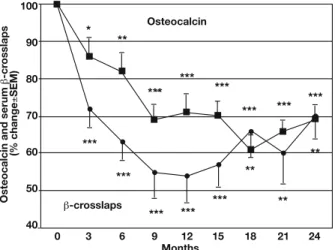
As shown in Figure 3, there was a progressive de-crease in bone serum `-crosslaps reaching a nadir between 9 and 12 months, and a slight increase
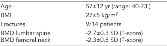
Table 1 - Patient characteristics at baseline (mean±SEM)
Age 57±12 yr (range: 40-73 )
BMI 27±5 kg/m2
Fractures 9/14 patients
BMD lumbar spine -2.7±0.3 SD (T-score) BMD femoral neck -2.3±0.8 SD (T-score)
during the second year of treatment at a level of 30% below the baseline. Osteocalcin decreased slower and remained stable at 30 to 40% below the baseline during the whole treatment. Corrected serum calcium increased slightly from 2.11±0.08 to 2.24±0.09 mmol/l (p<0.01) during the 2 yr of treat-ment. PTH, 25 (OH) vitamin D and 1,25 (OH) vita-min D did not change during the 2 yr of the study.
Fractures
Four fractures occurred in four men: 2 new sponta-neous vertebral fractures in 2 men with pre-existent vertebral fractures, and 2 traumatic fractures after a fall in 2 men (radius, metacarpus). The treatment was conservative in all cases.
Safety
There were no serious drug-related adverse events and 38 non-serious adverse events during the study. One patient dropped out after 1 yr and an-other after 18 months for reasons not related to ibandronate (one was depressed, one for unknown reason). Flu-like symptoms were reported in 3 pa-tients: in 2 patients after the first and second injec-tion and in 1 after each injecinjec-tion. Flu-like symptoms were controlled by self-administration of paraceta-mol for 1 or 2 days, and were not a cause of withdrawal. One patient experienced a transient skin rash 2 weeks after the last injection of iban-dronate. The patient was examined by an allergol-ogist, and no trigger was found. A transient rela-tive eosinophilia occurred in one patient, and a transient relative monocytosis in another patient, without evident causes.
DISCUSSION
The patients included in this study had severe os-teoporosis, considering the low BMD values and the prevalence of pre-existing fractures in two thirds of them (Table 1). Two-mg ibandronate given iv every 3 months for 2 yr increased BMD at lumbar spine, trochanter and femoral neck. The magnitude of this effect was similar to that report-ed in men with osteoporosis after 2 yr of treatment with 10-mg oral alendronate given daily: 6.7 vs 7.1% at the lumbar spine, 3.2 vs 4.3% at the trochanter, and 1.4 vs 2.5% at the femoral neck (4). These increases in BMD are also similar to those reported in postmenopausal women with osteo-Months L um b ar sp ine an dt ronc h an ter (% change ( SEM) 9 8 7 6 5 4 3 2 1 0 0 6 12 18 24 Lumbar spine ** ** ** *** *** *** * * Trochanter
Fig. 1 - Mean (±SEM) percent changes from baseline in the bone mineral density of the lumbar spine and the trochanter during 2 yr of treatment with iv ibandronate. Asterisks indicate significant dif-ferences from baseline (*p<0.05, **p<0.01, ***p<0.001).
Months
Femoral neck and total hip (% change
( SEM) 4 3.5 3 2.5 2 1.5 1 0.5 0 -0.5 -1 0 6 12 18 24 Femoral neck ** Total hip *
Fig. 2 - Mean (±SEM) percent changes from baseline in the
bone mineral density of the total hip and the femoral neck dur-ing 2 yr of treatment with iv ibandronate. Asterisks indicate sig-nificant differences from baseline (*p<0.05, **p<0.01).
Osteocalcin and serum
` -cr osslaps (% change ( SEM) 100 90 80 70 60 50 40 Months 0 3 6 9 12 15 18 21 24 Osteocalcin ** `-crosslaps * ** ** ** *** *** *** *** *** *** *** *** *** *** ***
Fig. 3 - Mean (±SEM) percent changes from baseline in serum
osteocalcin and `-crosslaps during 2 yr of treatment with iv iban-dronate. Asterisks indicate significant differences from base line (*p<0.05, **p<0.01, ***p<0.001).
porosis after 2 yr of therapy with iv pamidronate or oral alendronate or risedronate (6, 13, 14). Serum`-crosslaps, a marker of bone resorption, de-creased by 30 to 45%, and osteocalcin, a marker of bone formation, by 30%; these values were ob-tained 3 months after the last bolus of ibandronate, just before the next injection. The magnitude of the decline of the markers of bone remodelling was slightly less pronounced to that published with bis-phosphonates in men or in women (4, 6, 9, 13, 14). In these studies, osteocalcin decreased by 35 to 40%, even by 50% (6, 13). The comparison with markers of bone resorption is difficult. We used a serum marker, while the above mentioned studies used different urinary markers. The latter decreased by 26 to 40% in some studies (6, 14), and by 50 to 74% in other studies (4, 9, 13). The pattern of the decline of the markers was slightly different to that described in other studies with bisphosphonates. The maximum decrease was reached only after 9 to 12 months, while it is usually obtained already after 3 to 6 months. However in women with os-teoporosis, the injection of 2-mg ibandronate ev-ery 3 months decreased osteocalcin progressively with a nadir only after 9 months (7). In that study, urinary C-telopeptide decreased by nearly 70% 1 month after the injection of 2-mg ibandronate, but increased again at nearly 50% at 2 months. This in-termittent decline of the resorption markers raises the question if 3 months are an appropriate interval between two iv doses of ibandronate. A significant association between pharmacokinetics of oral iban-dronate and clinical response in bone turnover and bone mass in women with post-menopausal os-teoporosis was shown (15). A recent preliminary re-port confirms that, after 1 yr of treatment, the in-crease of BMD and the suppression of bone mark-ers achieved with 2-mg iv ibandronate given every 3 months in post-menopausal osteoporosis were similar to those achieved with oral bisphosphonates (16). These results suggest that 3 months are an ap-propriate interval between two iv doses of iban-dronate, when 2 mg are given.
The increase of the corrected serum calcium level probably reflects the positive effect of supplemen-tation with calcium and vitamin D. An immediate and transient drop in serum calcium and rise in PTH have been invariably found with iv bisphosphonates. We could not observe such modifications, since the measurements were repeated only after 3 months, just before the next infusion of ibandronate. Flu-like symptoms were the only side effect reported. They were well controlled with paracetamol, and were not a cause of withdrawal. Flu-like symptoms were described in approximately 20% of women with
osteoporosis treated with either 20-mg iv iban-dronate, or a perfusion of 30-mg pamidronate (6, 7). Our study has several limitations: it is an uncontrolled study, and the number of included patients was small. In conclusion, this pilot study confirms that iv iban-dronate is a promising treatment for osteoporosis in men. The increases of BMD and the suppression of bone markers achieved with 2 mg given every 3 months were similar to the results obtained with oral bisphosphonates. The convenience of a bolus injection every 3 months and the good tolerance make this treatment a promising alternative to oral bisphosphonates.
ACKNOWLEDGMENTS
Results of this study have been presented in part at the follow-ing congresses and published as abstracts: World Congress on Osteoporosis, Chicago, USA, 2000 [Osteoporosis Int 2000, 11 (Suppl. 2): 188 (P492)]; 1stJoint Meeting of the International
Bone and Mineral Society and the European Calcified Tissue Society, Madrid, Spain, 2001 [Bone 2001, 28: S238 (P615S)].
REFERENCES
1. Cooper C, Campion G, Melton LJ III. Hip fractures in the el-derly: a word-wide projection. Osteoporos Int 1992, 2: 285-9. 2. O’Neill TW, Felsenberg D, Varlow J, Cooper C, Kanis JA, Silman AJ. The prevalence of vertebral deformity in European men and women: the European Vertebral Osteoporosis Study. J Bone Miner Res 1996, 11: 1010-8. 3. Eastell R, Boyle IT, Compston J, et al. Management of male osteoporosis: report of the UK Consensus Group. QJM 1998, 91: 71-92.
4. Orwoll E, Ettinger M, Weiss S, et al. Alendronate for the treatment of osteoporosis in men. N Engl J Med 2000, 343: 604-10.
5. De Groen PC, Lubbe DF, Hirsch LJ, Daifotis A, Stephenson W, Freedholm D. Esophagitis associated with the use of alendronate. N Engl J Med 1996, 335: 1016-21. 6. Thiebaud D, Burckhardt P, Melchior J, et al. Two years’
ef-fectiveness of intravenous pamidronate (APD) versus oral fluoride for osteoporosis occurring in the postmenopause. Osteoporos Int 1994, 4: 76-83.
7. Thiebaud D, Burckhardt P, Kriegbaum H, et al. Three month-ly intravenous injections of ibandronate in the treatment of postmenopausal osteoporosis. Am J Med 1997, 103: 298-307. 8. Ralston SH, Thiebaud D, Herrmann Z, et al. Dose-response study of ibandronate in the treatment of cancer-associat-ed hypercalcaemia. Br J Cancer 1997, 75: 295-300. 9. Riis BJ, Ise J, von Stein T, Bagger Y, Christiansen C.
Ibandronate: a comparison of oral daily dosing versus in-termittent dosing in postmenopausal osteoporosis. J Bone Miner Res 2001, 16: 1871-8.
10. Gluer CC, Blake G, Lu Y, Blunt BA, Jergas M, Genant HK. Accurate assessment of precision error: how to measure
the reproducibility of bone densitometry techniques. Osteoporos Int 1995, 4: 262-70.
11. Garnero P, Grimaux M, Demiaux B, Delmas PD. Measurement of serum osteocalcin with a human specific two-site radioim-munoassay. J Bone Miner Res 1992, 7: 1389-97.
12. Kaufmann Didisheim N. Définition de l’intervalle de référence de la calcémie dans une population hospitalière. Doctor’s thesis, Faculty of medicine, University of Lausanne, Switzerland, 1989.
13. Pols HAP, Felsenberg D, Hanley DA, et al. Multinational, placebo-controlled, randomised trial of the effects of al-endronate on bone density and fracture risk in post-menopausal women with low bone mass: results of the FOSIT study. Osteoporos Int 1999, 9: 461-8.
14. Harris ST, Watts NB, Genant HK, et al. Effects of rise-dronate treatment on vertebral and nonvertebral fractures in women with postmenopausal osteoporosis: a ran-domised controlled trial. JAMA 1999, 282: 1344-52. 15. Ravn P, Neugebauer G, Christiansen C. Association
between pharmacokinetics of oral ibandronate and clinical response in bone mass and bone turnover in women with postmenopausal osteoporosis. Bone 2002, 30: 320-4.
16. Adami S, Delmas P, Felsenberg D, et al. Three-monthly 2 mg intravenous ibandronate bolus injections significantly increase BMD in women with postmenopausal osteo-porosis. IOF World Congress of Osteoporosis, Lisbon, Portugal, 2002, p S14-5 (abstract).