HAL Id: hal-01223120
https://hal.archives-ouvertes.fr/hal-01223120
Submitted on 2 Nov 2015HAL is a multi-disciplinary open access archive for the deposit and dissemination of sci-entific research documents, whether they are pub-lished or not. The documents may come from teaching and research institutions in France or abroad, or from public or private research centers.
L’archive ouverte pluridisciplinaire HAL, est destinée au dépôt et à la diffusion de documents scientifiques de niveau recherche, publiés ou non, émanant des établissements d’enseignement et de recherche français ou étrangers, des laboratoires publics ou privés.
From medical imaging to numerical simulations
Christophe Prud’Homme, Vincent Chabannes, Marcela Szopos, Alexandre
Ancel, Julien Jomier
To cite this version:
Christophe Prud’Homme, Vincent Chabannes, Marcela Szopos, Alexandre Ancel, Julien Jomier. From medical imaging to numerical simulations. Advanced technologies and treatments for Glaucoma, Oct 2015, Milano, Italy. �hal-01223120�
From medical imaging to numerical
simulations
Presenting Author: Christophe Prud'homme Center of Modeling and Simulation of Strasbourg Institut de Recherche Mathématique Avancée CNRS and Université de Strasbourg
7, rue René Descartes, 67084 Strasbourg Cedex, France, [email protected]
Author 1: Vincent Chabannes Laboratoire Jean Kuntzmann
CNRS and Université Grenoble Alpes [email protected] Author 2: Marcela Szopos
Center of Modeling and Simulation of Strasbourg Institut de Recherche Mathématique Avancée CNRS and Université de Strasbourg
7, rue René Descartes, 67084 Strasbourg Cedex, France, [email protected]
Author 3: Alexandre Ancel
Center of Modeling and Simulation of Strasbourg Institut de Recherche Mathématique Avancée CNRS and Université de Strasbourg
7, rue René Descartes, 67084 Strasbourg Cedex, France, [email protected]
Author 4: Julien Jomier Kitware
26 rue Louis Guérin, 69100 Villeurbanne, France [email protected]
Acknowledgements The authors hereby acknowledge the members of the ANR Vivabrain who contributed to the following work as well as PRACE and GENCI for awarding access to the resource Curie based in France at TGCC and the computing center of U. Strasbourg.
Abstract background
issuesIn the last 20 years there have been lots of progress in 3D medical imaging (such as Magnetic Resonance Imaging, MRI, and X-ray Computed Tomography, CT) and in particular in modalities to visualise vascular structures. The resulting images have been successfully used in various clinical applications, in particular for cerebrovascular pathologies (e.g., neurosurgery planning; stenoses, aneurysm or thrombosis quantification; arteriovenous malformation detection and follow-up, etc.). The complexity of the processing and analysis of these images (size, information vs noise, artifacts, etc) led to the development of imaging tools such as vessel filtering, segmentation and quantification. There is however, until now, no database of synthetic images and associated ground-truths (segmented data) available in cerebrovascular images contrary to morphological brain image analysis (e.g. brainweb).
In the ANR Vivabrain project, we combine the skills of several communities: computer science, applied mathematics, biophysics, and medicine to remedy the aforementioned observation. In particular we focus on complex multi-disciplinary problems such as (i) t
he handling of
inter-individual cerebrovascular variability
, (ii)the generation of computational
meshes
, (iv)t
he simulation of blood flows in the complete cerebrovascular system 3D+time (3D+t)
including calibration and validation
and (iv) the accurate simulation of the physical
processes involved in MRA acquisition sequences in order to finally obtain realistic virtual
angiographic images.
Abstract methods
We propose an interdisciplinary pipeline starting from realMR angiographic data to finally lead to the generation of virtual MR angiographic data. During the process, leading to simulated data, realistic 3D (anatomical) and 3D+t (hemodynamic) models are obtained. The pipeline involves successive steps, to wit
● Extraction of vascular volumes from real MRA images : this step mainly deals with image processing and analysis. This requires the development of methodological and applicative techniques, in the fields of filtering, segmentation and interactive correction, in particular in the context of mathematical morphology and discrete topology,
● 3D+t simulation of blood flow in complex (arterial and venous) models . First, this requires proper computational meshes which is currently a challenge. Then, it requires not only state of the art, but also novel numerical methods to process these problems that are large scale, coupled, highly non-linear, multi-physics and multi-scale models (in particular with respect to blood modelling and rheology). Finally, it requires validation steps allowing for calibration and uncertainty quantification and
● Simulation of MR acquisition of angiographic data from these 3D+t models . This requires the modelling of physical phenomena related to the specific MR sequences devoted to visualise moving structures, in order to reproduce the signal and noise finally leading to the formation of MRA data, on the basis of the 3D and 3D+t models previously generated.
Abstract results
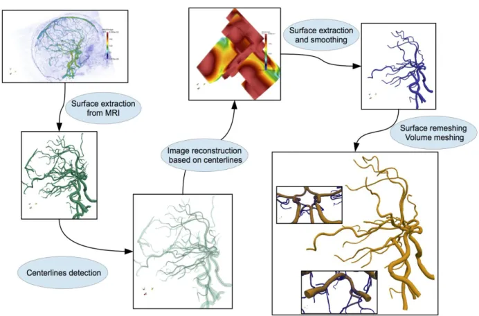
Figure 1: from medical images (top left) to computational mesh (bottom right)
Figure 2(left): Blood flow simulation in the computational mesh generated previously superposed with initial images, coloring using blood flow pressure
We have developed the open-source platform AngioTK that implements the aforementioned1 pipeline based on the following open-source software technologies VTK and ITK(Imaging), RORPO(filtering), VMTK(extraction), Gmsh(mesh generation), Feel++(numerical simulations) and Jemris(MRI simulations). AngioTK implements the glue between these components and automates most of the steps. Indeed very little human intervention is required to complete the pipeline. One of the major contribution of AngioTK is on the area of centerline computation. This ingredient is
crucial for many subsequent steps of the pipeline and not only geometry reconstruction but also in numerical simulations as well as post-processing.
Numerical simulations require model/parameter calibration and they can be used to pursue uncertainty quantification to assess the numerical and modeling choices such as mesh size, boundary conditions or newtonian vs non-newtonian modeling. This requires a tremendous amount of computations and access to state of the art high performance computing software (Feel++) and computational facilities (PRACE-Tier0, Genci-Tier1 and mesocenters-Tier2).
Abstract conclusion
We have developed a complete pipeline from medical images to numerical simulations using state of the art and beyond methodologies. It is robust and requires very little human intervention. There is however still some important research and developments before it is used on a daily basis to answer questions from medical doctors. For example (i)generating virtual is still in early development and is currently not available on reconstructions and simulations show in the previous section, (ii) in assessing the blood flow modeling or (iii) in providing fast (real-time) simulation of such systems.
However the platform is readily available for research projects such as Eye2Brain (IUPUI, Glick Institute, U. Strasbourg) which exploits the eye as a unique window on the brain for the early diagnosis of neuro-degenerative diseases. In this context, AngioTK provides the foundation of a reliable and efficient computational framework allowing for a computer-aided interpretation of clinical data.