Cochlear Hair Cell Regeneration from Neonatal Mouse
Supporting Cells
by
Naomi F. Bramhall
Doctor of Audiology (2007), Master of Science (2006), Bachelor of Science (2004) Speech and Hearing Sciences, University of Washington
Bachelor of Science (2003) Biology, McGill University
SUBMITTED TO THE HARVARD-MIT DIVISION OF HEALTH SCIENCES AND TECHNOLOGY IN PARTIAL FULFILLMENT OF THE REQUIREMENTS FOR THE
DEGREE OF
DOCTOR OF PHILOSOPHY IN
SPEECH AND HEARING BIOSCIENCE AND TECHNOLOGY AT THE
MASSACHUSETTS INSTITUTE OF TECHNOLOGY SEPTEMBER 2012
© 2012 Massachusetts Institute of Technology. All rights reserved.
1 10
Signature of the Author:
Harvard-MIT Division of Health Sciences and Technology August 31, 2012 Certified by:
Accepted by:
Albert Edge, PhD Associate Pr or of Otolaryngology, Harvard Medical School Thesis Supervisor
Arup Chakraborty, PhD Director, Institute for Medical Engineering and Sciences, Robert T. Haslam Professor of Chemical Engineering, Chemistry and Biological Engineering, Massachusetts Institute of Technology
I~cx~v~s
-, ICochlear Hair Cell Regeneration from Neonatal Mouse Supporting Cells
by Naomi F. Bramhall
Submitted to the Harvard-MIT Division of Health Sciences and Technology on August 31, 2012 in Partial Fulfillment of the
Requirements for the Degree of Doctor of Philosophy in Speech and Hearing Bioscience and Technology
ABSTRACT
Unlike lower vertebrates, capable of spontaneous hair cell regeneration, mammals experience permanent sensorineural hearing loss following hair cell damage. Although low levels of hair cell regeneration have been demonstrated in the immature mammalian vestibular system, the cochlea has been thought to lack any spontaneous regenerative potential. Inhibition of the Notch pathway can stimulate hair cell generation in neonatal mammals, but the specific source of these new hair cells has been unclear. Here, using in vitro lineage tracing with the supporting cell markers Sox2 and Lgr5, we show that Lgr5-positive inner pillar and 3rd Deiter's cells in gentamicin-damaged organs of Corti from neonatal mice give rise to new hair cells following treatment with a Notch inhibitor. These new hair cells are generated primarily through direct transdifferentiation of supporting cells, although a small number show evidence of proliferation. Inner pillar cells show the greatest transdifferentation capability, giving rise to immature outer hair cells, and transdifferentiating in response to damage even in the absence of Notch inhibition. In vivo pharmacologic inhibition of Notch and in vivo lineage tracing with Sox2 during genetic Notch inhibition provide generally consistent results, although additional new hair cells develop in the inner hair cell region. These data suggest a spontaneous capacity for hair cell regeneration in the neonatal mammalian cochlea. In addition, the data identify Lgr5-positive supporting cells as potential hair cell progenitors, making them an attractive target for future hair cell
regeneration treatments.
Thesis Supervisor: Albert Edge, PhD
Acknowledgements
Anybody who has been seriously engaged in scientific work of any kind realizes that over the entrance to the gates of the temple of science are written the words: "Ye must have faith."
Max Planck
This thesis describes the successful experiments that resulted from my Ph.D. research. Unrecognized are the months and months of experiments that failed before all the technical problems had been overcome. While I am thankful for the support and advice I received related to the work you see on these pages, I am most deeply grateful for the friends and colleagues who helped me keep faith when my research was faltering.
First and foremost, I would like to express my gratitude to my mentor and research supervisor, Albert Edge, for his patient guidance, faith in me, and reassurance when things were not going as planned.
I would like to thank my thesis committee: Charlie Liberman for chairing the committee, asking valuable questions, and always encouraging me to improve the quality of my images; Cynthia Morton for accepting me as a summer research student my first summer and providing warmth and support ever since; and Matt Kelley for graciously sharing his wealth of expertise and for his willingness to participate in committee meetings from afar.
I also want to acknowledge Valerie Street, my mentor for what started off as my Au.D. research project, but evolved into my master's thesis. It was her warm mentorship and enthusiasm for research that encouraged me to pursue a Ph.D.
I could not have written this thesis without the kindness and support of the members of the Eaton Peabody Laboratory. It was over lunchtime conversation in the cafeteria that I came up with many ideas for troubleshooting and improving my research. I want to thank Dianna Sands for fixing a variety of administrative problems I encountered along the way, and for being willing to defend me. To my labmates in the Edge lab who were always eager to help, I thank you
-specifically, Masato Fujioka and Aurore Brugeaud for training me when I first joined the lab; Fuxin Shi for patiently listening to my research woes and providing both friendship and useful advice; my baymate, Mingjie Tong, for valuable feedback through my thinking out loud; and Judith Kempfle, our lab social organizer, for always reminding us to have fun.
I have great appreciation for the SHBT faculty, students, and staff for all their treasured help and support over these last five years. I'm particularly grateful to my classmates, who helped me survive the first year coursework and have been a continuous source of encouragement along the
way: Ann Hickox, Annalisa Pawlosky, Andrew Schwartz, Bennett Bullock, Christine Hsieh, Lara Thompson, Leah Acker, Matt Crema, and Shanqing Cai.
Deepest gratitude is due to my family and friends: My mom, for her undying support and encouragement no matter how many times I changed my mind about what I wanted to do, and her continuous attempts to understand what my mice were doing; my boyfriend, Brad Buran, for his patient understanding and sound advice, his technical support, and his ability to make even the worst days seem better; and my friend and audiology classmate, Candace Kukino, for her constant faith in my potential as a researcher and for pushing me to get a Ph.D.
Finally, I would like to express my gratitude to the American Speech-Language-Hearing Foundation for awarding me with a scholarship, the NIH for providing training grant funding, and Bertrand Delgutte and Louis Braida for allowing me to continue to receive training grant support throughout my Ph.D.
Table of Contents
Introduction
8
Hair cell regeneration in lower vertebrates ...
8
Hair cell regeneration in mammals...
10
The role of the Notch pathway in the organ of Corti...
11
Inhibition of the Notch pathway in birds ...
11
Inhibition of the Notch pathway in mammals...
13
Mammalian supporting cell transdifferentiation ...
13
Lineage tracing of supporting cells...
14
Thesis overview ...
15
Chapter 1: Using lineage tracing during generation of new hair cells
17
Introduction ...
17
M ethods ...
. 2 1
R esu lts ...
. . 23
D iscussion ...
. . 26
Chapter 2: Lineage tracing of supporting cells following gentamicin
damage
28
Introduction ...
. . 28
Results ...
31
Discussion... 35
Chapter 3: Lineage tracing of supporting cells during Notch
inhibition of damaged organ of Corti explant cultures
38
Introduction... 38M ethods ... 41
Results ... 43
Discussion... 50
Chapter 4: Are new hair cells generated through proliferation?
56
Introduction... 56M ethods ... 58
Results ... 59
Discussion... 61
Chapter 5: In vivo lineage tracing during Notch inhibition
64
Introduction... 64M ethods ...
65
Results ...
67
Discussion... 72
Discussion
76
Regeneration of hair cells following dam age... 76Inner pillar and
3rdDeiter's cells transdifferentiate into hair cells... 76
Some new apical hair cells are generated through proliferation... 77
Hair cell generation greatest in regions with the least damage... 78
Production of inner versus outer hair cells...
79
The role of Lgr5 expression in transdifferentiation capability ...
79
Transdifferentiation capability of inner pillar cells...
81
Future w ork ...
84
Sum m ary ...
. . 85
Introduction
Sensorineural hearing loss resulting from damage to the hair cells of the inner ear is a commonly occurring communication disorder affecting approximately 31 million Americans (Kochkin, 2005). Currently, there is no method of replacing the damaged hair cells to restore hearing. The available treatment options for hearing loss consist of hearing aids and cochlear implants, which are not capable of providing many important features of natural hearing. The discovery of hair cell regeneration in lower vertebrates (Cotanche, 1987; Cruz et al., 1987) raised the hope of developing a method to stimulate regeneration in mammals and restore hearing function in individuals with hearing loss.
Hair cell regeneration in lower vertebrates
Hair cell regeneration in birds following acoustic trauma was first demonstrated in 1987 (Cotanche, 1987; Cruz et al., 1987). After hair cell damage either through acoustic trauma or
ototoxic drug treatment, both supporting cells and newly developed hair cells exhibited markers of proliferation, suggesting the new hair cells were a product of supporting cell re-entry into the cell cycle (Corwin and Cotanche, 1988; Ryals and Rubel, 1988). However, it was later shown that the first hair cells to appear following damage develop in the absence of supporting cell division (Adler and Raphael, 1996). This suggested a second possible mechanism of hair cell regeneration, whereby supporting cells change cell fate and directly transdifferentiate into new hair cells without dividing. Stone and Rubel (2000) noted morphology similar to a supporting
damage. They also found hair cells and supporting cells labeled for the proliferation marker bromodeoxyuridine (BrdU) were clustered together, suggesting these new cells came from a common progenitor, presumably a supporting cell. However, it has also been proposed that particularly in response to greater levels of hair cell damage, both supporting cells and hair cells might be replaced by migration of some other precursor cell population that retains proliferative capability (Girod et al., 1989).
Similar responses to hair cell damage have been observed in fish and amphibians, which are also capable of hair cell regeneration (Balak et al., 1990; Lombarte et al., 1993). Balak et al. used a laser to ablate hair cells specifically in the lateral line of the salamander tail without damaging the supporting cells. Using time-lapse microscopy, they showed increased mitotic activity in supporting cells after hair cell damage and eventual conversion of some supporting cell progeny into hair cells. In bullfrog vestibular hair cells, Baird et al. (1996) observed only a low level of BrdU labeling accompanying the development of new hair cells following treatment with the ototoxic drug gentamicin and noted that newly developing hair cells were surrounded by a lower number of supporting cells than normally found surrounding mature hair cells. This was further evidence suggesting new hair cells could be generated by direct transdifferentiation from supporting cells without proliferation. Similar to what has been reported in chicks, in regions of hair cell loss, they also observed cells with intermediate morphology such as supporting cells with cuticular plates and immature hair bundles.
Hair cell regeneration in mammals
Unlike lower vertebrates, mammals do not experience hair cell regeneration following damage. Two methods of inducing hair cell production in mammals are over-expression of the transcription factor A tohI and inhibition of the Notch signaling pathway.
Mutant mouse embryos lacking AtohI expression fail to develop cochlear and vestibular hair cells, suggesting Atohl is necessary for hair cell differentiation (Bermingham et al., 1999). Overexpression of A tohl leads to the formation of additional hair cells in embryonic and neonatal mammals (Gubbels et al., 2008; Kelly et al., 2012; Zheng and Gao, 2000). Izumikawa et al. (2005) found viral infection of the deafened adult guinea pig inner ear with A tohI led to the development of new outer and inner hair cells and some functional recovery of hearing as
measured by the auditory brainstem response (ABR). Although they claimed to have generated new inner hair cells with normal morphology that were the major contributors to the improved ABR, the possibility remains that the inner hair cells they observed were recovered rather than new hair cells. The outer hair cells had unusual morphology, displaying both hair cell and supporting cell characteristics, suggesting they were new hair cells that developed through transdifferentiation of supporting cells. In a follow-up study of guinea pigs with a flat cochlear epithelium devoid of supporting cells or hair cells, inoculation with the same A tohI virus did not result in hair cell regeneration, suggesting supporting cells are required for this process
The role of the Notch pathway in the organ of Corti
In both birds and mammals, Notch signaling during development is essential to establish patterning in the organ of Corti where no hair cell ever comes into direct contact with another (Figure 1). This patterning results from alternating expression of the Notch receptor and its ligands in adjacent cells, a mechanism called lateral inhibition (Lanford et al., 1999). The membrane-bound Notch ligands, Jagged2 (Jag2) and Delta-like 1 (Dlll), in hair cells bind to the Notch 1 receptor in adjacent supporting cells, activating the Notch pathway (Kiernan et al., 2005a; Lanford et al., 1999; Lanford et al., 2000; Morrison et al., 1999). Cleavage of Notchl by gamma secretase produces the Notch intracellular domain (NICD), which can then translocate to the nucleus, resulting in upregulation of transcription factors such as Hesi and Hes5 (Borggrefe and Oswald, 2009). This leads to inhibition of Atohl and prevents supporting cells from
becoming hair cells (Jeon et al., 2011; Lanford et al., 2000; Zine et al., 2001).
Inhibition of the Notch pathway in birds
Daudet et al. (2009) showed in undamaged chicken basilar papilla that treatment with the gamma secretase inhibitor DAPT, an inhibitor of the Notch pathway, did not result in increased
production of hair cells. However, four days after gentamicin-induced hair cell damage, several Notch pathway genes were significantly upregulated. They suggested the Notch pathway may play a role in regulating hair cell regeneration to prevent overproduction of hair cells at the expense of supporting cells. Consistent with this hypothesis, in contrast to the undamaged chicken basilar papilla, treatment with DAPT following streptomycin damage resulted in much higher numbers of hair cells than were generated through the natural regeneration process and a
A
Figure 1. The Notch pathway in the organ of Corti
(A) Notch ligands Jag2 and Dill are expressed in the hair cell membrane and bind to the Notch 1 receptor in adjacent supporting cells. This leads to cleavage of Notch 1 by gamma secretase, resulting in the Notch intracellular domain (NICD), which then translocates to the nucleus and activates transcription of genes such as Hes1 and Hes5 that inhibit Atohl. (B) Treatment with a gamma secretase inhibitor prevents cleavage of Notchl, inhibiting the Notch pathway and allowing for transcription of Atohl.
reduction in supporting cell numbers. This suggests Notch inhibition following damage in birds
can promote transdifferentiation of supporting cells into additional hair cells.
Inhibition of the Notch pathway in mammals
Although mammals do not typically show evidence of hair cell regeneration,
supernumerary hair cells in embryonic and neonatal mammals can be produced via inhibition of the Notch pathway, either through genetic manipulation or treatment with a Notch inhibitor (Doetzlhofer et al., 2009; Hayashi et al., 2008; Kiernan et al., 2005a; Takebayashi et al., 2007; Yamamoto et al., 2006; Zhao et al., 2011; Zine et al., 2001). Unlike birds, these effects of Notch inhibition are observed even in the absence of damage. Inhibition of the Notch pathway results in a reduction in the number of supporting cells that is correlated to an increase in hair cell number (Doetzlhofer et al., 2009; Kiernan et al., 2005a). Doetzlhofer et al. saw an increase in the number of hair cells, particularly in the apical region, when they treated postnatal mouse organ of Corti cultures with DAPT. In treated cultures, they also saw a decrease in the number of Deiter's cells as the number of hair cells increased.
Mammalian supporting cell transdifferentiation
Transdifferention into hair cells may only be possible in particular subtypes of
mammalian supporting cells. Doetzlhofer et al. (2009) suggested Deiter's cells were capable of transdifferentiation into hair cells following DAPT treatment, while pillar cells were not. Conversely, it has been proposed that pillar cells may be more capable of transdifferentiation than other supporting cells (White et al., 2006). Mutant mouse embryos with genetic reduction in
Notch expression show an increase in inner and outer hair cells and a reduction in Deiter's cell numbers (Kiernan et al., 2005a). However, there is also evidence of pillar cell proliferation even though pillar cell numbers remain stable, suggesting that pillar cells may also be
transdifferentiating into new hair cells. Although studies such as these have suggested
supernumerary mammalian hair cells are generated from supporting cells, there is only indirect evidence for this conclusion and a lack of agreement about the types of supporting cells that can transdifferentiate. It is important to conclusively determine whether specific subtypes of
supporting cells can differentiate into hair cells in mammals so that future efforts at stimulating hair cell regeneration can be targeted to the progenitor cells that give rise to new hair cells.
Lineage tracing of supporting cells
Lineage tracing using a supporting cell specific marker gene can provide direct evidence of whether supporting cells are capable of conversion into new hair cells. Lineage tracing is a method of permanently labeling a particular cell population so that it can be followed over time. Sox2 is a general marker of both adult and neonatal supporting cells, expressed in almost all supporting cell types and the greater epithelial ridge (GER) in neonates (Oesterle et al., 2008). Sox2 is a transcription factor required for prosensory formation of the inner ear (Kiernan et al.,
2005b) and is involved in both neurogenesis and proliferation and maintenance of stem cells (Episkopou, 2005). Lgr5 is a marker expressed in a smaller subset of neonatal supporting cells, including 3 Deiter's cells, inner pillar cells, inner border cells, and the GER (Shi et al., 2012). Lgr5-positive cells are of particular interest because Lgr5 is a known marker of intestinal stem cells and a target of the Wnt pathway (Barker et al., 2007). The highly conserved Wnt pathway has many roles in both developing and adult tissues including regulation of proliferation,
differentiation, and tissue homeostasis (Clevers, 2006). Lgr5 has also recently been found to be a marker of progenitor cells in the cochlea that can give rise to hair cells (Shi et al., 2012). Lineage tracing with both Sox2 and Lgr5 can be used not only to determine whether supporting cells are capable of transdifferentiation, but also to narrow down the relevant subpopulation.
Thesis overview
The goal of this thesis was to use lineage tracing with the markers Sox2 and Lgr5 to follow specific supporting cell populations over time while generating new hair cells through in vitro and in vivo Notch inhibition. Lineage tracing with the Cre-lox system enabled permanent labeling of supporting cells by preventing the label from being lost after changes in gene
expression when supporting cells became hair cells. Through Sox2 and Lgr5 lineage tracing, we demonstrated previously undetected low-level spontaneous transdifferentiation of inner pillar cells into new hair cells in response to gentamicin damage alone. Additionally, we showed Lgr5-positive inner pillar and 3rd Deiter's cells are progenitor cells capable of transdifferentiating into new hair cells in neonatal mice following gentamicin damage and Notch inhibition in vitro. We also provided evidence of low-level proliferation in new apical hair cells after damage with and without Notch inhibition. These in vitro lineage tracing results were supported by in vivo pharmacologic Notch inhibition and lineage tracing using genetic inhibition of Notch]. In vivo lineage tracing also indicated that supporting cells adjacent to inner hair cells may be able to transdifferentiate into new hair cells shortly after birth. Chapter 1 of this thesis describes lineage tracing using the Cre-lox system and the marker genes Sox2, Lgr5, and Pou4f3. Chapter 2 uses lineage tracing to explore the effects of gentamicin damage on neonatal mouse cochlear explant cultures. Through lineage tracing of supporting cells and hair cells, Chapter 3 investigates the
effects of Notch inhibition following damage. Chapter 4 examines the proliferative effects of damage and Notch inhibition on cochlear explants. Finally, Chapter 5 covers in vivo lineage tracing of neonatal mouse supporting cells during Notch inhibition.
Chapter 1
Using lineage tracing during generation of new hair cells
Introduction
Lineage tracing is a method of labeling particular cell populations and following them over time. This can be accomplished using the Cre-lox system, which involves crossing a Cre mouse line driven by a genetic marker specific to the cells of interest with a reporter mouse line (Figure 1). The reporter construct is driven by a ubiquitous promoter such as cytomegalovirus early enhancer element and chicken beta-actin promoter (CA G) and contains lox sites flanking a stop sequence upstream of a reporter gene. Cre recombinase cuts the DNA at the lox sites, excising the stop sequence whenever both the Cre and reporter constructs are expressed in the same cell. This allows for transcription of the reporter gene in the target cells. The advantage of using this system of labeling is that Cre recombination at the lox sites results in a permanent change to the DNA, allowing the cells of interest to continue expressing the reporter regardless of whether the marker gene continues to be expressed. This makes the Cre-lox system
particularly useful for labeling supporting cells that may turn into hair cells.
To perform lineage tracing of supporting cells, an appropriate supporting cell marker gene must first be identified. Lineage tracing of a general population of supporting cells can be achieved using a marker such as Sox2, a member of the highly conserved SOX family of
transcription factors expressed in almost all types of supporting cells. Although widely expressed during development for prosensory specification,
Cre-lox System
Cre $lislox
Supporting cells All other cells
Figure 1. Use of the Cre-lox system for lineage tracing
A supporting cell-specific Cre mouse line is mated with a reporter line driven by the ubiquitous promotor CAG, expressed in all cell types. In the supporting cells of double transgenic offspring, Cre cuts the reporter construct at the lox sites, excising the tdTomato gene and the stop sequence. This allows eGFP to be transcribed, permanently turning the supporting cells green. All other cell types, which do not express Cre, will remain red due to expression of tdTomato.
mouse organ of Corti Sox2 expression at the time of birth is confined to the supporting cells (Dabdoub et al., 2008; Oesterle et al., 2008). Sox2 is a general marker of both adult and neonatal supporting cells, expressed in almost all supporting cell types and the greater epithelial ridge (GER) in neonates (Oesterle et al., 2008). While a general supporting cell marker is useful for determining if supporting cells can transdifferentiate into new hair cells, a marker gene with a narrower pattern of expression is necessary to determine which supporting cell types are capable of transdifferentiation. Lgr5 was chosen as a second supporting cell marker for lineage tracing due to its expression in a smaller subset of neonatal supporting cells, including 3rd Deiter's cells,
inner pillar cells, inner border cells, and the GER (Figure 2, (Shi et al., 2012)).
Because Sox2 and Lgr5 are widely expressed in the prosensory epithelium during development, the Sox2-Cre and Lgr5-Cre constructs also contain a mutant form of the estrogen receptor (ER), responding only to the drug tamoxifen, to be used for induction of the Cre
construct. Administration of tamoxifen results in activation of Cre only in cells expressing Cre at that point in time. This allows for induction of the Cre construct to be postponed until after marker expression has been confined to supporting cells. Therefore, to localize Cre expression specifically to the supporting cells of interest, the Sox2-Cre and Lgr5-Cre constructs were induced by tamoxifen administration postnatally.
A third marker for lineage tracing was used to permanently label native hair cells as a means of distinguishing them from new hair cells. In contrast to Sox2 and Lgr5, which are genes
specifically expressed in supporting cells, Pou4J3 is expressed exclusively in the inner and outer hair cells by E 15.5 (Xiang et al., 1997). The Pou4J3-Cre construct is not inducible, meaning Cre
is always active and any cell expressing Pou4J3 will undergo recombination. This makes Pou4J3 lineage tracing useful for labeling all native hair cells.
Sox2 Hensen's
GER
Inner Inner InnerOue itr
Border Phalangeal Pillar Pillar Cells Cell Cell
Lgr5Hensen's
Cell CelC
GER
Border Phlaeal Pillar Pillar cls
Cell Cel
Figure 2. Neonatal expression of Sox2, Lgr5 and Pou4f3 in the organ of Corti
Schematic illustrates the expression of Sox2, Lgr5 and Pou4f3 in red. Sox2 is expressed in the Hensen's cell, Deiter's cells, inner and outer pillar cells, inner phalangeal cell, inner border cell and GER. Lgr5 is
expressed in the 3rd Deiter's cell, the inner pillar cell, the inner border cell and the GER. Pou4f3 is
This chapter describes reporter expression in both supporting cells and hair cells of the mouse organ of Corti resulting from recombination in Sox2-Cre, Lgr5-Cre, and Pou4'3-Cre lineage tracing.
Methods
Animals
The Sox2-Cre-ER mouse line (Arnold et al., 2011) was provided by Dr. Konrad
Hochedlinger, Harvard Medical School, Boston, MA. The Pou4f3-Cre mouse (Sage et al., 2006) was a gift from Dr. Douglas Vetter, Tufts University, Boston, MA. The Lgr5-EGFP-IRES-Cre-ER mouse line (Barker et al., 2007) as well as the CA G-tdTomato (Madisen et al., 2010) and CAG-tdTomato-EGFP (Muzumdar et al., 2007) reporter lines were obtained from Jackson Laboratory (stock #008875, 007914 and 007676 respectively). Sox2-Cre-ER and Pou4f3-Cre mice were genotyped by polymerase chain reaction (PCR) using primers to amplify the Cre gene (F 5'-TGG GCG GCA TGG TGC AAG TT-3', R 5'-CGG TGC TAA CCA GCG TTT TC-3'). Lgr5-EGFP-IRES-Cre-ER, CAG-tdTomato and CAG-tdTomato-EGFP mice were genotyped by PCR according to Jackson Laboratory recommendations.
Lineage tracing
Sox2-Cre-ER mice were crossed with CAG-tdTomato-EGFP or CAG-tdTomato reporter mice. Lgr5-EGFP-IRES-Cre-ER and Pou4f3-Cre mice were crossed with CAG-tdTomato mice. At P1 (or both PO and P1 for Lgr5 lineage tracing), an intraperitoneal injection of 100 tl
tamoxifen dissolved in corn oil (50 mg/ml) was administered to mothers of double transgenic (Cre- and reporter-positive) litters and delivered to the pups via the milk.
Organ of Corti dissection
Mice positive for both Cre and the reporter construct were dissected at P2 and cochleae removed from the temporal bones. Cochleae for organ of Corti culture were transferred to Hanks' Balanced Salt Solution (HBSS, Invitrogen) and organ of Corti isolated. The stria vascularis and basal hook region were removed and the organ of Corti plated and cultured as described below. Cochleae not attained for culture were immediately fixed for 10 min in 4% paraformaldehyde (PFA, Electron Microscopy Sciences). The organ of Corti was then isolated and the stria vascularis and basal hook region removed. This was followed by additional fixation in 4% PFA for 30 minutes and then storage in 1X Phosphate Buffer Solution (PBS) at 4*C until staining.
Organ of Corti culture
Organ of Corti were plated on glass coverslips coated with poly-L-ornithine (Sigma) and laminin (BD Biosciences) in 100 d Dulbecco's Modified Eagle Medium (DMEM, Invitrogen) with 10% fetal bovine serum (FBS, Invitrogen) and 25 pg/ml ampicillin (Sigma), and cultured at 37*C in 5% CO2 overnight. Cultures were then placed in serum-free DMEM:F 12 (Invitrogen)
with 1% B27 supplement (Invitrogen), 25 Ig/ml ampicillin, and 0.1% dimethyl sulfoxide (DMSO, Sigma) for 72 hours. After four days in vitro, cultures were fixed for 10 minutes with 4% PFA and stained for myosin VIla to identify hair cells and for either DsRed or GFP to enhance the endogenous signal from the reporter.
Antibodies
The following primary antibodies were used: anti-myosin VIa (mouse IgG,
anti-DsRed (rabbit IgG, Clontech, 1:500). Immunoreactivity was visualized using Alexa Fluor-conjugated secondary antibodies (Molecular Probes, 1:500).
Imaging and cell counting
Organ of Corti were analyzed using a Leica TCS SP5 confocal microscope. Organ of Corti cross-sections were obtained from confocal Z-stacks using Amira imaging software. High magnification fluorescent images of the organ of Corti were merged in Photoshop and counted manually with ImageJ software (NIH). Counts from each organ of Corti were divided into three regions (apex, middle and base) and hair cell counts were obtained per 100 [tm. Each counted region was 1900 t 100 tm. Sox2 lineage tracing recombination rates were estimated by counting the percentage of Sox2-tagged inner pillar cells in each region. LgrS lineage tracing
recombination rates were calculated for inner pillar and 3rd Deiter's cells.
Results
Sox2 neonatal expression
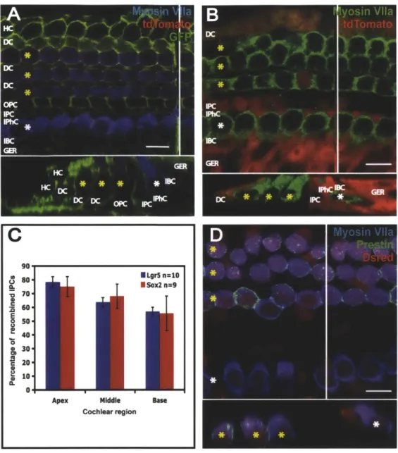
A confocal image of a whole mount from the apex of an organ of Corti from a P6
Sox2-Cre-ER/CA G-tdTomato-EGFP mouse that was exposed to tamoxifen at P1 through its mother's milk showed expression of GFP in Deiter's cells, inner and outer pillar cells, inner border cells, inner phalangeal cells, Hensen's cells and the greater epithelial ridge (Figure 3A). This pattern of recombination is similar to the Sox2 expression previously described by Oesterle et al. (2008). The Sox2-Cre recombination rate appeared relatively consistent across supporting cell types. The Sox2 lineage tag was also noted in some inner and outer hair cells. In five explants, the frequency of outer hair cells possessing the Sox2 tag was greatest in the apex (54.47% ± 4.99% of total
outer hair cells), infrequent in the middle region (2.43% - 1.43%), and rare in the base (0.14% 0.06%). The frequency of Sox2-tagged inner hair cells followed a similar pattern. Presumably, these Sox2-tagged hair cells are relatively immature native hair cells still expressing Sox2 to some degree at the time of tamoxifen exposure.
Lgr5 neonatal expression
The Lgr5 lineage tag was observed in an organ of Corti from an Lgr5-EGFP-JRES-Cre-ER double transgenic (positive for Cre and the reporter) mouse pup in the inner border, inner
phalangeal, inner pillar and 3'd Deiter's cells as well as the GER (Figure 3B). This pattern is consistent with previously reported neonatal expression of Lgr5, with the exception of the inner phalangeal cells, although closer examination of the published images reveals faint expression of Lgr5 in inner phalangeal cells (Shi et al., 2012). The observation of the Lgr5 lineage-tag in the inner phalangeal cells likely reflects the young age of these pups when they received tamoxifen, suggesting Lgr5 is expressed in inner phalangeal cells immediately after birth and is then quickly downregulated. There was no significant difference in the percentage of Lgr5-tagged inner pillar cells versus the percentage of Sox2-tagged inner pillar cells (Figure 3C). The frequency of Lgr5-tagged 3rd Deiter's cells was considerably higher than the frequency of Lgr5-tagged inner pillar
cells, with 86.36% ± 6.46% in the apex, 94.4% ± 2.84% in the middle and 96.74% - 1.52% in
the base. Some hair cells also exhibited the Lgr5 lineage tag. In seven explants, Lgr5-tagged outer hair cells were observed most frequently in the apex (19.80% ± 4% of total outer hair
cells), occasionally in the middle (1.95% ± 1.3%), and rarely in the base (0.22% ± 0.18%).
Lgr5-tagged inner hair cells were also seen, most frequently in the apex. This suggests that some relatively immature hair cells, usually in the apex, were still expressing Lgr5 at PO when tamoxifen was first delivered.
Pou4f3 neonatal expression
A confocal image of an organ of Corti explant culture from a Pou4J3-Cre/CA G-tdTomato mouse pup that was exposed to tamoxifen at P1 through maternal milk and dissected at P2
displayed the Pou4J3 lineage tag in all inner and outer hair cells (Figure 3D). The Pou43 tag was also observed in some supporting cells.
Figure 3. Reporter expression in Sox2, Lgr5 and Pou4f3 lineage tracing
(A-B, D) In confocal slices and cross-sections from neonatal organ of Corti, OHCs are labeled with yellow asterisks and IHCs with white asterisks. A white vertical line indicates the location of the
cross-section. The scale bar is 10 [tm. (A) The apex of a Sox2-Cre-ER/CA G-tdTomato-EGFP organ of Corti treated with tamoxifen at P1 shows reporter expression (stained for GFP, in green) in Hensen's cells,
Deiter's cells, inner and outer pillar cells, inner phalangeal cells, inner border cells, and the GER. (B) The apex of an Lgr5-EGFP-IRES-Cre-ER/CAG-tdTomato organ of Corti treated with tamoxifen at PO and P1 exhibits reporter expression (stained for DsRed, in red) in the 3 rd Deiter's cell, inner pillar cell, inner border cell, and the GER. (C) Mouse pups used for Sox2 lineage tracing received tamoxifen once at P1, while pups for Lgr5 lineage tracing received tamoxifen at both PO and P1. No significant difference in the number of inner pillar cells expressing the reporter was identified between the two lineage tracing
markers. (D) The mid-apex of a Pou4J3-Cre/CAG-tdTomato organ of Corti displays reporter expression (stained for DsRed, in red) in the IHCs and OHCs. Some supporting cells are also reporter-positive.
C
C . Middle Base Cochlear region ApexDiscussion
The Sox2 lineage tag was observed in all supporting cell types known to be
Sox2-positive. Similarly, all Lgr5-positive supporting cell types displayed the Lgr5 lineage tag. When Lgr5 lineage tracing mouse pups received two doses of tamoxifen rather than a single dose, the percentage of Sox2- and Lgr5-positive supporting cells that were lineage-tagged was similar. The lineage tag frequency for both Sox2 and Lgr5 was highest in the apex and lowest in the base.
Presumably due to immaturity and late downregulation of Sox2 and Lgr5, the lineage tag was observed in some hair cells for both Sox2 and Lgr5 lineage tracing. This was most
frequently observed in the apex and rarely found in the base. This complication of the lineage tracing made it more difficult to distinguish tagged native hair cells from tagged new hair cells derived from Sox2- or Lrg5-positive supporting cells. It was necessary to use other methods to verify the identity of a new lineage-tagged hair cell. Pou4J3 lineage tracing was one means of verifying Sox2 and Lgr5 lineage tracing results. Other methods are described in Chapters 2 and
Chapter 2
Lineage tracing of supporting cells following gentamicin damage
Introduction
Although mammals are generally believed to lack hair cell regeneration capability, there is some evidence of limited regenerative potential in the mammalian vestibular system and cochlea.
The mammalian vestibular system has been suggested to exhibit some regenerative capacity following damage (Forge et al., 1998; Kawamoto et al., 2009; Kuntz and Oesterle, 1998; Warchol et al., 1993). However, one obstacle in identifying new hair cells generated following damage has been the challenge of discriminating between newly regenerated hair cells and hair cells that persist or recover following damage. A recent study using viral infection of adult mammalian utricular supporting cells with an Atohl-GFP reporter virus provided more conclusive evidence of the generation of new hair cells after damage by facilitating detection of newly developed hair cells (Lin et al., 2011). Marking the new hair cells in this manner is similar in concept to lineage tracing, although it does not identify the hair cell progenitors.
Regeneration has also been investigated in mammalian cochlear hair cells. Kelley et al. (1995) damaged individual hair cells in embryonic and neonatal organ of Corti explant cultures using laser irradiation and then closely observed the lesion sites over several days. They
discovered that damaged outer hair cells in the embryonic cultures were quickly replaced with new hair cells exhibiting immature stereocilia bundles. They also found that the nuclei of the putative new cells appeared to move upward from below the hair cells to the hair cell nuclear
layer. The nuclei also were initially small, similar to supporting cell nuclei, but became larger and more spherical as they moved upward and the cells began to exhibit hair cell characteristics. In neonatal cultures, they saw only a very small number of what appeared to be regenerated hair cells at the lesion sites after the recovery period. In both embryonic and neonatal cultures, damaged inner hair cells did not appear to be replaced with new hair cells. However,
identification of new regenerated hair cells in this study was based primarily on morphological characteristics suggestive of immature hair cells, and they were unable to conclusively identify newly generated hair cells.
One of the biggest challenges in identifying regenerative capacity in the mammalian vestibular system and cochlea has been the difficulty of determining which hair cells are new. Most methods of damaging hair cells are incomplete, making it difficult to ensure complete loss of all native hair cells. This poses a challenge in distinguishing between regenerated hair cells and spared or repaired native hair cells. Lineage tracing provides a method not only for determining which hair cells are new, but also their origin. In this chapter, lineage tracing is employed in two ways to identify new hair cells following damage with the ototoxic drug gentamicin. One method uses the markers Sox2 and Lgr5 to follow supporting cells as they transdifferentiate into new hair cells and the other marks native hair cells with the hair cell marker Pou4J3 so they can be distinguished from new hair cells.
Methods
Animals
The Sox2-Cre-ER, Pou4f3-Cre, Lgr5-EGFP-IRES-Cre-ER, and CAG-tdTomato mouse lines were used as described in Chapter 1.
Lineage tracing
Sox2-Cre-ER mice, Lgr5-EGFP-IRES-Cre-ER, and Pou4J3-Cre mice were crossed with CA G-tdTomato reporter mice and treated with tamoxifen as indicated in Chapter 1.
Organ of Corti culture
Organ of Corti from P2 mice positive for both Cre and the reporter construct were isolated and plated as described in Chapter 1. One hour after dissection, cultures were treated with 50 IM gentamicin (Sigma) in DMEM with 10% FBS and 25 Rg/ml ampicillin. Organ of Corti were cultured overnight (16 hours) and then placed in serum-free DMEM:F12 (Invitrogen) with 1% B27 supplement (Invitrogen), 25 Rg/ml ampicillin, and 0.1% dimethyl sulfoxide
(DMSO, Sigma) for 72 hours. Controls were cultured under the same conditions without
gentamicin. After four days in vitro, cultures were fixed and stained for myosin VIla to identify hair cells and DsRed to enhance the endogenous signal from the reporter.
Antibodies
The following primary antibodies were used in addition to those indicated in Chapter 1: anti-Sox2 (goat IgG, Santa Cruz Biotechnology, 1:500) and anti-prestin (goat IgG, Santa Cruz Biotechnology, 1:400).
Imaging and cell counting
Organ of Corti were analyzed as described in Chapter 1. Additionally, counts of lineage-tagged outer hair cells were obtained for each region. Lineage-lineage-tagged hair cells in the pillar cell region were counted as outer hair cells. The number of tagged hair cells in the pillar cell region was also determined. Hair cell counts for the undamaged control group were compared to those from the gentamicin-treated group using the Student's t-test.
Results
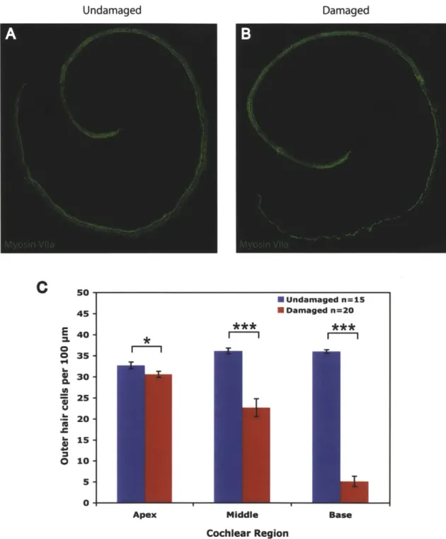
Treatment with gentamicin leads to outer hair cell loss in the middle and basal regions
Organ of Corti explant cultures treated with 50 pM gentamicin overnight and examined 72 hours later showed significant outer hair cell loss in all cochlear regions, with the greatest damage in the middle and basal turns (Figure 1). In some cultures, inner hair cell loss was also observed, particularly in the basal region.
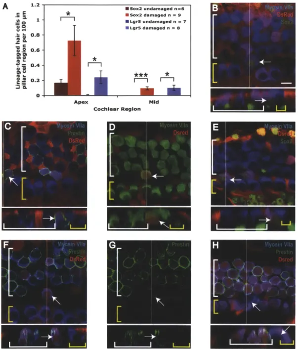
Sox2-positive supporting cells transdifferentiate into hair cells at low frequency after damage
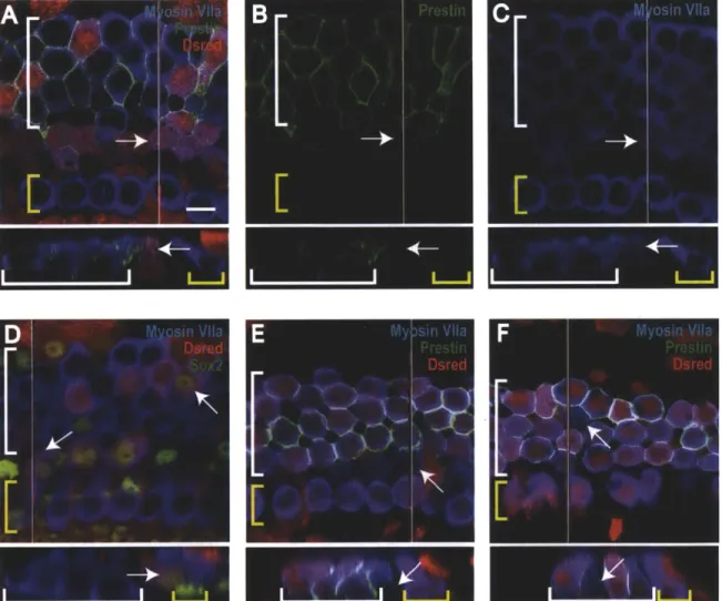
In the apex and middle turns of organ of Corti treated with gentamicin, significantly more Sox2-tagged hair cells were observed in the pillar cell region than in undamaged controls (red bars in Figure 2A). No lineage-tagged hair cells were found in the pillar cell region of the base. The unusual location and low frequency suggested these tagged cells were new hair cells. In addition, unlike native hair cells, these Sox2 lineage-tagged hair cells exhibited antibody staining for Sox2 in their nuclei (arrows in Figure 2B), consistent with immature hair cells (Kempfle et al., 2012). Organ of Corti were also stained with prestin to determine the identity of the new hair
cells. Prestin is a motor protein specific to the outer hair cells (Zheng et al., 2000). Many new hair cells in the pillar region displayed prestin staining, suggesting they were new outer hair cells (arrows in Figure 2C).
Lgr5-positive supporting cells transdifferentiate into hair cells following damage alone
Lineage tracing with Lgr5 was used to determine more precisely the subpopulation of supporting cells capable of transdifferentiation because Lgr5 expression is more restricted than Sox2 expression. Similar to results from Sox2 lineage tracing following gentamicin damage, occasional Lgr5 lineage-tagged hair cells were observed in the pillar cell region after damage (arrows in Figure 2D-G). In the apex and middle regions, significantly more Lgr5-tagged hair cells were observed in the pillar cell region following gentamicin damage than in undamaged controls (blue bars in Figure 2A). In the base, no lineage-tagged hair cells were found in the pillar cell region. Sox2-positive nuclei and prestin staining in the membranes of the lineage-tagged cells were consistent with new outer hair cells (arrows in Figure 2E-G). The expression pattern of Lgr5 and the pillar cell location of these new hair cells suggested they were generated by inner pillar cells.
Pou4f3 lineage tracing indicates the presence of new hair cells in the pillar cell region following damage
We further assessed the ability of supporting cells to generate new hair cells after damage by using Pou4J3 lineage tracing to label the original hair cells. As described in Chapter 1, in contrast to Sox2 and Lgr5, lineage tracing with Pou4J3 marks native hair cells. A small number of hair cells did not display the Pou4J3 lineage tag throughout the time course of the experiment, which was indicative of newly formed hair cells. New hair cells were noted in the pillar cell region
following gentamicin damage (arrows in Figure 2H), verifying Sox2 and Lgr5 lineage tracing results. This is in contrast to results from Chapter 1 showing all inner and outer hair cells were recombined in undamaged control cultures.
Undamaged * m 0 -1--Apex * Undamaged fl=15 II Undamaged n=15 I Damaged n=20
**
Cochlear RegionFigure 1. Gentamicin treatment causes hair cell damage
(A-B) Neonatal organ of Corti explant cultures stained for myosin VI1a to identify hair cells. (A)
Undamaged control explant, (B) Gentamicin-damaged explant. (C) Quantification of OHC numbers per
100 [tm for undamaged and damaged cultures showing a significant decrease in hair cells following
gentamicin treatment for all three cochlear regions. * P < 0.05, *** P < 0.001.
Damaged
C
2 0 .C 01 **m 50 45 - 40-35 -30 -25 -20 -15 - 10-5 Middle Base I*Sox2 undamaged n=6 " Sox2 damaged n = 9 *LgrS undamaged n = 7 " Lgr5 damaged n = 8 * *** * m m c E a 1 -= 0 .-60 U 84 0.8 120.6-'i Ch S0.4-.2 0.2-0 4
Figure 2. New hair cells are generated in the pillar cell region after gentamicin damage
(A) The number of lineage-tagged hair cells in the pillar cell region per 100 [tm in undamaged and damaged controls for Lgr5 (blue bars) and Sox2 (red bars) lineage tracing. * P < 0.05, *** P < 0.001. (B-H) Confocal slices and cross-sections from the mid-apex of neonatal organ of Corti explant cultures treated with gentamicin and lineage traced using the CA G-tdTomato reporter. The endogenous signal from the reporter was enhanced by staining for DsRed, shown in red. A white line on the whole mount image delineates the location of the cross section. IHCs are indicated by a yellow bracket and OHCs by a white bracket. Lineage-tagged hair cells can be identified by co-labeling with myosin Vlla and DsRed. Arrows point to new lineage-tagged (or untagged for Pou4J3 lineage tracing) hair cells in the pillar cell region. The scale bar is 10 [tm. (B-C) Sox2 lineage tracing images stained for Sox2 (B) and prestin (C). (D-G) Lgr5 lineage tracing images, stained for Sox2 (E) and prestin (F-G). (H) A culture from Pou4J3 lineage tracing showing an untagged hair cell in the pillar region.
A 1.2 * *m
I
Mid Apex Cochlear RegionDiscussion
Through lineage tracing of both supporting cells and hair cells we have conclusively shown for the first time the existence of limited regenerative capability in the neonatal
mammalian cochlea. Based on the location of these regenerated hair cells in the pillar cell region and their origins in the Lgr5 lineage, we can conclude that the new hair cells are produced through transdifferentiation of inner pillar cells. These results suggest a previously unrecognized similarity in regenerative potential between the adult mammalian utricle and the neonatal
mammalian cochlea.
New hair cells were detected in the apical and middle regions, but rarely seen in the base. Considering hair cell damage was greatest in the base, one possible explanation for the lack of new hair cells is that the extensive damage in the basal region may include supporting cells, preventing hair cell regeneration. Alternatively, this result may indicate a lower regenerative potential in the base that may correlate with earlier differentiation.
The adult mammalian retina experiences some intrinsic neural regeneration following damage, which is mediated by Wnt signaling (Osakada et al., 2007). It has also been
demonstrated that Wnt signaling leads to increased A tohI expression (Shi et al., 2010).
Considering that inner pillar cells express Lgr5, a downstream target of the Wnt pathway, it is plausible the spontaneous hair cell regeneration observed in the neonatal organ of Corti following gentamicin damage is stimulated by Wnt. Additionally, Wnt is a soluble rather than membrane-bound protein. Hair cell loss in the base of the organ of Corti may upregulate Wnt signaling, which could then stimulate transdifferentiation of inner pillar cells in the apex, even in the absence of damage in that region.
The observation that only inner pillar cells transdifferentiate in response to gentamicin damage, rather than all Lgr5-positive supporting cells, suggests that other signaling in addition to Wnt may stimulate inner pillar cells to become hair cells. Neonatal mouse pillar cells
specifically express Fgfr3, a fibroblast growth factor receptor (Jacques et al., 2007). In birds, Fgfr3 is transiently downregulated in supporting cells following gentamicin damage and then returns to normal levels after the completion of hair cell regeneration, indicating a role in preventing supporting cells from differentiating into hair cells (Bermingham-McDonogh et al., 2001). Consistent with this hypothesis, when the developing basilar papilla is treated with Fgf inhibitor SU5402, extra hair cells are produced and supporting cells are lost without evidence of proliferation, suggestive of supporting cell transdifferentiation into new hair cells (Jacques et al., 2012). Fgfr3 null mutant mice lack inner pillar cells and exhibit an extra row of outer hair cells (Hayashi et al., 2007; Puligilla et al., 2007). In mice, in vivo activation of Fgfr3 with Fgfl7 results in production of additional p75"*-positive (a pillar cell marker) cells and a loss of outer hair cells (Jacques et al., 2007). These findings suggest that decreased expression of Fgfr3 in the
inner pillar cells following damage may stimulate their transdifferentiation into outer hair cells. However, a previous study of the effects of damage on Fgfr3 expression in adult rats indicated
an increase in Fgfr3 expression following acoustic trauma (Pirvola et al., 1995). The response of Fgfr3 to damage may differ based on the age of the animal. In the present experiment, using neonatal mice, it is conceivable that Fgfr3 may be downregulated in inner pillar cells, which along with increases in Wnt signaling could allow a small number of inner pillar cells to transdifferentiate into new hair cells.
Chapter 3
Lineage tracing of supporting cells during Notch inhibition of
damaged organ of Corti explant cultures
Introduction
Previous studies in embryonic and neonatal mammals have suggested that new hair cells can be generated by transdifferentiation of supporting cells (Doetzlhofer et al., 2009; Hayashi et al., 2008; Kiernan et al., 2005a; Takebayashi et al., 2007; White et al., 2006; Yamamoto et al., 2006). One mechanism believed to stimulate supporting cell transdifferentiation is inhibition of the Notch pathway. Both supporting cells and hair cells develop from a common progenitor cell type (Fekete et al., 1998). Cell-to-cell contact has been implicated in activation of Notch in cells adjacent to hair cells, resulting in a supporting cell fate, while inhibition of the Notch pathway leads to a hair cell fate (Lanford et al., 1999; Yamamoto et al., 2006; Zine et al., 2000). Jagged2 (Jag2) and Delta-like 1 (Dlll) are membrane-bound Notch ligands expressed by developing mammalian hair cells (Kiernan et al., 2005a; Lanford et al., 1999; Lanford et al., 2000; Morrison et al., 1999). Binding of a ligand to a Notch receptor in an adjacent cell through cell-to-cell contact results in cleavage of the receptor by gamma secretase (Borggrefe and Oswald, 2009). The resulting Notch intracellular domain (NICD) can translocate to the nucleus where it
upregulates a number of genes such as Hes1 and Hes5 that inhibit Atohl and prevent a hair cell fate (Jones et al., 2006; Zine et al., 2001). It can therefore be expected that inhibition of the Notch pathway will result in increased Atohl expression in supporting cells, allowing for their transdifferentiation into hair cells.
Notch inhibition, either pharmacologic or genetic, is effective in generating
supernumerary hair cells in embryonic and neonatal mammals (Doetzlhofer et al., 2009; Hayashi et al., 2008; Kiernan et al., 2005a; Takebayashi et al., 2007; Yamamoto et al., 2006; Zhao et al., 2011; Zine et al., 2001). Mutations decreasing expression of the Notch receptor Notch] or the Notch ligands Jag2 and DlII result in extra inner and outer hair cells and a reduction in supporting cell number although this decrease does not completely account for the increase in hair cell number (Kiernan et al., 2005a). Hes1 and Hes5 knockout mice both exhibit extra hair cells in the organ of Corti, with additional inner hair cells found in Hes1 mutants and increased
outer hair cells in Hes5 mutants (Zine et al., 2001). Administration of DAPT to postnatal mouse organ of Corti explants results in an increase in the number of hair cells and a decrease in the number of Deiter's cells, suggesting that new hair cells may be generated by direct
transdifferentiation of supporting cells (Doetzlhofer et al., 2009).
While there is evidence to suggest supporting cells can transdifferentiate into new hair cells, there is a lack of agreement about which types of supporting cells possess this capability. Doetzlhofer et al. (2009) suggested that Deiter's cells were capable of transdifferentiation into hair cells following DAPT treatment, while pillar cells were resistant due to their expression of the Hes-related gene Hey2. They found that unlike Hes5, which is highly downregulated in Deiter's cells following DAPT administration, Hey2 levels were unchanged. Additionally, in organ of Corti explant cultures from Hey2 null mice, pillar cells disappeared in response to DAPT, presumably because the absence of Hey2 expression made them responsive to Notch inhibition and allowed them to transdifferentiate into hair cells. White et al. (2006) used
fluorescence-activated cell sorting (FACS) to isolate particular populations of neonatal sensory epithelial cells from Math l-GFP mice, which display green fluorescence in A tohI (also called
Math]) expressing cells, most notably the hair cells. Using FACS, they excluded Math l-GFP positive hair cells and sorted for p7 5NGFR to generate a population of cells enriched in pillar and Hensen's cells. Culture of the FACS-purified sensory epithelial cells led to the development of cells positive for Mathl-GFP, suggesting one or both of these supporting cells types had the capacity to generate hair cells. In a third study, mutant mouse embryos with decreased Notch ligand expression in the hair cells or reduced Notch receptor expression in the supporting cells demonstrated extra inner and outer hair cells and a decline in Deiter's cell numbers (Kiernan et al., 2005a). This study also noted that the pillar cell population remained unchanged despite evidence of proliferation, suggesting some pillar cells may have undergone transdifferentiation to form new hair cells. The lack of agreement in these studies on the role of Deiter's cells, pillar cells and Hensen's cells in hair cell differentiation highlights the need for a better method to determine which progenitor cells generate new hair cells.
Lineage tracing allowed us to identify which supporting cells are capable of
transdifferentiation. In addition, we were able to investigate the transdifferentiation capability of mammalian supporting cells in a damage model, which is more relevant to regeneration than the study of supernumerary hair cells. Hair cell damage may result in additional signaling to help promote generation of new hair cells. However, in previous studies, it was difficult to distinguish between hair cell regeneration and recovery from damage. One of the benefits of using lineage tracing is the ability to label new hair cells and determine their origins. By using lineage tracing with a supporting cell marker, hair cells descended from supporting cells can be identified. For supporting cell lineage tracing, we looked for cells that were positive for both the reporter, indicating Cre recombination, and a hair cell marker. This method pinpointed hair cells that
expressed Sox2 (or Lgr5) at the time of tamoxifen induction at PO or P1, indicating they transdifferentiated from supporting cells.
This chapter describes the use of Sox2 and Lgr5 lineage tracing to follow supporting cells and Pou4J3 lineage tracing to mark native hair cells in damaged organ of Corti explant cultures after treatment with LY411575, a gamma secretase inhibitor.
Methods
Animals
Sox2-Cre-ER, Pou4f3-Cre, Lgr5-EGFP-IRES-Cre-ER, tdTomato-EGFP and CAG-tdTomato mouse lines were used as described in Chapter 1.
Lineage tracing
Sox2-Cre-ER mice, Lgr5-EGFP-IRES-Cre-ER, and Pou4J3-Cre mice were crossed with CAG-tdTomato or CAG-tdTomato-EGFP reporter mice and treated with tamoxifen as indicated in Chapter 1.
Organ of Corti culture
Organ of Corti from P2 mice positive for both Cre and the reporter construct were isolated and plated as described in Chapter 1. One hour after dissection, cultures were treated with 50
sM
gentamicin (Sigma) in DMEM with 10% FBS and 25 [tg/ml ampicillin. Organ of Corti were cultured overnight (16 hours) and then placed in serum-free DMEM:F 12 (Invitrogen) with 1% B27 supplement (Invitrogen), 25 [tg/ml ampicillin, and either 5 IM DibenzazepineUndamaged controls were cultured under the same conditions without gentamicin or LY411575. LY411575 treatment continued for 72 hours. After a total of four days in vitro, cultures were fixed for 10 minutes with 4% paraformaldehyde (PFA, Electron Microscopy Sciences) and stained for myosin VIla to identify hair cells and either DsRed or GFP to enhance the endogenous signal from the reporter.
Antibodies
The following primary antibodies were used in addition to those indicated in Chapter 1: anti-Sox2 (goat IgG, Santa Cruz Biotechnology, 1:500), anti-prestin (goat IgG, Santa Cruz Biotechnology, 1:400), and anti-GFP (rabbit IgG, Invitrogen, 1:500).
Imaging and cell counting
Organ of Corti were analyzed as described in Chapter 1. Additionally, counts of lineage-tagged outer hair cells were obtained for each region. Lineage-lineage-tagged hair cells in the pillar cell region were counted as outer hair cells. Hair cell counts for undamaged and damaged control groups were compared to those from the gentamicin/LY411575-treated group using the Student's t-test. Lineage-tagged hair cell counts for cultures treated with gentamicin and
gentamicin/LY411575 were corrected for residual tagging of native hair cells by subtracting the mean number of tagged hair cells in undamaged controls in each region (apex, middle and base).
Results
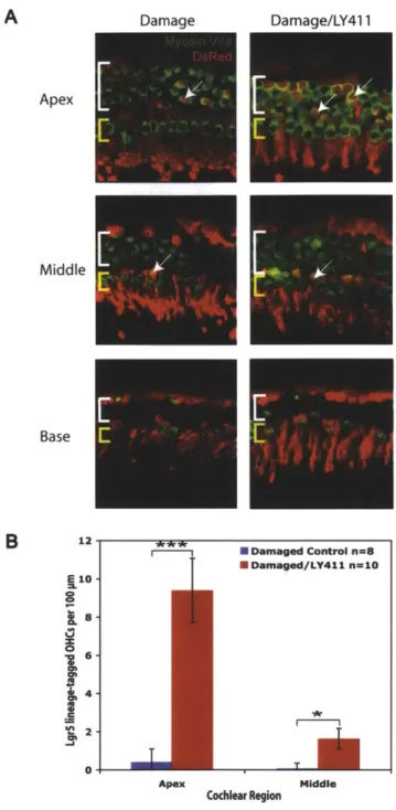
Damage followed by Notch inhibition results in an increase in hair cell numbers
Treatment with 50 tM gentamicin, an ototoxic antibiotic, followed by LY411575 resulted in significantly more hair cells in all cochlear regions than seen in gentamicin-damaged controls (Figure 1). These findings suggest LY411575 treatment subsequent to gentamicin damage results either in the production of new hair cells or aids in the recovery of hair cells from damage. However, hair cell numbers in the middle and base of explant cultures treated with LY411575 were significantly lower than in undamaged controls (Figure IB), suggesting Notch inhibition does not fully compensate for gentamicin damage.
Notch inhibition following damage leads to transdifferentiation of Sox2-positive supporting cells into new hair cells
Notch inhibition after gentamicin damage resulted in significantly more Sox2 lineage-tagged hair cells compared to gentamicin damaged controls in all three cochlear regions (Figure 2A). Sox2-tagged hair cells were found both in the pillar cell and outer hair cell regions, but with greater frequency in the pillar cell region (arrows in Figure 2B-G). Although Sox2-tagged hair
cells were often observed in the apex of controls as described in Chapter 1, new lineage-tagged hair cells were identified by their unusual location, Sox2 staining of their nuclei, or the presence
of immature stereocilia bundles. Many new hair cells in both the pillar and outer hair cell regions showed prestin expression (white arrows in Figure 2B-C), suggesting they were new outer hair cells. Prestin is a motility protein found uniquely in outer hair cells (Zheng et al., 2000). Other new hair cells did not appear to be prestin positive after three days of LY411575 treatment
A
Apex Middle Base 45- 40-E 35- 30- 23- 20- Sis-0 10- 5-0-Undamaged Damaged Dama
m Apex N Undamaged n=15 ISIDamaged n=20 * Damaged/LY411 n=20 ** Middle Cochlear Region Base
Figure 1. Damage followed by Notch inhibition leads to an increase in hair cell numbers
Organ of Corti explant cultures were damaged with gentamicin for 16 hours followed by 72 hours of LY411575 treatment. (A) Images show hair cells in the apex, middle and base of an undamaged control, a damaged control, and a damaged/LY411575-treated explAnt culture. (B) Hair cell counts in the apex, middle and base show significantly more OHCs per 100