HAL Id: hal-01328481
https://hal.inria.fr/hal-01328481
Submitted on 8 Jun 2016HAL is a multi-disciplinary open access archive for the deposit and dissemination of sci-entific research documents, whether they are pub-lished or not. The documents may come from teaching and research institutions in France or abroad, or from public or private research centers.
L’archive ouverte pluridisciplinaire HAL, est destinée au dépôt et à la diffusion de documents scientifiques de niveau recherche, publiés ou non, émanant des établissements d’enseignement et de recherche français ou étrangers, des laboratoires publics ou privés.
Comprehensive in vitro Proarrhythmia Assay (CiPA):
Pending issues for successful validation and
implementation
Icilio Cavero, Jean-Michel Guillon, Veronique Ballet, Mike Clements,
Jean-Frédéric Gerbeau, Henry Holzgrefe
To cite this version:
Icilio Cavero, Jean-Michel Guillon, Veronique Ballet, Mike Clements, Jean-Frédéric Gerbeau, et al.. Comprehensive in vitro Proarrhythmia Assay (CiPA): Pending issues for successful validation and implementation. Journal of Pharmacological and Toxicological Methods, Elsevier, 2016, 81, pp.21-36. �10.1016/j.vascn.2016.05.012�. �hal-01328481�
Comprehensive in vitro Proarrhythmia Assay (CiPA): Pending issues for
successful validation and implementation
Icilio Cavero
a, *, Jean-Michel Guillon
b, Veronique Ballet
b, Mike Clements
cJean-Frédéric
Gerbeau
dand Henry Holzgrefe
ea 54 Rue de la Glacière, 75013 Paris, France
b Sanofi, Preclinical Safety, Pharmacology, 13 quai Jules Guesde, 94403 Vitry sur Seine, France cGE Healthcare Life Sciences, Maynard Centre, Forest Farm, Whitchurch, Cardiff CF14 7YT, UK
dInria Paris and Sorbonne Universités UPMC Paris 6, France
e 13765 Seabiscuit Drive, Reno, NV, USA
Abstract
Introduction: The Comprehensive in vitro Proarrhythmia Assay (CiPA) is a nonclinical Safety
Pharmacology paradigm for discovering electrophysiological mechanisms that are likely to confer proarrhythmic liability to drug candidates intended for human use.
Topics covered: Key talks delivered at the ‘CiPA on my mind’ session, held during the 2015 Annual
Meeting of the Safety Pharmacology Society (SPS), are summarized. Issues and potential solutions relating to crucial constituents [e.g., biological materials (ion channels and pluripotent stem cell-derived cardiomyocytes), study platforms, drug solutions, data analysis, etc.] of CiPA core assays are critically examined.
Discussion: In order to advance the CiPA paradigm from the current testing and validation stages to a
research and regulatory drug development strategy, systematic guidance by CiPA stakeholders is necessary to expedite solutions to pending and newly arising issues. Once a study protocol is proved to yield robust and reproducible results within and across laboratories, it can be implemented as qualified regulatory procedure.
*Corresponding Author
Keywords
Comprehensive in vitro proarrhythmia assay (CiPA) Field potential data sampling and analysis
Induced pluripotent cell cardiomyocytes (hiPSC-CMs) Multi electrode array (MEA)
Patch clamp technologies Pending issues and solution Validation
Abbreviations: AP, action potential; ADP90, action potential duration at 90% repolarization; CaV, voltage dependent calcium channel; CDI, Cellular Dynamics International; CiPA, comprehensive in
vitro proarrhythmia assay; CROs, contract research organizations; CSA, consortium for safety
assessment; DMSO: dimethyl sulfoxide; FP, field potential; FPD, field potential duration; FPDcB, FPD corrected according to the Bazett formula; FPDcF, FPD corrected according to the Fridericia formula; GEVI: genetically-encoded voltage indicators; GLP: good laboratory practices; HESI, Health and Environmental Sciences Institute; HPF, high pass filter; hiPSC-CMs, human induced pluripotent stem cell cardiomyocytes; HTS, high throughput screening; ICH, International Conference on Harmonization; ICaL, L-type (long-lasting voltage-gated) depolarizing calcium current; IKr, rapidly activating delayed rectifier potassium current; IhERG, human ether-a-go-go-related gene potassium
current; IKs, slowly activating delayed rectifier potassium current; IK1, inward rectifier potassium current; Ito, transient outward potassium current; INaFast, fast depolarizing sodium current; INaLate, late depolarizing sodium current; ICWG, ion channel working group; ILSI, International Life Sciences Institute; hiPSC-CMs,: human induced pluripotent stem cell derived cardiomyocytes; ISI, interspike interval; ISWG, in silico working group; LQTS, long QT syndrome; JiCSA, Japan iPS Cardiac Safety Assessment consortium; JSPS, Japan Safety Pharmacology Society; LQT, long QT interval syndrome; LPF, low pass filter; MEA, multi-electrode arrays; MWG, myocyte working group; NaV, voltage dependent sodium channel QMS, Quality Management System; QTc, QT interval corrected according to the Fridericia algorithm; SD: Standard deviation; SEM: standard error of the mean; SPS, Safety Pharmacology Society; TdP, Torsade de Pointes arrhythmia; TQT, thorough QT study, Vmax, maximal velocity of depolarization; VSD, Voltage-sensitive dyes; VSO, Voltage-sensitive optical devices; WG, working group.
Corresponding author. Tel: +33768236797 E-mail address: [email protected]
1.
Introduction
CiPA is a novel Safety Pharmacology paradigm undergoing systematic evaluation for fitness to discover candidate drugs with the potential to trigger ventricular arrhythmic events in humans. If these evaluation efforts succeed, CiPA will become a Safety Pharmacology screening tool for drug research and development purposes (Cavero & Holzgrefe, 2015).
The CiPA paradigm has been designed to provide an accurate and comprehensive assessment of the cardiac ventricular electrophysiological properties of candidate drugs for identifying mechanisms that may mediate life-threatening ventricular proarrhythmic events. This preclinical approach can be considered as an extension of the currently applied ICH S7B guideline strategy (ICH, 2005a) which is designed to detect whether a candidate drug adversely affects the physiological function of the cardiac channel encoded by the ether-à-go-go related gene (hERG) which conducts the delayed rectifier K+ current (IKr). This concern arises primarily from clinical findings that drug-induced hERG inhibition can provoke a particular type of polymorphic ventricular arrhythmia called torsade de pointes (TdP). This tachyarrhythmia, at times, culminates in irreversible ventricular fibrillation. However, within the large number of drugs demonstrating potent IKr channel blocking activity in the in vitro patch clamp assay, some are free of proarrhythmic effects in integrated nonclinical assays, as well as in man (Kannankeril, Roden, & Darbar, 2010; Vandenberg, Perry, Perrin, Mann, Ke, & Hill, 2012).
CiPA is an initiative sponsored by a multi-partner international consortium which includes the FDA, HESI, CSRC, SPS, Japan National Institute of Health Sciences, Health Canada, European Medicines Agency, Pharmaceutical and Japan Medical Devices Agency, Japan iPS Cardiac Safety Assessment Group, academic electrophysiologists, in silico modelers, pharmaceutical industry associations, contract research organizations (CROs), stem cell manufacturers, and companies producing hardware and software for Safety Pharmacology research.
The CiPA core components are:
1) In vitro patch clamp assays in stably expressed recombinant human ion channels. The aim of these studies is to evaluate the effects of candidate drugs on key depolarizing and repolarizing ion currents participating in the formation of human ventricular action potential (AP).
2) An in silico AP assay which is performed to verify whether the results obtained in the previous investigations engender either phenotypic indicators signaling proarrhythmic liability on the human ventricular AP [e.g., prolongation of AP duration (e.g;, ADP90), EADs (Gintant, 2008)] evidenced on the AP profile generated by a mathematical model of the human ventricular myocyte AP (O'Hara, Virag, Varro, & Rudy, 2011) or mechanism-related proarrhythmic metrics (section 2.3) allowing the latter model to classify each drug candidate as no/low, intermediate or high proarrhythmic risk agents.
3) An in vitro assay designed to investigate the electrophysiological effects of candidate drugs in ventricular cardiomyocytes derived from human induced pluripotent stem cells (hiPSC-CMs). The aim is to confirm or cast doubt on the in silico predictions and to broaden the cardiac safety
assessment of the candidate drug to include additional proarrhythmic mechanisms not discoverable by the ion channel assay investigation or in silico analysis.
The CiPA initiative encompasses also the E14 guideline (ICH, 2005b) since it has the potential to de-emphasize the clinical thorough QT/QTc study (TQT) and replace it with an intensive Phase ECG investigation (Cavero, Holzgrefe, & Clements, 2016).
The first part of this article provides extended summaries of key presentations on the CiPA paradigm given at the ‘CiPA on my mind session’ which was part of the scientific program of the 15th Annual Meeting of the Safety Pharmacology Society (SPS) held in Prague in October 2015.
The second part of this report threads through issues concerning biological material, experimental platforms, measured parameters, and data analysis approaches that need to be resolved to qualify each CiPA core assay for Safety Pharmacology research and regulatory purposes. While available solutions for these issues are mentioned in this report, it will be the role of the CiPA Steering Committee and the various CiPA Working Groups and Teams to recommend the best possible solutions for bench stakeholders.
2.
Key presentations from ‘CiPA on your mind’ session held at the Annual Meeting of
the Safety Pharmacological Society
2.1. CiPA introduction. Ongoing activities (CiPA Steering Team) and updates. Dr. G. Gintant, Abbvie,North Chicago, IL, USA
The 2015 CiPA hiPSC-CM pilot study program was designed to test whether a set of reference drugs yielded reproducible effects on field potentials (FP) measured with multi-electrode arrays (MEA) and action potentials (AP) obtained by using voltage-sensitive optical dye (VSD) (Cavero & Holzgrefe, 2014). The knowledge acquired from these investigations will provide indications for optimizing experimental protocols with regard to the selection of the most informative parameters to measure, sampling times during the execution of a protocol, analytical approaches, and data reporting formats.
In September 2015, the FDA Cardiac Safety Committee (CSC), through an FDA Broad Agency Announcement (BAA) awarded a grant to HESI for ‘Validating human stem cell cardiomyocyte technology for better predictive assessment of drug-induced cardiac toxicity’ (HESI, 2014). This endowment will be used to finance studies to characterize the electrophysiological properties of
hiPSC-CMs in studies using MEA and VSD platforms.
HESI funding will also support a pilot Phase I study (to be completed by the end of 2016) of 12 reference drugs selected by the CiPA Clinical Subteam) which includes compounds with no/low (diltiazem, mexiletine, ranolazine and verapamil), intermediate (chlorpromazine, cisapride, terfenadine, and ondansetron), and high proarrhythmic risk (quinidine, bepridil, dofetilide, and sotalol). These drugs were selected from a larger set of 28 drugs [Table 1 in (Gintant, Sager, & Stockbridge, 2016)] which the CiPA Clinical Translation Working Group proposed for achieving the validation of the components of the CiPA paradigm (Cavero & Holzgrefe, 2015). The drugs within
this set have different physico-chemical properties and cover a wide spectrum of electrophysiological endpoints (in particular, the degree of torsadogenic risk, multichannel blockade, and variable degrees of hERG channel blockade).
The results obtained from ongoing studies will be used to test whether the CiPA in vitro ion channel assays provide appropriate markers of risk (metrics) for training the in silico AP model to recognize and assign an appropriate level of proarrhythmic risk to each of the 12 initial reference drugs. Phase II studies will investigate the entire collection of 28 drugs.
The CiPA Steering Committee has accepted a proposal from the Japan iPS Cardiac Safety Assessment (JiCSA) group to sponsor and coordinate, within Japan, an experimental study of 60 compounds with varying degrees of proarrhythmic potential for testing and evaluating the CiPA
hiPSC-CM assays.
The CiPA Steering Committee and the ICH S7B and E14 Working Groups have initiated discussions to define the possible place of the CiPA paradigm within the upcoming revisions of these regulatory documents. For instance, the E14 Q&As (R3), released by the ICH in December 2015 (ICH, 2015) opens the avenue to perform intensive ECG studies, in replacement of the thorough QT/QTc study, conducted within Phase I safety clinical investigations. The latter trials, if are designed in accordance with the rich cardiac electrophysiological knowledge generated by the CiPA assays, can be expected to accelerate and improve the human assessment of the proarrhythmic safety profile of candidate drugs (Cavero, et al., 2016).
2.2. Ion channel project update. Dr. Bernard Fermini Global Safety Pharmacology, Pfizer, Groton, CT, USA and Dr. Najah Abi Gerges, AnaBios Corporation, San Diego, CA, USA,
The Ion Channel Working Group (ICWG), under the auspices of the Safety Pharmacology Society, was charged with the following tasks:
1) Selection of cardiac ventricular ion channels requiring experimental scrutiny because drug-induced perturbations of their physiological function have been found to mediate proarrhythmic events in patients (Kannankeril, et al., 2010; Vandenberg, et al., 2012);
2) Establishing best practice protocols for manual and automated patch-clamp studies to determine whether candidate drugs disrupt human ventricular ion channel functions;
3) Performing pilot studies to identify those ion channel parameters that can best predict a proarrhythmic liability of candidate drugs (Fermini, Hancox, Abi-Gerges, Bridgland-Taylor, Chaudhary, Colatsky, et al., 2016).
For the first core assay of the CiPA paradigm, the ICWG selected three depolarizing [INaFast (NaV1.5 peak current), INaLate (NaV1.5 late current), ICa (Cav1.2),] and three repolarizing [(Ito (KV4.3), IKr (hERG), and IKs (KVLQT1/KCNE1)] cardiac ion channels and IK1 (Kir2.1) channel which can behave as a depolarizing or repolarizing current according to the phase of the cardiac cycle to which it contributes. This selection was made by taking into consideration the key role played by the channel in
shaping the ventricular depolarization and repolarization phases of the ventricular AP profile. A definitive triage will be made according to experimental data collected by ICWG and the in silico working groups (ISWG) and by taking into consideration of the results of an SPS inquiry concerning these channels most often investigated by the pharmaceutical industry for assessing the cardiac safety of candidate drugs which are INaFast, ICaL, IKr, and IKs.
For each of the seven ion channels selected by the ICWG, two sets of experimental protocols have been prepared:
1) Physiological protocols. These are designed to mimic the behaviors of an ion channel during the cardiac action potential by employing AP-like current stimuli to activate channels for generating IC50 values as well as data on voltage-, rate- and use-dependence, and channel activation, inactivation, and deactivation rates. The physiological protocol for IKr has been tested in manual patch clamp platforms and the results of the study have been recently published (Crumb, Vicente, Johannesen, & Strauss, 2016). The data obtained from the latter (pilot) and the ongoing post-pilot (investigating the 12 reference drugs listed in section 2.1) studies will be used by the
in silico working Group (ISWG) to train the O’Hara-Rudy model (O'Hara, et al., 2011) to assign
the correct proarrhythmic liability (no/low, intermediate, and high).
2) Dynamic block protocols (Milnes, Witchel, Leaney, Leishman, & Hancox, 2010) are designed to generate parameters (e.g., drug channel affinity (IC50), kinetics, and state-dependence of the drug-channel interaction) of potential interest for the in silico assay. Pilot IKr studies on dofetilide, cisapride, and verapamil, applying this protocol have already been carried out at room and physiological (37°C) temperatures by using manual patch clamp setups. Additional, ongoing patch clamp pilot studies on the effects of other reference hERG blocking drugs according to a dynamic protocol are expected to be completed within 2Q 2016. It is of interest to note that results reported by (Milnes, et al., 2010) indicate a marked ability of cisapride, but not dofetilide, to interact with the hERG channel on a dynamic pulse-by-pulse basis. These authors note that that if a candidate drug were to undergo voltage-dependent cycles of dissociation and reassociation, its effects might be overestimated by hERG-blocking assays using certain stimulus frequencies and voltage protocols. For these drugs (e.g., cisapride, droperidol, haloperidol, amiodarone) the time period between resting and gated (open/inactivated) states should be well-controlled during concentration-response studies and this may be achieved by using continuous voltage clamp applying a sustained-depolarization [Fig. 6 in (Milnes, et al., 2010)].
The ability of cisapride to rapidly associate and dissociate from the hERG channel during the AP cycle may account for the large variability of its potency as blocker of the hERG channel reported in the literature [Table 2 in (Milnes, et al., 2010)]. In contrast, dofetilide is a slow
hERG blocker which becomes and remains associated to the hERG channel throughout the AP
information on the development of channel blockade produced by a drug candidate and may account for possible variation in a drug IC50 determined by using different channel activation voltage protocols, and possibly, at different study temperatures.
The protocols tested in pilot and post-pilot studies are consensus protocols. However, the protocols, that will be officially proposed by the ICWG for the CiPA ion channel assays, will be best practice in nature since they will incorporate the knowledge acquired during the testing and validation studies.
All manual patch protocols, starting from those on hERG channels, will be adapted for high throughput screening (HTS) platform application and optimized to yield data of the same high quality as those obtained or obtainable with the manual patch clamp technique.
2.3. In silico modeling: update and next steps. Dr. Sara Dutta, Division of Applied Regulatory Science OCP/OTS/CDER/FDA, Silver Spring, MD, USA
The CiPA ISWG has been entrusted with selecting, training, and making publicly available a human (adult) ventricular AP computational model that has the power to detect the proarrhythmic potential of candidate drugs from patch clamp results generated in the CiPA cardiac ion channel assays.
The O’Hara-Rudy model (O'Hara, et al., 2011) was selected as a starting point for in silico simulations by a large panel of leading cardiac modeling experts because it is the cardiac cell electrophysiological model most tightly linked to human ventricular cell data obtained from over 100 undiseased human hearts. This choice is further supported by the following desirable features characterizing the model:
1) Simplicity of use;
2) Transparency of assumptions;
3) Direct relationship to experimentally derived and verifiable datasets; 4) Freedom from intellectual property protection;
5) Implementation as a community resource that will not require specialized hardware or software to be used.
The chief challenge for in silico programming scientists is to provide a model that can predict the clinical proarrhythmic risk of candidate drugs from in vitro ion channel results generated in patch clamp studies. In order to attain this goal, the ICWG should find reasonable solutions to the following issues:
1) Identification of key ion channels and parameters for accurate in silico prediction of the proarrhythmic risk of a drug candidate;
2) Selection of the most appropriate experimental protocol for generating, in the envisaged assay system (manual or automatic patch clamp), the most robust ion channel results for in silico purposes;
3) Definition of quality control criteria for data input into the computational model (Elkins, Davies, Brough, Gavaghan, Cui, Abi-Gerges, et al., 2013).
Issues relating to the use of the O’Hara-Rudy model (O'Hara, et al., 2011), which may still require attention, include:
1) Optimization of the basic model performance;
2) Identification of the best method to represent drug-channel interactions; 3) Enabling the ready use of patch clamp data as a model input;
4) Definition of the underlying principles to evaluate the fitness-for-purpose of simulation protocols. Of particular importance, the in silico assay-generated information on the clinical proarrhythmic risk of a candidate drug should:
1) Be mechanism-based (e.g., either producing phenotypic manifestations on the reconstructed AP such as EADs, or linked to one or more functional parameters, such as the value of the net repolarization current or amount of current necessary to trigger EADs);
2) Enable the rank categorization of a candidate drug into no/low, intermediate, and high proarrhythmic risk in relation to reference drugs assigned to these three classes by the CiPA Clinical Translational Working Group (Cavero & Holzgrefe, 2015);
3) Be usable for regulatory requirements.
In order to be compatible with the O’Hara-Rudy model (O'Hara, et al., 2011), patch clamp data should be obtained in ion channel preparations that generate data qualitatively and quantitatively similar to those measured in native adult cardiomyocytes since this model has been built according to electrophysiological data generated in preparations from human adult hearts.
Possible issues concerning datasets obtained in patch clamp studies are:
1) Certain cloned ion channels used for patch clamp studies may be expressed without their native complementary channel subunits that may play a critical role for physiological ion channel kinetics;
2) High throughput screening (HTS) patch clamp platforms may use bath solutions that differ from the native environment within which human cardiac ion channels function;
3) Currently available automated patch clamp platforms do not routinely provide the possibility of performing studies at physiological temperature, a feature necessary to observe rapid drug-induced channel blockade. This may explain why the IC50 values of certain drugs differ substantially from those obtained at room temperature. For example, this is the case for erythromycin and d,l-sotalol which exhibited an IC50 against IKr of 1410 and 810 µM, respectively, at 24 °C and 199 and 278 µM at 37°C (Kirsch, Trepakova, Brimecombe, Sidach, Erickson, Kochan, et al., 2004). In order to overcome this potential issue, the ICWG is generating data with the 12 reference compound set (2.1) at 24 and 37°C in order to support an ISWG project aimed at developing a computational routine that can use 24°C data to predict the behavior of a drug at 37°C. However, most of IKr blockers studied to date appears to be characterized by IC50 values that are often virtually the same when determined at either 24° C or 37° C (Li, Dutta, Sheng, Tran, Wu, & Colatsky, 2016). However, it should be noted that for
certain drugs block kinetics (and, therefore, risk prediction) may be vastly different at room and physiologic temperatures.
A candidate proarrhythmic metric under evaluation is the value of the residual net repolarizing current (Inet = IKr+ IKs + INaLate + ICaL which can be derived from the effects of a candidate drug on the repolarization phase under the ‘worse case’ EAD scenario which can be achieved by applying a current patch clamp step protocol. When Inet crosses 0 during repolarization, EADs are likely to occur [details in (Starmer, 2005)].
Another proarrhythmic metric under evaluation obtainable from patch clamp studies is Ibias (µA/µF) current which is the threshold current necessary to trigger EADs in the absence and presence of a candidate drug. For instance, a drug concentration producing a 50% block of the hERG channel reduced the control value of Ibias (0.734) by 50% (0.375) (Gray & Huelsing, 2008).
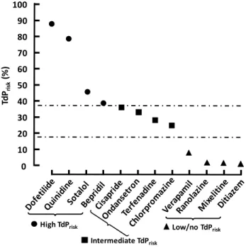
A new and potentially interesting
proarrhythmic metric referred as a ‘TdPrisk’ (Fig 1) is firstly provided by calculating the area under the time-current profile (AUC) of three key ventricular AP currents (IKr, ICaL, and INaLate) determined by the application of an AP patch clamp protocol. Then, the ratios of the AUCs calculated during control condition and AUC under drug (at clinical concentrations) exposure were computed. Model inputs included experimental manual patch clamp IC50 data obtained at 37°C, amount of block for each channel produced by drug exposures achieved in patients, and current conductance scaled accordingly by the computational model (Crumb, et al., 2016). The TdPrisk metric is obtained in the following manner: TdPrisk= a(AUCICaL)Control/(AUCICaL)Drug + b(AUCINaLate)Control/(AUCINaLate)Drug + c c(AUCIKr)Control/(AUCIKr)Drug + d. The parameters a, b, c, and d are calculated using the proportional odds model (Brant, 1990). The input of the TdPrisk metric into the O’Hara-Rudy model provided a correct classification (Fig. 1) of the clinical proarrhythmic risk of the reference training set of 12 drugs.
Fig. 1. Proarrhythmic metric (TdPrisk) derived using three
variables (see details in the text), namely, the AUC under
IKr, ICaL and INaLate current contours generated in AP patch
clamp studies under vehicle (control) and drug exposure (at human concentrations) conditions. Figure redrawn from a slide presented by Dr. Sara Dutta at the 2015 Annual Meeting of the Safety Pharmacological Society. The results of this figure should be considered exploratory in nature, since the data used to build it were not obtained using CiPA ion channel protocols and do not take into consideration hERG blocking kinetics. The authors of this work are in the process of further refining this metric and evaluating its performance using CiPA ion channel protocols and training compounds (personal communication)
Td Prisk (% ) 80 90 10 60 30 20 50 40 High TdPrisk 70 100 Low/no TdPrisk Intermediate TdPrisk 0
The ability of the in silico assay to link accurately ion channel pharmacology to clinical risk prediction requires ion channel datasets characterized by minimal variability and high reproducibility within and across laboratories and platforms.
The in silico CiPA shared community resource for public access will be accompanied by ISWG-prepared documentation concerning:
1) Quality of patch clamp data necessary to obtain reliable information from the AP model;
2) Boundaries (in terms of compound actions) within which the model predictions can be considered reliable;
3) Model assumptions;
4) A description of how data should be input into the model to perform the computational analysis. Envisaged future development of the in silico model includes:
1) Improvement of basic performance by refining the contribution of certain ionic currents, namely IK1 and INaLate;
2) Use of drug-channel interaction data obtained from the dynamic block protocol for generating reliable proarrhythmic predictions for drugs specifically blocking IKr;
3) Enabling the use of patch clamp data obtained at 24 °C to satisfy the model requirement for 37°C; 4) Inclusion of disease state models by introducing the potential pathological influences of key cardiovascular changes due for example to bradycardia, hypokalemia, co-medications, and certain inherited LQT syndromes;
5) Influence of data obtained by applying different patch protocols (current/voltage clamp) on simulation outputs;
6) Additional considerations for use of in silico models as discussed in a recently published review article (Davies, Wang, Mirams, Caruso, Noble, Walz, et al., 2016).
2.4. Current situation and future plan of Japan iPS Cardiac Safety Assessment (JiCSA) consortium. Yuko Sekino, National Institute of Health Sciences, Tokyo, Japan
The JiCSA (Jicsa, 2016) is a joint consortium (funded by the Japan Ministry of Health, Labour and Welfare) under the leadership of the Japan National Institute of Sciences, the Japanese Pharmacology Society, pharmaceutical companies, CROs, academia, and research institute representatives.
JiCSA established an active collaboration with the CiPA Steering Committee for participating in the testing and validation of core CiPA assays.
The key objectives of the JiCSA are:
1) Standardization and validation of Safety Pharmacology methods for identifying drugs which have the potential of inducing proarrhythmic events;
2) Validation of the hiPSC-CM assay for regulatory purposes.
In order to ensure the reproducibility of results of hiPSC-CMs electrophysiological investigations, the JiCSA is funding and conducting a study in four independent laboratories. The protocol of this
study calls for the use of the same hiPSC-CM line (CDI-iCells Cardiomyocytes) and the same experimental platform (MED64) for determining the effects of candidate drugs. In particular, to constrain sources of potential assay variability, technicians received special training concerning all experimental protocol provisions such as hiPSC-CM culture density, period to wait for the experimental procedure following cell seeding, drug sampling times for data points following drug exposure, temperature within the experimental well, and instrumentation calibration settings [e.g., high pass (HPF) and low pass (LPF) filters fixed at 0.1 Hz and 5 kHz, respectively]. A pilot study carried out according to these experimental conditions investigated five concentrations of reference hERG blockers added in a cumulative manner (observing 20 min intervals between 2 contiguous concentrations) to six independent experimental wells seeded with hiPSC-CMs. For statistical analysis, parameters derived from the field potential (FP) signals included interspike interval (ISI) in ms, field potential duration (FPD) in ms, early afterdepolarizations (EAD) incidence, and cell monolayer beating arrest. FPD values were corrected by using Fridericia [(FPDcF= FPD / (ISI / 1000)1/3] or Bazett (FPDcB = FPD / (inter-spike interval / 1000)1/2]) formula (Nakamura, et al., 2014; Asakura et al, 2015).Steady state FP signals (golden waves) within the exposure time selected for measuring ISI and FPD were characterized by an FPA ≥200 µV, an FPDcF ≥340 ms, and a second peak amplitude following the FP spike ≥ 15 µV. This study provided evidence that FPD was of greater amplitude at 0.1 Hz than with a 1.0 Hz high pass filter. The use of 1 Hz high pass filter (HPF) reduced FPD prolongation and obscured EAD detection produced by hERG blockers. The application of the above protocol by four geographically separated research sites to the study of E-4031 yielded the following results: a 3 nM concentration produced a 10% increase in FPDcF in all laboratories, 10 nM produced EADs in 2/6 preparations in 2 laboratories and in 5/6 preparations in the remaining laboratories. However, all four laboratories observed EADs in the 6 CM preparations after 10 min exposure at 30 nM.
The JiCSA has asked its NIHS member to discuss with the Medical Devices Agency (PMDA) and the MHL, the regulatory acceptance of electrophysiology datasets produced by Japan Pharma in studies using hiPSC-CMs. At the level of the ICH organization and international regulatory institutions, JiCSA consortium envisages a gateway role for the acceptance of the CiPA assays for drug development and regulatory purposes. Finally, it plans to relay all data generated by the studies carried out under its sponsorship to the Pharmacological Evaluation Institute of Japan (PEIJ), an institution supported by a grant from the Japanese government for the development of widely applicable data validation platforms.
2.5. Current situation and future plans in Japanese activity from the Kirishima meeting. Dr Atsushi Sugiyama, Department of Pharmacology, Faculty of Medicine, Toho University, Ota-ku, Tokyo, Japan
In January 2014, representatives of the Japanese health regulatory authority, pharmaceutical industry, and academia met in Kirishima (Japan) to discuss recent scientific advances and technologies for assessing the cardiac safety of candidate drugs (stem cell, computational, clinical, and integrated cardiac safety pharmacology-based methods). The meeting produced a consensus report (Sekino, 2014) in which it is stressed, inter alia, the desire to involve Japanese scientists in the evaluation of the CiPA paradigm. Additionally, the meeting participants considered that the CiPA strategy should be enriched of an in vivo nonclinical assay as currently recommended by the ICH S7B guideline (ICH, 2005b) and an early clinical ECG investigation to replace the ICH E14 (ICH, 2005b) TQT study (Cavero & Holzgrefe, 2014; Sugiyama, 2014) for ensuring the cardiac electrophysiological safety of candidate drugs for human use. At this meeting, the following dedicated working groups were designated: Stem Cell Pharmacology, In Silico Pharmacology, Clinical Pharmacology, and Integrated
Cardiac Safety Pharmacology. The common mission of these groups is to contribute via appropriate
initiatives to the determination of the human safety of candidate drugs selected for clinical investigation.
The specific assignments of each group are briefly indicated here below.
1) The Stem Cell Pharmacology Group has already established formal collaboration with the JiCSA for the development and standardization of experimental protocols to predict adverse drug reactions in nonclinical electrophysiological studies using hiPSC-CMs (see section 2.4).
2) The In Silico Pharmacology Group in charge with thee activity on the development of a novel three dimension (3D), multi-scale, and multi-physics computational heart model (UT Heart) to correctly predict the proarrhythmic potential of candidate drugs from a 12-lead human ECG generated from nonclinical electrophysiological data obtained in automated patch clamp studies. In pilot studies, the UT Heart model satisfactorily predicted the expected human ECG effects of E-4031 and verapamil.
3) The Clinical Pharmacology Group is tasked with the elaboration of Phase I ECG protocols that incorporate CiPA generated knowledge for the rapid, early clinical assessment of the proarrhythmic risk of candidate drugs in volunteers.
4) The Integrated Safety Pharmacology Group is in charge of the integration of CiPA generated datasets into the nonclinical integrated risk assessment scheme recommended by the ICH S7B (ICH, 2005b). Additionally, it evaluates the predictability of candidate drug clinical proarrhythmic liability from the integrated analysis of nonclinical in vitro and in vivo data in conjunction with human ECG datasets obtained during Phase I investigations.
3.
The CiPA core assays: potential issues and possible solutions
Safety Pharmacology studies should be designed, executed, and analyzed according to best scientific principles which entails, whenever possible, the adoption and scrupulous application of best practice
protocols in the execution of experimental procedures, particularly if these are carried out for regulatory purposes.
The following sections examine issues requiring resolution to render CiPA core assays fit for the discovery of proarrhythmic mechanisms of candidate drugs.
Ion channel assays 3.1.
The objective of the CiPA ion channel assays is to determine whether candidate drugs disrupt the physiological function of any ion channels playing a fundamental role in the generation of a key electrical current contributing to the generation of the human ventricular AP.
3.1.1 Biological material for the CiPA ion channel assay
The core structural component of any cardiac ion channel is the pore-forming α-subunit protein which functions as pathway for the entry into, and the exit of ions (Na+, K+ and Ca2+) from, the CM. This protein is often complemented by one or more accessory proteins (subunit β, γ, δ) which modulate the pore activity.
Ion channel functions can be easily explored in channel-proteins heterologously expressed in host cells. Whereas this expression process generally succeeds in the permanent expression of the ion conducting α-subunit protein, it does not routinely result in the co-expression of the multiple proteins needed to fully recapitulate the functions of the native channel.
For the CiPA ion channel assay, the cloned cardiac ion channels should ideally possess the protein (primary, secondary, and tertiary) structure of native channels present on ventricular myocytes of healthy adult individuals, be stably expressed and functional within the host cells, and conduct a current density sufficiently large to allow its accurate measure in automated patch clamp platforms.
3.1.2 Study of ICaL in heterologously expressed Ca2+ channels
While various heterologous cell lines express IKr, IKs, and INaFast (Nav1.5) populations which conduct currents of elevated amplitude (≥ nA/cell), this is not generally the case for ICaL (Cav1.2) (Lory, Varadi, Slish, Varadi, & Schwartz, 1993; Nishimura, Takeshima, Hofmann, Flockerzi, & Imoto, 1993). An additional experimental issue with heterologously expressed ICaL is that, with dialysis of the intracellular constituents in the whole-cell recording setup, ICaL amplitude rapidly declines, making it difficult to accurately determine the time-course of the electrophysiological effects of candidate drugs (Bean, 1984). However, CaV1.2 screening conditions (peak current amplitude maximization and rundown attenuation) have recently been optimized for an automated planar patch clamp platform (PatchXpress® 7000A) in an HEK-293 cell line stably expressing a human L-type cardiac Ca2+ channel assembly of α1C, α2δ, and β2a proteins along with the Kir2.3 inward rectifier K+ channel. The concurrent expression of Kir2.3 is a crucial strategy, adopted for maintaining host cell resting potential below the threshold resting current activating CaV1.2. Indeed, this feature facilitates an effective culture of the HEK-293 cell line without the need of using Ca2+ channel blockers to prevent the excessive influx of extracellular Ca2+ that is detrimental to cell health and growth. The selected
automated patch platform routinely achieved GΩ seals and could record relatively small current amplitudes. Additionally, the assay conditions were optimized to yield IC50 values that were similar to those obtained in manual patch clamp studies for six established Ca2+ antagonists [diltiazem IC50: 44 (automated patch champ) and 36 (manual patch clamp) µM, gallopamil: 17 and 1.1 µM, isradipine 2.2 and 8 nM, nifedipine 22 and 16 nM, nimodipine 0.11 and 0.7 µM; verapamil 15 and 44 µM] and a Ca2+ agonist [S(-)-Bayk8644: 3.1 and 8.2 nM)] [Table 1 in (Balasubramanian, Imredy, Kim, Penniman, Lagrutta, & Salata, 2009)].
It should also be recalled that published potency (IC50) values for verapamil as a blocker of Cav1.2 are rather heterogeneous. These differences are likely to depend not only from the voltage used to activate the channel and the presence or absence of subunits in the cloned Cav1.2, but also from the ion species (Ca2+ or Ba2+) used in the extracellular solution as charge carrier, and other experimental conditions such as study temperature (Dilmac, Hilliard, & Hockerman, 2004).
Recently, a new CaV1.2 cell line by NMITT (Nmitt Pharmaservices, 2016) has been claimed to stably express all three subunits (α1c/β2/α2δ) present in the native CaV1.2 channel, have high success rates and reliable performance in automated patch clamp assays, afford rundown-free recordings, easy maintenance in cell culture, and possibility of custom adjustment of expression levels.
These developments mandate that the cell line for the CiPA Ca2+ channel assay should be selected after appropriate pharmacological characterization in automated patch clamp platforms according to the ICWG protocols.
3.1.3 Recording the small INaLate
INaLate is a small fraction of the classic “peak” INa current carried by NaV1.5 which is generated by voltage-dependent Na+ channels entering in burst-gating mode (repeated openings and closures) during myocyte depolarization. Genetic mutations causing defective INa channel inactivation, myocardial ischemia, and drug treatments can upregulate INaLate and expose patients to arrhythmic risk (Chevalier, Amuzescu, Gawali, Todt, Knott, Scheel, et al., 2014). Drugs like ranolazine, which is a blocker of this current, can reduce or prevent the proarrhythmic propensity inherent to hERG channel blockade (Gupta, Khera, Kolte, Aronow, & Iwai, 2015). In contrast, drugs enhancing INaLate can favor the development of arrhythmic events. This underscores the importance of the inclusion of INaLate in the CiPA ion channel panel.
Because of the small amplitude of INaLate, agents which impair sodium channel inactivation such as veratridine or ATX-II, are routinely employed to overcome the small amplitude of this current in order to facilitate a more accurate characterization of candidate drug effects on this current. Nevertheless, it should be noted that it remains to be demonstrated that toxin-enhanced currents fully replicate native INaLate.
A recent report describes the successful use of the CytoPatch™ automated patch-clamp platform and a HEK293 cell line stably expressing human NaV1.5 to determine INaLate (Chevalier, et al., 2014). The
baseline amplitude of this current averaged -10.4 ± 2.2 pA (n=18) during the last 100 ms of 300 ms depolarizing pulses to -10 mV from an initial holding potential of -100 mV, delivered at 0.33 Hz. Ranolazine (30 µM) reduced this current by 58.4 ± 3.5% (N=18) after a 3 min incubation. In contrast, in cells exposed for 5 min to 1 µM of veratridine INaLate was increased by 269.1 ± 16.1 % (n=28) from a baseline value of -12.4 ± 1.9 pA.
3.1.4 Studying IKs in automated patch clamp platforms
Under baseline conditions, the repolarizing current IKs contributes minimally to the repolarization phase of the cardiomyocyte AP which is primarily mediated by IKr. However, IKs exerts a major and vital function during sympathetic tone elevation leading to enhanced cardiac chronotropic and inotropic activity. Indeed, in these physiological settings, the increase in IKs limits the degree of QT interval prolongation that would result from the augmented CaV1.2 currents in the absence of the IKs intervention. Therefore, IKs functions as cardiac repolarization reserve antiarrhythmic mechanism precluding excessive, potentially dangerous repolarization lengthening (Jost, Papp, & Varro, 2007).
Mutations in the gene encoding hKCNQ1 are responsible for the most common form of congenital long QT (LQT) syndrome (characterized by loss of channel function and referred to as LQT1 syndrome) which predisposes affected patients to excessive QT interval prolongation during surges in adrenergic stimulation (e.g., occurring during intensive exercise and emotions) that can trigger lethal TdP events. Indeed, in these patients, IKs fails to increase as a counterbalance to the enhanced depolarization mediated by β-adrenergic stimulation to the heart (Jost, et al., 2007).
HEK-293 cells transfected with the hKCNQ1 and hKCNE1 genes stably express IKs channels with voltage- and temperature-dependent gating characteristics, pharmacological responses, and β-adrenergic modulation, as exhibited by IKs in native human CMs (Wu, Naiki, Ding, Ohno, Kato, Zang, et al., 2014).
ICWG protocols should be tailored, tested, and validated for the ability to study IKs expressed in suitable cell models in automated patch clamp platforms that allow the adoption of desired pulsing rates.
3.1.5 Points to consider for the selection of automated patch clamp equipment and studies
Automated patch clamp platforms for CiPA ion channel assays should ideally:
1) Provide easily obtained knowledge-rich proarrhythmic metrics for the in silico assay; 2) Offer the possibility of studying candidate drugs at 37°C and at desired pacing rates; 3) Allow sampling of well solutions for the determination of drug candidate concentrations; 4) Allow superfusion of well preparations to uniformly expose CMs to desired concentrations; 5) Offer the possibility of rapidly evaluating possible rundown phenomena;
6) Ensure satisfactory reproducibility of results within and across platforms/cell lines/laboratories; 7) Provide easily programmable features for standardizing dataset output.
Available automated patch clamp plates used for cell growth and experimental procedures require validation for fitness-for-purpose. Dr. Fermini, during his presentation at the 2015 SPS meeting (section 2.1), mentioned that the IC50 of cisapride varied from 28 to 62 nM when determined in two different types of plates, despite the application of the same assay protocol by the same technician. Hence, each key component of the automated patch clamp platform requires careful evaluation for data reproducibility and reliability.
3.1.7 Solubility of test articles for automated patch clamp
Solubility of test articles may pose problems specific to HTS patch clamp platforms. Indeed, current HTS plates are generally passive microfluidic (low volume, small size) systems (Wikipedia, 2016) that are characterized by high shear stress affecting the conformation of patched cells and delaying homogenous solubility of the tested compounds within the experimental well solution (Cioffi, Moretti, Manbachi, Chung, Khademhosseini, & Dubini, 2010; Kraly, Holcomb, Guan, & Henry, 2009). The latter issue is routinely addressed by formulating poorly soluble compounds with substances [e.g., dimethyl sulfoxide (DMSO)] that render test articles more readily bioavailable to the patched cells. However, to be successful this strategy should be accompanied by means to rapidly achieve the homogenous mixing of small quantities of drugs or solvent containing solutions (see section 3.3.5). Finally, to ensure that homogenous mixing is achieved, the platform should allow sampling of well solutions for the determination of test substance concentration to correctly assess the potency of the candidate drug under evaluation.
3.1.8 Data management
Patch clamp dataset analysis should be standardized in terms of models used for IC50 (ICx and Hill coefficient of the concentration-response curve) calculations, minimal number of assays/data points to obtain meaningful, robust data, and correction methods for possible rundown phenomena.
3.1.9 Expectations from ion channel patch clamp generated data
Ion channel patch clamp datasets generated by the ICWG-organized pilot studies with a set of 12 reference drugs will be relayed to the ISWG to train the O’Hara-Rudy model (O'Hara, et al., 2011) to identify key parameters (metrics) that have the ability to cause modifications in the computationally derived AP signaling proarrhythmic potential. From these liability fingerprints, the computational model, once appropriately trained and calibrated for fitness-for-purpose, will be able toautomatically attribute a low/no, intermediate, or high proarrhythmic risk label to any candidate drug.
In silico assay 3.2.
Potential issues and solutions pertaining to CiPA in silico assay were discussed by Dr. Sara Dutta (section 2.3). In addition to these, the ISWG should consider developing simulations to assess the potential proarrhythmic danger of candidate drugs which block the IKs channel (Towart, Linders, Hermans, Rohrbacher, Van Der Linde, Ercken, et al., 2009), given the role of this channel in the cardiac repolarization process particularly during cardiac activation (Liu, Du, & Li, 2012).
hiPSC-CM assay 3.3.
The objective of the CiPA hiPSC-CM assay is not only to verify the proarrhythmic predictions obtained from the in silico simulations but also to discover proarrhythmic mechanism(s) that the ion channel and in silico assays did not, or were not powered (e.g., disruption of cardiac channel expression and trafficking) to discover.
3.3.1 Qualification of hiPSC-CMs for CiPA purpose
Commercially available hiPSC-CM cell lines exhibit different mechanical, electrophysiological, and pharmacological properties (Ji, Kang, & Rampe, 2014; Millard, Chvatal, & Ross, 2015). Hence, in order to be adopted for the CiPA assay, they need to be carefully characterized for fitness-for-purpose, i.e. for their ability to generate the sought knowledge in a reproducible manner within and across laboratories. Issues deserving targeted attention are:
1) Definition of requisite features of hiPSC-CM cell lines to be adopted for the CiPA paradigm; 2) CiPA-specific procedures concerning cell seeding and growth in HTS plate wells;
3) Desirable properties of cardiac ion channel expressed by hiPSC-CMs for the CiPA studies; 4) Drug testing solution preparation;
5) Homogeneous mixing of µL quantities of solutions containing testing agents within the experimental well and precautions to ensure that drug candidates are not adsorbed to platform wares;
6) Adoption, if necessary for experimental purposes, of experimental platforms offering pacing capability;
7) Study design;
8) Data point sampling timing; 9) Dataset analysis approaches.
3.3.2 hiPSC-CM cell line features
The phenotypic structure of current CMs is structurally and functionally characteristic of the embryonic and/or fetal development stage as documented by electrophysiological, calcium handling, and metabolic signatures exhibited by these cell preparations. Indeed, in contrast to native adult CMs,
hiPSC-CMs have small cellular size, lack T-tubule structures and well-formed sarcomeres, and
possess multiple nuclei, poor overall calcium storage and handling capability, and relatively small populations of mitochondria. In addition, their metabolic processes depend entirely upon glycolytic activity, and have intrinsic contractile automaticity (Ivashchenko, Pipes, Lozinskaya, Lin, Xiaoping, Needle, et al., 2013; Keung, Boheler, & Li, 2014; Robertson, Tran, & George, 2013; Roden & Hong, 2013). The lack of working load for hiPSC-CMs in culture conditions is responsible for the absence of the development of a cellular sarcomeric organization during the maturation process which, in turn, results in a poorly organized syncytium lacking adult phenotypic characteristics.
Intensive research efforts are ongoing for obtaining hiPSC-CMs exhibiting adult phenotypic features. Currently, any small progress toward the latter goal is obtained by hiPSC-CM production process changes which, in certain cases, are accompanied by changes in the pharmacological properties of hiPSC-. If the latter occurs, it infringes a central tenet of safety disciplines which stipulates that the biological material used for a safety test, particularly whenever performed for regulatory purposes, should, over time, provide reliable and reproducible datasets within and across laboratories. In order to fulfil this requirement, the production process of any hiPSC-CM line, if adopted for CiPA safety investigations, should be constrained to preserve the properties exhibited during the qualification process. To this end, it is incumbent on the manufacturers of hiPSC-CMs to ensure the consistency of cell products from batch to batch within the requirements of a Quality Management System (QMS) certified according to the International Organization for Standardization (ISO) principles. This entails, inter alia, that for experimental replication purposes, the manufacturer complies with ISO standards and maintains in its archive system (and, if requested, provides) documentation concerning the entire manufacturing process of any hiPSC- CM batch (World Health Organization, 2011). As a corollary to these basic principles, on material delivery the manufacturer has to inform investigators of any change in the production process of the hiPSC-CMs since this may require additional pharmacological validation assaysto assess whether the modified biological material responds to reference drugs in the same manner as established during the CiPA validation exercise.
3.3.3 Cell seeding and growth into HTP plate wells
The growth of hiPSC-CMs seeded into experimental wells uses solutions generally supplied by cell manufacturers. For proprietary reasons, the composition of these solutions is not always detailed on the container labels. It is, however, a safety assay requirement that the composition of any solutions used during the experimental procedure [for biological material preparation (monolayer differentiation) or assay execution] should be part of the experimental protocol. For example, if such culture or assay solutions contain proteins of animal or human origin (serum), they may modify candidate drug availability to the biophase sites.
Whenever possible, the use of protein-free differentiation and recording media should be considered, particularly if these media work well for the selected hiPSC-CM line. For example, commercial media such as mTeSR (Stemcell™ Technologies, 2016) and Essential 8TM (Thermofisher Scientific, 2016), are claimed to increase reproducibility of hiPSC-CM culture and differentiation by eliminating the use of serum, serum replacements, or conditioned media. Relative to recombinant growth factors, chemically synthesized small molecules incorporated into protein-free media reduce variability and cost and enhance the robustness of the hiPSC-CM assays. Additionally, these substrates stabilize certain properties of the cell culture systems and improve hiPSC-CM maturation processes when complemented with soluble cues of 3D design (e.g. substrate physical form optimizing cell-to-cell contacts) that may facilitate hiPSC- CM electrophysiological maturation (Denning, Borgdorff,
Crutchley, Firth, George, Kalra, et al., 2015; Patel, Celiz, Rajamohan, Anderson, Langer, Davies, et al., 2015).
Several hiPSC-CM manufacturers (e.g., Axiogenesis™, Pluriomics™) have developed proprietary free media for their cell lines. End-users have also adopted personalized recipes for protein-free media used in their experimental procedures [e.g., for CDI hiPSC-CMs (Harris, 2015). As such, the CiPA MWG should consider recommending the use of protein-free media for CiPA hiPSC-CM assays if the use of these solutions provides datasets of enhanced value compared to traditional, protein-containing media.
3.3.4 Measuring platforms
Platforms currently available for HTP determination of the electrical activity of hiPSC-CMs use MEA or VSD technologies (Cavero & Holzgrefe, 2015).
MEA is a multi-well plate technology for label-free, real-time electrophysiological characterization of cardiomyocyte drug responses by recording extracellular field potentials (FP) from cell monolayers or clusters. Each well bottom is fitted with multiple recording electrodes [e.g., Maestro MEA system has a 16 electrode grid (Axion Biosystems, 2016b)].
MEA technology can be used to determine effects (concentration-response curves, time-course over a prolonged period) of test articles on FP. Endpoints from FP recordings include amplitude (FPA in µV), duration (FPD in ms), beat period (BP in s) from which BR (contractions per min = 60/BP] can be calculated. Arrhythmic events are quantified by the number of experimental preparations showing rhythm irregularities. Classifications of these irregularities have been proposed according to specific parameters (Gilchrist, Lewis, Gay, Sellgren, & Grego, 2015).
Due to the variability of the FP shape, a critical issue concerns the selection of the control FP wave(s) to enable clear measurements of FP parameters (in particular FPD). The changes produced by a candidate drug on the baseline FP are used to determine proarrhythmic safety or liability of a candidate drug. To this end, four approaches appear applicable:
1) Golden electrode. The underlying rationale is that monolayers of hiPSC-CMs are typically linked in an electrically coupled syncytium beating as a whole unit. As a result, the characteristics of the culture recorded at a given electrode could be considered to represent the activity detectable by any other electrodes within the well. Therefore, data points from a single electrode (i.e., the ‘golden electrode’) exhibiting an excellent FP wave may be presumed to be representative of the entire preparation. A requirement for this approach is that the same electrode should be used for sampling baseline and drug or vehicle exposure data [Fig. 7 in (Clements, 2016)].
2) Well-wide mean. This refers to the mean response across all well electrodes. The morphology of the FP waves from the entire set of MEA electrodes is generally rather heterogeneous. In particular, some electrodes can display prominent repolarization waves and others none at all. This can result in the misidentification of a weak repolarization wave, in particular, by automatic detection software with possible inaccuracy in FPD measurements [Fig. 7 in (Clements, 2016)].
The Cardiac Data Aggregation Tool within the AxIS software (Axion Biosystems, 2016a) allows setting user-specific boundaries for improving the accuracy of well-wide statistics.
3) Golden FP wave. This is defined by the following criteria: FPA ≥200 µV, FPDcF ≥340 ms, and a second wave following the FP spike of ≥ 15 µV amplitude. This approach is used by Japanese investigators, as described in section 2.4. A requirement for this approach, as noted for the Golden electrode, is that the same electrode should be used for sampling data under baseline and drug or vehicle exposure conditions.
4) Golden class. This method is proposed by NOTOCORD Systems through their NOTOCORD- Field Potential Screener (fps) (Notocord Systems, 2016). For each electrode, it clusters, FP waveforms recorded from all electrodes within a well according to morphology to ensure coherent measurements made over short sampling periods throughout the whole experimental time. Waveforms within each cluster (or class) are then averaged to improve the signal/noise ratio. The user has the possibility to select his quality control criteria for the first analysis period (used then throughout the experiment) for BR, acceptable maximal variability, cluster number, and beat number. These criteria allow the automatic selection of all electrodes that can be analyzed. Then, on each analyzable electrode, the software uses the most populated clusters for the calculation of the desired biomarkers (e.g., FPA, FPD, BP)
The variability in the FP shape of the preparation generally recorded from the electrodes within the experimental well may have multiple origins, some of which are preparation specific (e.g., position of the electrode within the well in relation to the pacemaker driving the preparation, ground position, cell organization within the sheet, and differences in resistance and capacitance between myocytes and electrodes).
The most relevant issues with MEA technology requiring appropriate resolution include:
1) Determination of the time necessary to achieve homogenous mixing of the test article solution with the drug-free solution within the well. This may be a function of the solubilizing medium (e.g., use or not of DMSO) and amounts of containing solution selected to replace the drug-free solution;
2) Validation of electronic amplifier circuits and all connected hardware (e.g. electrode forms and position in relation to grounds) necessary to measure FP waveforms;
3) Use of a low frequency (0.1 Hz) digital filter for FP measurements in order to avoid information loss on recorded FP waveforms (Asakura, Hayashi, Ojima, Taniguchi, Miyamoto, Nakamori, et al., 2015);
4) Identification of recording electrodes within the well for capturing FP waves clearly exhibiting the parameters to be measured;
5) Selection of the most pertinent FP endpoint parameters for further analysis;
Voltage-sensitive dyes (VSD) and genetically-encoded voltage indicators [GEV (Q-State Biosciences, 2016)], together referred to as voltage sensitive optical probes (VSOs) can be used to measure the integrated AP generated by multicellular hiPSC-CM monolayers (Zhang, Gintant, & Pierson, 2014). Parameters that are generally calculated from these AP are BR, depolarization Vmax, APD30, APD50, APD90, AP triangulation (APD90-APD30) and number of proarrhythmic events such as early after depolarizations (EADs). AP shapes recorded with VSDs and the associated AP metric values (e.g., ADP90) may be specific to each hiPSC-CM line.
3.3.5 There are several theoretical limitations to VSD and GEVI technology which have been succinctly summarized by Leyton-Mange and colleagues (Leyton-Mange et al., 2014): 1) Fluorescence signals report relative, not absolute, membrane potentials; 2) The slow temporal response of these VSOs results in loss of high frequency elements and leads to small but systematic overestimation of APD, although, VSD ANEPPS dyes have a faster temporal response to GEVIs (e.g. ArcLight). However, for the moment, this slow temporal response is not a critical issue since currently available hiPSC-CM preparations exhibit low Vmax values than primary hiPSC-CMs (Robertson et al., 2013); 3) Phototoxicity associated with VSOs can limit the duration of experimental recordings. Comparatively, GEVIs have been reported to have a lower phototoxicity than VSDs (ArcLight vs. di-4-ANEPPS and di-8-ANEPPS); 4) Additionally, a possible interaction of VSOs with the candidate drug should also be taken into consideration when using these technologies (Hardy et al., 2009).Experimental platform offering the possibility of pacing the preparations
Platforms offering the possibility of pacing hiPSC-CM sheets [e.g., (Axion Biosystems, 2016b; Multichannel Systems, 2016)] should be selected to determine the electrophysiological effects of drugs blocking IKs. Indeed, the activation of this channel occurs at high driving rates (Braam, Tertoolen, Casini, Matsa, Lu, Teisman, et al., 2013; Liu, et al., 2012). As such, the determination of the effects of a candidate drug at low and high pacing rates can provide key information on the potential proarrhythmic liability of a candidate drug. It should be noted that for spontaneously beating
hiPSC-CM sheets, low pacing refers to a threshold rate just above the baseline rate (e.g., for CDI-iCell hiPSC-CM spontaneously contracting at ~60-times/min, stimuli delivered each 900-800 ms will raise
the rate by 10-20 contractions). The baseline heart rate of hiPSC-CM sheets depends not only on the individual cell line under study but also on experimental protocol provisions (e.g., presence or not of glucose in the recording well solution).
The 48-well MEA optical stimulation system Lumos® is claimed to provide artifact-free pacing (Axion Biosystems, 2016c). In this regard, it should be mentioned that other optical electrophysiology platforms for driving and recording the activity of hiPSC-CMs are now available and are based on proprietary ultrasensitive optogenetic reporters and actuators that convert APs into visible fluorescence flashes easily detectable by microscopy. These methodologies can also be used to deliver excitatory photo impulses to drive the preparation at a desired frequency (Q-State Biosciences, 2016).
The populations of cells constituting hiPSC-CM sheets are likely to be heterogeneous in nature (Herron, et al., 2012) as the channel properties of each cell type depends on the progression from the initial embryonic to the successive fetal and final adult status. For example, the maximum diastolic potential (MDP) of commercially available hiPSC-CMs is relatively low or depolarized [e.g., more than -75 mV (Ma, Guo, Fiene, Anson, Thomson, Kamp, et al., 2011; Peng, Lacerda, Kirsch, Brown, & Bruening-Wright, 2010)]. This accounts for the sustained conducting (open) state exhibited by Na+ channels in commercially available hiPSC-CMs, in contrast to the closed or inactivated state displayed by these channels in healthy native resting CMs. These electrophysiological properties, characteristic of immature hiPSC-CMs, remain virtually unchanged even after maintaining CMs in established culture systems for prolonged periods. It is possible that the composition of culture media favors the persistence of the immature phenotype observed in currently available hiPSC-CM lines [Fig. 1 in (Drawnel, Boccardo, Prummer, Delobel, Graff, Weber, et al., 2014)]. Additionally, the structure of ion channels and the functional role of associated regulatory sub-units in hiPSC-CM lines remain to be determined. In order to shed light on ion channel expression issues, the new CRISPR Genome Editing technology could be used since it offers tools for easy site-specific DNA deletions, insertions, inversions, and replacements. These genomic manipulations would help researchers to understand the gene function in various cellular contexts, to explore the mode of gene regulation at endogenous loci, and, most importantly, to model human disease conditions in in vitro cellular and in vivo systems (Shui, Hernandez Matias, Guo, & Peng, 2016) since patients harboring genetic disease are the most vulnerable human subjects to drug-induced proarrhythmic events.
A further issue with spontaneously beating hiPSC-CM sheets is whether either multiple synchronized or master pacemakers are responsible for the innate automaticity of pulsating monolayer syncytia. This information would be extremely useful for improving the analysis of FP waveforms, the shape of which varies with the contractile state of the preparation. A potential solution for this issue could be the adoption of standardized pacing protocols for hiPSC-CM assays. Alternatively, preparations exhibiting continuously changing sites of contraction initiation could be excluded from analysis. The acquisition software that accompanies the Maestro 48-/96-well MEA system allows the identification of the well site where the contractile wave starts (Axion Biosystems, 2016a).
The relative contribution of individual ion channels to FP shaping is poorly understood. The current knowledge on this matter is primarily provided by pharmacological maneuvers using channel blockers which are presumed to bind selectively and specifically to the channel under investigation (Ma, et al., 2011). However, the relative specificity and selectivity of these agents are properties which have not been established in hiPSC-CM preparations, but rather in conventional in vitro/in vivo preparations fully expressing functional and structurally organized human and non-human ion channels that have been studied under well-defined experimental conditions. Hence, the selectivity and efficacy of reference pharmacological agents remain to be established in immature hiPSC-CMs.
![Fig. 3. Conversion of 4 in silico FPs [obtained by means of a computational model developed from electrophysiological data obtained in hiPSC-CMs (Paci, et al., 2013)] into AP profiles (Zitoun, et al., 2015)](https://thumb-eu.123doks.com/thumbv2/123doknet/14269966.490294/28.892.121.803.509.907/conversion-obtained-computational-developed-electrophysiological-obtained-profiles-zitoun.webp)
