HAL Id: hal-02898979
https://hal.archives-ouvertes.fr/hal-02898979
Submitted on 11 Nov 2020
HAL is a multi-disciplinary open access
archive for the deposit and dissemination of
sci-entific research documents, whether they are
pub-lished or not. The documents may come from
teaching and research institutions in France or
abroad, or from public or private research centers.
L’archive ouverte pluridisciplinaire HAL, est
destinée au dépôt et à la diffusion de documents
scientifiques de niveau recherche, publiés ou non,
émanant des établissements d’enseignement et de
recherche français ou étrangers, des laboratoires
publics ou privés.
The orphan receptor GPR88 blunts the signaling of
opioid receptors and multiple striatal GPCRs
Thibaut Laboute, Jorge Gandía, Lucie P. Pellissier, Yannick Corde, Florian
Rebeillard, Maria Gallo, Christophe Gauthier, Audrey Léauté, Jorge Diaz,
Anne Poupon, et al.
To cite this version:
Thibaut Laboute, Jorge Gandía, Lucie P. Pellissier, Yannick Corde, Florian Rebeillard, et al.. The
orphan receptor GPR88 blunts the signaling of opioid receptors and multiple striatal GPCRs. eLife,
eLife Sciences Publication, 2020, 9, �10.7554/eLife.50519�. �hal-02898979�
*For correspondence: julie.le-merrer@inra.fr (JLM); jerome.becker@inra.fr (JAJB) †These authors contributed equally to this work
‡These authors also contributed equally to this work
Competing interests: The authors declare that no competing interests exist. Funding:See page 25 Received: 25 July 2019 Accepted: 30 January 2020 Published: 31 January 2020 Reviewing editor: Volker Do¨tsch, Goethe University, Germany
Copyright Laboute et al. This article is distributed under the terms of theCreative Commons Attribution License,which permits unrestricted use and redistribution provided that the original author and source are credited.
The orphan receptor GPR88 blunts the
signaling of opioid receptors and multiple
striatal GPCRs
Thibaut Laboute
1†, Jorge Gandı´a
1†, Lucie P Pellissier
1,2, Yannick Corde
1,
Florian Rebeillard
3, Maria Gallo
4, Christophe Gauthier
2, Audrey Le´aute´
1,
Jorge Diaz
3, Anne Poupon
2, Brigitte L Kieffer
5,6, Julie Le Merrer
1,6‡*,
Je´roˆme AJ Becker
1,6‡*
1
Deficits of Reward GPCRs and Sociability, Physiologie de la Reproduction et des
Comportements, INRA UMR-0085, CNRS UMR-7247, Universite´ de Tours, Inserm,
Nouzilly, France;
2Biology and Bioinformatics of Signalling Systems, Physiologie de
la Reproduction et des Comportements, INRA UMR-0085, CNRS UMR-7247,
Universite´ de Tours, Nouzilly, France;
3Cellular Biology and Molecular Pharmacology
of central Receptors, Centre de Psychiatrie et Neurosciences, Inserm UMR_S894
-Universite´ Paris Descartes, Sorbonne Paris Cite´, Paris, France;
4Department of
Experimental and Health Sciences, Pompeu Fabra University, Barcelona Biomedical
Research Park, Barcelona, Spain;
5Department of Psychiatry, Douglas Mental Health
University Institute, McGill University, Montreal, Canada;
6Institut de Ge´ne´tique et
de Biologie Mole´culaire et Cellulaire, CNRS UMR 7104, Inserm U1258, Universite´ de
Strasbourg, 1 rue Laurent Fries, Illkirch, France
Abstract
GPR88 is an orphan G protein-coupled receptor (GPCR) considered as a promising therapeutic target for neuropsychiatric disorders; its pharmacology, however, remains scarcely understood. Based on our previous report of increased delta opioid receptor activity in Gpr88 null mice, we investigated the impact of GPR88 co-expression on the signaling of opioid receptors in vitro and revealed that GPR88 inhibits the activation of both their G protein- and b-arrestin-dependent signaling pathways. In Gpr88 knockout mice, morphine-induced locomotor sensitization, withdrawal and supra-spinal analgesia were facilitated, consistent with a tonic inhibitory action of GPR88 on mOR signaling. We then explored GPR88 interactions with more striatal versus non-neuronal GPCRs, and revealed that GPR88 can decrease the G protein-dependent signaling of most receptors in close proximity, but impedes b-arrestin recruitment by all receptors tested. Our study unravels an unsuspected buffering role of GPR88 expression on GPCR signaling, with intriguing consequences for opioid and striatal functions.Introduction
GPR88 is a striatal-enriched orphan G protein-coupled receptor (GPCR) whose expression varies over development in the brain of rodents, monkeys and humans (Ghate et al., 2007;Massart et al., 2009; Van Waes et al., 2011; Massart et al., 2016). Gene association studies in humans have uncovered a link between GPR88 function and several psychiatric, neurodevelopmental or neurode-generative disorders, including schizophrenia, bipolar disorder, speech delay and chorea (Alkufri et al., 2016; Del Zompo et al., 2014). In rodents, transcript levels of Gpr88 gene were found modified following exposure to various psychoactive drugs, such as mood stabilizers, antide-pressants, methamphetamine, L-DOPA and drugs of abuse (Conti et al., 2007;Ogden et al., 2004;
Brandish et al., 2005;Le Merrer et al., 2012;Becker et al., 2017). GPR88 thus appears a promis-ing target for the development of innovative treatments for CNS pathologies. In mice, deletion of the Gpr88 gene alters primarily striatal physiology, striatum-centered brain networks and striatal-dependent behaviors, with notably severe deficits in motor coordination and skill learning, hyperac-tivity, stereotypies and altered reward-driven behaviors (Meirsman et al., 2016a;Meirsman et al., 2016b; Rainwater et al., 2017; Ben Hamida et al., 2018;Arefin et al., 2017; Quintana et al., 2012). GPR88 function, however, extends beyond striatal-mediated responses, in accordance with extra-striatal GPR88 expression (Ehrlich et al., 2018a) and widespread modifications in brain con-nectivity, gene expression and behavioral responses in Gpr88 null (Gpr88-/-) mice (Meirsman et al., 2016a;Arefin et al., 2017). Thus, GPR88 has a major influence on brain physiology and controls a vast repertoire of behaviors; the molecular bases of this control, however, remains poorly under-stood, mostly due to the paucity of pharmacological tools available to manipulate its activity.
At structural level, GPR88 appears as an atypical GPCR. Although considered as part of the class A (rhodopsin) family of GPCRs, GPR88 is distantly related to any well-known receptor of this class (Surgand et al., 2006; Vassilatis et al., 2003; Joost and Methner, 2002; Kakarala and Jamil, 2014) and displays an unusually short C-terminus (14 amino acids) and large non-homologous third intracellular loop (67 amino acids). Moreover, based on the sequence of its putative transmembrane binding pocket, GPR88 clusters with class C GPCRs, for which the endogenous ligands bind in the large N-terminus domain (Surgand et al., 2006). Intensive efforts in deorphanizing GPR88 have remained vain (Bi et al., 2015;Decker et al., 2017;Dzierba et al., 2015;Jin et al., 2014); mean-while, synthetic chemistry met significant difficulties in identifying potent agonists for this atypical receptor (Bi et al., 2015;Jin et al., 2014;Jin et al., 2018). With only a handful of synthetic ligands available to target GPR88, its pharmacology remains poorly explored. Although this receptor was shown to display constitutive and ligand-induced activity on Gai/o/z inhibitory protein coupling in heterologous and native cells (Meirsman et al., 2016a;Dzierba et al., 2015;Jin et al., 2018), its ability to recruit b-arrestins and intracellular trafficking have not been investigated yet.
The atypical pharmacology of GPR88 questions the role of an endogenous ligand in its function. Interestingly, it was proposed that orphan GPCRs may influence the signaling of other GPCRs in a ligand-independent manner through hetero-oligomerization, as shown for GPR50 (Levoye et al., 2006a;Levoye et al., 2006b). Remarkably, we have evidenced in a previous study (Meirsman et al., 2016a) an increase in [35S]-GTPgS binding of striatal membranes from Gpr88-/-mice in response to
stimulation by agonists of the muscarinic and opioid delta (dOR) and mu (mOR) receptors. Moreover, we noticed that behavioral features of these mutants intriguingly oppose several aspects of the phe-notype of mice lacking dOR (Oprd1-/-) and that pharmacological blockade of dOR ameliorate
behav-ioral deficits in Gpr88 knockout mice. Together, these data suggest that GPR88 represses the activity of opioid and muscarinic receptors under physiological conditions, with significant conse-quences on dOR-mediated behavioral responses.
In the present study, we first investigated in vitro whether GPR88 was able to come in close phys-ical proximity to the three opioid receptors, and if its co-expression with these receptors had an influence in their ability to activate G protein and b-arrestin-dependent signaling pathways. Having revealed a significant impact of GPR88 expression on mOR signaling, we then assessed behavioral responses to the mOR agonist morphine in mice lacking the Gpr88 gene. Intriguingly, the conse-quences of Gpr88 deletion on morphine-induced responses were different if not opposed depend-ing on the behavior tested. Considerdepend-ing that morphine-induced responses involve other GPCRs beyond mOR, we extended our in vitro studies to various GPCRs with striatum-enriched versus non-neuronal expression. We unraveled the ability of GPR88, when co-expressed with multiple other GPCRs, to bias their signaling by repressing G protein-dependent activation when closely interacting with them and b-arrestin recruitment independently from physical proximity.
Results
GPR88 comes in close physical proximity to opioid receptors and
inhibits their signaling
We previously showed that mOR and dOR-mediated G protein signaling is increased in striatal mem-branes of Gpr88-/-mice (Meirsman et al., 2016a), suggesting that, under physiological conditions,
GPR88 represses the activity of these two opioid receptors. Moreover, pharmacological blockade of dOR in Gpr88 null mice normalizes several aspects of their behavioral phenotype, consistent with excessive dOR activity in these animals. We thus hypothesized that GPR88 can form hetero-oligom-ers with opioid receptors and then influence their pharmacology.
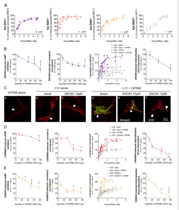
We first assessed whether GPR88 may come in close physical proximity to opioid receptors using bioluminescence resonance energy transfer (BRET1) saturation assay in HEK293FT cells to explore interactions between Luciferase Rluc8 (RLuc8)-tagged GPR88 and Venus-tagged GPCRs (see exam-ples of converse constructs inFigure 1—figure supplement 1). We evidenced that GPR88 displays specific and saturated BRET signals when co-expressed with the three opioid receptors, dOR, kOR and mOR, indicative of close physical proximity (within 10 nm) to them, in addition to itself (Figure 1A). These results indicate that GPR88 possibly forms hetero-oligomers with all opioid receptors.
We then tested, in heterologous cells, the consequences of co-expressing GPR88 with opioid receptors on their ability to activate G protein and b-arrestin dependent pathways as well as on their trafficking. Based on our previous results (Meirsman et al., 2016a), we first focused on dOR and tested the effects of SNC80 (10 mM) on dOR-mediated signaling in presence of increasing amounts of GPR88 (Figure 1B). While dOR cell surface expression (measured using anti-HA antibody, see Fig-ure 1—figFig-ure supplement 2A) remained constant (Figure 1—figure supplement 2B), growing lev-els of GPR88 expression led to a dose-dependent reduction in SNC80-induced dOR inhibition of cAMP accumulation (Gai/o-dependent pathway) and b-arrestin2 (b arr2) recruitment (and similarly b arr1, Figure 1—figure supplement 2B). We then monitored b arr2 recruitment at dOR in absence (basal) or presence (stimulated) of SNC80, to evaluate the probability of such recruitment in presence or not of GPR88 (30 ng of cDNA transfected). We evidenced that the probability of b arr2 recruitment at dOR was low under basal conditions and high when dOR was stimulated. GPR88 expression interfered with dOR/b arr2 interactions under stimulated conditions only. Furthermore, and consistent with dOR internalization being b-arrestin-dependent, we observed diminished loss of interaction between dOR and membrane-expressed Kras, and decreased interaction with early endo-some-expressed Rab5 and late endoendo-some-expressed Rab7 (Figure 1—figure supplement 2B), indi-cating decreased SNC80-induced dOR internalization and trafficking in presence of increasing amounts of GPR88. Finally, we used immunochemistry to further explore the consequences of GPR88 on dOR trafficking (Figure 1CandFigure 1—figure supplement 2C). We first verified mem-brane expression of GPR88 using a hemagglutinin (HA)-tagged construct (red,Figure 1C, Panel a, arrowhead). Similarly, HA-tagged dOR was mostly localized at the plasma membrane when not stim-ulated (red, Panel b, arrowhead) and SNC80 triggered its complete internalization in vesicular com-partments (Panel c, arrowhead). When HA-dOR (red) and GPR88-Venus (green) were co-transfected, they co-localized at plasma membrane under basal and activated conditions (Panels d and e, arrow-heads). Upon SNC80 agonist exposure, however, the typical internalization profile of dOR was lost compared to cells expressing dOR only (Panel f, arrowhead versus arrow). Altogether, these data indicate that co-expressing GPR88 with dOR is sufficient to affect G protein and b-arrestin depen-dent signaling pathways and trafficking of the latter.
We next explored the effects of the selective kOR agonist U50488H (10 mM) on kOR-mediated signaling in presence of increasing amounts of GPR88 (Figure 1D). GPR88 expression decreased U50488H-activated kOR pathway to Gai/o, although in our hands to a lesser extent than for dOR (decrease up to 27.7% of maximal effect versus 61.1% for dOR) and only for the highest dose of GPR88 cDNA, without altering kOR cell surface expression Figure 1—figure supplement 2D). GPR88, however, had a significant inhibitory effect on agonist-induced b arr2 recruitment at kOR (and b arr1,Figure 1—figure supplement 2D). When monitoring the affinity of b arr2 for kOR, we observed, as for dOR, an increase in such affinity under U50488H-stimulated conditions. GPR88 co-expression (30 ng of cDNA) had a limited influence on b-arr2 recruitment at kOR, contrasting with a significant inhibitory influence on internalization (Kras interaction) at a high dose of GPR88 cDNA, and trafficking (Rab5 interaction) (Figure 1—figure supplement 2D). Finally, we evaluated mOR sig-naling in response to its agonist DAMGO (10 mM) when co-expressed with increasing amounts of GPR88 (Figure 1—figure supplement 2E). GPR88 expression had a significant dose-dependent inhibitory effect on DAMGO-activated Gai/o pathway and b arr2 recruitment of mOR (see also b arr1,Figure 1—figure supplement 2E), mOR expression remaining stable (Figure 1—figure sup-plement 2E). Focusing on b arr2/mOR interactions, we observed again that b arr2 had significantly
Figure 1. GPR88 comes in close physical proximity to opioid receptors and inhibits their signaling and trafficking in vitro. (A) BRET1 saturation experiments were performed in transfected HEK293FT cells using constant quantity of GPR88-Rluc8 with increasing amounts of Venus-tagged opioid receptors dOR, kOR and mOR or V2R. Saturated BRET signals indicate close physical proximity (within 10 nm) to the target GPCR, thus possible hetero-or homo-oligomers. Such saturation is not observed with the V2R (right panel), D1 hetero-or CXCR4 recepthetero-ors (seeFigure 4), showing that close proximity is not detected for all GPCRs. See reverse constructions for dOR, kOR inFigure 1—figure supplement 1(B) Co-expressing GPR88 with dOR (wild-type or Rluc8-tagged, 30 ng of cDNA) blunts (left to right panels): SNC80 (dOR agonist, 10 mM)-induced inhibition of cAMP production (cAMP sensor: CAMYEL) (H4,39=28.7, p=0.0000) and Ypet-b-arrestin 2 (b-arr2) recruitment (H5,28=18.1, p=0.0028), Ypet-b-arr2 recruitment at dOR under stimulated but not basal conditions and, finally, SNC80-induced dOR internalization (internalization sensor: Kras-Venus) (H3,12=7,8, p=0.0492) in HEK293FT cells. (C) Confocal microscopy images show (arrow head) that (a) HA-GPR88 (red) is localized at cell surface (non-permeabilized cells), (b) under basal conditions, HA-dOR (red) is localized at cell surface, (c) SNC80-stimulation induces HA-dOR internalization, (d) under basal conditions, HA-dOR (red) and GPR88-Venus (green) co-localize at cell surface, (e) under SNC80 stimulation, GPR88 inhibits dOR internalization, (f) which results in different patterns of HA-dOR (red) distribution in GPR88-Venus (green) expressing versus non-expressing cells (arrow head versus arrow, respectively). (D) Co-expressing GPR88 with kOR (wild-type or Rluc8-tagged, 30 ng of cDNA) decreases (left to right panels): U50488H (kOR agonist, 10 mM)-induced inhibition of cAMP production (CAMYEL) (H4,25=17.9, p=0.0013) and Ypet-b-arr2 recruitment (H4,19=15.9, p=0.0031), Ypet-b-arr2 recruitment at kOR under stimulated but not basal conditions and U50488H-induced kOR internalization (Kras-Venus) (H4,18=13.6, p=0.0087) in HEK293FT cells. (E) Co-expressing GPR88 with mOR (wild-Figure 1 continued on next page
more affinity for mOR under stimulated versus basal conditions. Expressing GPR88 reduced the affin-ity of b arr2 for DAMGO-stimulated mOR to that measured under basal conditions. In line with these observations, DAMGO-induced internalization (loss of interaction with Kras) and trafficking (interac-tion with Rab5) of mOR were markedly impacted by the expression of increasing amounts of GPR88. These results indicate that GPR88 can bias the signaling of all three opioid receptors, with mOR appearing to be the most severely impacted.
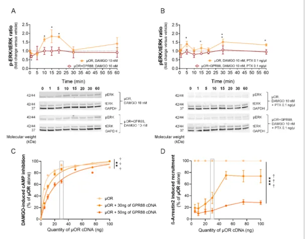
To further characterize the influence of GPR88 on mOR signaling, we tested the effects of GPR88 co-expression on mOR-induced phosphorylation of ERK in HEK293FT cells. We verified that DAMGO-induced mOR stimulation produced an increase in the phospho-ERK/total ERK total (pERK/ tERK) ratio that was suppressed in the presence of GPR88 (Figure 2A). Addition of pertussis toxin (PTX) markedly reduced the early phase of DAMGO-induced phosphorylation of ERK, showing its dependence on G protein recruitment (Gi/o). PTX, however, failed to suppress a later phase of
DAMGO-induced response, while GPR88 expression did (Figure 2B) (see gels inFigure 2—figure supplement 1). This later phase possibly corresponded to incomplete blockade of G-protein recruit-ment by PTX and/or to G protein-independent phosphorylation of ERK. Thus co-expressing GPR88 with mOR represses phosphorylation of the ERK complex through Gi/oprotein, further demonstrating
the inhibitory influence of GPR88 on G-protein dependent mOR signaling.
We then evaluated how the inhibitory effects of GPR88 on mOR signaling were influenced by the level of expression of the target receptor. To this aim, we first evaluated DAMGO-induced cAMP inhibition by increasing amounts of mOR in presence of a fixed amount of GPR88 (0, 30 or 50 ng of cDNA). We showed that the inhibitory effect of GPR88 on mOR signaling weakens when mOR amounts increase, with a complete restoration for high doses of mOR (Figure 2C). Raising the expression of GPR88 shifted this curve to the right, without modifying the maximal effect (full resto-ration). In contrast, increasing the amount of mOR was not able to completely overcome the inhibi-tory effect of GPR88 (30 ng of cDNA) on b-arr2 recruitment at mOR; notably, no further recruitment was observed over 50 ng of mOR cDNA transfected. Expressing more GPR88 led to near complete suppression of mOR-dependent b-arrestin recruitment (Figure 2D). In conclusion, GPR88 inhibitory action on G-protein dependent mOR signaling was more sensitive to mOR level of expression than its blunting effects on b-arrestin recruitment, suggesting different mechanisms of interaction.
We finally assessed whether GPR88 activation can modulate its ability to interfere with mOR sig-naling. GPR88 and mOR sharing inhibitory effects on cAMP production (recruitment of a Gi/o
pro-tein), this made impossible to directly disentangle mOR-dependent signaling from GPR88-dependent activity. However, when subtracting Compound 19-induced cAMP inhibition to the signal measured in presence of both DAMGO and Compound 19, we observed a tendency for an exacer-bation of GPR88 blunting effects on mOR-dependent signaling. We then tested the effects of the agonist Compound 19 on GPR88-mediated inhibition of b-arrestin recruitment at mOR in presence of DAMGO. GPR88 similarly repressed DAMGO-induced b-arrestin recruitment by mOR in presence or absence of Compound 19 (Figure 2—figure supplement 2).
Figure 1 continued
type or Rluc8-tagged, 30 ng of cDNA) inhibits (left to right panels): DAMGO (mOR agonist, 10 mM)-induced inhibition of cAMP production (CAMYEL) (H5,34=27.7, p=0.000) and Ypet-b-arr2 recruitment (H4,23=18.2, p=0.0011), Ypet-b-arr2 recruitment at mOR under stimulated but not basal conditions and DAMGO-induced mOR internalization (Kras-Venus) (H4,23=19.7, p=0.0006) in HEK293FT cells. Inhibition of cAMP production was determined in presence of 250 mM IBMX and 5 mM forskolin. Data are presented as mean ± SEM of n = 3–9 independent experiments (performed in triplicates). BRET1 values are presented as net BRET (normalized as the percentage of maximal BRET values) or induced BRET (normalized as the percentage of maximal BRET values in absence of GPR88) by Venus/Rluc8 BRET ratio. Asterisks: Kruskal-Wallis ANOVA, multiple comparison of mean ranks, *p<0.05, **p<0.01, ***p<0.001. Confocal imaging: representative pictures among n = 10 pictures. Receptor cell surface expression and additional data regarding OR trafficking in presence of GPR88 are displayed inFigure 1—figure supplement 2.
The online version of this article includes the following figure supplement(s) for figure 1:
Figure supplement 1. BRET saturation assays using Venus-tagged GPR88 and Rluc8-tagged target receptors. Figure supplement 2. GPR88 inhibits opioid receptor signaling and trafficking in vitro.
Figure 2. GPR88 dampens mOR-mediated signaling in vitro: ERK phosphorylation and effects of mOR expression levels. (A) Upper panel: in HEK293FT cells, DAMGO stimulated mOR (0.5 mg cDNA) activity, resulting in an increase in the phospho-ERK/ERK total (pERK/tERK) ratio peaking 15 min after DAMGO stimulation; this response was suppressed when GPR88 (1 mg cDNA) was co-expressed with mOR. Lower panel: representative western blotting images. (B) Upper panel: addition of pertussis toxin (PTX, 0.1 ng/ml, overnight) blocked the early phase of DAMGO-induced response, demonstrating its dependence on Gi/oprotein recruitment, but failed to inhibit a later component of ERK phosphorylation. GPR88 co-expression completely blocked DAMGO-induced phosphorylation of ERK. Lower panel: representative western blotting images. Levels of phosphorylated-ERK (pERK) and total-ERK (tERK) were normalized to the loading control protein glyceraldehyde 3-phosphate dehydrogenase (GAPDH). Data are presented as mean ± SEM of n = 5 independent experiments. Kruskal-Wallis ANOVA, multiple comparison of mean ranks *p<0.05, **p<0.01 (see gels in
Figure 2—figure supplement 1and statistics inSupplementary file 1). (C) Increasing the amount of mOR (wild-type, ng of cDNA) expressed in HEK293FT cells allowed to overcome the inhibitory effects of GPR88 (wild-type) on DAMGO (mOR agonist, 10 mM)-induced inhibition of cAMP production (CAMYEL), and a complete rescue of signaling was observed for the highest doses of mOR cDNA transfected (GPR88 effect: F2,21=86.7, p=0.0018; mOR effect: F7,147=90.9, p=0.0000; GPR88 x mOR interaction: F7,147=3.1, p=0.00030). Although increasing the amount of GPR88 (30 to 50 ng of cDNA) shifted the mOR dose response to the right, it was not able to prevent full restoration of mOR signaling at high doses of mOR. (D) Increasing the amount of mOR (wild-type) expressed in HEK293FT cells allowed only a partial overcoming of the inhibitory effects of GPR88 (wild-type) on DAMGO (mOR agonist, 10 mM)-induced Ypet-b-arr2 recruitment; no further recruitment was detected for doses of mOR cDNA over 50 ng (GPR88 effect: F2,6=86.7, p=0.000037; mOR effect: F6,36=32.3, p=0.0000; GPR88 x mOR interaction: F6,36=15.7, p=0.0000). Increasing the amount of GPR88 (from 30 to 50 ng of cDNA transfected) nearly suppressed b-arr2 recruitment at mOR. Data for 15 ng of mOR cDNA transfected are framed in gray, to allow comparison withFigure 1E. Data are presented as mean ± SEM of n = 3–12 independent experiments (performed in triplicates). BRET1 values are presented as induced BRET (normalized as the percentage of maximal BRET values in absence of GPR88) by Venus/Rluc8 BRET ratio. ANOVA
(repeated measure), stars: GPR88 effect, daggers: GPR88xmOR interaction; one symbol: p<0.05, two symbols: p<0.01, three symbols: p<0.001. Effects of GPR88 activation by synthetic agonist Compound 19 on its inhibitory action at mOR signaling in vitro are presented inFigure 2—figure supplement 2. The online version of this article includes the following figure supplement(s) for figure 2:
Figure supplement 1. Gels from western blot experiments; ERK phosphorylation assay in HEK293FT cells.
Figure supplement 2. Effects of Compound 19 on GPR88 signaling pathways and GPR88-mediated blunting of G-protein dependent signaling and b-arrestin recruitment by mOR.
Deletion of Gpr88 gene in mice modifies mOR-mediated responses in a
behavior-specific manner
Consistent with the above in vitro data, we previously observed an increase in mOR-dependent [35 S]-GTPgS binding in striatal membranes from Gpr88-/-mice (Meirsman et al., 2016a), suggesting that
G protein coupling by mOR was facilitated in the absence of GPR88 expression. We thus hypothe-sized that Gpr88 deletion should result in exacerbated morphine-induced mOR-dependent responses in mice. To test this proposition, we assessed several behavioral responses to morphine challenge in Gpr88-/-mice. The aim of this behavioral screening was to compare morphine-induced
behavioral responses depending on the brain regions involved (where GPR88 and mOR receptors are likely or not to co-localize under physiological conditions), the signaling pathways tackled (G pro-tein or b-arrestin-dependent pathways) and GPCR populations engaged (beyond mOR).
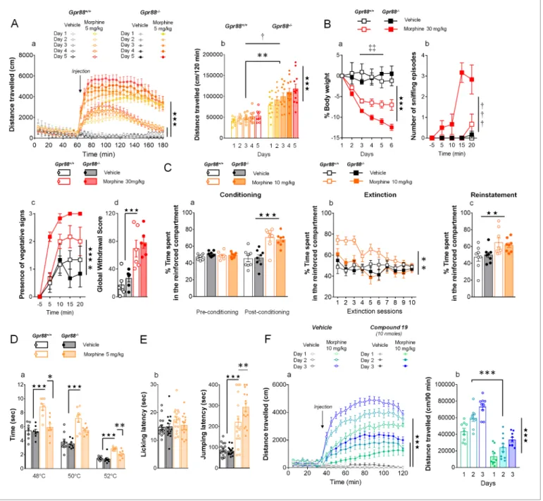
First, we measured morphine-induced locomotor activation and its sensitization, two behavioral outcomes tightly depending on striatal function, in Gpr88 null mice and their wild-type controls after they received daily injections of morphine (IP, 5 mg/kg). The locomotor activity of mutant and con-trol mice was similar under basal (habituation) and vehicle-treated conditions. In both mouse lines, morphine injections induced a significant rise in locomotor activity that progressively sensitized upon repeated administration (Figure 3A, panel a). Locomotor activation and sensitization, however, were markedly exacerbated in Gpr88-/-compared to Gpr88+/+mice (panel b). These results thus indicate that Gpr88 deletion facilitates morphine-induced locomotion and sensitization.
Next, we administered morphine (IP, 30 mg/kg) daily for 6 days to a cohort of Gpr88+/+ and
Gpr88-/- mice before triggering pharmacological withdrawal using naloxone (SC, 1 mg/kg). With-drawal syndrome involves notably activation in the periacqueductal gray, amygdala and nucleus accumbens. We observed that morphine induced-weight loss (measured daily during morphine exposure) was more pronounced in knockout versus control mice (Figure 3B, panel a). Following mOR blockade, mutant mice displayed significantly more sniffing episodes (panel b) and exhibited vegetative signs of withdrawal (pilorerection, ptosis and teeth chattering) quicker than wild-type con-trols (panel c), despite comparable global withdrawal scores (panel d) (see more signs inFigure 3— figure supplement 1). These data point to exacerbated effects of morphine on withdrawal symp-toms, with late signs of withdrawal appearing sooner in Gpr88 null mice.
Then, we submitted Gpr88-/-mice and their controls to a conditioned place preference (CPP)
par-adigm, in which vehicle or morphine injections (SC, 10 mg/kg) were paired with a compartment of the CPP apparatus. This conditioned behavior relies essentially on mOR activation in the ventral teg-mental area. Mutant and wild-type animals acquired similarly preference conditioning to morphine (Figure 3C, panel a). We then exposed the mice to an extinction protocol, during which exploration of the CPP apparatus was no longer paired with vehicle or morphine injections. Under these condi-tions, Gpr88-/-mice extinguished place preference to morphine quicker than their Gpr88+/+ counter-parts (panel b). In a last trial, we re-administered morphine (or vehicle) to reinstate place preference: both mouse lines similarly reinstated preference for the reinforced compartment (panel c). Thus, Gpr88 deletion had no effect on morphine-induced CPP and reinstatement, suggesting preserved morphine reward in knockout animals, but reduced the lapse of time they needed to extinguish this conditioning.
In a next step, we assessed the nociceptive thresholds of Gpr88 mutants and controls in the tail immersion and hot-plate tests under vehicle or morphine (IP, 5 mg/kg) challenge. The first test involves spinal responses whereas thermic nociception depends on central amygdala function. Responses to nociceptive stimuli did not differ between Gpr88-/-and Gpr88+/+ mice under vehicle conditions. In the tail immersion test, however, while we detected significant analgesic effects of morphine across temperatures in Gpr88+/+animals, this response was markedly blunted in Gpr88 -/-mice (Figure 3D, panel a). In contrast, in the hot-plate test, jumping latency under morphine chal-lenge, but not licking latency, was longer in mutant compared to wild-type animals, indicating that supraspinal morphine-induced analgesia was facilitated in Gpr88 null mice (panel b). Thus, the behavioral consequences of Gpr88 deletion on morphine-induced responses differed depending on the behavior assessed, pointing to brain substrates, signaling pathways and additional GPCRs involved as key modulators of these responses.
Having shown above that mice lacking Gpr88 display a drastic increase in their locomotor sensiti-zation to repeated morphine administration, and that this effect was highly significant since the
Figure 3. Gpr88 null mice display modified mu-opioid mediated behavioral responses. (A) In a locomotor sensitization paradigm (n = 5 to 11 mice per treatment and genotype) morphine induced an increase in locomotor activity that sensitized upon repeated administration in Gpr88+/+and Gpr88 -/-mice (panel a); morphine-induced locomotion and sensitization, however, were significantly greater in mutant -/-mice (panel b) (Genotype effect: F1,27=13.5, p=0.0010; Treatment: F1,27=99.9, p=0.0000; Genotype x Treatment interaction: F1,27=14.6, p=0.0007; Day: F4,108=7.9, p=0.0000; Day x Treatment: F4,108=10.2, p=0.0000; Day x Genotype x Treatment = 2.9, p=0.0253); solid stars: treatment effect (two-way ANOVA with one repeated measure – day), asterisks: genotype effect, dagger: Day x Genotype x Treatment interaction. (B) Upon exposure to escalating doses of morphine (n = 6 per treatment and genotype), Gpr88-/-lost more body weight than controls (panel a; Treatment: F1,20=43.7, p=0.0000; Day: F4,80=13.6, p=0.0000; Day x Genotype: F4,80=5.1, p=0.001; Day x Treatment: F4,80=9.9, p=0.0000; body weight was measured daily upon morphine treatment); when withdrawal was triggered by acute naloxone administration (1 mg/kg), mutant animals displayed sniffing episodes (panel b; Genotype/Treatment: F1,20=25.5, p=0.0000; Genotype x Treatment: F1,20=23.1, p=0.0001; Time: F3,60=10.5, p=0.0000; Time x Genotype/Treatment: F3,60=9.3, p=0.0000; Time x Genotype x Treatment: F3,60=8.3, p=0.0001) and vegetative signs of withdrawal (panel c; Genotype: F1,20=5.3, p=0.0324; Treatment: F1,20=5.3, p=0.0324; Time: F3,60=14.4, p=0.0000) quicker than their Gpr88+/+counterparts, despite similar final withdrawal scores (panel d; Treatment: F1,20=38.9, p=0.0000); solid stars: treatment effect (two-way ANOVA with one repeated measure – day/5 min time bin), double daggers: Time x Genotype interaction, daggers: Time x Genotype x Treatment interaction, asterisk: genotype effect. More withdrawal signs are displayed inFigure 3—figure supplement 1. (C) In a CPP paradigm (n = 8 per treatment and genotype), Gpr88-/-mice acquired preference for a compartment associated to morphine administration (10 Figure 3 continued on next page
second administration of morphine, we aimed at evaluating mOR signaling by assessing pERK/tERK ratio in brain regions from Gpr88-/- versus Gpr88+/+ mice, in an attempt to capture b-arr2/pERK recruitment, shown to play a crucial role in morphine-induced locomotor sensitization (Tao et al., 2017; Urs et al., 2011; Valjent et al., 2005; Valjent et al., 2010). We exposed Gpr88+/+ and Gpr88-/-mice to morphine administration (IP, 5 mg/kg) on two consecutive days in the same environ-ment (Valjent et al., 2010). We observed on the second day a robust locomotor sensitization in mutant animals that was not observed in wild-type animals (Figure 3—figure supplement 2A). 60 min after second morphine injection, however, we failed to detect differences in the pERK/tERK ratio between Gpr88-/-and Gpr88+/+ animals in striatal regions (caudate putamen and nucleus
accum-bens), or in the periaqueductal gray (Figure 3—figure supplement 2B) (see gels inFigure 3—figure supplement 3). Therefore, under these experimental conditions, we could not detect a significant impact of Gpr88 deletion on mOR-mediated b-arr2/pERK ex vivo.
Finally, we addressed the question of potential effects of GPR88 pharmacological activation on its ability to interfere with mOR signaling by evaluating the consequences of Compound 19 adminis-tration (icv, 10 nmoles) on morphine (IP, 10 mg/kg)-induced locomotion and sensitization in wild-type mice (Figure 3F). Compound 19 markedly inhibited morphine-induced locomotion in these ani-mals along the three days of morphine exposure (Figure 3F, panel a). However, the amplitude of morphine-induced locomotor sensitization was similar in mice that received the GPR88 agonist or vehicle (Figure 3F, panel b), indicating that Compound 19 had no detectable influence on this pro-cess. Thus, pharmacological activation of GPR88 can potentiate some, although not all, inhibitory effects of this orphan receptor on mOR signaling in vivo.
GPR88 comes in close proximity to multiple GPCRs and interferes with
their signaling
Morphine-induced behavioral responses rely on complex molecular mechanisms and the activation of multiple GPCRs beyond mOR. We thus explored whether GPR88 may come in close physical vicin-ity of other GPCRs, focusing first on GPCRs with striatal expression, such as muscarinic (M1, M4), dopaminergic (D1, D2), adenosine (A2A) or orphan receptor GPR12 (Ho et al., 2018;Heiman et al.,
2008), and then extending our interest to GPCRs not known to be expressed in CNS neurons, namely the vasopressin V2R and chemokine CXCR4 receptors. Remarkably, GPR88 displayed satu-rated BRET signals with all tested striatal GPCRs except the dopamine D1 receptor (Figure 4A). In
Figure 3 continued
mg/Kg) similarly as Gpr88+/+animals (panel a; Treatment: F
1,28=45.8, p=0.0000; Conditioning: F1,28=15.7, p=0.0005; Conditioning x Treatment: F1,28=31.1, p=0.0000); they extinguished this conditioning quicker than wild-type counterparts (panel b; Genotype: F1,28=10.3, p=0034; Treatment: F1,28=6.5, p=0166; Genotype x Treatment: F1,28=5.0, p=0329; Session: F9,252=5.2, p=0.0000; Session x Treatment: F9,252=3.7, p=0.0002) and finally reinstated morphine place preference at comparable levels as the latter (panel c; Treatment: F1,28=9.2, p=0.0052); solid stars: Treatment effect (two-way ANOVA with one repeated measure – day/5 min time bins); asterisks: Genotype effect. (D) In the tail flick test (n = 7–9 per treatment and genotype), Gpr88-/-mice were significantly less sensitive to morphine analgesia at 48˚C and 52˚C (Genotype: F
1,83=22.0, p=0.0000; Treatment: F1,83=84.9, p=0.0000; Temperature: F2,83=162.5, p=0.0000; Genotype x Treatment: F1,83=15.9, p=0.0001; Treatment x Temperature: F2,83=5.4, p=0.0065); (E) in the hot plate test (n = 15–16 per treatment and genotype), morphine-induced analgesia was detected by increased jumping latency in treated animals (right panel); this effect was increased in Gpr88 null mice versus controls (Treatment: F1,59=79.7, p=0.0000; Genotype x Treatment: F1,59=4.0, p=0.0490); solid stars: Treatment effect (two-way ANOVA), asterisks: Genotype x Treatment interaction (Newman Keules post-hoc test). Increased morphine-induced locomotor sensitization in Gpr88 null mice was not associated with modified pERK/tERK ratio in three brain regions (Figure 3—figure supplements 2
and3). (F) We administered the GPR88 agonist Compound 19 (icv, 10 nmoles) to mice exposed to a morphine-induced locomotor sensitization paradigm (n = 8 to 9 mice per treatment condition). Exposure to morphine (IP, 10 mg/kg) induced an increase in locomotor activity that sensitized upon repeated administration in both vehicle and Compound 19-treated groups (panel a); pharmacological activation of GPR88 drastically reduced
morphine-induced locomotor activity but left the amplitude of sensitization unchanged (Morphine effect: F1,30=305.1, p=0.0000; Compound 19: F1,30=55.3, p=0.0000; Day: F2,60=33.1, p=0.0000; Day x Morphine: F2,60=31.6, p=0.0000; Day x Morphine x Compound 19: F2,60=2.1, NS), solid stars: treatment effect (two-way ANOVA with one repeated measure – day), asterisks: genotype effect. Data are presented as mean ± SEM. One symbol: p<0.05, two symbols: p<0.01, three symbols: p<0.001.
The online version of this article includes the following figure supplement(s) for figure 3:
Figure supplement 1. Additional morphine-induced withdrawal signs in Gpr88-/-versus Gpr88+/+mice.
Figure supplement 2. Increased morphine-induced locomotor sensitization in Gpr88 null mice was not associated with modified pERK/tERK ratio in three brain regions.
contrast, BRET signal was unsaturated for interactions with the two GPCRs identified as not expressed in CNS neurons, V2R and CXCR4 receptors (Figure 4B). Thus GPR88 appears to possibly form hetero-oligomers with multiple GPCRs in addition to opioid receptors, notably those GPCRs whose expression is enriched in striatal regions, where GPR88 is also the most expressed.
We next explored the functional consequences of GPR88 co-expression on the signaling of stria-tal and non-striastria-tal GPCRs. We co-expressed GPR88 with these GPCRs and assessed the effects of such co-expression on their ability to activate their G protein and b-arrestin dependent signaling pathways. Interestingly, increasing amounts of GPR88 expression interfered with the G protein medi-ated signaling of all GPCRs from which it comes close (dopamine D2 and muscarinic M1 and M4) except for the adenosine A2A receptor (Figure 5A, panels a,c,d,e,f). In contrast, co-expressing
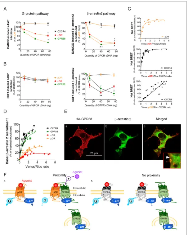
GPR88 with receptors for which we failed to evidence proximity to it, namely dopamine D1, vaso-pressin V2R and chemokine CXCR4 receptors, had no significant effect on their G protein dependent signaling, or even increased this signaling (D1 receptor) (Figure 5A, panels b,g,h). Of note, the impact of GPR88 expression on G protein mediated signaling was observed independently from the nature of the G protein coupled by the target receptor, either Gas (M1, M4), Gai/o (D2) or Gaq/11 (M4). When now focusing on b-arrestin recruitment, the picture was remarkably homogeneous: expressing increasing amounts of GPR88 dose-dependently dampened b-arr2 recruitment by all GPCRs tested (Figure 5B, panels a-g). Moreover, GPR88 expression markedly blunted CXCR4-dependent b-arr2 recruitment under both basal and stimulated conditions (Figure 5B, panel h). Taken together, our functional results indicate that GPR88 expression represses the G protein-dependent signaling of multiple GPCRs, more likely when it possibly forms hetero-oligomers with them, whereas it inhibits b-arr2 recruitment for all GPCRs tested.
GPR88 blunts b-arrestin recruitment by other GPCRs independently
from physical proximity
In an attempt to better understand how GPR88 impacts the signaling of other GPCRs, we compared the consequences of co-expressing GPR88 on the activation of the G-protein dependent pathway and the recruitment of b-arrestins by mOR, with which GPR88 potentially forms hetero-oligomers (Figure 6A), and CXCR4, for which we could not detect proximity to GPR88 (Figure 6B). Moreover,
Figure 4. GPR88 comes in close proximity to multiple GPCRs. BRET1 saturation experiments were performed in transfected HEK293FT cells using constant quantity of GPR88-Rluc8 with increasing amounts of (A) Venus-tagged striatal GPCRs: dopamine D1 and D2, adenosine A2A, muscarinic M1 and M4, and orphan receptor GPR12, (B) Venus-tagged non neuronal GPCRs: chemokine CXCR4 and V2R (seeFigure 1). (C) Venus-tagged GPR88. Saturated BRET signals indicate close physical proximity (within 10 nm) to the target GPCR. GPR88 comes in close proximity and possibly forms hetero-oligomers with D2, A2A, M1, M4, GPR12 and itself, but not D1, V2R and CXCR4 receptors. Values (mean ± SEM) from n = 3–4 independent experiments (performed in triplicates) are presented as net BRET (normalized as the percentage of maximal BRET values) by Venus/Rluc8 BRET ratio.
the effects of GPR88 co-expression were compared to those of co-expressing the alternative GPCR (CXCR4 for mOR, and reciprocally) or kOR, as examples of GPCRs forming or not heterodimers with the target receptor. Indeed, we verified that mOR and kOR closely interact (Figure 6C, upper panel), as previously reported (Fujita et al., 2014), and evidenced that CXCR4 likely forms hetero-oligomers with kOR but not mOR (Figure 6C, middle and right panels, respectively). Consistent with no detected proximity, CXCR4 co-expression had no deleterious effect on DAMGO-induced mOR sig-naling, and conversely mOR co-expression did not modify SDF1-mediated CXCR4 sigsig-naling, on either
Figure 5. GPR88 biases the signaling of multiple GPCRs. We evaluated the consequences of GPR88 co-expression on the signaling of adenosine A2A, dopamine D1, dopamine D2, muscarinic M1, muscarinic M4, vasopressin V2R and CXCR4 receptors in HEK293FT cells. BRET1 assay was used to assess the activation of the (A) G protein dependent pathway (cAMP sensor: CAMYEL; calcium sensor: aequorin-GFP) or (B) the recruitment of Ypet b-arrestin 2 (b-arr2) by Venus-tagged receptors in presence of increasing amounts of GPR88 cDNA transfected. (A) GPR88 co-expression blunted the G protein mediated signaling of all GPCRs from which it comes close (panels c-f; dopamine D2: H3,12=9.6, p=0.0223; muscarinic M1: H3,16=13.2, p=0.0041; M4 calcium: H3,35=24.0, p=0.0000; M4 cAMP: H3,12=12.9, p=0.0048), except the adenosine A2Areceptor (panel a; H3,12=1.2, p=0.7602). GPR88 had no significant impact on, or even facilitated, G protein dependent signaling of receptors for which we failed to evidence proximity to it, namely D1 (panel b, increased activity in presence of GPR88; H3,20=9.7; p=0.0211), V2R and CXCR4 receptors (panels g,h; V2R: H3,16=1.2, p=0.7602; CXCR4: H3,12=7.6, p=0.0546). (B) In contrast, GPR88 co-expression compromised the ability of all GPCRs tested to recruit Ypet-b-arr2 when activated by their agonist, independently from previously evidenced close proximity to GPR88 (panels a-g; A2A: H3,12=9.7, p=0.0211; D1: H3,12=9.6, p=0.223; D2: H3,16=13.2, p=0.0080; M1: H3,15=10.4, p=0.0151; M4: H3,16=13.2, p=0.0041; V2R: H3,16=10.9, p=0.0123; CXCR4: H3,16=13.2, p=0.0041). Focusing on CXCR4, co-expressing GPR88 diminished the probability of Ypet-b-arr2 recruitment at this receptor under both stimulated and basal conditions (panel h). Specific agonists used to stimulate GPCRs were: CGS21680 (A2A, 10 mM), SKF81297 (D1, 10 mM), quinpirole (D2, 10 mM), carbachol (M1 and M4, 10 mM), AVP (V2R, 10 mM) and SDF1 (CXCR4, 125 mM). Data are presented as mean ± SEM of n = 3–4 independent experiments (performed in triplicates). BRET1 values are presented as induced BRET (normalized as the percentage of maximal BRET values in absence of GPR88) normalized by Venus/Rluc8 BRET ratio. Asterisks: Kruskal-Wallis ANOVA, multiple comparison of mean ranks, *p<0.05, **p<0.01.
Figure 6. GPR88 impedes the recruitment of b-arrestins at other GPCRs independently from physical proximity. (A) We evaluated the functional consequences of expressing CXCR4, kOR and GPR88 with mOR on DAMGO-induced cAMP production and b-arr2 recruitment by mOR. CXCR4 co-expression had no deleterious impact on G-protein-dependent signaling and the recruitment of b-arrestins (cAMP: H3,16=6.6, p=0.0840; b-arr2: H3,16=2.8, p=0.427); in contrast, kOR and GPR88 expression dampened the activation of both, with GPR88 inhibiting cAMP production by mOR more efficiently than kOR (cAMP - kOR: H3,12=10.1, p=0.0176; GPR88: H3,12=10.5, p=0.0145; b-arr2 - kOR: H3,16=13.8, p=0.0031; GPR88: H3,16=13.5,
p=0.0036). (B) We then assessed the consequences of co-expressing mOR, kOR and GPR88 with CXCR4 on SDF1-induced cAMP production and b-arr2 recruitment by CXCR4. None of the co-expressed receptors had a significant impact on cAMP production by CXCR4 (mOR: H3,12=7.3, p=0.064; kOR: H3,12=2.6, p=0.4593; GPR88: H3,12=2.9, p=0.4077); as regards b-arr2 recruitment, mOR co-expression had no influence (H3,12=0.75, p=0.8604), whereas both kOR (H3,16=12.7, p=0.0053) and GPR88 (H3,12=9.8, p=0.0203) reduced it, with GPR88 having a more significant influence than kOR at the highest dose transfected. (C) BRET1 saturation experiments evidenced close proximity between mOR and kOR (upper panel), CXCR4 and kOR (middle panel) but not CXCR4 and mOR (lower panel). (D) Ypet-b-arr2 recruitment was higher at CXCR4 and GPR88 than kOR and mOR under basal conditions. (E) Confocal microscopy images show that (a) HA-GPR88 is localized at cell surface and also in the cytosol, following a patchy distribution (permeabilized cells), (b) b-arr2 is expressed in a diffuse pattern near the cell membrane and almost all the cytoplasm and (c) Colocalisation of GPR88 and b-arr2 is Figure 6 continued on next page
the G protein- or b-arrestin-dependent pathways (Figure 6A–B). In contrast, kOR expression repressed agonist-mediated G protein activation and b-arr2 recruitment by mOR, with which it forms hetero-oligomers (Figure 6A). Such expression however had no significant impact on G protein-mediated signaling by CXCR4 but significantly inhibited b-arr2-dependent recruitment by this recep-tor (Figure 6B). Compared to kOR, GPR88 had a more significant inhibitory impact on G protein dependent mOR signaling but similarly blunted mOR-dependent b-arr2 recruitment (Figure 6A). Finally, although GPR88 had little influence on SDF1-induced G protein activation by CXCR4 (see also Figure 5G), it was able to repress CXCR4-dependent b-arr2 recruitment more than kOR, despite no detectable physical proximity to CXCR4 (Figures 4B and 6B). Together, these results indicate that GPR88 can interfere with the signaling of other GPCRs when coming in close vicinity of them, but also in the absence of detected proximity.
A mechanism through which GPR88 may dampen GPCR recruitment of b-arrestins would be by sequestering them away from the plasma membrane, as shown for vasopressin V2R and neurokinin NK1 receptors upon pharmacological activation (Klein et al., 2001;Schmidlin et al., 2002). Under these conditions, however, V2R and NK1 display a high affinity for b-arrestins and an intracellular dis-tribution. Here, we evaluated the affinity for b-arrestins of GPR88 compared to mOR, dOR and CXCR4 under basal conditions (no agonist). The probability that b-arr2 was recruited at GPR88 and CXCR4 in the absence of pharmacological stimulation was higher than at the two opioid receptors (Figure 6D). We then used immunochemistry to assess the respective intracellular distributions of HA-GPR88 and b-arr2 after membrane permeabilization. We observed, in addition to location at cell membrane, a significant, patchy, intracellular distribution of GPR88 (Figure 6Ea), suggesting its presence in vesicular compartments. Diffuse cytosolic distribution of b-arr2 (Figure 6Eb) was found colocalized with GPR88-expressing patches (Figure 6Ec) and appeared depleted around the bright-est ones (framed, arrow head), suggbright-esting sequbright-estration by GPR88. Thus, GPR88 can bias the signal-ing of other GPCRs independently from oligomerization, possibly by trappsignal-ing b-arrestins in intracellular compartments.
Discussion
In the present study, we evidence for the first time physical proximity between GPR88 and the three opioid receptors, and a negative impact of GPR88 expression on opioid-receptor mediated G pro-tein and b-arrestin recruitment in vitro, mOR-dependent signaling appearing the most severely affected. In mice, Gpr88 deletion produced different effects on morphine-induced (mOR-dependent) responses depending on the behavior assessed, exacerbating morphine-induced locomotor sensiti-zation, withdrawal syndrome and supra-spinal analgesia while blunting morphine effects on spinal nociceptive responses and leaving CPP unchanged. Importantly, we also detected close proximity between GPR88 and multiple other GPCRs with enriched striatal expression but not with GPCRs not expressed in CNS neurons. GPR88 co-expression was able to inhibit G protein dependent signaling of most GPCRs from which it comes close, and only these. In contrast, such co-expression resulted in blunted b-arrestin recruitment by all GPCRs tested.
The first finding from this study is that GPR88 closely interacts with opioid receptors and that its expression impedes opioid signaling in vitro. Saturated BRET signals of GPR88 in presence of dOR, kOR and mOR indicate physical proximity with opioid receptors and suggest potential hetero-oligo-merization. Remarkably, although GPR88 displayed similar saturated BRET signals in presence of the
Figure 6 continued
clearly seen at the level of cytoplasmic patches, with the intensity of b-arr2 expression appearing lower around the densest GPR88-expressing patches (framed, arrow head). (F) Schematic representation of the putative mechanisms of GPR88 inhibitory action at GPCR signaling. Panel a: GPR88 dampens G-protein-mediated signaling of GPCRs to which it comes close (such as mOR), likely by interfering with G-protein coupling, and inhibits the recruitment of b-arrestins by sequestering them in intracellular compartments. Pharmacological activation of GPR88 may potentiate its inhibitory action on
G-protein dependent signaling only. Panel b: when no proximity is detected between GPR88 and the target receptor (such as CXCR4), only the effects on b-arrestin recruitment are observed. Data (A–D) are presented as mean ± SEM of n = 3–4 independent experiments (performed in triplicates). BRET1 values are presented as net BRET (normalized as the percentage of maximal BRET values) or induced BRET (normalized as the percentage of maximal BRET values when the target receptor is expressed alone) by Venus/Rluc8 BRET ratio. Asterisks: Kruskal-Wallis ANOVA, multiple comparison of mean ranks, *p<0.05, **p<0.01. Confocal imaging (E): representative pictures among n = 10 pictures. Legend toSupplementary file 1.
three opioid receptors, the orphan receptor seemed to have a differential influence on their signal-ing. GPR88 co-expression had only a modest impact on kOR-mediated G protein pathway activation compared to dOR and mOR. Conversely, dOR appeared to be the less affected among opioid recep-tors on its ability to recruit b-arrestins, as seen from agonist-induced b-arr2 recruitment and receptor internalization. Finally, mOR function seemed to be the most severely altered by GPR88 co-expres-sion, on both G protein and b-arrestin-dependent pathways.
In order to better characterize the effects of GPR88 expression on opioid signaling, we focused on GPR88-mOR functional interactions. We first confirmed the inhibitory effect of GPR88 expression on G-protein dependent signaling of mOR, by evidencing a suppression of Gi/oprotein-dependent
mOR-induced phosphorylation of ERK in presence of GPR88. We then evaluated how mOR expres-sion levels impact the effects of GPR88 co-expresexpres-sion on G-protein dependent signaling and b-arrestin recruitment by mOR. We observed a weakening of the inhibitory action of GPR88 on mOR-mediated G-protein dependent signaling when mOR expression increased over GPR88 expression. Thus, direct mOR-GPR88 interactions seem necessary for GPR88 to exert its inhibitory effects; more-over, the regular pace of the rightwards shift of mOR signaling dose response when GPR88 amounts increase suggests a 1:1 (or n/n) stoichiometry in this interaction. In contrast, increasing mOR amounts failed to fully restore mOR-mediated b-arrestin recruitment in presence of GPR88, suggesting that a pool of b-arrestins remained out of reach of mOR, regardless mOR expression levels. Therefore, GPR88 effects on mOR-mediated G-protein dependent signaling and b-arrestin recruitment likely involved different mechanisms of interaction. Interestingly, differential relative inhibitory effects of GPR88 on the signaling of the different opioid receptors (and other GPCR partners) possibly reflected differential sensitivity to variations in the expression of the partner receptor, G-protein sig-naling being more sensitive than b-arrestin recruitment to such variations. Finally, we tested whether agonist-induced activation of GPR88 influences its effects on mOR signaling in vitro. Activation of GPR88 by Compound 19 tended to exacerbate the inhibitory action of GPR88 on mOR-mediated G-protein dependent signaling, but had no detectable effect on b-arrestin recruitment by mOR. These results suggest that GPR88 is more efficient in blunting GPCR partner’s G-protein dependent signaling, but not b-arrestin recruitment, if in an active-like conformation.
The physiological consequences of GPR88 expression on opioid signaling are not straightforward to predict from in vitro experiments, as inhibition of G protein signaling argues for a reduction of (dOR and mOR) opioid signaling, whereas dampened b-arrestin recruitment suggests a retention of opioid receptors at the cell surface and maintained signaling but loss of b-arrestin-mediated func-tional effects. We took advantage of analyzing the phenotype of Gpr88 null mice to get insight into interactions between GPR88 and opioid signaling in vivo. Our previous findings of increased dOR-mediated G protein signaling in striatal membranes of Gpr88-/-mice together with a phenotypic pro-file opposing that of mice lacking dOR (Le Merrer et al., 2013) and partially normalized by dOR antagonist administration (Meirsman et al., 2016a) plead for an excessive dOR activity in these ani-mals, consistent with a main inhibitory impact of GPR88 on the G protein dependent signaling of this receptor. In the present study, we evaluated the in vivo consequences of Gpr88 deletion on mOR signaling by challenging knockout animals with morphine across several experimental para-digms. Morphine-induced responses in Gpr88-/-knockouts were differentially modified depending on the behavior assessed, likely due to differences in the brain regions, signaling pathways and GPCR populations engaged to mediate these responses. The prominent factor influencing the effects of Gpr88 deletion, however, was likely the brain substrates underlying behavioral responses, and whether GPR88 is expressed in these structures under physiological conditions. Indeed, Gpr88 deletion exacerbated morphine-induced responses that involved mOR activation in brain regions where Gpr88 expression is usually enriched (Massart et al., 2016; Ehrlich et al., 2018a;
Becker et al., 2008), namely locomotor activity and sensitization, depending on mOR signaling in the striatum (Tao et al., 2017;Charbogne et al., 2017), withdrawal symptoms involving mOR activity in the nucleus accumbens (Williams et al., 2001;Le Merrer et al., 2009) and supra-spinal analgesia measured in the hot-plate test, modulated notably by mOR activity in the central amygdala (Pavlovic et al., 1996;Pavlovic and Bodnar, 1998). In contrast, morphine-induced CPP and rein-statement were not modified in Gpr88-/-mice. Interestingly, morphine-induced CPP involves
primar-ily mOR signaling in the ventral tegmental area (Charbogne et al., 2017;Le Merrer et al., 2009) where GPR88 is scarcely expressed (Ehrlich et al., 2018a;Ehrlich et al., 2018b). This could account for unchanged morphine CPP in the absence of Gpr88 expression. Of note, Gpr88 null animals
extinguished CPP faster than controls, in agreement with previously evidenced facilitation of hippo-campus-dependent learning processes (Meirsman et al., 2016a). A second factor that plausibly influenced morphine-induced responses in Gpr88-/-mice was the signaling pathway involved in
mOR-mediated effects. Indeed, locomotor sensitization, markedly exacerbated in knockout mice, was evi-denced to depend tightly on b-arrestin recruitment not only by mOR, but also by the D1 dopamine receptor (Tao et al., 2017;Urs et al., 2011;Borgkvist et al., 2008;Becker et al., 2001). As regards these behavioral responses, the phenotypic profile of Gpr88 mutant animals is thus consistent with a relief of GPR88 break on b-arrestin recruitment by mOR, and D1. Now focusing on nociception, mor-phine-induced analgesia was significantly reduced in the tail immersion test, which tackles spinal function (Ramabadran et al., 1989). Gpr88 expression in spinal cord is discrete (Massart et al., 2016; Becker et al., 2008). Its deletion, however, may have allowed massive mOR-mediated b-arrestin recruitment under morphine stimulation, which was shown to be detrimental to spinal mor-phine analgesia (Yang et al., 2011). Finally, behavioral responses to morphine in Gpr88-/-mice may
not be the consequence of GPR88 removal on mOR function only but also on the signaling of other GPCRs involved in the expression of these responses. Notably, morphine-induced locomotor sensiti-zation involves striatal dopamine and cholinergic receptors (Tao et al., 2017;Valjent et al., 2005;
Ruan et al., 2019), and the effects of GPR88 deletion on their pharmacology likely contributed to the exacerbation of this phenomenon in mutant animals. The involvement of multiple GPCR players in morphine-induced responses may have participated to our failure in detecting the expected increase in pERK/tERK ratio in Gpr88 knockout mice sensitized to morphine; modified kinetics of ERK phosphorylation in vivo versus in vitro, however, represents a more likely explanation for this failure. In conclusion, altered behavioral responses to morphine challenge in Gpr88 null mice are consistent with an inhibitory influence of GPR88 co-expression on mOR signaling, especially for behaviors engaging striatal regions, thus demonstrating the predictive value of in vitro experiments.
Further confirming such predictive value, in vivo administration of Compound 19 markedly inhib-ited morphine-induced stimulation of locomotor activity in wild-type mice, resulting in opposed effects compared to Gpr88 deletion on this parameter. GPR88 activation, however, had no influence on the amplitude of locomotor sensitization measured upon repeated morphine administration. Interestingly, while acute stimulant effects of morphine were shown to involve both G protein activa-tion and b-arrestin recruitment by mOR (and D1 receptors), sensitizaactiva-tion, as menactiva-tioned above, relies essentially on the latter (Tao et al., 2017;Urs et al., 2011;Girault et al., 2007). In light of Com-pound 19’s effects on morphine-induced locomotor responses, it thus seems that pharmacological activation of GPR88 in vivo can potentiate the inhibitory effects of GPR88 on mOR-dependent G pro-tein activation, but not b-arrestin recruitment, in agreement with in vitro results (Figure 2—figure supplement 2). Whether this applies to other GPCR partners of GPR88, however, would deserve fur-ther investigation. Of note, these results furfur-ther argue for the inhibitory effects of GPR88 on the G-protein dependent signaling of a partner GPCR requiring GPR88 to be in an active-like conforma-tion, as previously described for many interacting GPCRs (Vilardaga et al., 2008;Goudet et al., 2005;Pin et al., 2019).
A second major finding from this study is that GPR88 can bias the signaling of multiple GPCRs beyond opioid receptors. In vitro, we detected physical proximity between GPR88 and muscarinic M1 and M4, dopamine D2, adenosine A2Aand GPR12 receptors, suggesting that GPR88 can form
hetero-oligomers with them. Such close interactions are plausible in vivo as all these partner recep-tors share a common enriched expression in the striatum and extended amygdala, notably in medium spiny GABAergic neurons (Ferre´ et al., 2007;Surmeier et al., 2007;Pellissier et al., 2018;
Ignatov et al., 2003), where GPR88 is also expressed. In contrast, we could not evidence proximity between GPR88 and dopamine D1 receptor, another GPCR with enriched striatal expression, and two GPCRs not known to be expressed in CNS neurons, the vasopressin V2R and chemokine CXCR4 receptors. Interestingly, these last results suggest that GPR88 forms close associations with other GPCRs in a selective manner. Further studies will aim at determining the molecular interface involved in interactions between GPR88 and other GPCRs.
At functional level, GPR88 dampened agonist-induced G protein pathway activation of most receptors to which it comes close, with the exception of adenosine A2Areceptors. These in vitro
effects are consistent with our previous observation of facilitated dOR, mOR and muscarinic receptor agonist-induced [35S]-GTPgS binding in striatal membranes from Gpr88-/- mice (Meirsman et al.,
although not sufficient, to predict an inhibitory effect of GPR88. This result suggests that GPR88 exerts its influence on G protein recruitment through hetero-oligomerization, either by triggering conformational changes of the target partner less favorable to G protein coupling, as proposed for the angiotensin II AT2receptor (AbdAlla et al., 2001), or by steric hindrance of this coupling, as
shown for the orphan receptor GPR50 (Levoye et al., 2006a). Interestingly, GPR88 displays an atypi-cally long third intracellular loop, which may impede G protein recruitment at partner GPCRs. Focus-ing now on b-arrestins, our in vitro work evidenced the ability of GPR88 to hinder their recruitment by all the GPCRs that we examined in the present study, independently from its physical proximity to these target receptors. Interestingly, previous reports have evidenced the ability of two GPCRs, the vasopressin V2R and neurokinin NK1 receptors, upon activation by a selective agonist, to sequester b-arrestins in endosomes and dampen their recruitment by a partner GPCR (Klein et al., 2001;Schmidlin et al., 2002). GPR88 fulfills two conditions required to exert a similar effect on b-arrestin trafficking, under basal conditions: it demonstrates high affinity for b-b-arrestins (Figure 6D) and displays, besides localization at cell membrane, a substantial, patchy, intracellular expression (Massart et al., 2016), suggesting its presence in vesicular compartments, where it colocalizes with b-arrestins (Figure 6E). Interestingly, the failure of high levels of mOR expression to rescue mOR-mediated b-arrestin recruitment in presence of GPR88 is also consistent with GPR88 maintaining an intracellular pool of b-arrestins out of reach of its partner GPCRs (Figure 2D). Thus, a likely mecha-nism by which GPR88 inhibits the recruitment of b-arrestins at other GPCRs is by sequestering the formers in intracellular compartments, and this effect appears to affect GPCRs in a non-selective manner, as we could not find a co-expressed receptor for which it was not observed. Of note, CXCR4, which shares with GPR88 a high basal affinity for b-arrestins (Figure 6D) but shows a higher ratio of membrane versus intracellular expression (Watts et al., 2013), was not able to interfere with agonist-induced b-arrestin recruitment at mOR (Figure 6A), further highlighting the peculiarity of GPR88 action.
Experimental evidence supports the physiological relevance of previous in vitro findings. Indeed, mice lacking GPR88 were shown to display a diminished locomotor response to the stimulant effects of the dopamine D1 agonist SKF81297 (Quintana et al., 2012), matching with the increase in G pro-tein dependent activation of D1 receptors that we detected in presence of GPR88. Reduced D1 receptor activation in the striatum of Gpr88-/-animals may have contributed to decreased alcohol reward in a CPP paradigm (Ben Hamida et al., 2018;Cole et al., 2018), not observed for morphine CPP as it was possibly compensated by facilitated mOR signaling, and reduced foraging efficiency in these animals (Rainwater et al., 2017). Of note and as mentioned above, increased b-arrestin recruitment at D1 receptor likely played a crucial role in the facilitation of morphine-induced locomo-tor sensitization that we observed in Gpr88 null mice. Moreover, in these animals, the effects of D2 receptor activation on striatum-dependent locomotion, stereotypic behavior and catalepsy were shown to be exacerbated (Quintana et al., 2012;Logue et al., 2009), consistent with decreased signaling when GPR88 is co-expressed with the D2 receptor in vitro. Thus in vitro evidence that GPR88 interferes with dopamine receptor activity accurately predict pharmacologically-induced behavioral responses in vivo. More interestingly, these data further argue for a critical role of GPR88 in modulating the physiology of dopaminoceptive neuronal populations, primarily in the striatum and amygdala (Meirsman et al., 2016a;Ben Hamida et al., 2018;Quintana et al., 2012), where it would dampen GPCR activity. As a corollary of this, induction of Gpr88 expression may represent an adaptive modulatory mechanism in these populations, in accordance with the propensity of psycho-active drugs targeting dopaminoceptive substrates to stimulate Gpr88 transcription (Conti et al., 2007;Ogden et al., 2004; Brandish et al., 2005;Le Merrer et al., 2012;Becker et al., 2017). GPR88 could thus play a protective buffering role whenever striatal GPCRs are excessively activated, a role that may be lost or compromised in neuropsychiatric conditions (Alkufri et al., 2016;
Del Zompo et al., 2014;Ben Hamida et al., 2018).
In conclusion, the orphan receptor GPR88 dampens the signaling of multiple GPCRs, the conse-quences of such inhibition depending on their location in the CNS and the signaling pathways involved. This study further highlights the interest of GPR88 as a promising target to treat various CNS disorders, together with the complexity of its pharmacology. Future investigations might con-sider developing, besides ligands modulating GPR88 activity, novel compounds able to interfere with its ability to closely interact with other GPCRs, or to sequester b-arrestins, in order to influence opioid function and/or striatal physiology.
Materials and methods
Key resources table
Reagent type (species) or resource
information Designation
Source or
reference Identifiers Additional
Chemical compound, drug Arginine-Vasopressin (AVP) Tocris Bioscience, Bristol, UK Cat# 2935/1 Chemical compound, drug Carbamoylcholine chloride Tocris Bioscience, Bristol, UK Cat# 2810/100 Chemical compound, drug CGS21680 Tocris Bioscience, Bristol, UK Cat# 1063/10 Chemical compound, drug Coelenterazine H substrate Interchim, Montluc¸on, France Cat# R30783 Chemical compound, drug
Compound 19 Kindly synthetized by Domain Therapeutics Dzierba et al., 2015 Chemical compound, drug
DAMGO Tocris Bioscience,
Bristol, UK
Cat# 1171/1
Chemical compound, drug
Forskolin Tocris Bioscience, Bristol, UK
Cat# 1099
Chemical compound, drug
IBMX Tocris Bioscience,
Bristol, UK Cat# 2845/50 Chemical compound, drug Ionomycin calcium salt Tocris Bioscience, Bristol, UK Cat# 1704/1 Chemical compound, drug
Metafectene PRO Biontex, Mu¨nchen, Germany Cat#T040-5.0 Chemical compound, drug Morphine HCl Francopia, Antony, France Chemical compound, drug Naloxone Sigma-Aldrich, Saint-Quentin Fallavier, France Cat# N7758 Chemical compound, drug
Pertussis Toxin Tocris Bioscience, Bristol, UK Cat# 3097/50U Chemical compound, drug Quinpirole hydrochloride Tocris Bioscience, Bristol, UK Cat# 1061/10 Chemical compound, drug SKF81297 Tocris Bioscience, Bristol, UK Cat# 1447/10 Chemical compound, drug SNC80 Tocris Bioscience, Bristol, UK Cat# 0764/10 Chemical compound, drug Stromal cell-derived factor 1 (SDF-1) Tocris Bioscience, Bristol, UK Cat# 3951
Continued Reagent type (species) or resource information Designation Source or
reference Identifiers Additional
Chemical compound, drug
U50488H Tocris Bioscience, Bristol, UK Cat# 0495/25 Material Mithras2LB 943 Monochromator Multimode Microplate Reader Berthold Technologies GmbH and Co. KG, Bad Wildbad, Germany Material MACSQuant10 flow cytometer Miltenyi, Bergisch Gladbach, Germany Material Trans-Blot Turbo Transfer System Bio-Rad, Hercules, California, USA. Material Odyssey CLx LI-COR, Lincoln, Nebraska, USA Material LSM 700 laser scanning confocal microscope Zeiss, Oberkochen, Germany
Material Infrared floor Videotrack; View Point, Lyon, France Material Computerized CPP boxes Imetronic, Pessac, France
Material Hot plate Ugo Basile,
Gemonio, Italia Antibody GAPDH (14C10) Rabbit mAb Cell Signaling, Leiden, Netherlands Cat# 2118 (1:2000)
Antibody Phospho-p44/42 MAPK (Erk1/2) (Thr202/ Tyr204) (D13.14.4E) XP Rabbit mAb Cell Signaling, Leiden, Netherlands Cat# 4370 (1:2000) Antibody p44/42 MAPK (Erk1/2) (3A7) Mouse mAb Cell Signaling, Leiden, Netherlands Cat# 9107 (1:2000)
Antibody Goat anti-rabbit IRDye800CW LI-COR, Lincoln, Nebraska, USA Cat# 926–3221 (1:15000) Antibody Goat Anti-mouse IRDye680CW LI-COR, Lincoln, Nebraska, USA Cat# 926–68070 (1:15000)
Antibody Goat polyclonal anti-HA HRP-conjugated antibody Bethyl Laboratories, USA Cat#A190-138P (1:10000)
Antibody Anti-HA tag antibody - ChIP Grade
Abcam, Cambridge, UK
Cat# ab9110 (1:300)
Antibody Cy3 AffiniPure Donkey Anti-Rabbit IgG (H+L) Jackson ImmunoResearch Europe Ltd, Cambridgeshire, Uk Cat# 711-165-152 (1:300) Cell line (Human) HEK 293FT cell line ThermoFisher Scientific Inc, Waltham, Massachusetts, USA Cat# R70007
Continued on next page
Continued Reagent type (species) or resource information Designation Source or
reference Identifiers Additional
Commercial assay or kit SuperSignal ELISA Femto Maximum Sensitivity Substrate ThermoFisher Scientific Inc, Waltham, Massachusetts, USA (1:10) Commercial assay or kit Trans-Blot Turbo RTA Midi Nitrocellulose Transfer Kit Bio-Rad, Hercules, California, USA Cat# 1704271 Commercial assay or kit DAPI Hardset mounting medium Vectashield Vector laboratories, Burlingame, California, USA Cat# H-1500 Strain, strain background (Mus musculus) Gpr88+/+and Gpr88-/-, hybrid 50% C57BL/6J–50% 129Sv genetic background
Meirsman et al., 2016a
Strain, strain background (Mus musculus) C57BL/6JRj Janvier Labs, Le Genest-Saint-Isle, France
Plasmids
Plasmid encoding the different human receptor cDNAs, GPR88 (GPR88), OPRK1 (kOR), OPRM1 (mOR), OPRD1 (dOR), ADORA2A (A2A), DRD1 (D1), DRD2 (D2), GPR12 (GPR12), CHRM1 (M1),
CHRM4 (M4) were purchased at Missouri S and T cDNA resource center, USA. All the receptors were tagged at their C-terminus with either Venus or Rluc8 BRET partners in pcDNA3. CXCR4-Venus, CXCR4-Rluc8, AVPR2-Rluc8 (V2R), b-arrestin1-Venus, b-arrestin2-Venus, YPet-b-Arrestin2,
Rab5-Venus, Rab7-Venus, Kras-Venus, CAMYEL (cAMP sensor using YFP-Epac-Rluc), Aequorin-GFP (calcium-sensitive photoprotein) constructs in pcDNA3/pcDNA3.1 were generous gifts from MA Ayoub (Ayoub et al., 2013). mOR, dOR, kOR and GPR88 were tagged at the N-terminus with mGluR5 signal peptide and the hemagglutinin (HA) epitope tag (MYPYNVPNYA) in pcDNA3.1 expression vector (HA-mOR, HA-dOR, HA-kOR and HA-GPR88.
Cell line
In vitro experiments in this study were performed using the HEK293FT cell line (ThermoFisher Scien-tific Inc, Waltham, Massachussets, USA) that received the RRID CVCL_6911. Test for mycoplasma revealed no contamination.
Chemical and drugs
DAMGO, U50488H, SNC80, CGS21680, AVP (arginine vasopressin), IBMX, Forskolin, stromal cell-derived factor 1 (SDF-1 or CXCL12), carbamoylcholine chloride (carbachol), quinpirole hydrochloride, SKF 81297, ionomycin calcium salt, were purchased from Tocris Bioscience (Bristol, UK) and diluted in DMSO (dime´thylsulfoxyde) at 10 2M (except SDF1 at 125 mM and IBMX at 200 mM) for frozen stock aliquots and coelenterazine H substrate from Interchim (Montluc¸on, France) was diluted in 100% ethanol and kept at 20˚C. Protease/Phosphatase Inhibitor Cocktail were purchased from Cell Signaling Technology (Leiden, Netherlands). Pertussis toxin (PTX), purchased from Tocris, were diluted in water at 0.1 mg/ml and stored at 4˚C. Morphine HCl was purchased from Francopia (Paris, France). Phenylmethanesulfonyl fluoride (PMSF) was diluted in isopropyl alcohol at 200 mM and stored at 20˚C. Compound 19 was generously synthetized by Domain Therapeutics (Illkirch, France). For in vitro studies, it was diluted in DMSO (dime´thylsulfoxyde) at 10 2M and frozen at 20˚C. For in vivo studies, Compound 19 was kept as a powder at 4˚C and diluted in a saline solu-tion before ICV administrasolu-tion (NaCl 9%).