DISCONTINUOUS DNA SYNTHESIS IN MAMMALIAN CELLS by
Henry Bennet Horwitz B. A., Yeshiva University
(1966)
Ordination, M. H. L., Yeshiva University (l969)
SUBMITTED IN PARTIAL FULFILLMENT OF THE REQUIREMENTS FOR THE DE GREE OF DOCTOR OF PHILOSOPHY
at the
MASSACHUSETTS INSTITUTE OF TECHNOLOGY October, 1975
( 'I~(";!-" h~:?"(! 1- I 0 ' ( ; { }n /
,i
Signature of Author. · .. · .. • · .. · f :-"'~~~;t:~~~
if
~;'~i~~;.
October, 1975 Certified by.... '7~. • . . • . . . '.• J • • • • • • • • • • • • • • • • • • • • • • • • • • • • • • • • Thesis Supervisor Accepted by II~ .- • • • • • • • • • . • • • • • • • •,~.\• • • • • • • • • • • • • • • • • • • • • • Chairman, Departmental Committee on Graduate Students2
ABSTRACT
Many of the techniques used to analyze mammalian DNA on sucrose gradients give artifactual results. When a cell lysate is treated with alkali and sedimented through an alkaline sucrose gradient without purifying the DNA, an artifactual peak of acid pre cipitable radioactivity appears in the Okazaki peak region. Several methods used to purify DNA before centrifugation remove this artifact, but also cause break-down of the bulk DNA; while some methods do not even remove the artifact. Two procedures were found that seem to remove the arti-factual peak without degrading the DNA.
Treating the DNA in a way that does not produce any of these arti-factual results allows observation of the course of DNA synthesis in mammalian cells. It seems that the DNA on both sides of the repli-cation fork is first synthesized in the form of very sho~t fragments (~ 100 nucleotides long) , equivalent to the Okazaki fragments of
bacteria. These fragments are joined to growing replicons which show up on alkaline sucrose gradients as an IIintermediate'1 peak of DNA. The replicons, when completed, join together to form 11bulkll
DNA. Inhibitors of protein synthesis severely reduce the incorporation of (3 H ) thymidine without causing much change in the shapes of the sedimentation profiles. Nascent DNA seems to move from the Okazaki peak to the intermediate peak at the same rate whether or not these
inhibitors are present. Various explanations of this result are proposed, and the implications of each scheme are considered.
Inhibitors of DNA synthesis cause dramatic changes in the sedimentation profiles of pulse labeled DNA. Hydroxyurea causes a build-up of small fragments and the disappearance of the intermediate peak. A constant percentage of the acid-precipitable radioactivity appears in the Okazaki peak over a wide range of pulse-times. Total incorporation decreases to about 2% of the normal rate.
At first, Ara C also causes a build-up of small fragments, but an intermediate peak eventually appears. The pattern of DNA synthesis appears similar to that of normal synthesis, but on a greatly expanded time scale. Incubating with FUdR also causes a build-up of small frag-ments, but no intermediate peak appears. Pulse labeling these cells with thymidine (instead of deoxycytidine) produces very unusual sedimentation profiles, not at all similar to the DNA sedimentation profiles of uninhibited cells.
Rationalizations for all these findings are offered and the possible implications of the results obtained by FUdR inhibition are considered.
3
ACKNOWLEDGEMENTS
I wish to thank Joel Huberman for his guidance, advice, criticism and encouragement throughout the course of this work. His enthusiasm kept me interested in my project when nothing was going right and his steadiness kept me calm when everything was going wrong. But most of all, I value his friendship.
Anwar Waqar is a true friend. He helped me whenever I needed it, and always did it cheerfully.
Howard Edenberg, besides helping me analyze my most con-fusing results, critically reviewed my thesis. He also did all the electron microscopy work shown in this thesis.
Everyone in the Huberman group provided interesting discussions, some of which had to do with biology. Eva Aufreiter, Janice Fraser, Dan Perlman and Bob Zara all helped make my stay at M. 1. T. a rewarding experience.
Tom Maniatis helped me beyond my wildest expectations. Not only did hp o;ive me marker DNAs and show me how to run gels, he ran all the gels for me.
Alice Tsai provided able technical assistance. She was always cheerful ano did much toward making the lab a pleasant place to work in.
While I was at M. I. T. I re ceived support from the Biophysics Training program of the National Institute of Health.
4
lowe an eternal debt of gratitude to Rabbi Joseph Dov Soloveitchik and Rabhi Aaron Soloveichik for, among many other things, having
trained me to be logical.
lowe thanks to Fay Feldman and Benny Kraut for convincing me that I would make faster progress if I only did one thing at a time.
My son, Yosef, is a constant source of wonder, awe and joy. His birth made everything else easier to bear.
To my parents lowe much more than I can say. They gave me life and supported me for thirty years. They have been the standards on which I have tried to base my actions. Their love and understanding was appre ciated even more than their financial support.
Frieda has been my wife for five years. She has sustained me, improved me, encouraged me and loved me. Her faith made me return to the lab when the outlook was really bleak. But most of all, she makes me happy, and that makes everything else possible.
Last of all, I thank Murphy's Law, which showed me that my situation wasn't unique.
TABLE OF CONTENTS TITLE PAGE . . ABSTRACT . . ACKNOWLEDGEMENTS TABLE OF CONTENTS LIST OF FIGURES LIST OF TABLES. DEDICATION . . . INTRODUCTION·
I. Discontinuous DNA synthesis A. Bacterial DNA synthesis.
1. Okazaki fragments
2. Enzymes needed for joining . . B. Okazaki fragments in other organisms
1 2 3 5 11 16 17 18 18 18 18 19 20 5
c.
Size of Okazaki fragments. . . 21D. Similarities between prokaryotic and eukaryotic
fragments . . . 22
1. Are the fragments single or double stranded? 22
2. RNA primers . . . . 3. Are both strands made dicontinuously? .
23 24
E. Eukaryotic Okazaki fragments seem to chase into
larger DNA . . . 26
II. Inhibitor s . . . . A. Inhibition of protein synthesis.
1. Effect on DNA synthesis 2. Me chanism of action. . B. Inhibitors of DNA synthesis . C. E ffe ct of inhibition. .
MATERIALS & METHODS. I. Mate rials . . II. Solutions III. Methods 6 Page 28 28 28 30
.
31 33A.
Growing the ce lIsB. Pulse and pulse-chase labeling C. Inhibitors. . . .
D. Stopping the pulse and preparing the DNA for
centrifugation . . . 37
1. Total lysate method . 37
2. Proteinase K-chloroform-ethanol method 3. Nuclear isolation method .
E. Sucrose gradient centrifugation . .
38 38 38
F.
G.
H. CsCl equilibrium centrifugation . . Polyacrylamide gel electrophoresis . . Electron microscopy. . . .39 40 41
7
Page
RESULTS . . 42
I.
Artifacts 42A.
Aggregation & breakdown. 42B. Determination of acid-precipitable radioactivity 45
1. Leakage through filters . . . 45 2. Adsorption of nucleosides or nucleotides to
cellular components . . . 48
c.
3. Elimination of artifactual adsorbtion a. Unsuccessful methods .
b. Proteinase K-chloroform-ethanol method c. Nuclear isolation method
Sedimentation conditions . . 1. Amount of DNA . . . . Z. Speed of centrifugation . . 3. pH 61 61 74 79 86
86
9196
D. Denaturing conditionsII. Discontinuous DNA synthesis . . A. Pulse labeling at 37°C . . .
96
· 107 . . 107B.
c.
D.Pulse-chase labeling done at 37°C . . Pulse labeling at 25°C . .
Size of Okazaki fragments
· lIZ
120 · 127
8
B. Sedimentation . . . . I. Artifacts
II. Discontinuous synthesis of DNA . . . . • .
1. Entanglement of different-sized strands
132 132 149 149
. · . .
154·
.
.
165 165 172.
·
.
.
188 188 188 188 190 190 191 191 192 192 193 193 194 a. Pulse labeling with deoxycytidine1. Hydroxyurea.. 2. Ara C
3. FUdR
1. Why they weren't seen by other groups . . Inhibitors of DNA synthesis .
Okazaki fragments. . . .
B.
A. Adsorbtion . . .
1. The "Okazaki peak" 2. Nascent DNA complex . .
3. Further problems in analysis.
2. Spee d of centr ifugation C. Denaturation. . . .
A.
b. Pulse labeling with thymidine DISCUSSION .
III. Inhibitors. . . . A. Inhibitors of protein synthesis.
2. 3. 4.
Size of Okazaki fragments. . . . . Does the size change with time? Are both sides made discontinuously
196 196 198 9
B.
c.
Joining the fragments. . . . Kinetics of synthesis and joining.
200 201 D. Nascent DNA passes through an intermediate size
class . . . 203
1.
2.
Discontinuous movement. .
Is the intermediate peak an artifact?
204 204
E.
The time course of discontinuous synthesis . . 2061. Okazaki fragments are precursors to intermediate
DNA . . . 206 2. The intermediate peak is composed of growing
replicons. . . 206
III. E ffe ct of inhibitors 212
A.
Inhibitors of protein synthesis.1. Reduction in the rate of replicon initiation
212 212 2. Reducation in the rate of chain elongation.
a. Initiation of replicons must also be raised. b. All elongation steps are affected . . . . . c. Only the initiation of Okazaki fragments
is affe cte d .
213 213 214
. 215 d. Both the initiation and elongation of fragments
are affe cte d. . . 2 17 e. Intermediate strands take longer to be come
3. Relation of my data to the work of other groups . . . 218
10
B. Inhibitors of DNA synthesis 220
1. Hydroxyurea . . . 220
a. A11 steps involved in DNA synthesis are
affected equally. . . 220 b. Initiation of fragments is affected 221
i. Rate of joining that of initiation
re duced more than
221
ii. Which part of the joining step is affected? 222 iii. This imbalance increases the pool of
Okazaki pieces. . . 223
iv. An increase in polymers. 223
v. What causeS the reduction in incorporation? . 224 c. Other effe cts of inhibition.
2. Ara C. • • . . . • . . . . • • .
a. Intermediate peak eventually appears b. Okazaki peak gets bigger . . .
3. F U d R . . . . • 225 226 • 226 . • 227 . • 228 a. Changes observed at different concentrations . . 228
b. Elongation is inhibited more than initiation c. The Okazaki peak grows bigger rapidly . .
d. Thymidine does not immediately stop the FUdR inhibition . . . . BIBLIOGRA PHY . . . . AUTOBIOGRAPHICAL NOTE • 228 • 229 230 231 · 250
LIST OF FIGURES
FIGURE
11
1 Profiles of DNA sedimented through neutral and
alkaline sucrose gradients. . . 43 2 Profiles of sucrose gradients analyzed in different
ways III • • • • • • • • • • • • • • • • .. It " • • • ... • 46 3 4 5 6 7 8 9 10 11 12
Sucrose gradient profiles of pulse labeled DNA pre-pared by NaOH lysis . . . . Sucrose and CsCl gradient profiles of DNA prepared by total lysate method . . . . Sucrose gradient profiles of DNA prepared by NaOH lysis, when radioactivity was added before or after lysis . . . . Sucrose and CsCl gradient profiles of DNA prepared
by lysis with SDS .
Sucrose and CsCl gradient profiles of pulse labeled DNA lysed with NaOH . . . . Sucrose gradient profiles of DNA from cells lysed before the addition of radioactivity . . . . Sucrose and CsCl gradient profiles of pulse labeled DNA purified by dialysis. . . . Sucrose and CsCl gradient profiles of DNA from cells lysed before the addition of radioactivity and purified by
dialysis and CsCl centrifugation .
Sucrose and CsCl gradient profiles of pulse labeled DNA purified by dialysis and CsCl centrifugation. . . . Sucrose gradient profiles of DNA purified by pronase,
chloroform extraction and dialysis . . . . 49 52 55 57 59
62
6567
70 7212 FIGURE 13 14 15 16 17 18 19 20 21 22 23 24
Sucrose gradient profiles of pulse labeled DNA purified by enzyme digestion and dialysis, with and without chloroform extraction. . . . Sucrose and CsCl gradient profiles of pulse labeled DNA purified by proteinase K, chloroform extraction and ethanol pre cipitation. . . . Sucrose gradient profile of pulse labeled DNA from cells lysed before the addition of radioactivity,
purified by proteinase K and ethanol precipitation . . . Sucrose gradient profiles of DNA purified by enzyme and chloroform extraction, with or without ethanol
precipitation .
Sucrose and CsCl gradient profiles of pulse labeled DNA purified by nuclear isolation . Sucrose gradient profiles of differing amounts of pulse labeled DNA . . . . Sucrose gradient profiles of differing amounts of
DNA. . . .
Sucrose gradient profiles of pulse labeled DNA Sucrose gradient profiles of DNA centrifuged at different speeds . . . . Profiles of DNA sedimented through alkaline sucrose
gradients of differing pH .
Profiles of pulse labeled DNA sedimented through alkaline sucrose gradients of differing pH . . . . Sucrose gradient profiles of pulse labeled DNA denatured in various ways . . . .
75
77 80 82 84 87 89 92 94 97 99 10113 FIGURE
26
27 28 29 30 31 32 33 34 35Electron micrograph of DNA denatured at 50°C. Sucrose gradient profiles of pulse labeled DNA denatured at different concentrations of NaOH . Sucrose gradient profiles of DNA pulse labeled at 37°C and purified by nuclear isolation . . . . . Sucrose gradient profiles of DNA pulse labeled at 37°C and purified by proteinase K, chloroform extraction and ethanol pre cipitation . . . . Sucrose gradient profiles of pulse chased DNA.
Sucrose gradient profiles of pulse labeled and pulse chased DNA . . . . Sucrose gradient profiles of DNA pulse labeled at room temperature and purified by nuclear
isolation . . . . Sucrose gradient profile of DNA pulse labeled for 15 seconds at room temperature . Sucrose gradient profiles of DNA pulse labeled at room temperature and purified by proteinase K, chloroform extraction, and ethanol . . .
Sucrose gradient profile of Okazaki peak
105 108 110 113 115 118 121 123 125 128 36 Sucrose gradient and gel profiles of pulse
labeled DNA . • • . . • . . . . • • • • . • . . . . 130 37 Sucrose gradient profiles of the DNA from cells
labeled for various times . . . 133 38 Sucrose gradient profiles of pulse labeled DNA from
cells preincubated with emetine . . . 136 39 Sucrose gradient profiles of pulse labeled DNA from
14
FIGURE
40 Sucrose gradient profiles of pulse labeled DNA
from cells preincubated with cycloheximide . . 141 41 Sucrose gradient profiles of pulse labeled DNA
from cells preincubated with various concentrations
of emetine . . . 143 42 Sucrose gradient profiles of the pulse labeled DNA
from cells preincubated with various concentrations
of emetine . . . 145 43 Sucrose gradient profiles of the DNA from cells
preincubated with emetine and pulse labeled for
various time s 147
44 Sucrose gradient profiles of pulse labeled DNA from
cells preincubated with hydroxyurea . . . 150 45 Sucrose gradient profiles of pulse chased DNA from
cells preincubated with hydroxyurea . . . 152 46 Sucrose gradient profiles of pulse labeled DNA from
cells preincubated with various concentrations of
hydroxyure a . . . 155 47 Sucrose gradient profiles of pulse labeled DNA from
cells preincubated with 500 pglml hydroxyurea. . 157
48 Sucrose gradient profiles of the DNA from cells preincubated with hydroxyurea and pulse labeled
for various time s . . . 159
49 Sucrose gradient profiles of the Okazaki peaks from cells preincubated with hydroxyurea and pulse labeled
for various time s. . . 161 50 Sucrose gradient profiles of pulse labeled DNA from
cells preincubated with 1 x 10- 5 MAra C . . . 163 51 Sucrose gradient profiles of pulse labeled DNA from
FIGURE
52 Sucrose gradient profile s of pulse labeled DNA from cells preincubated with Ara C .
15
168 53 Sucrose gradient profiles of Okazaki peaks from
cells preincubated with Ara C
.
.
.
.
·
170 54 Sucrose gradient profiles of pulse labeled DNAfrom cells preincubated with 2 x 10- 7 M FUdR.
·
. · .
173 55 Sucrose gradient profiles of pulse labeled DNAfrom cells preincubated with 5 x 10- 6 M FUdR.
·
·
175 56 Sucrose gradient profiles of pulse labeled DNAfrom cells preincubated with 1 x 10- 5 M FUdR.
·
.
·
177 57 Sucrose gradient profiles of DNA from cellspreincubated with 1 x 10- 6 M FUdR and pulse
labeled with thymidine. . . 179
58 Sucrose gradient profile of DNA from cells preincubated with 5 x 10- 6 M FUdR and pulse
labeled with thymidine 181
59
60
61
62
Sucrose gradient profile of DNA from cells preincubated with 1 x 10- 5 M FUdR and pulse
labeled with thymidine .
Sucrose gradient profiles of pulse labeled DNA from cells preincubated with FUdR and then thymidine. . . . An explanation of "intermediate strands"
An explanation for the apparent increase in the size of intermediate strands . . . .
184
186
207
16
LIST OF TABLES Table
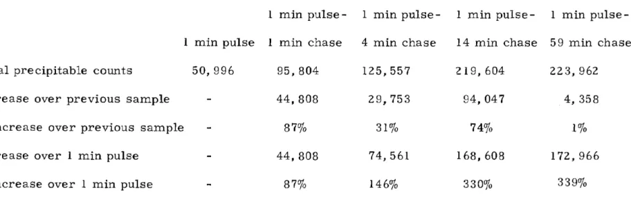
L Increase in total amount of acid precipitable radioactivity during chases of a 1 minute
pulse . . . 11 7 II Percentage of normal uptake remaining after
To my parents
18
INTRODUCTION
I. Discontinuous DNA synthesis
Discontinuous DNA synthesis in bacteria was first demonstrated in bacteria by Sakabe and Okazaki (1966), who showed that short
strands of DNA (about 1000 nucleotides long) are rapidly synthesized and then apparently converted to longer strands by a joining process. Sakabe and Okazaki suggested that these short strands could explain the apparent discrepancy between the fact that one strand of the DNA
helix must grow in the 3'---75' direction and the fact that known DNA
polymerases synthesize DNA only in the 5'~31 direction. They proposed that these pieces are made on at least one strand in the
5'~31 direction and then joined together in an overall sequence of
3'~5'.
A. Bacterial DNA synthesis 1. Okazaki fragments
Since then discontinuous DNA synthesis has been repeatedly demonstrated in bacteria (Okazaki et al., 19 68a, b; Yudelevich et al. ,
1968; Geider & Hoffman-Berling, 1971; Wang & Sternglanz, 1972; Dingman et al., 1974) and in phage (Sadowski et al., 1968; Polsinelli et al. J 1969; Sugino & Okazaki, 1972), and eviden ce was obtaine d showing
that all the Okazaki pieces are made in the 51----+3' direction (Sugino & Okazaki, 1972). Only Werner and coworkers disagree with this scheme,
19
claiming that Okazaki fragments are made through a process of random 11crystallization" of nucleotides on the parental template rather than sequentially in a 51~31 direction (Diaz et a1., 1975; Diaz & Werner, 1975). However, their arguments seem very unclear and it is not at all certain that their pulse times were short enough to insure that most of the nascent Okazaki pieces were not labeled along their whole length.
2. Enzymes needed for joining
Bacterial Okazaki fragments chase into bulk- sized DNA
(Sadowski et a1., 1968), and some work has been done on elucidating the enzymes involved. DNA ligase is considered to be necessary
for joining the fragments together (Hosoda & Mathews, 1968; Newman &
Hanawalt, 1968a, b; Okazaki et a1., 1968a; Sugimoto et a1., 1968; Nozawa &Nizuno, 1969; Pauling & Hamm, 1969; Olivera & Lundquist, 1971; Sugino & Okazaki, 1972; Gottesman et a1., 1973a, b). There have been reports that the peak of acid-precipitable radioactivity appearing near the top of the gradient when the DNA is sedimented through alkaline sucrose seems to slowly grow larger even when the ligase is supposedly absent or inactivated. However, in these experi-ments, there is probably some residual ligase acti vity or, in the case of phage, the bacterial ligase may be doing the joining (Newman &
Hanawalt, 1968; Sugimoto et a1., 1968; Pauling & Hamm, 1969; Olivera &
20
at low concentrations of deoxynucleotides (see below) do not require the NAD (nicotinamide-adenine dinucleotide)-dependent ligase to join into larger pieces (Hess et al., 1973), but there is probably some other ligase that joins these pieces.
DNA polymerase I is apparently also required for joining (Sadowski et al., 1968; Yudelevich et al., 1968; Kuempel & Veomet, 1970; Okazaki et al., 1971), and Konrad & Lehman (1974) have demon-strated that a necessary activity is that of the 5'~31 exonuclease associated with the polymerase. Pol A cells also seem to be able to slowly join Okazaki fragments into long DNA in vitro (Geider & Hoffman-Berling, 1971; Okazaki et al., 1973), but probably in such cases there is residual enzyme activity.
Recently a new mutant of E. coli has been obtained which makes fragments much smaller than normal-sized Okazaki fragments (Konrad &
Lehman, 1975). One implication of this sof (small Okazaki fragments) mutation is that the first intermediate in E. coli DNA synthesis may be smaller (4-5S) than originally detected (lOS), and the mutation may cause some ligase deficiency that makes these pieces observable.
B. Okazaki fragments in other organisms
When I first started this project, there were several researchers who did not find Okazaki fragments in higher organisms (Tsukada et al. , 1968; Lehmann & Ormerod, 1969, 1970; Habener et al., 1969b; Berger & Irvin, 1970; Hyodo et al., 1970; M. S. Horwitz, 1971). Now, however,
21
there is general agreement that Okazaki fragments are made by mammalian cells in vivo (Painter, 1968; Mueller, 1969; Painter &
Schaefer, 1969b; Schandl & Taylor, 1969, 1971; Ensminger & Tamm, 1970; Nuzzo et al., 1970; Sato et a1., 1970; Taylor et a1., 1970, 1973a; Hyodo et a1., 1971; Probst et a1., 1971; Cheevers et a1., 1972; Fox et a1. , 1973; Goldstein & Rutman, 1973; Berger & Huang, 1974; Friedman et a1. , 1975; Gautschi & Clarkson, 1975; Mendelsohn et a1., 1975; Tseng &
Goulian, 1975a) and in vitro (Kidwell & Mueller, 1969; Fox et al., 1973; Friedman, 1974; Hershey & Taylor, 1974; Tseng & Goulian, 1975a). They are also seen in other eukaryotes: sea urchins (Baker, 1971), Drosophila melanogaster (Kriegstein & Hogness, 1974) and Physarum polycepha1um (Waqar & Huberman, 1975a); in higher plants (Sakamaki et a1., 1975); and in viruses: CELO (Bellet & Younghusband, 1972), Adenovirus (Vlak et a1., 1975), Polyoma (Cheevers et a1., 1972; Magnusson, 1973; Pigiet et a1., 1973, 1974; Hunter & Francke, 1974; Francke & Hunter, 1974a, b; Otto & Reichard, 1975) and SV40 (Fareed &
Salzman, 1972; Fareed et a1., 1973; Salzman & Thoren, 1973; Salzman et al., 1973; Qasba, 1974a, b; DePamphilis & Berg, 1975). In this
thesis I present evidence confirming the existence of Okazaki fragments in mammalian cells.
C. Size of Okazaki fragments
The Okazaki fragments made by bacteria and phage are generally assumed to be between 1000 and 2000 nucleotides in length (7-118)
22
and there are some reports of much smaller fragments (Geider &
Hoffman- Berli ng, 1971; Wang & 8t ernglanz, 1972, 1974; Hess et a1. , 1973; Konrad & Lehman, 1975; Lark & Wechsler, 1975) that mayor may not be precursors to the 7-118 pieces.
For the Okazaki fragments of other organisms there have been a wide range of si ze s reported, ranging from 1000-2 000 nucl eoti de s (Kidwell & Mueller, 1969; Mueller, 1969; Painter & Schaefer, 1969; 8chandl & Taylor, 1969, 1971; Taylor et a1., 1970, 1973a; Baker,
1971; Bellett & Younghusband, 1972; Cheevers et a1., 1972; Goldstein &
Rutman, 1973; Friedman, 1974; Sakamaki et al., 1975; Vlak et a1., 1975), to 300 nucleotides (Nuzzo et a1., 1970) to 50-200 nucleotides (Hyodo et a1. , 1971; Fareed & Salzman & Thoren, 1973; Francke & Hunter, 1974a, b; Kriegstein & Hogness, 1974; Pigiet et a1., 1974; Qasba, 1974b; Friedman et a1., 1975; Gautschi & Clarkson, 1975; Mendelsohn 8t a1., 1975; Tseng &
Goulian, 1975a) to 10-20 nucleotides (Schandl & Taylor, 1969,1971;
Taylor et a1., 1970; Schandl, 1972). I n this thesis I present evidence that the Okazaki fragments of mammalian cells are about 100 nucleotides long, and I discuss why some researchers found other values.
D. Similarities between prokaryotic and eukaryotic fragments
1. Are the fragments single or double stranded?
Although there seems to be a large difference between the size of the Okazaki fragments in prokaryotes and eukaryotes, several of the other physical characteristics of Okazaki fragments appear similar.
23
The question of whether the fragments are single or double stranded before denaturation has not been decided in either case. Some report that they are mainly si ngle stranded after lysis (Okazaki et al., 19 68a; Painter & Schaefer, 1969; Sato et a1., 1970; Cheevers et a1., 1972; Mendelsohn et a1., 1975) while others say they are not (Tsukada et a1. , 1968; Yudel evich et a1., 1968; Fareed & Salzman, 1972; Gautschi &
Clarkson, 1975). Probably the fragments are observed as single or double stranded on sucrose gradients depending on the severity of the procedures used to lyse the cells, and these experiments shed little light on the question of what state the fragments are in inside the living ce 11s.
2. RNA primers
Evidence that Okazaki fragments are primed by stretches of RNA has been obtained in a wide variety of organisms. The existence of such RNA primers would resolve the difficulty presented by the fact that no known DNA polymerase can function without a primer. RNA
covalently attached to Okazaki fragments has been detected in E. coli (Sugino et a1., 1972; Hirose et al., 1973; Okazaki et a1., 1973, 1975; Sugino & Okazaki, 1973), colicinogenic Factor El (Blair et a1., 1972), phage (Brutlag et a1., 1971; Miller, 1972; Schekman et a1., 1972; Wickner et a1., 1972), Physarum (Waqar & Huberman, 1973, 1975a),
polyoma (Magnusson et a1., 1973; Sadoff & Cheevers, 1973; Hunter & Francke, 1974b; Pigiet et a1., 1974; Reichard et a1., 1974), SV40 (Qasba, 1974b),
24
and mammalian cells (Sato et al., 1972; Fox et al., 1973; Taylor et al. , 1973b; Neubart & Bases, 1974; Tseng & Gaulian, 1975b; Qaqar &
Huberman, 1975b). Evidence for RNA has been found either by CSzSO4 equilibrium density gradient centrifugation or by nearest neighbor
analysis. However, it has been shown that even non-covalently bound RNA can cause a density shift in CSZSO
4'
perhaps due to the formation of an association of RNA and DNA based on non-specific aggregation or limited reannealing after a denaturation step (Mendelsohn et al., 1975). A few groups do not find RNA, but speculate that their pulse times may have been too long to observe attached RNA or that RNA may have been removed during treatment of the DNA samples (Berger and Huang, 1974; Gautschi and Clarkson, 1975).3. Are both strands made discontinuously?
Having only one strand of the DNA helix made discontinuously is sufficient to resolve the problem posed by the inability of polymerases to synthesize DNA in the 3'~51 direction. Whether the other strand is also made discontinuously is still very much an open question. On the basis of the assumption that more than 50% of acid-precipitable radioactivity in small pieces after a short pulse-label indicates that both strands are being synthesized discontinuously, many groups have claimed to observe totally discontinuous synthesis (Okazaki et al. , 1968a, h, 1973; Sadowski et al., 1968; Yudelevich et al., 1968; Sugino & Okazaki, 1972; Fareed et al., 1973; Goldstein & Rutman, 1973;
25
Gautschi & Clarkson, 1975; Kurosawa & Okazaki, 1975; Mendelsohn et a1. , 1975; Tseng & Goulian, 1975a). Others observe less than 500/0 of the
radioactivity in the small pieces even after short pulses, and conclude that only one strand is made discontinuously (Painter & Schaefer, 1969; Hyodo et a1., 1970, 1971; Iyer and Lark, 1970; Eisenberg & Dernhardt, 1974; Francke & Hunter, 1974a; Friedman, 1974; Hershey & Taylor, 1974; Louarn & Bird, 1974).
Even when this question is examined in light of the annealing properties of the Okazaki fragments, contradictory conclusions arise. Examining either the ability of the fragments to self-anneal or the extent of their annealing to one or both of the parent DNA strands, some groups conclude that both sides of the helix replicate discontinu-ously (Tomizawa & Ogawa, 1968; Okazaki & Okazaki, 1969; Polsinelli et a1., 1969; Sugimoto et a1., 1969; Ginsberg & Hurwitz, 1970;
Fareed & Salzman, 1972; Sugino & Okazaki, 1972; Fareed et a1., 1973; Laipis & Levine, 1973; Pigiet et a1., 1973; Vlak et a1., 1975). However, since some of the bacteria and phage studied replicate their DNA bi-directionally, only the self-annealing experiments can be considered as strong evidence. Another group finds that even this crite'rion
indicates only semi-discontinuous synthesis (Francke & Hunter, 1974a; Francke & Vogt, 1975). As I will point Qut in the Discussion section, this is not an easy question to resolve. It is made even more difficult by the fact that the 5'----,)l3' strand may be replicated more or less
26
discontinuously depending on mutations (Lark, 1972; Louarn & Bird, 1974) or on other conditions affecting the rate of synthesis (Herrmann et al., 1972; Olivera & Bonhoeffer, 1972).
E. Eukaryotic Okazaki fragments seem to chase into large r DNA Proof that eukaryotic Okazaki fragments chase into larger DNA is not as easily obtainable as it is in the case of bacteria, both because of the relatively long time required for added nucleosides to equilibrate in the intracellular pool and because of the large amount of additional incorporation that usually occurs during in vivo chases. However, many groups have obtained evidence that eukaryotic
Okazaki fragments also chase into bulk DNA. Such evidence has been obtained both in in vivo studies (Painter & Schaefer, 1969; Schandl & Taylor, 1969; Baker, 1971; Cheevers et aL, 1972;
Fareed & Salzman, 1972; Laipis & Levine, 1973; Magnusson, 1973a; Salzman & Thoren, 1973; Salzman et al., 1973; Berger & Huang, 1974; Qasba, 1974b; Gautschi & Clarkson, 1975; Mendelsohn et al., 1975; Tseng & Goulian, 1975a; Vlak et al., 1975) and in more rigorous in vitro experiments (Kidwell & Mueller, 1969; Mueller, 1969; Magnusson, 1973; Magnusson et aL, 1973; Francke & Hunter, 1974a, b; Qasba,
1974a; Hunter & Francke, 1975; Otto & Reichard, 1975; Tseng &
Goulian, 1975a; Fraser, unpublished). F. Intermediate-sized DNA
27
bacteria and mammalian cells are similar, there are several differences. One difference is that nascent DNA in mammalian
cells is sometimes found both as Okazaki fragments and in a size class larger than Okazaki fragments but smaller than non-replicating bulk DNA. Several groups have reported a peak of nascent DNA
sedimenting at a position between the Okazaki and bulk peaks (Painter, 1968; Taylor & Miner, 1968; Habener et al., 1969b; Kidwell & Mueller, 1969; Mueller, 1969; Schandl & Taylor, 1969; Berger & Irvin, 1970; Hyodo et al., 1970, 1971; Lehmann & Ormerod, 1971; Chiu & Rauth, 1972, Gautschi 8t al., 1973; Goldstein & Rutman, 1973; Berger &
Huang, 1974; Kohn et al., 1974; Friedman et al., 1975; Rajalakshmi &
Sarma, 1975; Sakamaki et al., 1975). This peak will be referred to as an "intermediate peak!!. Other groups do not see a discrete peak of intermediate DNA, but do observe some DNA smeared across that region, even though they sometimes do not discuss it (Friedman &
Mueller, 1969; Nuzzo et al., 1970; Sato et al., 1970; Cheevers et al. , 1972; Friedman, 1974; Gautschi & Clarkson, 1975; Mendelsohn et al. , 1975). In spite of these reports the exact nature of what is going on is not clear. There are reports of no intermediate DNA being seen (Painter & Schaefer, 1969; Gautschi & Kern, 1973), of peaks being
reported and then disclaimed (Hyodo et al., 1970, 1971), of intermediate peaks being seen that are actually only single fractions in an alkaline sucrose gradient (Friedman et al., 1975), of graphs purporting to show
28
peaks that really don't exist (Fukiwara, 1972), of peaks that appear in vivo but not in vitro (Tseng & Goulian, 1975a), of intermediate peaks that appear only when the bulk peak doesn't (Ensminger & Tamm, 1970), and of peaks that are made up of small double-stranded pieces joining together while the single-stranded pieces move directly from the Okazaki peak to the bulk peak (Baker, 1971). In this thesis I will demonstrate that the intermediate peak is reproducible and provide an explanation for it.
II. Inhibitor s
In the past few years many studies have been done on the effect of inhibitors of protein or DNA synthesis on DNA replication. Such experiments are conducted with the hope of elucidating the exact process that is being affected by the inhibitors and thereby examining the
relationships among the processes that are involved in DNA synthesis. A. Inhibition of protein synthesis
1. Effect on DNA synthesis
In bacteria, protein synthesis is required for initiation of DNA replication but is not needed to support replication in progress (Lark, 1969; Brown et al., 1970). Yeast seem to be affected in the same way: Protein synthesis is needed for the initiation, but not continuation, of DNA synthesis (Hereford & Hartwell, 1973;
Williamson, 1973; Slater, 1974). In Physarum, protein synthesis seems to be required for the continuation of DNA synthesis as well
29
(Muldoon et al., 1971; Bersier & Braun, 1974), but it has been suggested that it is actually needed at only 10 discrete time points during S phase to allow the initiation of replicative units which begin at those 10 points (Muldoon et al.. 1971).
Protein synthesis is required during Gl for vertebrate cells to enter S phase (Taylor, 1965; Terasima & Yasukawa, 1966;
Schneiderman et al., 1971; Highfield & Dewey, 1972) and the inhibition of protein synthesis also inhibits DNA synthesis in progress in
ChIarella (Wanka & Moors, 1970), Tetrahymena (Gale et al., 1972), polyoma (Yu et al., 1975), chick red blood cells (Weintraub & Holtzer, 1972) and mammalian cells (Taylor, 1965; Young, 1966; Brega et al. , 1968; Mueller, 1969; Weiss, 1969; Brown et al.. 1970; Ensminger & Tamm, 1970; Chung & Coffee, 1971; Hand & Tamm, 1972, 1973; Gautschi & Kern, 1973; Hori and Lark, 1973; Gautschi, 1974).
In many instances, when protein synthesis is reduced by 950/0 or more, DNA synthesis is quickly reduced to less than 20% of the normal amount (Brega et al., 1968; Ensminger & Tamm, 1970; Gautschi & Kern, 1973; Gautschi, 1974), but in other cases it is reduced only to between 500/0 and 20% of the control (Ennis & Lubin,
1964; Young, 1966; Grollman, 1969; Chung & Coffee, 1969; Hyodo et al. , 197I; Seale & Simpson, 1975). Weintraub and Holtzer (1972) have
suggested that DNA synthesis seems to be immediately reduced by 50% and later und€rgoes further exponential decline.
30
2. Mechanism of action
The mechanisms of action of both the protein and DNA inhibitors have been examined at the molecular level. Cycloheximide apparently inhibits translocation of peptidyl-tRNA from the ribosome A site to the P site. It thus prevents the movement of ribosomes along mRNA but does not accelerate release of nascent polypeptide chains or cause polyribosome breakdown (Ennis & Lubin, 1964; Ensminger & Tamm, 1970; Gale et a!., 1972). This movement is mediated by transfer factor II, which may be the site of action of cycloheximide. It has been shown that the cause of the drop in DNA synthesis is not the inhibition of deoxynucleotide kinase or DNA polymerase (Taylor, 1965; Wanka & Moors, 1970). There is some speculation that cycloheximide's inhibition of DNA synthesis is due to its effect on histone synthesis (Weintraub, 1972), on a protein that controls the initiation of new rounds of genome replication (Yu et a1., 1975), or on a protein that acts on initiation of DNA replication at the membrane complex (Fugiwara, 1972).
Emetine resembles cycloheximide in its mode of action. It inhibits protein synthesis at the transfer level by inhibiting peptide chain elongation (Grollman, 1968; Gale et a1., 1972).
Puromycin is a structural analog of aminoacyl-adenosine, the 3' terminus of aminoacyl-tRNA. It can therefore substitute for the aminoacyl-tRNA bound to the ribosome A site by taking part in
31
the ribosome peptide bond-forming reaction, and can then accept the nas cent peptide chain. Since puromycin binds only weakly to ribosomes, the resultant peptide-puromycin molecule usually falls off the
ribosome almost at once (Nathans, 1967; Grollman, 1968; Ensminger & Tamm, 1970; Gale et al., 1972). As a secondary consequence of
releasing nascent peptides from ribosomes, puromycin causes
degradation of polyribosomes both in vivo and in vitro (GroUman, 1968; Gale et al., 1972).
B. Inhibitors of DNA synthesis
The inhibitors of DNA synthesis act much more directly on
steps involved in DNA replication. 5-fluorouracil deoxyriboside (FUdR) seems to act at the level of thymidylate synthetase, blocking the
conversion of dUMP to dTMP (Cleaver, 1969; Hand & Tamm, 1973; Manteuil & Girard, 1974). Apparently in vivo it is converted to 5-fluorodeoxy- UMP, which is a strong and specific inhibitor of
thymidylate synthetase (Gale et al. J 1972). It has been suggested
that a polymerase used to fill in the gaps between Okazaki fragments is preferentially inhibited by this reduced level of [dTTPJ .
Hydroxyurea has been reported to act by inhibiting ribonucleotide diphosphate reductase (Frenkel et al. J 1964; Neuhard, 1967; Bjursell &
Reichard, 1973; Hand & Tamm, 1973). This inhibition is not
32
but may be due to an inactivation of protein B2 of the reductase complex (Krakoff et al. I 1968), and may be based on the ability of hydroxyurea to chelate metal ions (Moore, 1969). There is one re-port that the inhibition caused by hydroxyurea is due to a lack of dGTP rather than a general lack of intranuclear dNTP pools (Skoog & Bjursell, 1974).
The mode of action of cytosine arabinoside (Ara C) has apparently been more difficult to determine. Some groups claim it blocks the conversion of CDP to dCDP (Evans et a1., 1964; Cleaver, 1969; Manteuil & Girard, 1974), while others say it has no effect on the ribonucleotide reductase activity (Moore & Cohen, 1967; Skoog &
Nordenskjold, 1971). Momparler (1969) claimed that Ara C acts by causing chain termination and is found mainly at the 31-hydroxyl terminus, while others say it can be incorporated internally into the DNA chain as a substitute for deoxycytidine and does not cause chain termination (Graham & Whitmore, 1970a, b; Hunter & Francke, 1975). It may be that the nucleotide added to a growing chain immediately after a molecule of Ara C is incorporated at a much slower rate, so when synthesis is stopped a disproportionately large percentage of the nascent chains have Ara C at the end position. This would give rise to the false assumption that Ara C causes chain termination.
33
c.
Effect on inhibitionBesides looking at the site of or the molecular bases for inhibition, many investigators have examined the apparent gross effect of
inhibitors of protein or DNA synthesis. They have focused primarily on the question of whether it is a form of DNA initiation or of DNA
chain elongation that is primarily responsible for the overall reduction in DNA synthesis. This question has been raised mainly in relation to inhibition of protein synthesis, but there are some reports that both FUdR and hydroxyurea markedly reduce the rate of DNA chain
elongation (Hand & Tamm, 1973) while 2-4 dinitrophenol (an uncoupler of oxidative phosphorylation) acts by reducing the number of operating replicons without affecting the average rate of chain elongation
Gautschi et al., 1973).
Most investigators report that cycloheximide does not inhibit the initiation of new replicons, but rather severely reduces the rate of chain elongation (Weintraub, 1972b; Weintraub & Holtzer, 1972; Gautschi & Kern, 1973; Gautschi et al., 1973; Hand & Tamm, 1973). Others report that the rate of elongation is not changed (Ensminger &
Tamm, 1970; Fujiwara, 1972).
When puromycin is used as the inhibitor of protein synthesis, the effect on DNA synthesis is much less clear. Some claim that, as with cycloheximide, the rate of chain elongation is significantly reduced without any change in the pattern of initiation (Gautschi, 1974). Others
34
say that the elongation rate is affected, but not enough to account for the drop in overall DNA synthesis (Hand & Tamm, 1972, 1973). Still other groups say that the rate of elongation is not reduced at all, so the inhibition of protein synthesis must be stopping initiation of new replicons (Ensminger & Tamm, 1970; Hori & Lark, 1973).
Although many of the above studies were carried out using
DNA autoradiography as the main investigative tool, sucrose gradient analysis has also begun to provide much useful information. The most spectacular effect of inhibition seems to be that the DNA inhibitors cause a bUild-up of small pieces that mayor may not be the same size as Okazaki fragments (Graham & Whitmore, 1970b; Laipis &
Levine, 1973; Magnusson, 1973; Magnusson et al., 1973; Salzman &
Thoren, 1973; Berger & Huang, 1974; Manteuil & Girard, 1974;
Hunter& Francke, 1975; Vlak et al., 1975). With inhibition of protein synthesis a build-up in the percentage of smaller-than-normal-sized pieces has also been reported (Gautschi & Kern, 1973; Weiner et al. , 1974). However, other reports indicate it is mainly the growth of intermediate-sized DNA into molecules of bulk length that is slowed down (Hyodo et al., 1971; Gautschi, 1974; Seale & Simpson, 1975) and one group even sees a reduction in the amount of small pieces in proportion to the other size classes (Fugiwara, 1972). In this thesis I will provide further insight into the mechanism of action of these inhibitors on DNA synthesis.
35
MATERIALS & METHODS
1. Materials
Chinese hamster ovary cells were obtained from David Baltimore. Joklik-modified minimum essential medium and non-essential amino acids came from Grand Island Biological Co. Fetal calf serum came from Microbiological Associates.
(14C ) thymidine (30 mCi/mmole), (3 H) thymidine (40-60 Ci/mmole), and (3 H ) deoxycytidine (30 Ci/ mmole) w'ere purchased from New England Nuclear. The unlabe led thymidine and deoxycytidine were purchased from Calbiochem.
Pronase was obtained from Calbiochem, proteinase K from EM Laboratories, Inc., NP-40 from Shell Chemicals and Angio-CONRAY (sodium iothalamate, 80% (w / v) solution) from Mallinckrodt Chemical Works.
Emetine, puromycin, cycloheximide, hydroxyurea, and cytosine arabinoside (Ara C) came from Sigma Chemical Co. FUdR came from Hoffman-La Roche.
32
The short ( P)-labeled DNA markers were generous gifts from Dr. Tom Maniatis. The P22 DNA marker was a gift from
Michael Mulholland and the phage A DNA marker was a gift from David Bottstein. The small (,...,..300 nucleotides) (14 C )-labeled DNA marker w'as made by labeling CRO cells overnight with (14 C ) thymidine, extracting and purifying the DNA, dissolving it in
sse
and sonicating it.36
II. Solutions
Buffer A (Gross-Bellard et a1., 1973): 10 mM tris-HeI, 10 mM EDTA, 10 roM NaCl, 0.50/0 sodium dodecylsulfate, pH 8.
sse:
0.15 M NaCl, 0.015 M Na Citrate.TD: 0.137 M NaCl, 0.005 M KCl, 0.007 M NaH
zP04, O. OZ5 M Tris, pH 7.4.
III. Methods
A. Growing the ce11s
eHO cells were maintained in spinner bottles and grown on plastic petri dishes (60 x 15mm, Falcon) in Joklik-modified MEM supplemented with non-essential amino acids and 7% fetal calf serum. Cells were grown in a 5% COz atmosphere at 37°C.
B. Pulse and pulse-chase labeling
For labeling of bulk DNA, (14 C ) thymidine was added overnight at O. OZ5 J.1Ci! m!. Pulse labeling was done by pouring off the medium in which the cells had been growing, mixing it 1: 1 with fresh medium, putting back a reduced volume (1. 5 ml) of this mixture and allowing the cells to grow for another 1/2 hour at either 37°C or room
temperature (22-26°C). At the end of this time a syringe was used to add (3 H ) thymidine at 100-300 fJ.eUml.
Chases were done by pouring off the label-containing medium, washing the cells twice in 5 ml volumes of the medium mixture which had been made O. 7 roM in cold thymidine and 10 fJ. M in deoxycytidine,
37
and then letting the cells continue to incubate in this medium for the indicated times.
Pulses or pulse-chases were terminated in anyone of several ways, depending on the subsequent treatment of the lysate (see below).
c.
InhibitorsIf pulse labeling was to be done in the presence of inhibitors of protein or DNA synthesis, the inhibitor was added to the conditioned medium
on the plate, and the cells were incubated for 90 minutes at 37DC.
The me dium was then poure d off and 1. 5 ml of a mixt ure of new and conditioned medium containing the same concentration of inhibitor was added. The plates were further incubated in a 50/0 CO
2 atmosphere at room temperature for 30 minutes and then pulsed.
D. Stopping the pulse and preparing the DNA for centrifugation Three main methods were used to lyse the cells and treat the lysate:
1) Total lysate method. Pulses were stopped by lJouring off the medium and pouring on 1 ml of either O. 2 N NaOH or a low con-centration (0. 25-1. 0%) of SDS or sarkosyl. In either case the solution was 10 roM in EDTA. If SDS or sarkosyl was used the lysate was made O. 2 N in NaOH. The lysate was then heated at 50DC for 45
minutes to an hour, cooled to room temperature, layered on gradients and analyzed by sucrose gradient centrifugation.
38
2) Proteinase K - chloroform - ethanol method. Pulse s were stopped by pouring off the medium and pouring on 3 ml of Buffer A containing 150/-Lg of proteinase K. Before being used, the proteinase K was autodigested overnight at 37°C in buffer A to inactivate the nucleases. After the lysate was incubated at 37°C for one hour, it was rolled for half an hour with an equal volume of chloroform containing 4% isoamyl alcohol. After centrifuging the resulting suspension the aqueous layer was removed. Two volumes of ice-cold ethanol were added, the mixture was gently shaken, stored at -20°C for half an hour, and centrifuged. The resulting pellet of DNA was washed three times in 70% ethanol and dissolved in O. 2 N NaOH. The solution was then heated at 50°C for 45 minutes to an hour, cooled to room temperature, layered on gradients and analyzed by sucrose gradient centrifugation.
3) Nuclear isolation method. Pulses were stopped by pouring off the medium and pouring on 3 ml of ice cold TD containing O. 65%
Nonidet P-40. The resulting nuclei were scraped off the dish with a rubber policeman and after 10 minutes were pelleted, washed once with TD, and suspended in 1 ml of TD which was then made
o.
2 N in NaOH. The solution was then heated for 45 minutes - one hour at 50°C, cooled to room temperature, layered on gradients and analyzed by sucrose gradient centrifugation.E. Sucrose gradient centrifugation
39
sucrose in Angio-CONRAY (Bottstein, 1968). For the SW27 rotor, gradients were 34 ml of 5 -2 0% sucrose solution over a 2 ml shelf; for the SW41, 10. 8 ml of solution over a O. 5 ml shelf, and for the
SW50. 1, 4.2 ml of solution over a O. 3 ml shelf. Gradients were O. 9 M in NaCl and 1 mM in EDT A. If alkaline gradients were required the pH was adjusted to 12.2 - 12.3 with NaOH. All the lysate from one plate was layered onto one gradient. Gradients were centrifuged under conditions described in the figure legends. Fractions were collected by punching a hole in the bottom of the tube. In most cases, fractions of O. 2 ml were collected directly on filter discs (Schleicher & Schuell, Inc.). The papers were dried, washed three times in cold 1 M HCI and twice in ethanol, dried, and counted in a liquid scintillation counter. When certain fractions were to be analyzed further, or when gradients were run in the SW27 rotor, fractions were collected in tubes and O. 1 ml aliquots were dried onto filter papers which were then processed as
de scribed above.
F. CsCl equilibrium centrifugation
CsCl was added to the sample, the solution was made O. 01 M in EDTA and the pH was adjusted to
>
12. 1. Final density was about 1. 730-1. 750. The solution (2. 5 ml for the SW50. 1 rotor and 5 ml for the 50 rotor) was put in a cellulose nitrate tube which was then filled to the top with mineral oil. Gradients were spun in an SW50. 1 rotor or a 50 rotor under conditions described in the figure legends.40
Gradients were collected and counted as discussed for sucrose gradients.
G. Polyacrylamide gel electrophoresis
All gels were made and run in the laboratory of Dr. Tom Maniatis. 50/0 gels were made by deionizing 100 ml of formamide with 5 grams of mixed bed ion exchange resin (20-50 mesh, Bio Rad AG 501-X8)
for 1 hour, filtering it through a Millipore filter and then mixing 50-60 ml of it with 3. 1875 grams of acrylamide, 0.5625 of bis -acrylamide, and O. 18 grams of tris. The pH was adjusted to 9. 0 with NaOH, the volume was brought up to 74 ml with formamide, the pH was adjusted again, and the solution was filtered through a Whatman #I filter. 150 p.g of TEMED (N, N, N', N'-tetramethyl ethylene diamine) was added and the solution was heated at 37°C. 100 mg of ammonium persulfate dissolved in 1 ml of water was added to the solution and stirring was begun
immediately. When the solution was thoroughly mixed, it was poured into a slab gel, 2mm thick x 160mm long.
The DNA pellet, which had been ethanol precipitated and washed several times with 700/0 ethanol, was dried, taken up in 50 Ml of
deionized formamide and layered on the gel. DNA pieces of known size were layered onto wells of the slab gel and the gel was run at 200 volts and 8 milliamps for about 10 hours at room temperature. The gel w'as sliced into 2mm slices and the fractions were put into a I ml 9: I NCS:water solution and heated at 37°C for 3 hours. After
41
cooling, 15 ml of toluene containing PPO and POPOP was added and the samples were counted.
H. Electron microscopy
Ele ctron microscopy was done by Howard J. Edenberg. Samples were taken from the alkaline solutions that had been heated for various times, and allowed to cool to room temperature. Within 20-30
minutes, 20 Ml samples (in 0.2 or 0.5 M NaOH) were diluted with a mix to give 100 Ml of solution containing 0.5 M ammonium acetate, O. 2 M tris (pH 8. 5) and 10 fJg of cytochrome C, and spread on a hypophase of O. 2 lVI ammonium acetate. The grids were stained with a fresh solution of 5 x 10⁻⁵ M uranyl acetate in 95% ethanol and rotary shadowed (10: 1) angle with 80% Pt/20% Pd. Photographs were taken at 10, OOOX in a JEOL microscope and printed at a 2X enlargement.
42
RESULTS
1. Artifacts
A. Aggregation and breakdown
Since there were so many discrepancies in the published reports and there was disagreement even as to the basic question of discontinuous synthesis, it w'as very important to choose experimental techniques
that would alter the DNA under observation as little as possible.
As examples of the difficulties encountered while using different DNA purification techniques, Figure 1 show's two different problems that arise when a cell lysate is sedimented through a neutral sucrose gradient and the peak fractions are then denatured and sedimented through alkaline sucrose gradients. Figures lA and 1C show the sedimentation profiles of DNA from eRG cells pulse labeled for short times at 37°C and run on neutral sucrose gradients. In both cases the pulse labeled and bulk labeled DNA sedimented together, and the peak fractions show'ed considerable DNA aggregation. (Because of this aggregation, the DNA peak position is not reproducible.) In Fig. 1B the aggregated DNA was sedimented through alkaline sucrose after being denatured by raising the pH to 13. The DNA was still clumped and the pulse labeled DNA ran together with the bulk DNA to the bottom of the gradient. In Fig. ID the DNA run through neutral sucrose was manipulated by hand and vortexed to break up the clumping before being centrifuged again. As a result, the bulk labeled DNA was
43
Figure 1. Profiles of DNA sedimented through neutral and alkaline sucrose gradients. CHO cells, prelabeled overnight with (14 C ) thymidine, were pulse labeled with (3 H ) thymidine, lysed and
centrifuged through a neutral sucrose gradient. The peak fractions were pooled, denatured with NaOH, and centrifuged through an alkaline sucrose gradient. In one case (D) an attempt was made to break up the aggregated DNA before it was sedimented through the alkaline sucrose (see text). (A) and (C) are the neutral sucrose
gradients, centrifuged in an SW27 rotor at 20, 000 rpm for 80 minutes at 10°C. (B) and (D) are the alkaline gradients, centrifuged in an SW27 rotor at 22, 500 rpm for 14 hours at 10°C.
(A) 75 second pulse, neutral sucrose gradient. (B) 75 second pulse, alkaline sucrose gradient. (C) 5 minute pulse, neutral sucrose gradient. (D) 5 minute pulse, alkaline sucrose gradient.
44
-A
C
r:-I
540
I r-lI-
•
18 8 15 50/-
12 6 \ 9 4-
•
6---
40 10 i•
I - N3~.
28l
P
2
I 0 ~4 -
A
\
I I I ... I ( ) ) ( ~ 0, . 0 O-~ 0 0"'Ua..
•
8
0
s:
-
U I: 4-
x "., 0 I I'\) ... 0 3 - 3 ""-6 0.6•
2\
2 4-
•
\ -.,
2• •
'.'\..
,
...
-.
.
I I I I I ;a- D 6 /2 18 24 30° O· 12 18 24 30°fractions.
45
severely broken dovvn and no valid comparisons could be made between it and the pulse labeled DNA.
B. Determination of acid-precipitable radioactivity
1. Leakage through filters
The above are examples of artifacts which cause DNA to sediment either more rapidly or more slowly than it should on the basis of its molecular weight. Serious problems also arise with techniques of analysis that cause a peak of radioactivity to appear where there is actually either no more than a background level of DNA or no DNA at all. An example of this is shown in Figure 2. A
14
culture of eRG cells, labeled overnight with C thymidine, was lysed with 1% sarkosyl, denatured, and sedimented through an
alkaline sucrose gradient. The gradient was then collected in 1. 5 ml 3
An equal amount of ( H) -lambda DNA was added to each fraction and each fraction was thoroughly mixed. O. 1 ml aliquots from each fraction were put on filter papers which were then dried, washed with 1 N Hel and then ethanol and dried again. Then 200 /-lg of bovine serum albumen (ESA) were added to each tube as a carrier. The tubes were mixed, made 5% in TCA, mixed again and allowed to settle for 10 minutes at O°C. The contents of each tube were then poured through a glass fiber filter which was then washed with TeA and ethanol and allowed to dry. Both sets of filters were then counted. The filters
-0-0- ,
Figure 2. Profiles of sucrose gradients analyzed in different ways. Cells prelabeled overnight with (14 C ) thymidine were lysed with 1% sarkosyl (10 mM in EDTA), denatured and centrifuged through an alkaline sucrose gradient in an SW27 rotor at 20, 000 rpm for 16 hours at 10°C. The gradient was collected and (3 H )-labeled phageA DNA was added to each fraction in equal amounts.
(A) O. 1 ml aliquots from each fraction were put on filter papers, which were then processed
as described in the text.
(B) 100 pg of BSA were added to the remaining contents of each tube and the fractions were
processed by filtration as described in the text.
3 14
H: - 0 - 0 - , C.
foi::>.
; ) )
•
,
) ) )200
A.
Analysis by Washing1000
2000
B.
Analysisby
Filtration3000
W
i'-...
...,
150I
\/ \
•
i \/.\/. .,
1\.... •
. . . . .' \ I.
~
Q
IT
1050
3H
CPM
500
50
25
14CCPM
15°°1
lOaD'T
....
\
•
\
•
/
.\,.
.
/
.
...
,
.
\
iV
V
•
·~·V
2000
1000
100
50
14CCPM
5
10
15
20
25
5
10
15
20
25
Fraction number from bottom
hI'::..
48
the same amount of tritium-labeled DNA in each fraction, with the downward slope probably due to quenching caused by the cell debris found near the top of the gradient.
However, the filters through which the fractions had been
poured (Fig.
Za
show a sharp peak near the top of the gradient. Since these filters should have 10-15 times as many counts as the onescontaining aliquots, it can be assumed that across a large part of the gra.dient most of the DNA waS pulled through the filters, but at the top the cell debris trapped the DNA onto the filters.
2. Adsorption of nucleosides or nucleotides to cellular components Finally a method was chosen which appeared at first to give reliable results. This is the method of Nuzzo et a1. (1970) in which the cells are lysed with O. 2M NaOH containing 10 mM EDTA.
The lysate is heated at 50°C for 30-45 minutes in order to
thoroughly denature the DNA, gently layered onto an alkaline sucrose gradient and centrifuged under the conditions described in the figure legends. Figure 3 shows the sedimentation profile of acid-precipitable radioactivity from cells pulse labeled for short times at 37°C as well as cells in which the pulse was chased for periods up to one hour. After the shortest pulse (Fig. 3A) all the nascent DNA seems to be in the form of very short strands comparable to the Okazaki fragments of bacteria. As the pulse time increases (Figs. 3B, C, D) label gradually appears in longer strands so that a peak is formed in the
49
Figure 3. Sucrose gradient profiles of pulse labeled DNA prepared
by NaOH lysis. Cells, prelabeled overnight with (14C) thymidine, were pulse labeled for varying lengths of time with (3 H) thymidine at 37°C and lysed with 0.2 M NaOH containing 10 mM EDTA (see Methods, D). The lysate was heated at 50 DC for 30-45 minutes and then centrifuged through an alkaline sucrose gradient in an SW27 rotor at 25, 000 rpm for 16 hours at 0 DC. Chases were done in the presence of nonradioactive thymidine.
50
-A
15 seconds 6 12•
t
1\
20
,"""~
i \
4
8I
I - 10 2 .4
",j.J"'" ••
!r"·~~
I
•
Io
ID
2 minutes 16•
8-
•
•
/'...w'.
cf
-
~ t"~'
i \
C\I,
"
12 - (')b
••
•
I •~
/
\
""U :3 I 10 ~><
~. ~/
.
8 -
I•
4
x\
q
a..
•
,
.
-
U5
I•
•
4
~...
,
2 -I•
0 rt) ~ 0 0o·
E
F
1 minute pulse 59 minute chase~
83
151.5
J
6 2 \0l.o~
•
\
\•
~/
\•
•
\
5
05•
2
\
'-i 0-51
middle of the gradient. After a 30 second pulse the peak of putative short strands is still clearly visible, while after 1 minute it is no longer distinguishable as a separate peak and after 2 minutes it is
completely obscured by a peak of larger strands. (This latter peak will be referred to as the intermediate peak.) A s the pulse is chased
with medium containing an excess of cold thymidine the intermediate peak moves down the gradient (Fig. 3E) and after 1 hour the pulse-labeled DNA sediments almost as rapidly as the bulk DNA (Fig. 3F).
These results led to the assumption that a pattern of discontinuous DNA synthesis in mammalian cells had been confirmed (Huberman & Horwitz, 1973). However, it turned out that the acid-precipitable radioactivity in the" Okazaki peak" did not band at the density of DNA in cesium chloride gradients (Fig. 4). CHO cells were pulse-labeled for 2 minutes at 25°C and sedimented through an alkaline sucrose gradient (Fig. 4A). The peak fractions (#24-25) were then centrifuged to equilibrium in a CsCI buoyant density gradient. More than 750/0 of the acid-precipitable counts went to the top of the gradient (Fig. 4B). This suggests that the radioactivity in the Okazaki peak does not represent short DNA fragments, but rather may simply be a small fraction of the cells' (3 H ) thymidine pool adhering to some cellular component that floats in
CsC1-Further confirmation of this hypothesis was obtained by running controllysates containing CHO cells lysed before the addition of
Figure 4. Sucrose and CsCl gradient profiles of DNA prepared by total lysate method. Cells were prelabeled overnight with (14 C ) thymidine and pulse labeled for 2 minutes with (3 H )_ thymidine at 25 °C. The total cell lysate was centrifuged on an alkaline sucrose gradient
(A) in an SW27 rotor spun at 25,000 rpm for 16 hours at O°C. The peak fractions (#24-25)
were centrifuged to equilibrium in an alkaline CsCl gradient (B) to which short ( 300 nucleotides) pieces of (14 C )-labeled DNA had been added as a marker. The gradient was spun in an SW50. 1 rotor at 31, 000 rpm for 48 hours at 20°C.
c.n
') l ")
..
) ) -) ) ) ) l280
A
I
200L
B
•
-
•
I
I
-120
•
-C\I I~ 24~~
-PI ~.'.
12 ()
-
80
120
( ) ) ( ~ ( )!
~;ff\
I
\
10 -c
~60
100
()
~a...
8 )(
u
40
-0•
80
~ - I6
'i ",
20
60
,...
0-4
040
-2
20
4
8
12
16
20 24
28
4
8
12
16
Fraction number from bottom
c.n
54
radioactivity. Figure 5 shows the results of such an experiment. In one case (Fig. 5A) CHO cells were pulse-labeled with (3 H ) thymidine for 15 seconds at 37°C. For the controls, cells were first lysed by the addition of O. 2 M NaOH and then (3 H) thymidine was added to one lysate (Fig. 5B) and (32p ) TTP to the other lysate (Fig. 5C). All three lysates were sedimented through sucrose gradients and all the sedimentation profiles look the same, even though neither of the controls (Fig. 5B,C) could have incorporated the radioactive label into DNA. Thus it appeared that either thymidine or TTP was adsorbing to some cellular component that moved only slowly into the sucrose gradient. A similar artifact appears if the cells are lysed with
o.
50/0 SDS instead of NaOH. Figure 6 shows that the" Okazaki peak" observed under such conditions floats at the top of a CsCI gradient even if the gradient's density is adjusted in such a way that the DNA marker sediments to the bottom.It should be noted that although the" total lysate" method of analysis described above give s an artifactual "Okazaki peak," its intermediate
peak is partially real, as demonstrated in Figure 7. Cells were pulse labeled for 5 minutes, lysed with NaOH and sedimented through an alkaline sucrose gradient (Fig. 7A). Pooled fractions from the intermediate peak of 7A were then centrifuged to equilibrium in a CsCI buoyant density gradient (Fig. 7B). Although some of the radioactivity bands at the density of DNA, almost one-half of the
Figure 5. Sucrose gradient profiles of DNA prepared by NaOH lysis, when radioactivity was added before or after lysis. Cells were lysed with O. 2 M NaOH, heated and centrifuged on an
alkaline sucrose gradient in an SW50. 1 rctor spun at 45, 000 rpm for 19.5 hours at 20
°e.
(Note that under these conditions the Okazaki peak should sediment near the middle of the gradient. )(A) 3(H) thymidine was added to the cells for 15 seconds at 37°C before lysis. (B) (3H ) thymidine (200 ,uCi) was added after lysis with NaOH.
(C) (32 p ) TTP (50 /-lei) was added after lysis with NaOH.
-(t-(J-ct-, (3 H ) - total counts; -e-e-e-, (3 H ) - acid precipitable counts; - .6. -.6. -.6. _. (32 p ) - total
counts; -A-A-A-. (32 p ) - acid precipitable counts.
CJl CJl
l ) ~ l } ) l
•
42f--A
I
420B
lP.~.
I
I
C
20 / -120 36 •.• ("'\ 360 /.\. ;" UJ " U J . . . \ Ul~
N~
/\!
16 I~
j
gO" 16 I .:: lJ 30 • ..~ () 300I'
()
15 0 ,..., 1 " " T ' 1r'1 " " . I V I • l J 'it
lJ o ~o. 0 ~ x 24j
I \
12::. -:: 240/1 \
12~
-:: 60 • 12 "~
I \
O~
l · -~
(l. • J..(l. • . , , " . 0 (l. 0 <..> 18 /I
\,0
8 -; <..> 1801\
1/
\
8 : u 45l
....
j 9 r0 I · " - I () \ (l.!
...
,..., 12·"I /
r'1 1 2 0 ..1
.,
.!.
~ 30 6 ..I \ ...
l
· \
I / · w
I
, . \ / \ <) 4 I" 4 6f'\.JV..
!
60I \
'/
15,1
--13 ,a' ·...".1
I 0'<)'<) J. I .().CI·o
L(I-<Jo<I-()-~()o ct" 0 0 8:IJ.o<>.a-.().Q-c.-<t I I I 10 011IIJ6A la'-~ I I 106 12 18 24 6 12 18 24 30 6 12 18 24
Fraction number from bottom
(Jl 0 '
Figure 6. Sucrose and CsCl gradient profiles of DNA prepared by lysis with SDS. Cells were pulse labeled with (3 H ) thymidine for 20 seconds at 37°C and lysed with
o.
50/0 SDS. After adding NaOH to a final concentration of O. 2 M and heating the lysate at 50°C for 45 minutes, the sample was centrifuged through an alkaline sucrose gradient.(A) in an SW50. 1 rotor at 45,000 rpm for 10 hours at 20°C. The peak fractions (#17-22) were collected and centrifuged to equilibrium in an alkaline CsCI gradient.
(B) The gradient was spun in an SW50. 1 rotor at 30, 000 rpm for 48 hours at 20°C, and 14
( C) labeled bulk DNA was added as marker.
CJ1 -:r
58 14C