HAL Id: hal-02130026
https://hal.archives-ouvertes.fr/hal-02130026
Submitted on 10 Feb 2021
HAL is a multi-disciplinary open access
archive for the deposit and dissemination of
sci-entific research documents, whether they are
pub-lished or not. The documents may come from
teaching and research institutions in France or
abroad, or from public or private research centers.
L’archive ouverte pluridisciplinaire HAL, est
destinée au dépôt et à la diffusion de documents
scientifiques de niveau recherche, publiés ou non,
émanant des établissements d’enseignement et de
recherche français ou étrangers, des laboratoires
publics ou privés.
Nanoparticle through Silane Chemistry for Simultaneous
MRI/ 64 Cu PET Imaging
Vu-Long Tran, Vivek Thakare, Marco Natuzzi, Mathieu Moreau, Alexandra
Oudot, Jean-Marc Vrigneaud, Alan Courteau, Cédric Louis, Stéphane Roux,
Frederic Boschetti, et al.
To cite this version:
Vu-Long Tran, Vivek Thakare, Marco Natuzzi, Mathieu Moreau, Alexandra Oudot, et al..
Func-tionalization of Gadolinium Chelates Silica Nanoparticle through Silane Chemistry for Simultaneous
MRI/ 64 Cu PET Imaging. Contrast Media and Molecular Imaging, Wiley, 2018, 2018, pp.1-10.
�10.1155/2018/7938267�. �hal-02130026�
Research Article
Functionalization of Gadolinium Chelates Silica
Nanoparticle through Silane Chemistry for Simultaneous
MRI/
64
Cu PET Imaging
Vu-Long Tran,
1,2Vivek Thakare,
3,4Marco Natuzzi,
1Mathieu Moreau,
4Alexandra Oudot,
5Jean-Marc Vrigneaud,
5Alan Courteau,
5C´edric Louis,
2St´ephane Roux,
6Fr´ed´eric Boschetti,
3Franck Denat,
4Olivier Tillement,
1,7and François Lux
1,71Univ Lyon, Universit´e Claude Bernard Lyon 1, CNRS, Institut Lumi`ere Mati`ere, F-69622 Lyon, France 2Nano-H S.A.S., 2 Place de l’Europe, 38070 Saint Quentin Fallavier, France
3CheMatech, 2 rue Pauline Kergomard, Dijon, France
4Institut de Chimie Mol´eculaire de l’Universit´e de Bourgogne (ICMUB), UMR CNRS 6302,
Universit´e de Bourgogne Franche-Comt´e, 9 Avenue Alain Savary, 21078 Dijon, France
5Plateforme d’Imagerie et Radioth´erapie Pr´ecliniques, M´edecine Nucl´eaire,
Georges-Francois LECLERC Cancer Center-UNICANCER, 21000 Dijon, France
6Institut UTINAM, Universit´e de Bourgogne Franche-Comt´e, CNRS UMR 6213, 16 Route de Gray, 25030 Besançon, France 7NH TherAguix SAS, F69100 Villeurbanne, France
Correspondence should be addressed to François Lux; [email protected]
Received 30 April 2018; Revised 6 August 2018; Accepted 18 August 2018; Published 1 November 2018 Academic Editor: Mar´ıa L. Garc´ıa-Mart´ın
Copyright © 2018 Vu-Long Tran et al. This is an open access article distributed under the Creative Commons Attribution License, which permits unrestricted use, distribution, and reproduction in any medium, provided the original work is properly cited. Multimodal nanoprobes are highly demanded for biomedical imaging applications to enhance the reliability of the diagnostic results. Among different types of nano-objects, ultrasmall silica gadolinium nanoparticle (SiGdNP) appears as a safe, effective, and versatile platform for this purpose. In this study, a new method to functionalize SiGdNP based on silane chemistry has been reported. Two types of chelating silanes (APTES-DOTAGA and APTES-NODAGA) have been synthesized and grafted on SiGdNP by a simple one-step protocol. This functionalization strategy requires no other reactants or catalyzers and does not compromise the ultrasmall size of the particles. NODAGA-functionalized particle has been labeled with64Cu isotope and
injected intravenously to mice bearing TS/A carcinoma tumor for biodistribution study to demonstrate its potential as a bimodal MRI/PET imaging agent. A fully integrated MRI/PET system was used to simultaneously monitor the distribution of the particle. The results showed that the functionalized particle maintained properties of a renal clearable NP which could rapidly escape through kidneys and had low retention in other organs, especially liver, even though its accumulation in the tumor was modest.
1. Introduction
Biomedical imaging is composed of different sophisticated techniques ranging from ultrasonography (US), X-ray computed tomography (CT), magnetic resonance imaging (MRI), nuclear imaging, and optical imaging (OI). However, each of them has different drawbacks alongside with ad-vantages. Among them, MRI is gaining more and more popularity in hospitals for precise imaging thanks to high
spatial resolution and nonionizing nature of the radiation. Nevertheless, MRI is plagued by its low sensitivity and therefore, not an ideal technique for quantification of contrast agents. On the other hand, nuclear imaging, more specifically, single photon emission computed tomography (SPECT) or positron emission tomography (PET), offers precise quantification without being limited by penetrability issue as OI. Hence, more and more researches have been dedicated to the combination of MRI contrast agents and Volume 2018, Article ID 7938267, 10 pages
radioisotopes in one single object to correlate the images obtained by MRI and the ones acquired by nuclear imaging [1]. In this context, a bimodal probe which allows the lo-calization of disease sites by both techniques should be highly desirable. Our group has developed an ultrasmall silica gadolinium nanoparticle (SiGdNP) as a probe for multimodal imaging and radiosensitization [2]. It is made of a polysiloxane core and displays several macrocyclic chelators DOTA (1,4,7,10-tetraazacyclododecane-1,4,7,10-tetraacetic acid) on the surface. Most of them complexed gadolinium ions (Gd3+) and acted as positive contrast agent for MRI. This type of nanoparticle (NP) was made from biocompatible materials. Moreover, it displays an ultrasmall hydrodynamic diameter (<5 nm) and is covered by both positively (amines) and negatively charged (DOTA chelates of Gd) groups. The latter helps distribute the opposite charges evenly and further minimize protein adsorption [3]. These parameters allow it to be quickly eliminated through kidneys and prevent the deposit of chelates in organs like liver, spleen, lung, or bone marrow. This characteristic combined with the high affinity of DOTA for gadolinium helps in avoiding possible release of toxic Gd ions [4–6]. Meanwhile, different radioisotopes, e.g., 111In, 68Ga, and 89Zr, used for nuclear imaging can be complexed to free
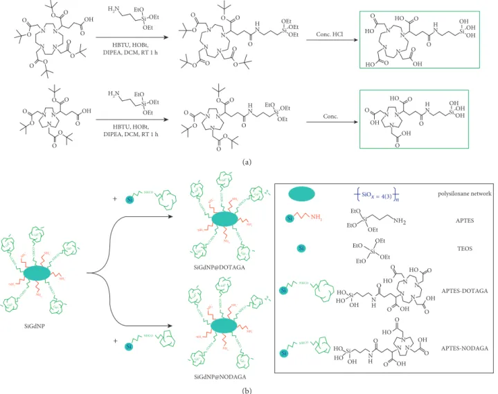
chelators that existed from the beginning or being grafted postsynthetically [7–9]. Common methods for grafting free chelators on SiGdNP were to use NHS esters [8, 10] or isothiocyanate derivatives [9] of chelators to react with amine groups on the surface of the particles [9]. The ma-terials required for these strategies have been well developed and commercialized. Moreover, these reactions are quite fast, straightforward, and well known. Nevertheless, there are still some limitations for these strategies. First of all, NHS esters and isothiocyanates are prone to be deactivated by hydrolysis. Hence, they cannot be stored for a long period of time even at low temperature in the desiccated condi-tion. Second, these activated species can cross-react with amines present in targeting ligands that we want to func-tionalize, for example, ε-amine of lysine of a peptide. This implies complicated protecting/unprotecting strategies such as using tert-butyloxycarbonyl (Boc) or 9H-fluoren-9-ylmethoxycarbonyl (Fmoc) protecting groups [11]. In this study, a new strategy based on silane chemistry was pro-posed. The macrocyclic chelators, i.e., DOTAGA (1,4,7,10-tetraazacyclododecane-1-glutaric acid-4,7,10-triacetic acid) and NODAGA (1,4,7-triazacyclononane, 1-glutaric acid-4,7-diacetic acid) were coupled with APTES (aminopropyl triethoxysilane) (Figure 1(a)). These species were then used to functionalize SiGdNP by reacting specifically with available silanol groups, Si-OH, on the surface of the particle in a single step without the presence of any other reactants (Figure 1(b)). This strategy has been used by Ciccione et al. to successfully graft precisely certain ratios of fluorophores and different targeting peptides on a 80 nm silica nano-particle to actively increase the internalization of nano-particles in cancer cells [12]. The activities of silanes are not affected by hydrolysis. Hence, it can be stored and used for a long period of time. In addition, the unreacted silanes are highly solu-ble so that they should be removed easily by filtration.
The functionalized particles were characterized with dif-ferent analytical methods to prove the presence of free chelators on the particles. By increasing the quantity of silane precursors, the amount of free chelators can be in-creased proportionally. Finally, a simultaneous bimodal imaging experiment using a fully integrated MRI/PET sys-tem was performed on murine tumor model to demonstrate the potential of this type of NP. Radioisotope of copper,
64Cu, was chosen as positron emitter for PET modality. This
isotope has a reasonable half-life (12.7 h) for pharmacoki-netic study of SiGdNP which has a 1 h half-life in patients’ blood (phase I trial NANO-RAD; ClinicalTrials.gov Iden-tifier: NCT02820454). In addition, the energy of positron emission from 64Cu is relatively low (656 keV), and the lowest-energy positrons from a commonly used radiometal. This allows obtaining images with resolution as high as the ones obtained with clinically prevalent18F isotope (640 keV) [13]. Regarding the choice of chelator, although the ther-modynamic stability constant for Cu complex of NOTA (original structure of NODAGA) is comparable to the one of DOTA (logKNOTA�21.6 vs. logKDOTA�22.2), [14] different
studies have shown higher in vivo stability of Cu complexes of NOTA, NODAGA, and their derivatives compared to the ones of DOTAGA and their derivatives [15–18]. Therefore, NODAGA functionalized particle was chosen to be radio-labeled with 64Cu for the biodistribution study.
2. Results and Discussion
2.1. Functionalization of NPs through Silane Chemistry.
Two types of chelating silanes, i.e., APTES-DOTAGA and APTES-NODAGA, were synthesized from the reaction be-tween APTES and either t-butyl protected DOTAGA ((t-Bu)4DOTAGA) or t-butyl protected NODAGA ((t-Bu)3
NO-DAGA) (Figure 1(a)). The peptide bond was formed by using HBTU (2-(1H-benzotriazol-1-yl)-1,1,3,3-tetramethyluronium hexafluorophosphate) as coupling agent. After purification, APTES-(t-Bu)4DOTAGA and APTES-(t-Bu)3NODAGA, were
deprotected by concentrated hydrochloric acid to obtain final products. The excess of acid was removed by evaporation. Solutions were lyophilized for storage.
Then, each type of silanes was mixed with SiGdNP in molar ratio Gd : chelator � 10 : 1.5. Due to the steric hin-drance of the macrocyclic chelators, the silanes, i.e., APTES-DOTAGA and APTES-NODAGA, are not likely to form stable particles. However, when being mixed with already-synthesized particles, they can form covalent siloxane bonds with free silanol groups or exchange with the already-existing silanized chelators on the surface of the particles. The remaining free silanes can be easily washed out by tangential filtration. The resulting NPs were referred to as SiGdNP @ D-1 (for DOTAGA functionalized particle) and SiGdNP @ N-1 (for NODAGA functionalized particle), re-spectively. Furthermore, lower temperature (40°C instead of
80°C) and lower total silanized chelator concentration
in-cluding the chelators on the surface of the particles and added free chelators (ca. 58 mM instead of ca. 115 mM) were applied to see the effects on the homogeneity of the final NPs. These particles were referred to as SiGdNP @ D-2 and
SiGdNP @ N-2. A series of samples with increasing amount of APTES-DOTAGA, i.e., Gd : chelator 10 :1.5, 10 : 2, and 10 : 4, was prepared to test the possibility of tailoring the amount of free chelators at the surface of the NP. These particles were referred to as SiGdNP @ D-2, 3, 4 accordingly. The experimental conditions of these particles were sum-marized in Table S1. Tables 1 and S2 show different char-acterization results of the NP before and after functionalization by chelating silanes at 80°C. DH of the
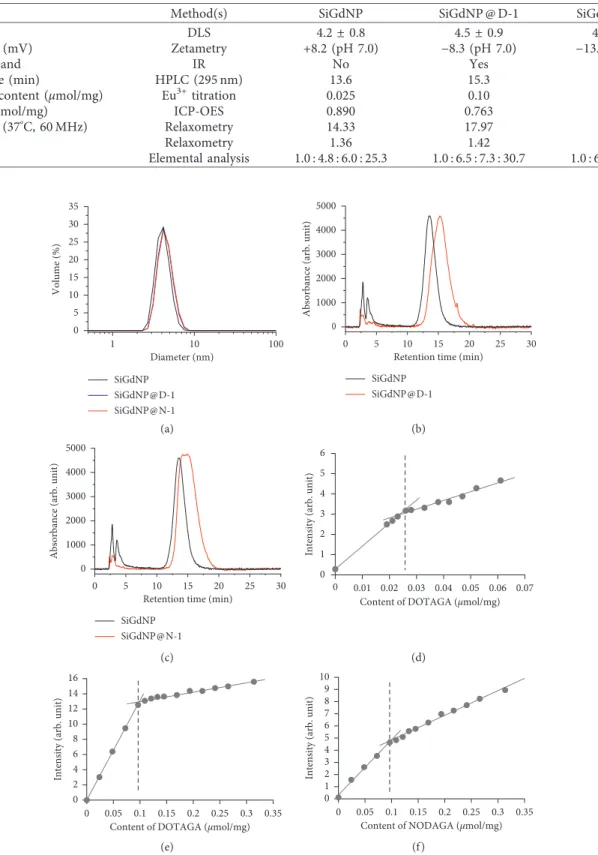
particles which only slight increase from 4.2 ± 0.8 nm for SiGdNP to 4.5± 0.9 nm for SiGdNP @ D-1 and SiGdNP @ N-1 indicated that the size was maintained ultrasmall (Figure 2(a)). On the other hand, other characteristics proved the presence of free chelators on the particles after the reaction. First of all, the zeta potentials at pH 7.0 de-creased from +8.2 mV for SiGdNP to−8.3 mV for SiGdNP @ D-1 or −13.7 mV for SiGdNP @ N-1. Vibration band at 1730 cm−1in IR spectra, corresponding to C O stretching vibration band of free carboxylic acid, was detected (Figure S5). The retention time (tR) in chromatograms
in-creased from 13.6 min for SiGdNP to 15.3 min for SiGdNP @
D-1 or 14.7 min for SiGdNP @ N-1 (Figures 2(b) and 2(c)). The content of free chelators quantified by Eu titration increased from 0.025 µmol/mg for SiGdNP to 0.10 µmol/mg for SiGdNP @ D-1 and SiGdNP @ N-1 (Figures 2(d)–2(f)). The principle of this titration method was described in details in a precedent paper. Briefly, a series of samples containing a fixed amount of NPs and an increasing amount of EuCl3 was prepared. Eu3+has two specific
phosphores-cence emission peaks at 594 nm and 616 nm when being excited at 395 nm [19, 20].
When Eu3+complexed with DOTAGA, it will be shielded
from the quenching effect of O-H oscillator of water and emits much stronger luminescence signals. When the amount of Eu3+
increases, the intensity increases linearly until no DOTAGA is available for chelation. Then, the intensity will increase more slowly due only to the phosphorescence of free Eu3+[8]. The
content of Gd measured by ICP-OES decreased from 0.89 µmol/mg for SiGdNP to 0.76 µmol/mg for SiGdNP @ D-1 and SiGdNP @ N-1. The longitudinal relaxivity (r1)
in-creased from 14.3 mM−1·s−1for SiGdNP to 18.0 mM−1·s−1for
SiGdNP @ D-1 or 18.2 mM−1·s−1 for SiGdNP @ N-1 at 37°C Conc. HCl HBTU, HOBt, DIPEA, DCM, RT 1 h N N O O O O O O N O O OH O N EtO OEt OEt Si H2N OEt OEt OEt Si O N O O O O N N N O O O N H O O HO O O Si OHOH OH N H N O HO N N N O HO OH O O N N O OH O O N O O EtO OEt OEt Si O O O O O O O N N N N H HBTU, HOBt, DIPEA, DCM, RT 1 h EtO OEt OEt Si H2N Conc. Si OH OH OH N H O HO O N N N OH O O OH (a) + + NHCO NHCO SiOx = 4(3) NH2 OEt OEt OEt O O O O O O N N N H O N H OH HO HOSi HO OH APTES-DOTAGA SiGdNP@DOTAGA SiGdNP@NODAGA SiGdNP APTES-NODAGA TEOS APTES polysiloxane network OH N HO HO OH HOSi O OH O OH HO N N N N EtO EtO EtO EtO Si Si Gd3+ Gd3+ NHCO NHCO NHCO NHCO Gd3+ Gd3+ Gd3+ Gd3+ Gd3+ Gd3+ Gd3+ Gd3+ Gd3+ Gd3+ Gd3+ Gd3+ NH3+ NH3+ NH 3+ +NH3 NH+3 NH3+ NH3+ NH2 NH3+ +NH3 NH+3 NH3+ NH3+ NH3+ +NH3 NH+3 NHCO NHCO NHCO NHCO NHCO NHCO NHCO NHCO NHCO NHCO NHCO NHCO NHCO n Si Si Si Si Si Si 2 2 (b)
Figure1: (a) The reaction scheme of the synthesis of APTES-DOTAGA (upper) and APTES-NODAGA (lower); (b) the reaction scheme of the functionalization of APTES-DOTAGA and APTES-NODAG on SiGdNP.
Table1: Summary of characterizations of NPs before and after functionalization with chelating silanes.
Properties Method(s) SiGdNP SiGdNP @ D-1 SiGdNP @ N-1
DH(nm) DLS 4.2± 0.8 4.5± 0.9 4.5± 0.9
Zeta potential (mV) Zetametry +8.2 (pH 7.0) −8.3 (pH 7.0) −13.7 (pH 7.0)
Free COOH band IR No Yes Yes
Retention time (min) HPLC (295 nm) 13.6 15.3 14.7
Free chelator content (µmol/mg) Eu3+titration 0.025 0.10 0.10
Gd content (µmol/mg) ICP-OES 0.890 0.763 0.763
r1(mM−1·s−1) (37°C, 60 MHz) Relaxometry 14.33 17.97 18.17 r2/r1 Relaxometry 1.36 1.42 1.42 Gd : Si : N : C Elemental analysis 1.0 : 4.8 : 6.0 : 25.3 1.0 : 6.5 : 7.3 : 30.7 1.0 : 6.1 : 7.2 : 30.8 1 10 100 0 5 10 15 20 25 30 35 Volume (%) Diameter (nm) SiGdNP SiGdNP@D-1 SiGdNP@N-1 (a)
Absorbance (arb. unit)
0 5 10 15 20 25 30 0 1000 2000 3000 4000 5000
Retention time (min) SiGdNP
SiGdNP@D-1
(b)
Absorbance (arb. unit) 1000 2000 3000 4000 5000 0 5 10 15 20 25 30 0
Retention time (min) SiGdNP SiGdNP@N-1 (c) 0 0 1 2 3 4 5 6 0.01 0.02 0.03 0.04 0.05 0.06 0.07 Content of DOTAGA (μmol/mg)
Intensity (arb. unit)
(d) 0 0 2 4 6 8 10 12 16 14 0.05 0.1 0.15 0.2 0.25 0.3 0.35 Content of DOTAGA (μmol/mg)
Intensity (arb. unit)
(e) 0 0 1 2 3 4 5 7 6 10 9 8 0.05 0.1 0.15 0.2 0.25 0.3 0.35 Content of NODAGA (μmol/mg)
Intensity (arb. unit)
(f)
Figure2: Physicochemical characterization during the functionalization of GdNP. (a) DLS diagrams of SiGdNP (black), SiGdNP @ D-1 (DOTAGA) (blue), and SiGdNP @ N-1 (NODAGA) (red). (b) HPLC chromatograms of SiGdNP (before functionalization) (black) and SiGdNP @ D-1 (after functionalization with DOTAGA at 80°C) (red). (c) HPLC chromatograms of SiGdNP (before functionalization) (black) and SiGdNP @ N-1 (after functionalization with NODAGA at 80°C) (red). The concentration of samples analyzed by HPLC was at 5 mM in Gd. Eu titration curves λex 395 nm, λem 594 nm of SiGdNP (d), SiGdNP @ D-1 (e), and
at 60 MHz (Table 1). This might be due to an increase in ro-tational correlation time resulting from the grafting of a ligand and/or simply the removal of less stable particles or small fragments in the starting population of particles. Meanwhile, the ratio r2/r1stayed around 1.4 (1.36 for SiGdNP vs. 1.42 for
both SiGdNP @ D-1 and SiGdNP @ N-1) indicating that the efficiency of the NPs as positive contrast agents was maintained. Results from elemental analysis also confirmed the increases of other elements, i.e., Si, N, and C compared to Gd (Table 1).
Two other syntheses (SiGdNP @ D-2, SiGdNP @ N-2) with the same ratio of SiGdNP and silanes (molar ratio Gd : chelator � 10 : 1.5) were conducted at lower initial concentrations of reactants (50 mM in Gd of SiGdNP and 8 mM of chelating silanes) and lower incubating tempera-ture (40°C). They have more or less similar characteristics
compared to previous particles except the homogeneities of the particles which were improved as indicated by full width at half maximum (FWHM) of NP peaks on chromatograms. This was especially true for NODAGA functionalized par-ticles where FWHM of SiGdNP @ N-1 and SiGdNP @ N-2 were 3.4 min and 2.5 min, respectively (Table S2, Figure 2(c), Table S3, Figures S2, S7(A), and S7(C)). But these subtle differences are not anticipated to change significantly the properties of the particles. Two other formulas with higher ratio of APTES-DOTAGA (SiGdNP @ D-3 molar ratio Gd : chelator � 10 : 2, and SiGdNP @ D-4 molar ratio Gd : che-lator � 10 : 4) were tested. The result of Eu titration showed that the amount of grafted free chelators was increased as more chelating silanes were added (Figure S4). The chro-matograms of these samples do not show any changes in the NP peaks indicating the preservation of homogeneous populations of NPs (Figure S5(B)). The relaxivities of these samples were very similar to each other and similar to other samples (r1 ∼ 16-17 mM−1·s−1 r2/r1 ∼ 1.4 at 37°C and
60 MHz) (Table S3).
2.2. Radiochemistry. SiGdNP @ N-1 was chosen for bimodal
MRI/PET imaging experiment due to the presence of gadolinium complexed DOTAGA and free NODAGA on its surface. The latter can complex with 64Cu more efficiently. Radiolabeling of the nanoparticles could be easily performed by incubating the nanoparticles with64Cu at 37°C for 45 min.
The radiochemical purity was determined to be 100% by ITLC using 0.1 M EDTA as a mobile phase. To further verify the stability of SiGdNP @ N-1(64Cu), it was incubated in human plasma and 45 mM EDTA solution over a period of 48 h (Figure 3). The particle retained the radiochemical purity of above 95% in both cases. This experiment confirms the in vitro stability of copper chelates on the particle under physiological condition in the presence of competitors that could potentially trigger the decomplexation or trans-chelation. It justifies the use of NODAGA particle for preclinical study.
2.3. Simultaneous MRI/PET Imaging Experiments. To
eval-uate the targeting potential and understand the pharma-cokinetics of the nanoparticles, the radiolabeled particles were intravenously injected into mice bearing TS/A tumor.
The dose of the nanoparticles was calibrated taking into consideration the amount of the nanoparticles needed for the radiosensitization during the radiotherapy based on previous experience with these nanoparticles [8, 9]. Since each SiGdNP @ N-1(64Cu) nanoparticle contains both paramagnetic gadolinium ions and positron emitter 64Cu, these NPs appear well suited for simultaneous MRI/PET imaging. Such a combination is very attractive because it allies the high resolution of MRI with the high sensitivity of PET to obtain at the same time accurate anatomical and quantitative information. Although examples of nano-particles designed for combining MRI and PET were de-scribed in the literature, the simultaneous MRI/PET imaging was rarely performed because of the difficulty in developing integrated MRI/PET imaging set-up and the recent arrival on the market of these fused instruments [21]. In many cases, MR images and PET images are collected separately with different animals. These experiments demonstrate that the agent can be followed by MRI or PET but cannot exploit the full potential of a combination between MRI and PET. The main advantage of the simultaneous MR/PET imaging lies in the fact that the biodistribution of SiGdNP @ N-1(64Cu) is monitored by both techniques after a single intravenous injection on the same animal. This strategy leads to higher reliability by excluding the problem of different introduced doses, variability in manipulation as well as differences in anatomy, physiology, and position of animals. In this way, this also facilitates the merging of the images since the superimposition of the images does not require complex colocalization method.
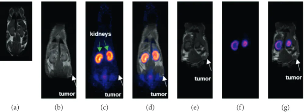
As can be seen from Figure 4 (as well as Figures S6 and S7), SiGdNP @ N-1(64Cu) provided enhanced contrast in both MRI and PET images. The PET images recorded 1 h and 24 h after intravenous injection clearly indicate that the SiGdNP @ N-1(64Cu) freely circulate since the undesirable accumulation in healthy tissues (except in the kidneys) is relatively low 1 h after the injection and almost impercep-tible 24 h post injection (pi). It must be pointed out that the
70 1 24 Time (h) % 64Cu inco rp or at ed 44 80 90 100 110
Figure 3: In vitro stability of SiGdNP @ N-1(64Cu) in plasma (blue) and EDTA solution (red).
presence of nanoparticles in kidneys can be assigned to the renal clearance mechanism since the simultaneous de-crease of the positive contrast enhancement and radio-activity in the kidneys is observed between 1 and 24 h pi (Figure S8) [5]. Despite the postfunctionalization which is accompanied by a slight increase of hydrodynamic di-ameter, the safe biological behavior of SiGdNP is pre-served [5]. This highlights the fact that this NP can be cleared via renal excretion and do not suffer from the problem of biopersistence in major organs as is observed with many other inorganic nanoparticles. From these images, it can be deduced that SiGdNP @ N-1(64Cu) is well suited for simultaneous MR/PET imaging. This was fur-ther illustrated by the biodistribution data at 24 h pi wherein the nanoparticles were observed to be majorly distributed in kidneys (Figure 5). The signal from the kidneys (24.2 ± 2.8% ID/g) was much higher than those in other tissues. However, radioactivity at tumor after 24 h was only 0.46 ± 0.01% ID/g. But this is commonly seen for such ultrasmall nanoparticle which is designed for being rapidly cleared from the body [10, 22, 23]. The modest imaging signal at tumor site 1 h after injection was of more concern (Figures 4(c) and 4(d)). This is contrary to the result we have obtained previously with AGuIX func-tionalized with Cyanine 5.5 fluorophore in similar TS/A tumor mice model [24]. Probably, the tumors in this study have not grown to sufficient size to manifest disorgani-zation of blood vessels and eventually considerable EPR effect [25].
3. Conclusions
We have described a straightforward and simple approach to functionalize ultrasmall silica gadolinium NP along with the physicochemical characterization of the products. Macro-cyclic chelating silane precursors have been synthesized by peptide coupling. They can be simply redispersed and grafted on the particle through siloxane bond formation. The amount of free chelators can be tailored according to the starting ratio of precursors. The influence of reaction con-ditions, i.e., temperature and initial silane concentration, on the homogeneity of the particle was studied. NODAGA functionalized particle was chosen to be labeled with 64Cu
before being intravenously injected to the mice bearing TS/A tumors. The radiolabeling process was conducted without difficulties. High complexation stability of labeled particle in harsh in vitro conditions was verified. MRI/PET images and biodistribution results demonstrate the renal clearability of the particle. However, the accumulation of the particle in the tumor was not significant. This calls for further in-vestigations probably using more mature tumor models in the future.
4. Experimental Section
4.1. Materials (See S1 for More Details). The SiGdNP
nanoparticles were synthesized by a top-down process de-scribed by Mignot et al. [26] and provided by NH TherAguix company (France).
4.2. Synthesis of Chelating Silanes
4.2.1. Synthesis of APTES-DOTAGA. DOTAGA(tBu)4
(0.9 g, 1.284 mmol) was weighted in 100 mL round bottom (a) (b) (c) (d) (e) (f ) (g)
Figure 4: Simultaneous MRI/PET coronal images of mice bearing TS/A tumors (white arrows) after being injected with SiGdNP @ N-1 (64Cu) nanoparticle. (a) Control T1-weighted MRI image of a healthy mouse; (b) T1-MRI; (c) PET, and (d) merged images of tumor bearing
mouse 1 h after injection; (e) T1-MRI; (f ) PET and (g) merged images of TS/A of tumor bearing mouse at 24 hours after injection.
Blood Liver Tumor Muscle Lungs Brain Kidney Spleen 0.0 0.5 1.0 1.5 2.0 20 22 24 26 28 % ID/g Tumor/blood Tumor/muscle 0 1 2 3 4 5 6 7 Ra tio
Figure 5: Biodistribution of SiGdNP-NODAGA(64Cu) at 24 h after injection in the mice bearing TS/A tumor. Insert: the ratio of signal from tumor tissue over the one from blood or muscle.
flask and was dissolved in 20 mL of DCM with stirring. DIPEA (1.14 mL, 6.55 mmol) was added into the above solution followed by coupling agents viz. HBTU (0.52 g, 1.37 mmol) and HOBt (0.18 g, 1.37 mmol) and the solution was left for stirring at RT for 15 min. APTES (0.3 g, 1.37 mmol) was added to the above solution directly using the 1 mL syringe, and the solution was stirred further at RT for 60 min after which the product formation was confirmed through MS. The above solution was mixed with 50 mL of citric acid solution (pH 2.5–3) in a separating funnel, and the organic layer was recovered. The organic layer was further mixed with 50 mL of 5% NaHCO3 in a separating funnel,
and the organic layer was recovered. The DCM solution was stirred with 5 g of MgSO4 for 10 min and filtered using
sintered funnel to receive the dry and clear DCM solution. Organic phase was evaporated under vacuum at 30°C to get
a viscous brownish residue, as an intermediate product (1.05 g, 78%). The intermediate formation was verified using HRMS (High resolution Mass Spectrometry),1H,13C NMR, and elemental analysis.
DOTAGA(tBu)4-APTES (1 g) was weighed into
a 100 mL round bottom flask and was mixed with 5 mL of concentrated HCl (∼12 M) and stirred for 10 min. Later, the acid was evaporated under vacuum at 35°C in 5–15 min to
get a dried residue. The dried residue was dissolved in 10 mL of water and lyophilized to get a light brown colored powder (850 mg, 81%). The product was verified using HRMS, 1H NMR and, elemental analysis.
4.2.2. Synthesis of APTES-NODAGA. NODAGA(tBu)3
(1.0 g, 1.84 mmol) was weighted in 100 mL round bottom flask and was dissolved in 20 mL of DCM under the hood with stirring. DIPEA (1.3 mL, 7.5 mmol) was added into the above solution followed by coupling agents viz. HBTU (1.0 g, 1.95 mmol) and HOBt (1.0 g, 1.95 mmol), and the solution was left for stirring at RT for 15 min. APTES (0.43 g, 1.95 mmol) was added to the above solution directly using the 1 mL syringe, and the solution was stirred further at RT for 60 min after which the product formation was confirmed through MS. The solution was mixed with 50 mL of citric acid solution (pH 2.5–3) in a separating funnel, and the organic layer was recovered. The organic layer was further mixed with 50 mL of 5% NaHCO3 in a separating funnel,
and the organic layer was recovered. The DCM solution was stirred with 5 g of MgSO4 for 10 min and filtered using
sintered funnel to receive the dry and clear DCM solution. Organic phase was evaporated under vacuum at 30°C to get
a viscous brownish residue, as an intermediate product (1.5 g, 99%). The intermediate formation was verified using HRMS,1H,13C NMR, and elemental analysis.
NODAGA(tBu)3-APTES was weighted into a 100 mL
round bottom flask and was mixed with 5 mL of concen-trated HCl (∼12 M) and stirred for 10 min. Later, the acid was evaporated under vacuum at 35°C in 5–15 min to get
a dried residue. The dried residue was dissolved in 10 mL of water and lyophilized to get a light brown-colored powder (1.2 g, 96%). The product was analyzed using HRMS, 1H NMR, and elemental analysis.
4.3. Characterization of Chelating Silanes (See S2 for More Details)
4.3.1. Intermediate Product (DOTAGA(t-Bu)4-APTES).
HRMS: calculated for C44H85N5O12Si: 926.585 [M+Na]+;
obtained: m/z � 926.584 [M+Na]+(Figure S9)
1H NMR (500 MHz, CDCl 3): δ 0.4–0.7 (m, 2H), 0.7–0.8 (m, 1H), 1.0 (dd, J � 9.0, 6.7 Hz, 1H), 1.1–1.2 (m, 9H), 1.3–1.5 (m, 32H), 1.5 (p, J � 7.8 Hz, 2H), 1.7 (d, 1H), 1.9–2.1 (m, 1H), 2.1–2.3 (m, 1H), 2.4–3.4 (m, 29H), 3.5–3.7 (m, 1H), 3.7–3.8 (m, 4H). 13C NMR (126 MHz, CDCl 3): δ 7.5, 7.8, 18.3, 20.4, 23.5, 25.9, 26.8, 27.8, 27.8, 27.9, 27.9, 27.9, 28.2, 28.3, 29.7, 33.0, 38.6, 42.1, 47.6, 49.8, 58.4, 63.6, 80.8, 82.3, 171.1, 173.2. Elemental analysis:
Calculated for C44H85N5O12Si·0.9HPF6(%): C: 51.03, H:
8.36, N: 6.76.
Observed (%): C: 51.86, H: 8.91, N: 8.38.
4.3.2. APTES-DOTAGA. HRMS: calculated for C22H41N5
O12Si: 596.259 [M+H]+; obtained: m/z � 596.261 [M+H]+; 618.242 [M+Na]+(Figure S10). 1H NMR (500 MHz, D 2O): δ 0.5–0.8 (m, 2H), 1.2–1.3 (m, 1H), 1.4–1.6 (m, 1H), 1.6–1.8 (m, 1H), 1.8–2.2 (m, 1H), 2.3–4.5 (m, 26H). Elemental analysis:
Calculated for C22H41N5O12Si·HPF6·2HCl (%): C: 32.44,
H: 5.44, N: 8.60.
Observed (%): C: 32.17, H: 6.54, N: 9.39.
4.3.3. Intermediate Product (NODAGA(t-Bu)3-APTES).
HRMS: calculated for C36H70N4O10Si: 747.493 [M + H]+;
obtained: m/z � 747.493 [M + Na]+(Figure S11)
1H NMR (500 MHz, CDCl 3): δ 0.5–0.6 (m, 2H), 1.0–1.1 (m, 1H), 1.1–1.2 (m, 7H), 1.4 (d, J � 7.2 Hz, 25H), 1.5–1.6 (m, 2H), 1.8 (d, J � 8.8 Hz, 1H), 1.9–2.1 (m, 1H), 2.2–2.5 (m, 2H), 2.6–3.3 (m, 22H), 3.7–3.8 (m, 4H), 6.2 (d, 1H). 13C NMR (126 MHz, CDCl 3): δ 7.8, 18.3, 18.4, 23.1, 26.2, 28.1, 28.2, 28.3, 29.7, 33.4, 38.6, 42.0, 53.7, 55.9, 58.4, 80.8, 165.7, 171.7, 172.8. Elemental analysis:
Calculated for C36H70N4O10Si·0.6HPF6(%): C: 51.81, H:
8.53, N: 6.71.
Observed (%): C: 52.58, H: 9.13, N: 8.85.
4.3.4. APTES-NODAGA. HRMS: calculated for C18H34N4
O10Si: 495.211 [M + H]+; obtained: m/z � 495.212 [M + H]+; 517.191 [M + Na]+(Figure S12). 1H NMR (500 MHz, D 2O): δ 0.41–0.80 (m, 2H), 0.93– 1.28 (m, 2H), 1.39–1.59 (m, 2H), 1.84–2.18 (m, 2H), 2.27–2.46 (m, 2H), 2.73 (s, 5H), 2.97–3.18 (m, 5H), 3.19–3.45 (m, 7H), 3.48–3.75 (m, 1H), 3.79–4.10 (m, 4H). Elemental analysis:
Calculated for C18H34N4O10Si·HPF6·HCl (%): C: 31.93,
H: 5.36, N: 8.28.
4.4. Grafting of APTES-DOTAGA or APTES-NODAGA on SiGdNP. For SiGdNP @ D-1 and SiGdNP @ N-1, 250 µmol
in Gd of SiGdNP was dispersed in water to obtain [Gd3+] � 200 mM for 1 h leading to a pH of the solution of 7.4. 29.8 mg (40 µmol) of APTES-DOTAGA or 24.7 mg (40 µmol) of APTES-NODAGA was dissolved in water. pH of the so-lution was adjusted to 9 by adding NaOH. This soso-lution was left under stirring for 1 h before water was filled to obtain [chelating silane] � 100 mM. It was then gradually added to the SiGdNP solution under stirring at room temperature before water was added to obtain [Gd3+] � 100 mM and [chelating silane] � 15 mM. The mixture was left stirring for 1 h before adjusting pH to 4.5. It was heated at 80°C and left
under stirring overnight.
For SiGdNP @ D-2, SiGdNP @ D-3, SiGdNP @ D-4, and SiGdNP @ N-2, similar protocol was applied with some modifications: [Gd3+] was decreased to 100 mM. 29.8 mg (40 µmol) or 37.2 mg (50 µmol) or 74.5 mg (100 µmol) of DOTAGA or 24.7 mg (40 µmol) of APTES-NODAGA were used, respectively. The concentration of chelating silane solutions before being added in NP solutions was decreased to 50 mM. At the end, they were heated at 40°C overnight. Detailed parameters can be found in
Table S1.
Then each solution was purified with tangential filtration through Vivaspin membranes (MWCO � 3 kDa). The pH of the solution should be adjusted to 2 by adding HCl solutions before the purification. The solution was centrifuged until half of the volume remains. This step was repeated by filling the tubes with hydrochloric acid (HCl) solution 10−2M and centrifuging again for at least 50 purification factor (puri-fication factor � starting volume/end volume). Then, the solution was filtered through 0.2 µm membrane to remove dust and large impurities. Finally, the solution was freeze-dried for long-term storage using a Christ Alpha 1-2 lyophilisator.
4.5. Characterization of Functionalized Nanoparticles (See S3 for More Details)
4.5.1. Dynamic Light Scattering (DLS) and Zeta Potential.
Hydrodynamic diameter distribution of the nanoparticle was measured by DLS with a Zetasizer Nano-S (633 nm He-Ne laser) from Malvern Instruments (USA).
4.5.2. Infrared Spectroscopy. The IR spectra of dry samples
were acquired on an IRAffinity-1, Shimadzu with an ATR platform by applying the attenuated total reflection Fourier transform infrared (ATR-FTIR) spectroscopy from 400 to 4000 cm−1.
4.5.3. High-Performance Liquid Chromatography (HPLC).
Gradient HPLC analysis was done by using the Shimadzu Prominence series UFLC system with a CBM-20A controller bus module, a LC-20AD pump, a CTO-20A column oven, and a SPD-20A UV-vis detector.
4.5.4. Phosphorescence Spectroscopy. Phosphorescence
mea-surements were carried out using a Varian Cary Eclipse fluorescence spectrophotometer, in the resolved time mode.
4.6. Relaxivity Measurement. Relaxivity measurements were
performed on a Bruker
®
minispec mq60NMR analyzer (Bruker, USA) at 37°C at 1.4 T (60 MHz).4.7. Elemental Analysis. The contents of elements in
SiGdNP @ D-1 and SiGdNP @ N-1 were analyzed by FILAB SAS., Dijon, France. Elemental analyses enabled the de-termination of the contents of C and N, while inductively coupled plasma-optical emission spectrometry (ICP-OES) was used to determine the contents of Gd and Si.
For other samples, the determination of the content of Gd was performed in the Institut Lumi`ere Mati`ere, Lyon by ICP-OES (with a Varian 710-ES spectrometer).
4.8. Radiochemistry and the Stability of Radiolabeled NPs.
Radiolabeling: SiGdNP @ N-1 (3.5 mg) was dispersed into 12.9 µL of 1 M, AcONH4buffer pH 5.8, and 12.9 µL of64Cu
(7 MBq) in a minitube. The nanoparticle dispersion was incubated at 37°C for 30 min. The radiochemical purity was
established by ITLC using 0.1 M EDTA as a mobile phase. Plasma Stability: 25 µL of the radiolabeled dispersion of nanoparticles was diluted with 45 µL of PBS. 10 µL of the resulting solution was mixed with 100 µL of human plasma in a Eppendorf tube. This tube was incubated at 37°C for
48 h, and aliquots were withdrawn at 24 h and 48 h to test the stability of the radiolabeled nanoparticles using ITLC.
EDTA challenge stability: 25 µL of the radiolabeled dispersion of nanoparticles was diluted with 45 µL of PBS. 10 µL of the resulting solution was mixed with 100 µL of a buffer containing EDTA (50 mM) and HEPES (0.5 M) pH 7 in a minitube. This tube was incubated at 37°C for 48 h, and
aliquots were withdrawn at 24 h and 48 h to test the stability of the radiolabeled nanoparticles using ITLC.
The analyses were performed in triplicate wherein the radiolabeled NPs were incubated in plasma (n � 3) and EDTA challenge solution (n � 3) separately in Eppendorf tubes. The samples were aliquoted from the same tubes at 24 h and 48 h and analyzed by ITLC. The values have been reported as average with standard deviation (n � 3).
Radiochromatograms were carried out with a Raytest miniGITA-Star c Radiochromatography (Raytest, Strau-benhardt, Germany) or with a Bioscan AR-2000 radio-TLC Imaging Scanner (Bioscan Inc., Washington, DC).
4.9. Animal Studies. All animal studies were conducted in
accordance with the legislation on the use of laboratory animals (directive 2010/63/EU) and were approved by accredited Ethical committee (C2ea Grand Campus no. 105). Female NMRI nude mice (6–8 weeks, Janvier Labs, France) were subcutaneously injected with 1.107TS/A cells (murine mammary adenocarcinoma).
Mice were maintained in ventilated housing units un-der controlled conditions of temperature (22 ± 2°C) and
photoperiod (12 h light/12 h dark) with free access to food and drink.
Three to five weeks after tumor cells injection, TS/A tumor bearing-mice (n � 3) were given 10 mg of SiGdNP @ N-1(64Cu) in PBS corresponding to radioactivity of 10.2 ± 0.3 MBq by intravenous injection. Size of the tumors varies from 41 to 157 mm3 with a mean value of 105 mm.
For biodistribution, at the end of 24 h, animals were euthanized by isoflurane anesthesia followed by pentobar-bital overdose. Blood, tumor, and organs were collected, weighed, and radioactivity in these samples was measured with a scintillation gamma-counter (Cobra 4180, Perkin Elmer, Waltham, MA, USA).
4.10. MRI-PET Imaging and Biodistribution Measurement.
Simultaneous MRI-PET imaging was performed at 1 and 24 hours after the injection of the radiolabeled nanoparticles on a fully integrated system (MR Solutions, Guilford, UK) consisting of a 7T dry magnet (Powerscan MRS-7024-PW) coupled with a SiPM dual-ring PET-I-802 insert.
Mice were anaesthetized with isoflurane (2% in oxygen) and positioned in a dedicated heating cradle. MRI and PET acquisitions were performed simultaneously. Animal res-piration was monitored with abdominal pressure sensor and dedicated software (PC Sam, SAII, Stony Brook, US).
List-mode data were collected in the PET system during 30–60 minutes. Images were reconstructed with the 3D ordered subset expectation maximization (OSEM) algo-rithm implemented in the system using 2 iterations, 32 subsets, and an isotropic voxel size of 0.28 mm. An energy window of 250–750 keV and a coincidence time window of 8 ns were applied to the list-mode data. The algorithm takes into account normalization and random and decay cor-rections. No attenuation correction was applied.
T1- and T2-weighted fast spin echo MR images were
acquired with respiratory gating and sequences in axial and coronal plans. T1 sequences were acquired with time of
repetition (TR) of 1000 ms, time of echo (TE) of 11 ms. T2
sequences were acquired with TR of 4000 ms, TE of 45 ms. Following parameters were used for both T1 and T2: flip
angle of 90°, 4 signal averages, 1 mm slice thickness, 0.15 ×
0.15 mm2transaxial pixel size, and 256 × 256 pixels matrix. Finally, the PET-MR fusion image was obtained using VivoQuant (Invicro, Boston, US). Each scan was then vi-sually interpreted.
For the gamma counting of the organs, the counter was cross-calibrated to the dose calibrator used to measure the injected dose, and the linearity range was determined for all the geometries used in ex vivo counting. Data were then converted to percentage of injected dose and to percentage of injected dose per gram of tissue.
Data Availability
The data used to support the findings of this study are in-cluded within the article and the supplementary information file. Any other data are available from the corresponding author upon request.
Conflicts of Interest
FL and OT have to disclose the patent WO2011/135101. This patent protects the AGuIX
®
NPs described in this publi-cation. FL and OT are employees from NH TherAguix that is developing the AGuIX®
NPs and possess shares of this company.Authors’ Contributions
Vu-Long Tran and Vivek Thakare contributed equally to this work.
Acknowledgments
The authors gratefully acknowledge European Union FP7-PEOPLE Initial Training Network ARGENT (Advanced Radiotherapy, Generated by Exploiting Nanoprocesses and Technologies), Project ID 608163, for the financial support. This work has also been supported by a French Government Grant managed by the French National Research Agency (ANR) under the program “Investissements d’Avenir” (reference ANR-10-EQPX-05-01/IMAPPI Equipex).
Supplementary Materials
Supplementary materials contains an experimental section (S1) describing the different chemicals products used for the synthesis and a description of the cheracterization methods of the silanes (S2) and of the functionalized nanoparticles (S3). Two figures are present (S9 and S10) to describe the MRI and the PET/MRI images after intravenous adminis-tration of the radiolabeled NPs. (Supplementary Materials)
References
[1] S. Kunjachan, J. Ehling, G. Storm, F. Kiessling, and T. Lammers, “Noninvasive imaging of nanomedicines and nanotheranostics: principles, progress, and prospects,”
Chemical Reviews, vol. 115, no. 19, pp. 10907–10937, 2015.
[2] F. Lux, A. Mignot, P. Mowat et al., “Ultrasmall rigid particles as multimodal probes for medical applications,” Angewandte
Chemie International Edition, vol. 50, no. 51, pp. 12299–
12303, 2011.
[3] P. J. Bonitatibus, A. S. Torres, B. Kandapallil et al., “Preclinical assessment of a zwitterionic tantalum oxide nanoparticle X-ray contrast agent,” ACS Nano, vol. 6, no. 8, pp. 6650–6658, 2012.
[4] L. Sancey, F. Lux, S. Kotb et al., “The use of theranostic gadolinium-based nanoprobes to improve radiotherapy effi-cacy,” British Journal of Radiology, vol. 87, no. 1041, article 20140134, 2014.
[5] L. Sancey, S. Kotb, C. Truillet et al., “Long-term in vivo clearance of gadolinium-based AGuIX nanoparticles and their biocompatibility after systemic injection,” ACS Nano, vol. 9, no. 3, pp. 2477–2488, 2015.
[6] H. Soo Choi, W. Liu, P. Misra et al., “Renal clearance of quantum dots,” Nature Biotechnology, vol. 25, no. 10, pp. 1165–1170, 2007.
[7] E. Miot-Noirault, A. Vidal, J. Morlieras et al., “Small rigid platforms functionalization with quaternary ammonium: tar-geting extracellular matrix of chondrosarcoma,” Nanomedicine:
Nanotechnology, Biology and Medicine, vol. 10, no. 8,
pp. 1887–1895, 2014.
[8] C. Truillet, P. Bouziotis, C. Tsoukalas et al., “Ultrasmall particles for Gd-MRI and68Ga-PET dual imaging,” Contrast
Media & Molecular Imaging, vol. 10, no. 4, pp. 309–319, 2015.
[9] C. Truillet, E. Thomas, F. Lux, L. T. Huynh, O. Tillement, and M. J. Evans, “Synthesis and characterization of 89Zr-labeled ultrasmall nanoparticles,” Molecular Pharmaceutics, vol. 13, no. 7, pp. 2596–2601, 2016.
[10] A. Detappe, E. Thomas, M. W. Tibbitt et al., “Ultrasmall silica-based bismuth gadolinium nanoparticles for dual magnetic resonance–computed tomography image guided radiation therapy,” Nano Letters, vol. 17, no. 3, pp. 1733–1740, 2017. [11] K. J. Jensen, P. T. Shelton, and S. L. Pedersen, Peptide Synthesis
and Applications, Humana Press, New York, NY, USA, 2nd
edition, 2013.
[12] J. Ciccione, T. Jia, J.-L. Coll et al., “Unambiguous and con-trolled one-pot synthesis of multifunctional silica nano-particles,” Chemistry of Materials, vol. 28, no. 3, pp. 885–889, 2016.
[13] E. W. Price and C. Orvig, “The chemistry of inorganic nu-clides (86I, 68Ga, 64Cu, 89Zr, 124I),” in The Chemistry of
Molecular Imaging, pp. 119–150, John Wiley & Sons, Inc.,
Hoboken, NJ, USA, 2015.
[14] A. Bevilacqua, R. I. Gelb, W. B. Hebard, and L. J. Zompa, “Equilibrium and thermodynamic study of the aqueous complexation of 1,4,7-triazacyclononane-N,N′,N″-triacetic acid with protons, alkaline-earth-metal cations, and copper (II),” Inorganic Chemistry, vol. 26, no. 16, pp. 2699–2706, 1987.
[15] A. F. Prasanphanich, P. K. Nanda, T. L. Rold et al., “[64Cu-NOTA-8-Aoc-BBN(7-14)NH2] targeting vector for positron-emission tomography imaging of gastrin-releasing peptide receptor-expressing tissues,” in Proceedings of the National
Academy of Sciences, vol. 104, no. 30, pp. 12462–12467, 2007.
[16] Y. Zhang, H. Hong, J. W. Engle et al., “Positron emission tomography imaging of CD105 expression with a 64Cu-labeled monoclonal antibody: NOTA is superior to DOTA,” PLoS One, vol. 6, no. 12, Article ID e28005, 2011. [17] S. C. Ghosh, K. L. Pinkston, H. Robinson et al., “Comparison
of DOTA and NODAGA as chelators for 64Cu-labeled immunoconjugates,” Nuclear Medicine and Biology, vol. 42, no. 2, pp. 177–183, 2015.
[18] J. Lux, A. G. White, M. Chan, C. J. Anderson, and A. Almutairi, “Nanogels from metal-chelating crosslinkers as versatile platforms applied to copper-64 PET imaging of tumors and metastases,” Theranostics, vol. 5, no. 3, pp. 277– 288, 2015.
[19] V. Jokanovi´c, M. D. Drami´canin, and ˇZ. Andri´c, “Nano-designing of SiO2: Eu3+ particles obtained by spray pyrolysis method and their luminescence properties,” Acta Chimica
Slovenica, vol. 53, pp. 23–28, 2006.
[20] L. Ma, L. Chen, J. Wen, Q. Wang, and C. Tan, “Eu3+chelate with phenanthroline derivative gives selective emission re-sponses to Cu(II) ions,” Journal of Organometallic Chemistry, vol. 696, no. 4, pp. 829–831, 2011.
[21] C. Catana, A. R. Guimaraes, and B. R. Rosen, “PET and MR imaging: the odd couple or a match made in heaven?,” Journal
of Nuclear Medicine, vol. 54, no. 5, pp. 815–824, 2013.
[22] S. Kotb, A. Detappe, F. Lux et al., “Gadolinium-based nanoparticles and radiation therapy for multiple brain mel-anoma metastases: proof of concept before phase I trial,”
Theranostics, vol. 6, no. 3, pp. 418–427, 2016.
[23] P. Bouziotis, D. Stellas, E. Thomas et al., “68Ga-radiolabeled AGuIX nanoparticles as dual-modality imaging agents for PET/MRI-guided radiation therapy,” Nanomedicine, vol. 12, no. 13, pp. 1561–1574, 2017.
[24] J. Morlieras, S. Dufort, L. Sancey et al., “Functionalization of small rigid platforms with cyclic RGD peptides for targeting tumors overexpressing αvβ3-integrins,” Bioconjugate
Chem-istry, vol. 24, no. 9, pp. 1584–1597, 2013.
[25] E. A. Sykes, Q. Dai, C. D. Sarsons et al., “Tailoring nano-particle designs to target cancer based on tumor patho-physiology,” in Proceedings of the National Academy of
Sciences, vol. 113, no. 9, pp. E1142–E1151, 2016.
[26] A. Mignot, C. Truillet, F. Lux et al., “A top-down synthesis route to ultrasmall multifunctional gd-based silica nano-particles for theranostic applications,” Chemistry-A European
Stem Cells
International
Hindawi www.hindawi.com Volume 2018 Hindawi www.hindawi.com Volume 2018 INFLAMMATIONEndocrinology
International Journal ofHindawi www.hindawi.com Volume 2018 Hindawi www.hindawi.com Volume 2018
Disease Markers
Hindawi www.hindawi.com Volume 2018 BioMed Research InternationalOncology
Journal of Hindawi www.hindawi.com Volume 2013 Hindawi www.hindawi.com Volume 2018 Oxidative Medicine and Cellular Longevity Hindawiwww.hindawi.com Volume 2018
PPAR Research
Hindawi Publishing Corporation
http://www.hindawi.com Volume 2013 Hindawi www.hindawi.com
The Scientific
World Journal
Volume 2018 Immunology Research Hindawi www.hindawi.com Volume 2018 Journal ofObesity
Journal of Hindawi www.hindawi.com Volume 2018 Hindawi www.hindawi.com Volume 2018 Computational and Mathematical Methods in Medicine Hindawi www.hindawi.com Volume 2018Behavioural
Neurology
Ophthalmology
Journal of Hindawi www.hindawi.com Volume 2018Diabetes Research
Journal ofHindawi
www.hindawi.com Volume 2018
Hindawi
www.hindawi.com Volume 2018
Research and Treatment
AIDS
Hindawi
www.hindawi.com Volume 2018
Gastroenterology Research and Practice
Hindawi www.hindawi.com Volume 2018