HAL Id: hal-03210093
https://hal.archives-ouvertes.fr/hal-03210093
Submitted on 27 Apr 2021
HAL is a multi-disciplinary open access
archive for the deposit and dissemination of sci-entific research documents, whether they are pub-lished or not. The documents may come from
L’archive ouverte pluridisciplinaire HAL, est destinée au dépôt et à la diffusion de documents scientifiques de niveau recherche, publiés ou non, émanant des établissements d’enseignement et de
Structural Characterization and Anti-infective Activity
of 9,10-Seco-29-norcycloartane Glycosides Isolated from
the Flowers of the Peruvian Medicinal Plant Cordia
lutea
Inés Castro, Nicolas Fabre, Sandra Bourgeade-Delmas, Nathalie Saffon, Chloé
Gandini, Michel Sauvain, Denis Castillo, Geneviève Bourdy, Valérie Jullian
To cite this version:
Inés Castro, Nicolas Fabre, Sandra Bourgeade-Delmas, Nathalie Saffon, Chloé Gandini, et al.. Struc-tural Characterization and Anti-infective Activity of 9,10-Seco-29-norcycloartane Glycosides Isolated from the Flowers of the Peruvian Medicinal Plant Cordia lutea. Journal of Natural Products, Ameri-can Chemical Society, 2019, 82 (12), pp.3233-3241. �10.1021/acs.jnatprod.9b00149�. �hal-03210093�
Structural Characterization and Anti-infective Activity of 9,10-Seco-29-norcycloartane
Glycosides Isolated from the Flowers of the Peruvian Medicinal Plant Cordia lutea
Inés Castro,§,† Nicolas Fabre,† Sandra Bourgeade-Delmas,† Nathalie Saffon,‡ Chloé Gandini,§,† Michel Sauvain,§,† Denis Castillo,§ Geneviève Bourdy,† and Valérie Jullian§,†,*
†UMR 152 PharmaDev, Université de Toulouse, IRD, UPS, France
‡Institut de Chimie de Toulouse, ICT FR 2599, Université Paul Sabatier-Toulouse III,
Toulouse 31062 Cedex 9, France
§Laboratorios de Investigación y Desarrollo, Facultad de Ciencias y Filosofía, Universidad
ABSTRACT: Six new secocycloartane glycosides (1- 6) were isolated from the ethanol
extract of the flowers of Cordia lutea Lam. (Boraginaceae) on the basis of bioassay-guided fractionation. Their structures were determined by the application of NMR and MS data analyses together with X-ray crystallographic analyses for compounds 1 and 2. Compounds 1
- 6 represent the first examples of 9,10-seco-29-norcycloartane glycosides. These compounds
showed significant in vitro anti-Helicobacter pylori activity, and no activity against either
Escherichia coli or Pseudomonas aeruginosa. Significant activity was observed for 5 and 6
against Staphylococcus aureus. All compounds displayed weak cytotoxicity against RAW 264.7 cells. The in vitro antileishmanial and antiplasmodial activities of 1-6 were also evaluated.
The genus Cordia (Boraginaceae) encompasses more than 300 species of trees and shrubs distributed in the tropical areas of Africa, America, and Asia, several of which possess different ethnopharmacological properties.1-3 In Peru, the genus Cordia is represented by approximately 30 species, among which Cordia lutea has significant medicinal use.
The dried flowers of C. lutea, also known as “flor de overo”, “overall” or “membrillejo”, are commercialized and widely used in Northern Peru for the treatment of liver diseases, kidney pain, and gastrointestinal disorders.4 However, there have only been two reported phytochemical investigations of C. lutea. Mayevych and coworkers described some ubiquitous flavonoids and lipidic compounds and verified the absence of pyrrolizidine alkaloids in the whole plant.5 A second study reported the LC-MS analysis of a C. lutea flower extract.6 This extract showed characteristic signals for the reported products, but some minor compounds remained unidentified.
Furthermore, we found interesting activity for the ethanol extract of C. lutea dried flowers against the bacteria Helicobacter pylori, a microaerophilic gram-negative bacterium that has been associated with severe gastrointestinal disorders in humans such as chronic gastritis, peptic ulcers, MALT-type lymphoma, and gastric cancer. The prevalence of infection is considered to be above 70% in developing countries, while it ranges from 20 to 50% in developed countries. Antimicrobial resistance is an important global health problem that leads to therapeutic failure.7 The limited knowledge about the chemical composition of the flowers of C. lutea, together with this interesting biological activity, prompted us to isolate the compounds responsible for the antibacterial effect.
Cycloartane-type triterpenoids and their glycosides have been described as major constituents of and are responsible for the biological activities of Cimifuga and Actae species (Ranunculaceae) and Astragalus species (Leguminosae).8-11 Collectively, they represent
triterpenoids featuring a seven-membered B ring.9, 13-14 Recently, an unusual 9,10-secocycloartane triterpenoid was isolated for the first time from black cohosh (Actaea
racemosa), a widely accepted herbal medicine used in the United States and Europe for the
treatment of climacteric symptoms.8
Herein is the first report of new glycosides derived from 4-methyl-9,10-secocycloartane triterpenoids, cordiasecosides A-F, 1-6, isolated from the dried flowers of C.
lutea. The antibacterial, antileishmanial, and antiplasmodial activities of these compounds
were evaluated, as well as the cytotoxicity activity against RAW 264.7 macrophages.
RESULTS AND DISCUSSION
Compounds 1-6 were isolated from the ethanol extract of the flowers of C. lutea by bioguided fractionation. Their structures were assigned via the analysis of spectroscopic data, including 2D NMR techniques.
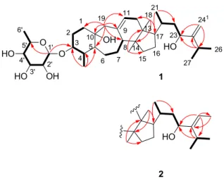
Figure 1. Structures of compounds 1-6.
Figure 3. Key NOESY correlations for compounds 1 and 2.
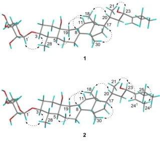
Figure 4. Molecular views of compounds 1 and 2. The thermal ellipsoids represent a 50%
probability level. For 1, only one of the two molecules in the asymmetric unit is shown, and solvent molecules (H2O) were omitted for clarity. For 2, solvent molecules (MeOH) and
Table 1. 13C NMR Spectroscopic Data (125 MHz, CDCl3 – Methanol-d4 9:1) for Compounds 1-6 (δ in ppm) Position Type 1 2 3 4 5 6 1 CH2 40.1 40.1 40.1 40.1 40.1 40.1 2 CH2 28.7 28.7 29.1 29.1 29.1 29.1 3 CH 85.2 85.2 85.6 85.6 85.5 85.5 4 CH 39.6 39.5 39.6 39.6 39.6 39.6 5 CH 52.4 52.3 52.5 52.5 52.5 52.5 6 CH2 31.9 31.9 31.8 31.8 31.8 31.8 7 CH2 26.5 26.5 26.5 26.5 26.5 26.5 8 CH 48.4 48.4 48.4 48.3 48.3 48.3 9 C 136.7 136.5 136.6 136.6 136.6 136.6 10 C 71.8 71.8 71.7 71.7 71.7 71.7 11 CH 124 124.1 124.1 124.1 124 124 12 CH2 38.3 38.3 38.3 38.3 38.3 38.3 13 C 44 44 44.1 44.1 44.1 44.1 14 C 48.7 48.7 48.7 48.7 48.7 48.7 15 CH2 32.9 32.9 32.9 32.9 32.9 32.9 16 CH2 27.9 27.9 27.8 27.9 27.8 27.9 17 CH 51.5 51.5 51.4 51.5 51.4 51.5 18 CH3 15.7 15.7 15.7 15.7 15.7 15.7 19 CH2 50.5 50.5 50.5 50.5 50.5 50.5 20 CH 33 33.2 33 33.2 33 33.2 21 CH3 17.9 17.9 17.9 17.9 17.9 17.9 22 CH2 43.4 44.8 43.7 44.8 43.7 44.8 23 CH 70.7 68.9 70.9 68.9 70.8 68.9 24 C 160.4 149.3 160.4 149.3 160.4 149.3 241 CH 105.5 118.1 105.5 118.1 105.5 118.1 242 CH3 - 12.9 - 12.9 - 12.9 25 CH 30.5 27.9 30.5 27.9 30.5 27.9 26 CH3 23.2 21.2 23.2 21.2 23.2 21.2 27 CH3 22.4 21.2 22.3 21.2 22.3 21.2 28 CH3 15.5 15.5 15.4 15.3 15.4 15.4 30 CH3 16.6 16.5 16.5 16.5 16.5 16.5 1’ CH 102.2 102.2 104.6 104.7 104.5 104.6 2’ CH 70.8 70.9 72.8 72.9 71.3 71.3 3’ CH 71.3 71.4 75.1 75.1 72.6 72.6 4’ CH 72.9 72.9 69.5 69.5 67.5 67.6 5’ CH/CH2a 68.2 68.1 64.6 64.6 64.8 64.9 6’ CH3 17.1 17.1 - - - - a: CH for 1-2, CH2 for 3-6.
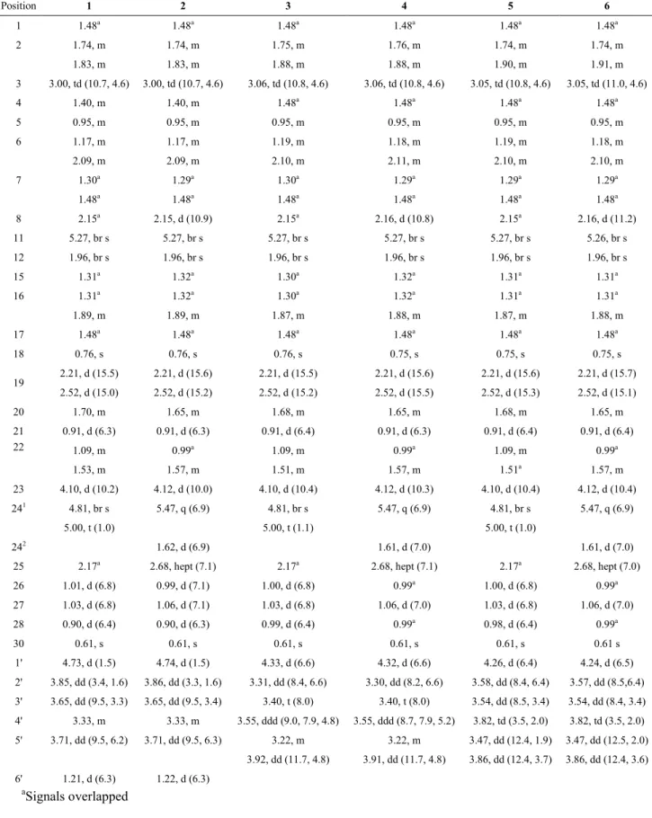
Table 2. 1H NMR Spectroscopic Data (500 MHz, CDCl3 – Methanol-d4 9:1) for Compounds 1-6 (δ in ppm) Position 1 2 3 4 5 6 1 1.48a 1.48a 1.48a 1.48a 1.48a 1.48a 2 1.74, m 1.74, m 1.75, m 1.76, m 1.74, m 1.74, m 1.83, m 1.83, m 1.88, m 1.88, m 1.90, m 1.91, m 3 3.00, td (10.7, 4.6) 3.00, td (10.7, 4.6) 3.06, td (10.8, 4.6) 3.06, td (10.8, 4.6) 3.05, td (10.8, 4.6) 3.05, td (11.0, 4.6) 4 1.40, m 1.40, m 1.48a 1.48a 1.48a 1.48a 5 0.95, m 0.95, m 0.95, m 0.95, m 0.95, m 0.95, m 6 1.17, m 1.17, m 1.19, m 1.18, m 1.19, m 1.18, m 2.09, m 2.09, m 2.10, m 2.11, m 2.10, m 2.10, m 7 1.30a 1.29a 1.30a 1.29a 1.29a 1.29a 1.48a 1.48a 1.48a 1.48a 1.48a 1.48a 8 2.15a 2.15, d (10.9) 2.15a 2.16, d (10.8) 2.15a 2.16, d (11.2) 11 5.27, br s 5.27, br s 5.27, br s 5.27, br s 5.27, br s 5.26, br s 12 1.96, br s 1.96, br s 1.96, br s 1.96, br s 1.96, br s 1.96, br s 15 1.31a 1.32a 1.30a 1.32a 1.31a 1.31a 16 1.31a 1.32a 1.30a 1.32a 1.31a 1.31a 1.89, m 1.89, m 1.87, m 1.88, m 1.87, m 1.88, m 17 1.48a 1.48a 1.48a 1.48a 1.48a 1.48a 18 0.76, s 0.76, s 0.76, s 0.75, s 0.75, s 0.75, s 19 2.21, d (15.5) 2.21, d (15.6) 2.21, d (15.5) 2.21, d (15.6) 2.21, d (15.6) 2.21, d (15.7) 2.52, d (15.0) 2.52, d (15.2) 2.52, d (15.2) 2.52, d (15.5) 2.52, d (15.3) 2.52, d (15.1) 20 1.70, m 1.65, m 1.68, m 1.65, m 1.68, m 1.65, m 21 0.91, d (6.3) 0.91, d (6.3) 0.91, d (6.4) 0.91, d (6.3) 0.91, d (6.4) 0.91, d (6.4) 22 1.09, m 0.99a 1.09, m 0.99a 1.09, m 0.99a 1.53, m 1.57, m 1.51, m 1.57, m 1.51a 1.57, m 23 4.10, d (10.2) 4.12, d (10.0) 4.10, d (10.4) 4.12, d (10.3) 4.10, d (10.4) 4.12, d (10.4) 241 4.81, br s 5.47, q (6.9) 4.81, br s 5.47, q (6.9) 4.81, br s 5.47, q (6.9) 5.00, t (1.0) 5.00, t (1.1) 5.00, t (1.0) 242 1.62, d (6.9) 1.61, d (7.0) 1.61, d (7.0)
25 2.17a 2.68, hept (7.1) 2.17a 2.68, hept (7.1) 2.17a 2.68, hept (7.0)
26 1.01, d (6.8) 0.99, d (7.1) 1.00, d (6.8) 0.99a 1.00, d (6.8) 0.99a 27 1.03, d (6.8) 1.06, d (7.1) 1.03, d (6.8) 1.06, d (7.0) 1.03, d (6.8) 1.06, d (7.0) 28 0.90, d (6.4) 0.90, d (6.3) 0.99, d (6.4) 0.99a 0.98, d (6.4) 0.99a 30 0.61, s 0.61, s 0.61, s 0.61, s 0.61, s 0.61 s 1' 4.73, d (1.5) 4.74, d (1.5) 4.33, d (6.6) 4.32, d (6.6) 4.26, d (6.4) 4.24, d (6.5) 2' 3.85, dd (3.4, 1.6) 3.86, dd (3.3, 1.6) 3.31, dd (8.4, 6.6) 3.30, dd (8.2, 6.6) 3.58, dd (8.4, 6.4) 3.57, dd (8.5,6.4) 3' 3.65, dd (9.5, 3.3) 3.65, dd (9.5, 3.4) 3.40, t (8.0) 3.40, t (8.0) 3.54, dd (8.5, 3.4) 3.54, dd (8.4, 3.4) 4' 3.33, m 3.33, m 3.55, ddd (9.0, 7.9, 4.8) 3.55, ddd (8.7, 7.9, 5.2) 3.82, td (3.5, 2.0) 3.82, td (3.5, 2.0) 5' 3.71, dd (9.5, 6.2) 3.71, dd (9.5, 6.3) 3.22, m 3.22, m 3.47, dd (12.4, 1.9) 3.47, dd (12.5, 2.0) 3.92, dd (11.7, 4.8) 3.91, dd (11.7, 4.8) 3.86, dd (12.4, 3.7) 3.86, dd (12.4, 3.6) 6' 1.21, d (6.3) 1.22, d (6.3) aSignals overlapped
Compound 1 was isolated as a white powder and formed colorless needles in MeOH. Its molecular formula was established as C36H60O7 by negative HRESIMS (m/z 649.4324, [M
+ HCOO]-) and showed seven indices of hydrogen deficiency. The IR spectrum showed an absorption band for hydroxy groups at 3385 cm-1.
The J-modulated 13C NMR (Table 1) and HSQC data of compound 1 showed 36 carbon signals, 30 of which were assignable to a triterpenoid skeleton and six to a hexosyl moiety. These carbons could be resolved as seven methyls, 10 methylenes, 14 methines, an oxygenated tertiary, and four quaternary carbons. The 13C NMR spectrum showed four olefinic carbons at δC 136.7 (C-9), 124.0 (C-11), 160.4 (C-24), and 105.5 (C-241).
Additionally, the 13C NMR spectrum of 1 showed two oxygen-bearing carbons at δC 71.8
(C-10) and 70.7 (C-23) as well as six additional carbon signals, which were in agreement with those previously described for a rhamnopyranosyl moiety.15 All these data indicated that one index of hydrogen deficiency was due to the sugar moiety and that two indices of hydrogen deficiency were due to two double bonds. Thus, a tetracyclic core is consistent with the remaining four indices of hydrogen deficiency.
The 1H NMR spectrum (Table 2) of 1 displayed methyl signals associated with a 29-norcycloartane skeleton including two angular methyl groups at δH 0.76 (s, H3-18) and 0.61
(s, H3-30) and four secondary methyl groups at δH 0.91 (d, J = 6.3 Hz, H-21), 1.01 (d, J = 6.8
Hz, H-26), 1.03 (d, J = 6.8 Hz, H-27) and 0.90 (d, J = 6.4 Hz, H-28). These signals suggested the presence of a single methyl substituent at C-4 as demonstrated for cycloeucalenol, a biosynthetic intermediate of 4-methylphytosterols.16,17
The 1H NMR spectrum also showed two diagnostic methylene proton signals for H2
-19. Although they are typically present in the extremely upfield region in the 29-norcycloartane skeleton, these proton signals were present as doublets at δH 2.21 and 2.52
with a large geminal coupling constant (15.0 Hz), indicating that compound 1 is a 9,10-seco-29-norcycloartane rhamnoside.9, 18
As expected from its 13C NMR data, the 1H NMR data (Table 2) also displayed signals for an olefinic methine proton at δH 5.27 (br s, H-11) and two terminal olefinic methylene
protons at δH 4.81 (br s, H-241) and 5.00 (t, J = 1.0 Hz, H-241). The locations of the two
double bonds (Δ9(11), Δ24(241)) were supported by the HMBC cross peaks between H2-19, C-8,
C-9, and C-11; between H2-241, C-23, and C-25; and between H-25, C-23, and C-24 (Figure
2).
The position of the oxymethine signal at δH 4.11 (d, J = 10.2 Hz, H-23) was deduced
via the presence of vicinal 1H-1H COSY correlations between H-20, H2-22, and H-23. Further
HMBC correlations between H-25, C-23, and C-24 confirmed the location of the hydroxy group at C-23 (Figure 2). Similarly, the presence of an oxygenated tertiary carbon at C-10 was supported by HMBC correlations between H2-19, C-1, C-5, and C-10. The presence of
two hydroxy groups and a rhamnosyl moiety were consistent with the HRESIMS data recorded in positive mode (Figure S2, Supporting Information). The ion at m/z 569 is attributable to the loss of two water molecules from the protonated molecule [M + H]+. The ion at m/z 423 is consistent with a consecutive neutral loss of the rhamnosyl unit, that is, [M + H - 2H2O - 146]+.19
The attachment of the rhamnopyranose moiety to C-3 was confirmed via the HMBC cross peak between the anomeric proton H-1'and C-3. The coupling constant of the anomeric proton at δH 4.74 (d, J = 1.5 Hz, H-1') indicated an anomeric α-configuration of the
rhamnopyranosyl unit.15
The presence of an axial (α)-oriented H-3 methine signal at δH 3.00 (dt, J = 4.6, 10.7
between H-3 and H-4.17, 20, 21 This implied the presence of an α-oriented H3-28. NOESY
correlations (Figure 3) between H-3 and H3-28 confirmed this assumption.
Further examination of the NOESY spectrum showed key correlations between H-5 and H-19a, H-11 and H-19b, H-8 and H3-18, H3-18 and H-20, and H-17 and H3-28.
Comparison of these key correlations with those in the literature indicated that H-5 and Hb-19
(δH 2.21, d, J = 15.5 Hz) were α-oriented and Ha-19 (δH 2.52, d, J = 15.0 Hz) was β-oriented.
Although the Flack parameter [0.2(2), CuKα radiation] was too large to be used to
determine the absolute configuration, the relative configuration of compound 1, including the
sugar moiety, was determined by X-ray crystallography (Figure 4), which showed the orientation of the 10-hydroxy group, and the relative configuration at C-23. Furthermore, the structure and absolute configuration of the monosaccharide moiety was identified as L
-rhamnose after hydrolysis, derivatization, and UHPLC/MS analysis of a mixture containing the same amount of 1 and 2 (rt of sample: 19.73 min, rt of L-rhamnose standard: 19.73 min, rt
of D-rhamnose standard: 11.80 min).22 This allowed definition of the structure of
cordiasecoside A (1) as (23R)-3β,10β,23-trihydroxy-24-methyl-9,10-seco-29-norcycloart-9(11),24(241)-diene-3-O-α-L-rhamnopyranoside.
The structural skeleton of compound 1 resembles that of podocarpaside D, for which a putative biosynthetic pathway has been published.18
Interestingly, when comparing the 13C NMR data of 1 with those of the structurally related 9,10-secocycloartane glycosides cimifoetidanolides A, B, C, and D, the chemical shifts of C-1, C-5, and C-10 (δC 40.1, 52.4, 71.8) are closer to those of cimifoetidanolides A
and C (δC 41.0, 53.1, 72.0), with a 10β-hydroxy group, than to those of cimifoetidanolides B
and D (δC 32.7, 57.4, 74.6), which are their corresponding C-10 epimers.9 Therefore,
Furthermore, regarding the C-23 absolute configuration, Ma et al. and Cheng et al. determined via Mosher’s method the (23S) absolute configuration in a steroid with the same side chain, and showed that in this case, H-23 appeared as a triplet with a coupling constant of 6.7 Hz.23,24 Chen et al. also described the (23R) epimer, where H-23 is a doublet with a coupling constant of 10.2 Hz. Therefore, in compound 1, the (23R) absolute configuration is consistent with the multiplicity and coupling constant observed for H-23 (d, J=10.2 Hz).
Compound 2was isolated as a white powder. The molecular formula, C37H62O7, was
deduced from the negative mode HRESIMS data (m/z 663.4481 [M + HCOO]-). The IR, 1H and 13C NMR data of 2, as well as the 2D NMR spectra, were similar to those of 1, except for the replacement of one of the terminal vinylic protons by a downfield-shifted methyl group. The latter was assigned as C-242 based on the 1H-1H COSY correlation between the methyl at δH 1.62 (d, J = 6.9 Hz, H3-242) and H-241 and HMBC correlations between H3-242 and C-24
and between H-241, C-23, C-242, and C-25 (Figure 2). The NOESY correlations (Figure 3) between H-23 and H-241 and between H3-242 and H-25 established the E configuration of the
Δ24(241)double bond. The monosaccharide unit was identified as L-rhamnose, and
single-crystal X-ray diffraction analysis using MoKα radiation permitted the determination of the
relative configuration only. The multiplicity and coupling constant of H-23 (d, J=10.0 Hz) are consistent with the (23R) absolute configuration. Consequently, the structure of cordiasecoside B (2) was defined as (24E,23R)-24-ethyl-3β,10β,23-trihydroxy-9,10-seco-29-norcycloart-9(11),24(241)-diene-3-O-α-L-rhamnopyranoside.
Compounds 3 and 5 were isolated as white powders and showed a molecular ion in the negative mode HRESIMS at m/z 635.4167 [M + HCOO]-, which corresponded to the molecular formula C35H58O7.Positive mode HRESIMS data of 3 and 5 (Figures S20 and S38,
units, that is, [M + H - 2H2O - 132] +, which may correspond to a pentosyl unit. The 1H and 13C NMR data of 3 and 5 (Tables 1 and 2), as well as the 2D NMR spectra, were highly
similar to those of 1, except for the signals of the sugar moiety. Thus, 3 and 5 are both analogues of 1 bearing a different sugar moiety. In the 13C NMR spectra of 3 and 5, five signals belonging to a carbohydrate moiety were observed, thus confirming the presence of a pentosyl moiety. The 2D 1H-1H COSY and HMBC correlations, as well as comparison with literature data, permitted the identification of a xylosyl unit for compound 3 and an arabinosyl moiety for compound 5.25 The NMR data of the xylosyl unit (1H and 13C) corresponding to C-2’, C-3’, and C-4’ were distinguishable from those of the arabinosyl unit. This was confirmed by the TLC Rf values of the monosaccharides obtained after acid
hydrolysis of 3 and 5. Hydrolysis, derivatization, and analysis by UHPLC/MS of a mixture containing equivalent amounts of 3 and 4, and a mixture containing equivalent amounts of 5 and 6, allowed the identification of D-xylose and L-arabinose, respectively, by comparing the
retention times observed for the samples (16.61 min and 16.99 min) with those observed for
D- and L-xylosyl standards (16.64 min, 15.86 min) and L- and D-arabinosyl standards (16.98
min, 17.32 min).22
The coupling constants of the anomeric protons in the 1H NMR spectra of 3 (δH 4.33,
d, J = 6.6 Hz) and 5 (δH 4.26, d, J = 6.4 Hz) indicated that the D-xylosyl and L-arabinosyl units
were in the β and α configurations, respectively.25 These stereochemical assignments were confirmed by NOESY correlations between H-1' and H-3 in both compounds 3 and 5.
Finally, comparison of the NOESY correlations of 3 and 5 with those of 1 indicated the same relative configuration. We assumed that the absolute configurations of their seco-norcycloartane skeleton were the same as in compound 1. Hence, the structure of cordiasecoside C (3) was defined as
(23R)-3β,10β,23-trihydroxy-24-methyl-9,10-seco-29-3β,10β,23-trihydroxy-24-methyl-9,10-seco-29-norcycloart-9(11),24(241)-diene-3-O-α-L
-arabinopyranoside.
Compounds 4 and 6 had the same molecular formula, C36H60O7, based on their
negative mode HRESIMS data (m/z 649.4323, [M + HCOO]-). The 1H and 13C NMR spectra of 4 showed a general resemblance to those of 3 except for the presence of an additional methyl group (C-242), as previously described for 2. Cordiasecoside D (4) was therefore characterized as (24E,23R)-24-ethyl-3β,10β,23-trihydroxy-9,10-seco-29-norcycloart-9(11),24(241)-diene-3-O-β-D-xylopyranoside. Similarly, when comparing the 1H and 13C
NMR data of 5 and 6, a methyl group linked to the double bond of the side chain was also identified. Therefore, cordiasecoside F (6) was characterized as (24E,23R)-24-ethyl-3β,10β,23-trihydroxy-9,10-seco-29-norcycloart-9(11),24(241)-diene-3-O-α-L
-arabinopyranoside.
Compounds 1-6 represent the first secocycloartane compounds isolated from the genus Cordia. They were identified as monoglycosylated triterpenoids with two nuclei in common and attached to rhamnopyranosyl, xylopyranosyl and arabinopyranosyl moieties. A putative biosynthetic pathway for the formation of this type of secocycloartane glycoside has been proposed.18 However, in addition to the formation of the seven-membered B ring, the cycloartane skeleton undergoes sterol-like 4α-demethylation and modification of the C-24 side chain by the introduction of a methyl group (compounds 1, 3, and 5) or two methyl groups (compounds 2, 4, and 6). These types of biosynthetic modifications have already been described for the bioformation of citrostadienol from cycloartenol during phytosterol biosynthesis.26
Fractions of different polarities were assessed on an in vitro culture of the bacteria
active against H. pylori; thus, it was further partitioned between MeOH (MIC = 62.5 µg/mL) and petroleum ether (MIC > 1000 µg/mL). Cordiasecosides A-F were responsible for the in vitro anti-H. pylori activity of the MeOH extract (Table 4). In a previous study, the anti-H.
pylori activity of plant extracts or fractions was classified as follows: strong (MIC < 10
µg/mL), strong-moderate (MIC = 10-100 µg/mL) and weak-moderate (MIC = 100-1000 µg/mL).27 Cordiasecosides C, D, E, and F exhibited moderately strong activity (MIC = 25 µM) comparable to the activity of cordiasecosides A and B (MIC = 50 µM). To gain information on their antibacterial selectivity against H. pylori, the cordiasecosides were also tested against Staphylococcus aureus, Escherichia coli, and Pseudomonas aeruginosa. Interestingly, the cordiasecosides were inactive against E. coli, and P. aeruginosa, which are Gram negative bacteria, as H. pylori, and only cordiasecosides E and F displayed significant activity on S. aureus, a Gram positive bacteria. Therefore, cordiasecosides displayed some selectivity against H. pylori.
All isolated compounds were also tested for their anti-parasitic activity against axenic amastigotes of Leishmania infantum and the chloroquinine-resistant FcB1 strain of
Plasmodium falciparum (Table 4). They displayed weak antiplasmodial activity with IC50
values ranging from 46.9 to 61.3 µM. All isolated compounds showed a moderate activity against the axenic amastigotes of L. infantum, with IC50 values ranging from 5.0 to 7.7 µM.
To compare the anti-infective potential of cordiasecosides A-F with their toxicity on healthy mammalian cells, and evaluate their selectivity, the cytotoxicities of cordiasecosides A-F were tested against a murine macrophage cell line (RAW 264.7) using the MTT assay (Table 4). They displayed IC50 values ranging from 19.6 to 35.1 µM, which indicated weak
Table 4. Anti-infective Activities of Compounds 1-6 No. H. pylori CIP 103995 MIC 72 h (µg/mL) S. aureus ATCC 25923 MIC 24 h (µg/mL) E.coli ATCC 25922 MIC 24 h (µg/mL) P. aeruginosa ATCC 27853 MIC 24 h (µg/mL) P. falciparum FcB1 IC50 (µM)a L. infantum IC50 (µM)a RAW Cells 267.7 CC50 (µM)b 1 31.3 >125 >125 >125 55.0 ± 6.5 6.7 ± 1.6 35.1 ± 4.5 2 31.3 >125 >125 >125 61.3 ± 1.7 5.2 ± 1.3 21.9 ± 1.5 3 15.6 62.5 >125 >125 >150 5.0 ± 0.8 24.2 ± 3.9 4 15.6 >125 >125 >125 46.9 ± 4.3 5.2 ± 1.3 30.3 ± 3.9 5 15.6 31.3 >125 >125 52.4 ± 9.4 7.7 ± 3.0 26.6 ± 1.7 6 15.6 31.3 >125 >125 53.7 ± 4.9 6.3 ± 1.3 19.6 ± 1.7
PC Clarithromycin 0.01 Tetracycline 0.5 Tetracycline 2 Tetracycline 16 Chloroquine 0.15 Amph. B 0.07 c Doxorubicin 0.80
aIC
50: 50% inhibitory concentration. bCC50: 50% cytotoxic concentration. PC: positive control. cAmphotericin B
In conclusion, we report here a family of compounds isolated for the first time from
C. lutea flowers, which presented interesting in vitro anti-H. pylori activity, but also show
some toxicity. Despite their lack of selectivity, the identification and characterization of these potential biomarkers paves the way for future pharmacological and quality control studies of this herbal remedy.
EXPERIMENTAL SECTION
General Experimental Procedures. Melting points were determined using an
Electrothermal IA 9200 melting point apparatus. Optical rotations were determined with a JASCO P2000 digital polarimeter. Infrared (IR) spectra were obtained on a Perkin Elmer Frontier FT/IR spectrophotometer. The NMR spectra were recorded on a Bruker Avance 500 MHz instrument with samples diluted in CDCl3 (δH 7.28 and δC 77.1) and CD3OD (δH 3.33
an LTQ Orbitrap XL mass spectrometer (Thermo Fisher Scientific). All spectra were acquired and processed using LCQ Xcalibur 3.0 software (Thermo Fisher Scientific). ESI parameters were tuned as follows: capillary temperature was fixed at 300 °C, sheath gas flow rate and auxiliary gas flow rates were, respectively, at 55 and 10 arbitrary units. The source voltage was at 3.5 kV, and the source current was 100 µA. Capillary and tube lens voltages were set to 50 and 120 V, respectively. A mass range of 100 to 2000 m/z with a resolution of 15000 [full width at half maximum (FWHM) of 400 m/z] was applied. All MS data were first acquired in full MS scanning mode followed by MS/MS data dependent in CID fragmentations at 35 arbitrary energy units.
Merck Silica gel 60 (15–40 µm) was used for medium pressure column chromatography (MPLC). Analytical thin-layer chromatography was achieved on precoated silica gel plates (Merck, Kieselgel 60 F254, 0.25 mm) using UV 254 nm and a 1%
vanillin/10% H2SO4 reagent in EtOH for visualization. Semipreparative high-performance
liquid chromatography (HPLC) was carried out using a LaChrom Merck Hitachi system consisting of a LaChrom L-7100 Pump, a L-7455 DAD, a D-7000 interface and employing a Phenomenex Luna 5 µm C18(2) 100 Å, 250 10 mm2 column.
Plant Material. Flowers from C. lutea Lam. were purchased from a local market in
Trujillo, Peru on May 2017. The plant was authenticated by the botanist Severo Baldeón. A voucher specimen was deposited at the National Herbarium of the San Marcos University in Lima, Peru under register No 032-2017-USM-MHN.
Extraction and Isolation. The air-dried and powdered flowers of C. lutea (900 g)
were extracted with 95% EtOH (4 9 L) at room temperature for 24 h to give 372 g of dry
residue (yield 33% w/w). Each 80 g of the dried residue was dissolved in 800 mL of distilled water, and the extracted solution was left for 12 h at 4 °C. The extracted solution was
chromatographed with an authentic standard of rutin. The aqueous residue was extracted sequentially with CH2Cl2. The CH2Cl2 extract (14 g) was suspended in 800 mL of
MeOH/H2O (90/10) and extracted with petroleum ether (4 800 mL). Evaporation under
reduced pressure of the upper and lower phase gave rise to 6 g and 8 g of residue, respectively. Each of the organic extracts (CH2Cl2,MeOH, and petroleum ether), the aqueous
residue, and the yellow precipitate were tested in the H. pylori assay. The MeOH fraction was shown to have the greatest activity against the H. pylori strain.
The MeOH extract (6 g) was fractionated by successive MPLC (silica gel 60) eluted with EtOAc containing increasing proportions of MeOH (200 mL 0%, 200 mL 10%, 200 mL 20%, 150 mL 30%, 60 mL 40% and 60 mL 50%) with the collection of 10 mL fractions. Based on TLC analysis, the eluted fractions were pooled into six fractions (1-6), each of which was tested in the H. pylori assay. Fractions 3 (762 mg), 4 (983 mg), and 5 (618 mg) all showed activity against the H. pylori strain.
Fraction 3 (762 mg) was chromatographed over silica gel 60 and eluted with EtOAc MeOH (9.5:0.5) to afford six subfractions. Subfraction 3-3 (235 mg) was
fractionated over silica gel and eluted with CH2Cl2 MeOH (9.5:0.5) to afford a mixture (78
mg) that was further separated by semipreparative RP C18 HPLC eluted with H2O CH3CN
(20:80) at 3 mL/min to isolate 1 (15 mg) and 2 (8 mg) as white amorphous solids. Fraction 4 (983 mg), was chromatographed over silica gel 60 and eluted with EtOAc MeOH (9:1) to
afford three subfractions. Subfraction 4-3 (280 mg) was fractionated over silica gel 60 and eluted with CH2Cl2 MeOH (9.5:0.5) to afford a mixture (96 mg) that was further separated
by preparative RP C18 HPLC eluted with H2O CH3CN (20:80) at 3 mL/min to isolate 3 (15
mg) and 4 (12 mg) as white amorphous solids and other minor fractions. Fraction 5 (618 mg) was chromatographed over silica gel 60 and eluted with CH2Cl2 MeOH (9.5:0.5) to afford a
H2O CH3CN (50:50) at 3 mL/min to isolate 5 (15 mg) and 6 (12 mg) as white amorphous
solids and other minor fractions.
Cordiasecoside A (1): colorless needles (MeOH); mp 253-254 °C; +2 (c 1,
MeOH); IR υmax 3385, 2922, 2873, 1374, 1045 cm-1; 1H and 13C NMR data, see Tables 1 and
2; HRESIMS m/z 649.4324 [M + HCOO]- Δppm = 0.4 (calcd for C37H61O9-, 649.4321).
Cordiasecoside B (2): white amorphous solid; +3 [c 0.5, CH
2Cl2–MeOH (9:1)];
IR υmax 3408, 2925, 2868, 1374, 1045 cm-1; 1H and 13C NMR data, see Tables 1 and 2;
HRESIMS m/z 663.4481 [M + HCOO]- Δppm = 0.5 (calcd for C38H63O9-, 663.4478).
Cordiasecoside C (3): white amorphous solid; +14 [c 0.4, CH
2Cl2-MeOH
(9:1)]; IR υmax 3418, 2920, 2871, 1374, 1040 cm-1; 1H and 13C NMR data, see Tables 1 and 2;
HRESIMS m/z 635.4167 [M + HCOO]- Δppm = 0.5 (calcd for C36H59O9-, 635.4165).
Cordiasecoside D (4): white amorphous solid; +15 [c 0.3, CH
2Cl2-MeOH
(9:1)]; IR υmax 3418, 2917, 2871, 1374, 1037cm-1; 1H and 13C NMR data, see Tables 1 and 3;
HRESIMS m/z 649.4323 [M + HCOO]- Δppm = 0.3 (calcd for C37H61O9-, 649.4321).
Cordiasecoside E (5): white amorphous solid; +26 [c 0.4, CH
2Cl2-MeOH (9:1)];
IR υmax 3418, 2920, 2871, 1374, 1050 cm-1; 1H and 13C NMR data, see Tables 1 and 2;
HRESIMS m/z 635.4170 [M + HCOO]- Δppm = 0.8 (calcd for C36H59O9-, 635.4165).
Cordiasecoside F (6): white, amorphous solid; +24 [c 0.4, CH
2Cl2-MeOH
(9:1)]; IR υmax 3413, 2920, 2871, 1374, 1050 cm-1; 1H and 13C NMR data, see Tables 1 and 3;
HRESIMS m/z 649.4323 [M + HCOO]- Δppm = 0.3 (calcd for C37H61O9-, 649.4321).
X-ray Crystal Structure Analysis and Crystallographic Data of compounds 1 and
2. Compounds 1 and 2 were crystallized by slow evaporation from MeOH at room
temperature. X-ray crystallographic diffraction data were measured for 1 with Cu Κ
106(2) K on a Bruker-AXS APEX II QUAZAR diffractometer equipped with a 30 W
air-cooled microfocus source, using MoKα radiation ( = 0.71073 Å). The data were integrated
with SAINT, and an empirical absorption correction with SADABS was applied.29 The structure was resolved by using the intrinsic phasing method (SHELXT-2015)30 and all nonhydrogen atoms were refined anisotropically using the least-squares method on .31 The
H atoms were refined isotropically at calculated positions using a riding model, except for the oxygen atoms. When possible, these H atoms were located by a difference Fourier map. For
1, the quality of the data did not allow us to locate all of the hydrogens on the oxygen atoms
of the solvent molecules (H2O). The crystal was found to be nonmerohedrally twinned
(matrix: 0 0 1, 0 - 1 0, 1 0 0), and the data generated were used in the final refinement (refined BASF = 0.1988).32 For 2, some residual electron density due to the presence of disordered solvent was difficult to modelize. Therefore, the SQUEEZE function of PLATON33 was used to eliminate the electron density contribution in the solvent region from the intensity data, and the solvent-free model was employed for the final refinement. Crystallographic data for the structures reported in this paper have been deposited with the Cambridge Crystallographic Data Centre with accession numbers CCDC 1891236 (1) and CCDC 1915222 (2). Copies of the data can be obtained, free of charge, upon application to the CCDC, 12 Union road, Cambridge CB2 1EZ, UK (fax: +44-(0) 1223-336033 or email: deposit@ccdc.cam.ac.uk). Crystal data for 1: 2(C36H60O7 H2O ,Mr = 677.84, monoclinic,
space group P21, , , ,
, Z = 4, T = 193(2) K, V = 3864.2(2) Å3,
, F (000) =
1476, Dc = 1.165 mg/m3. A total of 61058 reflections were collected in the range of
, with 12462 independent reflections ;
650.90, orthorhombic, space group P212121, , ,
, Z = 4, T = 106(2) K, V = 3901.6(9) Å3,
, F (000) =
1432, Dc = 1.108 mg/m3. A total of 81970 reflections were collected in the range of
, with 6597 independent reflections ; completeness
to was 98.8%; final indices , ; absolute structure parameter -0.2(7).
Acid Hydrolysis and Identification of the Sugar Moieties. A mixture (4.6 mg)
containing equivalent amounts of compounds 1 and 2 was refluxed in 2N aqueous CF3COOH
(3 mL) for 2 h at 100 °C. After extraction with CH2Cl2 (3 × 5 mL), the aqueous layer was
repeatedly evaporated to dryness with MeOH until it reached a neutral pH.34 The same procedure was accomplished with a mixture (4.7 mg) containing equivalent amounts of compounds 3 and 4 and a mixture (4.7 mg) containing equivalent amounts of compounds 5 and 6. These two last hydrolyzed mixtures were analyzed by TLC over silica gel (CHCl3–
MeOH–H2O, 8:5:1) by comparison with authentic samples. The following sugars were
detected: xylose for compounds 3 and 4 and arabinose for compounds 5 and 6. Furthermore, the absolute configuration of the sugar moiety was determined as previously described by Wang et al., with slight modifications.22 Briefly, derivatization of the monosaccharide enantiomers with L-cysteine methyl ester and phenylisothiocyanate converted them into
diastereoisomeric arylthiocarbamates, which were separated and detected by UHPLC/MS analysis. The three hydrolyzed mixtures (2 mg) were dissolved in 120 µL of a solution of L
-cysteine methyl ester in pyridine (0.3 M), and incubated for 1 h at 90°C, then 160 µL of a solution of phenyl isothiocyanate in pyridine (0.69 M) was added, and the mixture was incubated for 1 h at 90 °C. The reaction mixture was allowed to cool and diluted 20-fold with CH3CN and filtered before UHPLC/MS analysis. The injection volume was 6 µL. Each sugar
was subjected to the same protocol, with 240 µL of the L-cysteine methyl ester solution and
320 µL of the phenyl isothiocyanate solution. The reaction mixtures were diluted 60-fold with CH3CN and filtered before UHPLC/MS analysis. The injection volume was 3 µL. The
obtained solutions were analyzed with an ACQUITY UPLC BEH C18 column (100 × 2.1
mm, 1.7 µm), Waters. The column temperature was 35 °C, and the flow rate was 0.3 mL/min. Eluent A was a mixture of water and formic acid (0.05%). Eluent B was a mixture of CH3CN,
MeOH, and iPrOH (50/25/25 v/v) with 0.05% formic acid. The gradient consisted of linear mixtures of A and B, with 14% of eluent B at t = 0, reaching 30% at t = 22 min, and 90% at t = 25 min. Then, the return to initial conditions was achieved at t = 28 min and maintained for 2 min before the next injection.
Analysis of the positive extracted ion chromatograms at m/z 403 and 417 (corresponding to the [M + H]+ adduct for the arylthiocarbamates obtained from arabinose, xylose and rhamnose) obtained from the samples and standards allowed for the identification of the sugar moieties in compounds 1-6. The observed retention times were 19.73 min, 16.61 min, and 16.99 min for the samples obtained from 1 and 2, 3 and 4, and 5 and 6, respectively. These retention times were in agreement with those observed for L-rhamnose (19.73 min), D
-xylose (16.64 min), L-arabinose (16.98 min), compared to D-rhamnose (11.80 min), L-xylose
(15.86 min), D-arabinose (17.32 min) (see Supporting Information).
MIC Determination. H. pylori CIP 103995 (reference strain) was obtained from
Prof. Christine Roques (Laboratoire de Génie Chimique, UMR CNRS 5503, Département BioSym, Faculté de Pharmacie, Université Paul Sabatier). Reference strains E. coli ATCC 25922, P. aeruginosa ATCC 27853 and S. aureus ATCC 25923 were also tested. Stock cultures of the strains were stored at -80 °C in brain heart infusion broth (BHI) (BBL) containing 10% (w/v) glycerol until use. The H. pylori strain was grown in BHI agar (BBL)
at 37 °C under the microaerophilic conditions of 5% O2 and 10% CO2 for 72 h. Bacterial
growth was confirmed by Gram staining, urease, catalase and oxidase tests. Cultures of the other reference strains were performed in Mueller-Hinton agar. Samples were dissolved in dimethyl sulfoxide (DMSO) to an initial concentration of 100 mg/mL and further dissolved in BHI medium supplemented with 10% fetal bovine serum (FBS) for H. pylori and Mueller-Hinton broth for E. coli, S. aureus and P. aeruginosa strains. The minimal inhibitory concentration (MIC) was determined by the broth microdilution method in 96-well culture plates. Fifty microliters of the extracts, fractions or compounds at concentrations ranging from 3.9 to 250 µg/mL (using two-fold serial dilutions) were added to each well and incubated with 50 µL of the bacterial inoculum at a final concentration of 1 × 10⁶ CFU/mL. Clarithromycin at 0.0012 to 0.04 µg/mL (H. pylori) and tetracycline at 0.125 to 64 µg/mL (E.
coli, S. aureus and P. aeruginosa) were used as positive controls. The plates containing E. coli, S. aureus and P. aeruginosa were incubated for 24 h at 37 °C, while the plates
containing H. pylori were incubated for 72 h at 37 °C under microaerophilic conditions. Following incubation, the plates were examined visually and spectrophotometrically at 660 nm, from which the lowest concentration showing complete inhibition of growth was recorded as the MIC, according to the Clinical and Laboratory Standards Institute, CLSI (2007). Tests were performed in triplicate.
Antileishmanial Activity. L. infantum promastigotes (MHOM/MA/67/ITMAP-263,
CNR Leishmania, Montpellier, France, expressing luciferase activity) in log-phase growth
were centrifuged at 900 g for 10 min. The supernatant was removed carefully and replaced by the same volume of RPMI 1640 complete medium at pH 5.4 and incubated for 24 h at 24 °C. The acidified promastigotes were incubated for 24 h at 37 °C in a ventilated flask. Promastigotes were transformed into amastigotes. The effects of the tested compounds on the
were incubated at a density of 2 × 10⁶ parasites/mL in sterile 96-well culture plates with various concentrations of compounds dissolved in DMSO (final concentration of DMSO of less than 0.5% v/v), in duplicate. Appropriate controls, DMSO and amphotericin B, were added to each set of experiments. After a 48 h incubation period at 37 °C, each plate-well was then microscopically examined to detect any precipitate formation. To estimate the luciferase activity of the axenic amastigotes, 80 µL of each well was transferred to white 96-well plates, Steady Glow® reagent (Promega) was added according to the manufacturer's instructions, and plates were incubated for 2 min. The luminescence was measured in a Microbeta luminescence counter (PerkinElmer). The inhibitory concentration 50% (IC50) was defined as
the concentration of drug required to inhibit the metabolic activity of L. infantum amastigotes by 50% compared to the control. IC50 values were calculated by nonlinear regression analysis
processed on dose response curves, using TableCurve 2D V5 software. IC50 values reported
represent the mean value.
Antiplasmodial Activity. The FcB1 strain of P. falciparum (chloroquine-resistant
strain) was maintained in RPMI 1640 medium and 5% human serum (EFS, Toulouse, France). Parasitized RBCs were maintained in 25 cm2 culture flasks in a controlled atmosphere. As a control, a stock solution of chloroquine was dissolved in culture medium (stock solution: 1 mg/mL). For the drug assays, serial drug dilutions in triplicate (100 µL/well) were performed in RPMI medium (final concentration of DMSO was 0.5%) and added to P. falciparum culture media (2% hematocrit, 1% parasitemia, 100 µL/well) in a 96-well culture plates. Forty-eight hours after the beginning of incubation, the plates were washed three times with PBS and resuspended in 150 µL of PBS. Then, 100 μL of a lysis buffer containing 2× SYBR Green (Thermo Fisher) was added to 100 μL of each dilution of the parasitized RBCs + drugs in a dark 96-well plate and incubated in a dark place at room
fluorescence plate reader at 485 nm and 518 nm excitation and emission wavelengths, respectively. The fluorescence (after subtraction of the background fluorescence for the nonparasitized RBCs) values were plotted, and the IC50 values were determined by linear
regression analysis.
Cell Viability Assay. The evaluation of cell viability was measured using the MTT
assay according to Mosmann (1983) with slight modifications. Murine macrophage-like RAW 264.7 cells (5 104 cells/mL) in 100 µL of RPMI-1640 medium (Thermo Scientific)
supplemented with 10% fetal bovine serum (FBS), 2 mM L-glutamine and antibiotics (100
U/mL penicillin and 100 µg/mL streptomycin) were distributed in 96-well culture plates (Corning) and incubated at 37 °C and 5% CO2 for 24 h. The medium was replaced by 100 µL
of medium with compounds at different concentrations and incubated at 37 °C and 5% CO2
for 48 h. The medium was replaced with RPMI-1640, and 20 μL of 5 mg/mL MTT was added to each well followed by incubation for 4 h at 37 °C. Formazan crystals were solubilized by adding 100 μL of a solution containing 50% isopropanol and 10% sodium dodecyl sulfate (IPA/SDS, pH 5.4), and the absorbance was measured at 550 nm. Controls for cell growth (without treatment) in the maximum DMSO concentration (0.1% for all assays) and the fraction absorbance controls of all concentrations and medium controls were also included. The positive control doxorubicin (Sigma) was also included. The concentration of each fraction or pure compound that inhibited cell viability by 50% compared to the control (IC50) was determined by dose-response curve fitting using the GraphPad Prism software
(version 7 for Windows, GraphPad Software).
ASSOCIATED CONTENT
Supporting Information. HRESIMS for 1-6, 1H,13C, HSQC, COSY, HMBC and NOESY NMR spectra for 1-6, UHPLC/MS chromatograms of samples and standards allowing the
determination of the glycosidic moieties of 1-6, and the checkCIF files for X-ray analysis of 1 and 2 are available free of charge via the Internet at http://pubs.acs.org.
AUTHOR INFORMATION
Corresponding Author
*Tel: +33-562256886. E-mail: valerie.jullian@ird.fr
ORCID
Valérie Jullian: 0000-0002-1675-9105
Notes
The authors declare no competing financial interests.
ACKNOWLEDGMENTS
This research was supported by the Institut de Recherche pour le Développement of France (IRD) through The Allocations de Recherche pour une Thèse au Sud (ARTS) grant awarded to Castro I. The authors thank as well the logistic support of the LMI-LaVi (UPCH-IRD). The authors are grateful to Pierre Lavedan, Marc Vedrenne and Caroline Toppan from the NMR platform of the Institut de Chimie de Toulouse (ICT) for NMR analyses. Previous funding from the Peruvian Council of Science, Technology and Innovation (CONCYTEC) is acknowledged. The authors are indebted to the personnel of Universidad Nacional Mayor San Marcos, Lima, Peru, (UNMSM) herbarium for plant determination and the Servicio Nacional Forestal y de Fauna Silvestre (SERFOR) for issuing the C. lutea collecting permit (No 146-2016-SERFOR/DGGSPFFS).
REFERENCES
(2) Nakamura, N.; Kojima, S.; Lim, Y. A.; Meselhy, M. R.; Hattori, M.; Gupta, M. P.; Correa, M. Phytochemistry 1997, 46, 1139-1141.
(3) Dutra, R. C.; Campos, M. M.; Santos, A. R. S.; Calixto, J. B. Pharmacol. Res. 2016,
112, 4-29.
(4) Mostacero-León, J.; Castillo-Picón, F.; Mejía-Coico, F. R.; Gamarra-Torres, O. A.; Charcape-Ravelo, J. M.; Ramirez-Vargas, R. A. 1 ed.; Asamblea Nacional de Rectores: Trujillo, 2011.
(5) Mayevych, I.; López-Romero, M.; Cabanillas, J. Nat. Prod. Chem. Res. 2015, 3, 1-2. (6) Semple, H. A.; Sloley, B. D.; Cabanillas, J.; Chiu, A.; Aung, S. K. H.; Green, F. H. Y.
J. Complementary Integr. Med. 2016, 13, 163-173.
(7) Tirado-Hurtado, I.; Carlos, C.; Lancho, L.; Alfaro, A.; Ponce, R.; Schwarz, L. J.; Torres, L.; Ayudant, M.; Pinto, J. A.; Fajardo, W. Critical Reviews in Oncology/Hematology
2019, 134, 22-30.
(8) Jamróz, M. K.; Jamróz, M. H.; Dobrowolski, J. C.; Gliński, J. A.; Davey, M. H.; Wawer, I. Spectrochim. Acta, Part A 2011, 78, 107-112.
(9) Chen, J.-Y.; Li, P.-L.; Tang, X.-L.; Wang, S.-J.; Jiang, Y.-T.; Shen, L.; Xu, B.-M.; Shao, Y.-L.; Li, G.-Q. J. Nat. Prod. 2014, 77, 1997-2005.
(10) Bedir, E.; Calis, I.; Dunbar, C.; Sharan, R.; Buolamwini, J. K.; Khan, I. A.
Tetrahedron 2001, 57, 5961-5966.
(11) Kuang, H.; Okada, Y.; Yang, B.; Tian, Z.; Okuyama, T. Helv. Chim. Acta 2009, 92, 950-958.
(12) Azimova, S., Natural Compounds: Cycloartane Triterpenoids and Glycosides. 1 ed.; Springer: New York, 2013.
(13) Hao, D.-C.; Gu, X.-J.; Xiao, P.-G.; Liang, Z.-G.; Xu, L.-J.; Peng, Y. Chin. Herb. Med.
2013, 5, 81-95.
(14) Kuang, H.-X.; Wang, Q.-H.; Yang, B.-Y.; Wang, Z.-B.; Okada, Y.; Okuyama, T.
Helv. Chim. Acta 2011, 94, 2239-2247.
(15) Bravo B, J. A.; Sauvain, M.; Gimenez T, A.; Balanza, E.; Serani, L.; Laprévote, O.; Massiot, G.; Lavaud, C. J. Nat. Prod. 2001, 64, 720-725.
(16) Kocor, M.; St. Pyrek, J. J. Org. Chem. 1973, 38, 3688-3690.
(17) Kikuchi, T.; Kadota, S.; Matsuda, S.; Suehara, H. Chem. Pharm. Bull. 1986, 34, 3183-3201.
(18) Ali, Z.; Khan, S. I.; Fronczek, F. R.; Khan, I. A. Phytochemistry 2007, 68, 373-382. (19) Stevens, J. F.; Reed, R. L.; Morré, J. T. J. Agric. Food Chem. 2008, 56, 3945-3952. (20) Wen, L.; Weiming, C.; Zhi, X.; Xaotain, L. Planta Med. 1986, 52, 4-6.
(21) Qasim Khan, A.; Rasheed, T.; Najam-ul-Hussain Kazmi, S.; Ahmed, Z.; Malik, A.
Phytochemistry 1988, 27, 2279-2281.
(22) Wang, Y.-H.; Avula, B.; Fu, X.; Wang, M.; Khan, I. A. Planta Med. 2012, 78, 834-837.
(23) Ma, K.; Li, W.; Fu, H.; Koike, K.; Lin, W.; van Ofwegen, L.; Fu, H. Steroids 2007,
72, 901-907.
(24) Cheng, S.-Y.; Wen, Z.-H.; Wang, S.-K.; Chiang, M. Y.; El-Gamal, A. A. H.; Dai, C.-F.; Duh, C.-Y. Chem. Biodiversity 2009, 6, 86-95.
(25) Mabou, F. D.; Ngnokam, D.; Harakat, D.; Voutquenne-Nazabadioko, L. Phytochem.
Lett. 2015, 14, 159-164.
(26) Benveniste, P. Annu. Rev. Plant Biol. 2004, 55, 429-457. (27) Wang, Y. C. World J. Gastroenterol. 2014, 20, 10368-10382.
60-(30) Sheldrick, G. M., ShelXT. Acta Crystallogr. Sect A 2015, 71, 3-8. (31) Sheldrick, G. M., ShelXL. Acta Crystallogr. Sect. C 2015, 71, 3-8.
(32) Spek, A. L., PLATON (Twin Rot Mat). Acta Crystallogr. 2009, D65, 148-155. (33) Spek, A. L., Acta Crystallogr. Sect C 2015, 71, 9-18.
(34) Haddad, M.; Lelamer, A.-C.; Banuls, L. M. Y.; Vasquez, P.; Carraz, M.; Vaisberg, A.; Castillo, D.; Sauvain, M.; Rojas, R.; Kiss, R. Phytochem. Lett. 2013, 6, 128-134.