HAL Id: hal-02110945
https://hal.sorbonne-universite.fr/hal-02110945
Submitted on 25 Apr 2019
HAL is a multi-disciplinary open access
archive for the deposit and dissemination of
sci-entific research documents, whether they are
pub-lished or not. The documents may come from
teaching and research institutions in France or
abroad, or from public or private research centers.
L’archive ouverte pluridisciplinaire HAL, est
destinée au dépôt et à la diffusion de documents
scientifiques de niveau recherche, publiés ou non,
émanant des établissements d’enseignement et de
recherche français ou étrangers, des laboratoires
publics ou privés.
To cite this version:
Céline Mirjolet, Jean-Louis Merlin, Gilles Truc, Georges Noël, Juliette Thariat, et al.. RILA blood
biomarker as a predictor of radiation-induced sarcoma in a matched cohort study. EBioMedicine,
Elsevier, 2019, 41, pp.420-426. �10.1016/j.ebiom.2019.02.031�. �hal-02110945�
RILA blood biomarker as a predictor of radiation-induced sarcoma
in a matched cohort study
☆
C. Mirjolet
a, J.L. Merlin
b, G. Truc
a, G. Noël
d, J. Thariat
e,f, J. Domont
g, P. Sargos
h, S. Renard-Oldrini
i,
I. Ray-Coquard
j, X. Liem
k, C. Chevreau
l, J.L. Lagrange
m, M.A. Mahé
n, F. Collin
o, F. Bonnetain
c,1,
A. Bertaut
c, P. Maingon
a,p,⁎
aDepartment of Radiation Oncology, Georges François Leclerc Cancer Center, Dijon, France
bDepartment of Biopathology, Université de Lorraine, CNRS UMR 7039 CRAN, Institut de Cancérologie de Lorraine, Nancy, France c
Department of Statistics, Georges François Leclerc Cancer Center, Dijon, France
d
Department of Radiation Oncology, Paul Strauss Center, Strasbourg, France
e
Department of Radiation Oncology, Antoine Lacassagne center, Nice, France
f
Department of Radiation Oncology, François Baclesse Center, Caen, France
g
Department of Oncology, Gustave Roussy, Villejuif, France
hDepartment of Radiation Oncology, Institut Bergonié, Bordeaux, France i
Department of Radiation Oncology, Institut de Cancérologie de Lorraine, NANCY, France
j
Department of Radiation Oncology, Léon Berard Center, Université Claude Bernard, Lyon, France
k
Department of Radiation Oncology, Oscar Lambret Center, Lille, France
l
Department of Radiation Oncology, IUCT Oncopole, Toulouse, France
m
Department of Radiation Oncology, Henri Mondor Hospital, Créteil, France
n
Department of Radiation Oncology, ICO René Gauducheau, Saint Herblain, France
oDepartment of Biopathology, Georges François Leclerc Cancer Center, Dijon, France p
Department of Radiation Oncology, Sorbonne Université, GHU La Pitié Salpêtrière Charles Foix, Paris, France
a b s t r a c t
a r t i c l e i n f o
Article history:
Received 11 December 2018
Received in revised form 14 February 2019 Accepted 15 February 2019
Available online 1 March 2019
Purpose: Radiation-induced sarcoma (RIS) is a rare but serious event. Its occurrence has been discussed during the implementation of new radiation techniques and justified appropriate radioprotection requirements. New approaches targeting intrinsic radio-sensitivity have been described, such as radiation-induced CD8 T-lymphocyte apoptosis (RILA) able to predict late radio-induced toxicities. We studied the role of RILA as a pre-disposing factor for RIS as a late adverse event following radiation therapy (RT).
Patients and methods: In this prospective biological study, a total of 120 patients diagnosed with RIS were matched with 240 control patients with cancer other than sarcoma, for age, sex, primary tumor location and delay after radiation. RILA was prospectively assessed from blood samples usingflow cytometry.
Results: Three hundred and forty-seven patients were analyzed (118 RIS patients and 229 matched control pa-tients). A majority (74%) were initially treated by RT for breast cancer. The mean RT dose was comparable with a similar mean (± standard deviation) for RIS (53.7 ± 16.0 Gy) and control patients (57.1 ± 15.1 Gy) (p = .053). Median RILA values were significantly lower in RIS than in control patients with respectively 18.5% [5.5–55.7] and 22.3% [3.8–52.2] (p = .0008). Thus, patients with a RILA N21.3% are less likely to develop RIS (pb .0001, OR: 0.358, 95%CI [0.221–0.599].
Conclusion: RILA is a promising indicator to predict an individual risk of developing RIS. Our results should be followed up and compared with molecular and genomic testing in order to better identify patients at risk. A ded-icated strategy could be developed to define and inform high-risk patients who require a specific approach for primary tumor treatment and long term follow-up.
© 2019 The Authors. Published by Elsevier B.V. This is an open access article under the CC BY-NC-ND license (http:// creativecommons.org/licenses/by-nc-nd/4.0/). Keywords: Radio-induced sarcoma Predictive biomarker Apoptosis Lymphocyte 1. Introduction
More than half of patients with cancer undergo radiation therapy (RT) as part of their treatment strategy [1]. In many cases, RT prevents recurrence in the main tumor site and improves overall survival. Conse-quently, long term radio-induced toxicity is becoming an increasingly important issue. Sarcoma is a rare but serious adverse event following
☆ These results have been presented by a selected oral presentation during Amsterdam ECCO meeting in January 2017 (Sarcoma in irradiated area (SARI): radiation-induced CD8 T-lymphocytes apoptosis as a potential predisposition factor: Results of the SARI trial").
⁎ Corresponding author at: Department of Radiation Oncology, Sorbonne Université, GHU La Pitié Salpêtrière, Charles Foix, Paris, France.
E-mail address:philippe.maingon@aphp.fr(P. Maingon).
1
In Memorial.
https://doi.org/10.1016/j.ebiom.2019.02.031
2352-3964/© 2019 The Authors. Published by Elsevier B.V. This is an open access article under the CC BY-NC-ND license (http://creativecommons.org/licenses/by-nc-nd/4.0/).
Contents lists available atScienceDirect
EBioMedicine
radiation exposure, and overall survival at 5 years of patients develop-ing sarcoma is estimated between 10 and 36% [2–7]. The defining criteria for radiation-induced sarcoma (RIS) have changed over time [8–10]. The most recent version has suggested the following criteria: [1] patient should have received RT; [2] the neoplasm occurred within the radiation volume; [3] a latency of several years; [4] microscopic proof of sarcoma. This definition excludes patients with cancer syn-dromes such as Li-Fraumeni or Rothmund-Thomson [11]. The ten-year cumulative risk of developing RIS is described asb1% [5,6,12–14]. Radiation-induced sarcomas are generally observed 6 to 20 years after RT [14–17] with various histopathological types, such as angiosarcoma, fibrosarcoma, osteosarcoma, and leiomyosarcoma. The majority of pub-lished studies about post-radiation sarcomas include a limited number of patients, and most frequently report angiosarcoma post breast cancer management [18–23].
The risk-benefit ratio for RT is balanced between tumor control and injury to normal tissue. Widely accepted international guidelines such as QUANTEC recommend dose constraints for each at-risk organ in order to limit radio-induced toxicity [24]. There is wide variation be-tween patients in normal-tissue tolerance and therefore, in the fre-quency and intensity of radio-induced late toxicity. About 5–10% of patients receiving radiotherapy develop severe late toxicity (RIS
late complications. Foro et al. [30] confirmed the significant correlation between radio-induced apoptosis of CD4+ T lymphocytes and genito-urinary toxicity in 214 patients with prostate cancer treated with radio-therapy. Subsequent trials have confirmed these data for various tumor locations (breast, prostate, head and neck) [31,32]. More recently, a pro-spective multicenter French trial includingN500 breast-cancer patients suggested that RILA can predict the risk of radio-induced breastfibrosis [33].
Because RIS is a serious late radio-induced toxicity, the aim of the current study is to assess RILA as a predictive factor for the development of RIS.
2. Patients and methods 2.1. Methods
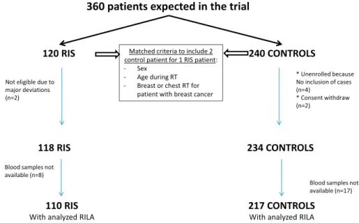
We designed a prospective study scheduled to include 120 patients with RIS who were matched for potential confounding factors with 240 control patients without RIS. For each RIS patient, two control pa-tients were included. The matching criteria were: sex, age during RT (± 5 years), breast or chest RT for patients with breast cancer.
A sample of 120 subjects from the case population and 240 subjects from the control population produce a two-sided 95.0% confidence in-terval with a precision comprised between 5 and 6% assuming a sample AUC between 0.70 and 0.80.
2.2. Inclusion criteria
Fifteen French centers, participating in the French Sarcoma Group (GSF-GETO), included patients in this prospective translational study. This trial was registered on ClinicalTrials.gov under the number NCT01504360. GSF-GETO selected patients alive with RIS from a na-tional register containing all individuals diagnosed with sarcoma and followed by a hospital participating in this trial. The initial sarcoma diag-nosis of RIS patients was systematically reviewed by expert pathologists in France.
The eligibility criteria for control patients were: no soft tissue sar-coma;N5 years complete remission following RT for cancer, adults aged over 18 years for the treatment of primary tumor. Controls were matched to RIS patients according to sex, age at initial radiotherapy (± 5 years) and for initial breast cancer, control patients treated initially for breast cancer had to have undergone the same type of surgery (mastectomy versus lumpectomy) as the associated RIS patient. Inclu-sion of control patients was performed during a standard follow-up consultation.
Specific eligibility criteria for RIS patients included: histologically proven soft tissue sarcoma developed in the previously treated area, adults aged over 18 years for the treatment of the primary tumor; primary tumor other than sarcoma as the initial cancer treated by RT. Inclusion of RIS patients was performed at the moment of RIS diagnosis or during a follow-up consultation.
Bone sarcomas were exclusion criteria for all patients. Patients were not receiving chemotherapy at the time of blood sampling in the pres-ent study, to avoid chemotherapy effect of on RILA results.
Radiation-induced sarcoma (RIS) is a rare but serious adverse event following RT, and overall survival at 5 years for patients veloping such a sarcoma is estimated between 10% and 36%, de-pending on tumor stage at the moment of diagnosis. The development of a biomarker which may predict the risk to develop a RIS will help clinicians to adapt patient treatment and follow up. Recently, a prospective multicenter French trial includingN500 breast-cancer patients suggested that RILA can predict the risk of radio-induced breast fibrosis.
Because RIS is a serious late radio-induced toxicity, the aim of the current study is to assess RILA as a predictive factor for the devel-opment of RIS.
Added value of this study
To our knowledge this is the first translational clinical trial specifi-cally designed to highlight a biomarker which may help to predict risk to develop a RIS. This national multicentric study allowed to analyze a large cohort of patients: 118 RIS patients and 229 matched control patients previously treated by RT free from RIS. Implications of all available evidence
Our study showed that RILA biomarker was significantly cor-related with a risk of RIS development. After validation of these results by an ongoing study in a second cohort of patients, this biomarker will provide clinicians a tool to personalize RT pre-scription in fragile or at risk populations and to personalize pa-tient at risk follow up, allowing treatment at an earlier stage and improvement of disease prognosis.
2.3. Inclusions and shipping procedures for blood samples
Before inclusion, all patients were given by clinicians and signed a written informed consent during a follow-up consultation.
For each included patient, one blood sample was collected in a 5 ml heparinized tube. Blood samples were shipped the same day for RILA analysis (Institut de Cancérologie de Lorraine, Nancy, France) at room temperature using a specialized transporter (Transporteo, Aulnay-sous-Bois, France).
2.4. Radio-induced CD8 T-lymphocyte apoptosis (RILA) analysis
RILA measurement was performed in the Department of Biopathology (Institut de Cancérologie de Lorraine, Nancy, France) after validation of the correlation with results obtained by the tory of Montpellier (IRCM, Inserm U1194, France), the reference labora-tory for this assay [34]. The measurement protocol has been previously described [29].
Briefly, blood was diluted 1:10 in RPMI 1640 medium containing 20% fetal bovine serum, irradiated at 0 Gy (sham samples) or 8 Gy using a linear accelerator (photons, 6 MeV, Clinac iX, Varian Medical Systems). After 48 h of incubation, cells were labeled using FITC-conjugated anti-CD8 monoclonal antibodies (Becton Dickinson, Rungis, France), red blood cells were lysed, and DNA was stained with propidium iodide. Samples were measured in duplicate using aflow cytometer (FACScalibur, Becton Dickinson, San Jose, CA), and data anal-ysis was performed using Cell Quest software (Becton Dickinson, San Jose, CA). Apoptotic CD8 T-lymphocytes were defined as positively-selected FITC-labeled cells (FL2N101) presenting a DNA content lower than 1n.
Each apoptosis evaluation was carried out in triplicate. The RILA measurements were expressed by the delta of RILA percentages after 8Gy minus RILA in non-irradiated controls.
2.5. Statistical analysis
Categorical variables are presented as number and percentage, and compared using the Chi2(using quartile and tertile analysis) or Fisher's
exact test. Continuous variables are described as mean ± standard devi-ation (SD) and/or median (range) and compared using the Student t or non-parametric Mann Whitney test in case of non-normal distribution. Normality was evaluated using Shapiro-Wilk normality test. The area
under the receiver operating characteristic (ROC) curve (AUC) modeling RIS occurrence according to RILA percentage was determined with a 95% confidence interval, using logistic regression. The RILA concentration with the highest Youden index was considered as the optimal cut-off value for the prediction of RIS. A sub-group analysis has been performed in patients with a primary breast cancer, which was the most frequent localization to develop a RIS. However, analysis was a secondary objec-tive and exclusively descripobjec-tive. Statistical analyses were performed using SAS version 9.4 (SAS Institute Inc., Cary, NC, USA). All tests were two sided and p-valuesb.05 were considered significant.
3. Results 3.1. Trial Profile
From November 2011 to July 2016, 120 RIS patients and 236 control patients were enrolled in this study. At baseline, two RIS patients were excluded for major protocol violation (as well as their corresponding four matched controls), two control patients withdrew consent and blood samples were not technically available for 25 patients (8 RIS patients and 17 control patients). Theflowchart of the study population is given inFig. 1.
3.2. Patient, disease and treatment characteristics
Study population characteristics are presented inTable 1. More than 80% of patients enrolled in the study were women, andN75% presented a primary breast tumor. The median age at initial RT was 53 years (range 20.6–91 years for RIS patients and 20–89 years for controls). As per the study design, patients and controls were matched for sex and age, and thus, no difference was observed between groups. No statistical differ-ences were observed between RIS and controls for total RT with 53.7 ± 16 Gy and 57.1 ± 15.1 Gy, respectively (p = .0535). The median total dose of RT was 50 Gy (range 8–150), including 1.8 to 3.9% brachy-therapy as part of the treatment for RIS and control patients, respec-tively. Overall, 37.3% of RIS patients and 43.3% of control patients received chemotherapy delivered in an adjuvant setting after initial RT (p = .294), while 47.1% of RIS patients and 54.8% of control patients re-ceived hormone therapy for their primary cancer (p = .224). For whole population, the median year of initial cancer diagnosis was 2006 [1966–2012]. Concerning RIS patients, the median time between pri-mary tumor treatment by radiotherapy and sarcoma diagnosis was
10.1 [1.8–46.0] years, and the median delay between RIS diagnosis and inclusion in this trial was 11.3 [0.2–352.8] months.
3.3. Analysis of differences in RILA for RIS and control patients
Median RILA was 18.5% (range 5.5–55.7%) and 22.3% (range 3.8–52.2%) for RIS and control patients, respectively, (p = .0008,
Table 2). This difference remained significant after analysis in quartiles (p = .002) or in tertiles using the limits described in two previous pub-lished studies (p = .0292 with limits of 16 and 24%; p = .0004 with limits of 12 and 20%) [29,33]. Using a quartile analysis, the proportion of RILA lesser than 12% was 18.2% for RIS patients versus 10.6% for con-trol patients. Additionally, the proportion of RILA higher than 20% was 38.2% for RIS patients versus 61.3% for control patients.
3.4. Analysis of differences in RILA for RIS and control patients with initial breast cancer
In a subgroup analysis of patients initially treated for breast cancer, median RILA was significantly different for RIS and control patients, at 18.4% (5.5 to 55.7%) and 22.3% (3.8 to 52.2%), respectively, (p = .0015;Table 3). This difference remained significant after analysis in quartiles (p = .0087) or in tertiles using the cut-offs 12% and 20% as
There was no significant association between hormone therapy for the initial cancer and RILA, either in the whole population or in the subpopulation of RIS patients (p = .159 and p = .905, respectively). Conversely, a significant relationship was observed between RILA and hormone therapy in control patients, whereby patients who received hormone therapy were found to have a lower percentage of RILA (p = .026) (Table 4). Concerning these patients, hormonal treatment deliv-ered for initial cancer, was stopped before blood sampling.
Finally, there was no significant relationship between age at the time of RT, and RILA results (pN .74).
3.6. RILA as predictor of secondary sarcoma risk
ROC curves analysis of all study patients or patients with primary breast cancer yielded a RILA cut-off of 21.3%. Thus, all patients with RILA≥21.3% had a comparatively lower risk of developing RIS (p b .0001, OR: 0.358 [0.221–0.599],Fig. 2A), and the results were similar in an analysis limited to patients with primary breast cancer (p = .0003, OR 0.358 [0.205–0.626],Fig. 2B).
The AUCs were 0.622 [0.569–0.679] for all patients and 0.624 [0.560–0.687] for patients with primary breast cancer (Fig. 2A and B, respectively).
4. Discussion
The aim of this translational multicenter study was to investigate the ability of RILA to predict the development of RIS. SARI is thefirst large-size prospective study exploring the biological characteristics of patients who developed RIS after a primary cancer, in contrast with
Male 20 (18.2%) 33 (15.2%) Female 90 (81.8%) 184 (84.8%) Primary tumor localization 0.692 (Fisher exact) Breast 87 (75.9%) 163 (75.1%)
Head and Neck 8 (7.4%) 20 (9.2%)
Pelvis 7 (6.5%) 19 (8.8%)
Other 13 (12%) 15 (6.9%)
RIS histological types
Angiosarcoma 66 (60%)
Leiomyosarcoma 10 (9.1%)
Pleiomorphic sarcoma 9 (8.2%
Uncertain differentiation sarcoma 6 (5.5%)
Liposarcoma 4 (3.6%)
Rhabdomyosarcoma 4 (3.6%)
Others 10 (9.1%)
RTa
total dose 0.0535 (Mann-Whitney)
Mean (standard deviation) 53.7 (16.0) 57.1 (15.1)
Missing data 27 8
RTa
Fraction quantity 0.145 (Mann-Whitney)
Mean (standard deviation) 23.1 (6.3) 24.0 (5.5)
Missing data 76 67
Brachytherapy 2 (1.8%) 8 (3.7%) 0.5046 (Fisher exact)
Chemotherapy 41 (37.3%) 94 (43.3%) 0.294 (Chi2
)
Hormone therapy 52 (47.1%) 119 (54.8%) 0.224 (Chi2
)
Surgery 96 (88.9%) 188 (86.6%) 0.564 (Chi2)
Time from primary tumor to RIS diagnosis (years)
median [min-max] 10.1 [1.8–46.0]
aRadiation therapy. b
Radio-induced sarcoma
Table 2
RILA analysis in patients with RIS and controls. RISc
Controls p-value (test)
N = 110 N = 217 RILA (%)a mean (SDb ) 19.63 (9.1) 22.44 (8.4) 0.0008 (Mann-Whitney) median [min-max] 18.5 [5.5–55.7] 22.3 [3.8–52.2]
Quartile analysis 0.002 (Chi2
)
b15.4 35 (31.8%) 44 (20.3%)
[15.4–21.1[ 36 (32.7%) 47 (21.7%)
[21.1–26.3[ 19 (17.3%) 64 (29.5%)
≥26.3 20 (18.2%) 62 (28.6%)
Tertile analysis according to (Ozsahin et al. 2005) 0.0292 (Chi2 ) b16 38 (34.55%) 48 (22.12%) [16–24] 45 (40.91%) 92 (42.40%) N24 27 (24.55%) 77 (35.48%)
Tertile analysis according to (Azria et al. 2015)
0.0004 (Chi2)
b12 20 (18.18%) 23 (10.60%)
[12−20] 48 (43.64%) 61 (28.11%)
N20 42 (38.18%) 133 (61.29%)
aRadiation-Induced CD8 T-Lymphocyte Apoptosis. b Standard Deviation.
c
previous publications, which focused mainly on radio-induced breast angiosarcoma [35–37]. Our results found a median latency from RT to the diagnosis of RIS of 10.1 years, consistent with other studies indicat-ing a period between 6 and 20 years [16–18,38–40].
We initially selected adult patients with sarcoma within a group of RIS patients to avoid potential confounding factors for sarcoma develop-ment. None of our patients had identified risk factors for developing RIS such as unspecific genetics factors (Li-Fraumeni-Syndrome [41]), young age at radiotherapy or concomitant chemotherapy associated with sec-ondary carcinoma [39,40]. To minimize the role of potential prior risk factors, RIS patients were matched with control patients exhibiting similar characteristics (age at RT, sex and breast cancer localization to reduce RT dose and variability of fractionation). The proportion of
patients receiving chemotherapy was similar between these two co-horts. This result allowed to reduce the impact of this type of treatment on RIS development, as previously described for anthracycline mole-cules, as well as other cytotoxic agents, a known risk factor associated with secondary sarcomas in childhood cancer survivors [42]. However, in our study we didn't collect chemotherapy details for all patients. Therefore, the relationship between anthracycline treatment and RIS development was not specifically evaluated in our cohort.
In order to develop personalized medicine in RT, clinicians need to be able to predict potential radio-induced adverse events such as sec-ondary sarcomas, which are rare but deleterious. In the literature, the five-year survival rate for RIS patients is estimated to be between 10 and 36% [2–7], lower than the survival rate for patients with sporadic sarcomas (from 54% to 76%) [11,15,36,43]. Previous studies have also revealed that variations in prognostic criteria yield a significant differ-ence in overall survival of RIS patients. For example, sarcoma-related survival was significantly better for patients with a RIS tumor of b5 cm (p = .008) [35]. Firstly, patients at risk should be offered alternative therapeutic options to minimize the risk of severe late events. Secondly, patients at risk of developing RIS should have a personalized follow-up, which could enable screening and earlier diagnosis of tumors, thus of-fering a better chance of cure. A personalized follow up of patients un-dergoing RT would require a robust biological marker able predict the development of RIS. Ideally, this marker should be inexpensive, have a simple quantification method and allow a large scale use. The RILA bio-marker has already been described as a predictor of late RT-related ad-verse event, such as the risk of grade≥ 2 radio-induced breast fibrosis using the CTCAE validated scale [33]. Currently, the pioneer team that developed the RILA parameter, didn't completely elucidate the mecha-nism linking RILA and radio-induced toxicity. It has been suggested that this relationship could be due to a slow apoptotic response and en-hancement of cytokine production, leading to tissue inflammatory cell infiltration [29]. A specific study, using a proteomic approach, is pres-ently ongoing to evaluate the underlying mechanism [33].
Although a large majority of our patients had a primary breast cancer, our analysis was performed in the whole population, as established by the protocol. Identical results were observed when the analysis was re-stricted to the breast cancer sub-population. Concerning other initial cancer localizations, as head and neck and pelvis, results could not be
Table 4
Relationship between clinical parameters and treatment other than RT and RILA in RIS and control patients.
Whole population RISe
Controls
n RILA (%) mean (SD)a,b
n RILA (%) mean (SD)a,b
n RILA (%) mean (SD)a,b
Chemotherapy Yes 135 20.7 (7.6) 41 19.1 (7.8) 94 21.4 (7.4) No 192 22.1 (9.5) 69 20.0 (9.9) 123 23.2 (9.1) P value (test) 0.353 (MWc ) 0.853 (MWc ) 0.218 (MWc ) Hormonotherapy Yes 171 20.8 (8.5) 52 19.4 (9.1) 119 21.4 (8.3) No 155 22.2 (9.0) 57 19.8 (9.3) 98 23.7 (8.4) P value (test) 0.104 (MWc ) 0.968 (MWc ) 0.018 (MWc ) Age during RTd b 53 years old 143 21.2 (7.3) 41 18.7 (6.7) 102 22.2 (7.3) ≥ 53 years old 158 21.8 (10.2) 47 29.5 (11.9) 111 22.8 (9.3) p value (test) 0.893 (MWc ) 0.398 (MWc ) 0.760 (MWc ) Delay between RTd
and sarcoma diagnosis
b 10 years NA NA 53 19.9 (9.6) NA NA
≥ 10 years NA NA 57 19.4 (8.7) NA NA
p value (test) 0.879 (MWc
)
a
Radiation-Induced CD8 T-Lymphocyte Apoptosis.
b Standard Deviation. c Mann Whitney. d Radiation Therapy. eRadio-Induced Sarcoma. Table 3
RILA analysis for RIS and Control patients with a breast cancer as primary disease.
RISc Controls p-value (test)
N = 82 N = 163
RILA (%)a
mean (SD)b 19.1 (9.2) 22.36 (8.6) 0.0015
(Mann-Whitney) median [min-max] 18.4 [5.5–55.7] 22.3 [3.8–52.2]
Quartile analysis 0.0087 (Chi2)
b15.1 30 (36.6%) 34 (20.9%)
[15.1–20.7[ 24 (29.3%) 37 (22.7%)
[20.7–26.2[ 14 (17.1%) 46 (28.2%)
≥ 26.2 14 (17.1%) 46 (28.2%)
Tertile analysis according to (Ozsahin et al. 2005)
b16 30 (36.59%) 38 (23.46%) 0.0564 (Chi2
)
[16–24] 33 (40.24%) 69 (42.59%)
N24 19 (23.17%) 56 (33.95%)
Tertile analysis according to (Azria et al. 2015) b12 17 (20.73%) 17 (10.43%) 0.0017 (Chi2 ) [12–20] 35 (42.68%) 48 (29.45%) N20 30 (36.59%) 99 (60.12%) a
Radiation-Induced CD8 T-Lymphocyte Apoptosis.
b
Standard Deviation.
c
rigorously validated due to lack of statistical power. Another subgroup analysis concerning only patients with non-breast cancer as primary dis-ease didn't highlight significant relationship between RIS and RILA (Sup-plementary Table 1). These results were be able to be explain by a specific relationship between RIS and RILA only for breast cancer as pri-mary disease or by a lack of statistical power due to a low numbers of an-alyzed patients for this analysis (28 RIS patients and 54 control patients). Given that for RIS patients, blood was collected after RIS diagnosis and that for control patients, blood was collected during follow-up and the majority of these patients had a relapse free status, we cannot totally exclude that RILA fraction might be a marker of tumor presence in general, rather than a predictor of RIS. To decrease this risk, we com-pared our cohort results with other trials evaluating RILA for patients with or without cancer at sample time. In our RIS cohort, RILA median was 18.5% [5.5–55.7] and in our control patient cohort the RILA median was 22.3% [3.8–52.2], in a large breast cancer cohort (n = 456; [33]) the RILA median was 15.2% [0.7–52.8], and finally in the EPOPA cohort [44] which analyzed 224 patients treated by radiotherapy for a prostate can-cer since several years, the majority of these patients presented a re-lapse free status, the RILA median was 15.3% [4.4–37.7]. These data didn't highlight an increase in RILA in patients without cancer, suggest-ing that the difference of RILA between our two cohorts were indepen-dent of cancer presence at the moment of RILA analysis.
In our present study, with an AUC of 0.62, the 21.3% RILA cut off was low in sensitivity and specificity and, consequently, we cannot promote the use of this biomarker alone. It will be of key importance to build a specific nomogram model combining relevant data such as genetic markers, to improve sensitivity and specificity for event prediction on a population basis. Afinal validation process in a prospective random-ized phase III trial would be necessary before introducing RILA testing in routine practice for patients with significant long time overall sur-vival probability after radiotherapy. For patients identified with a high risk of developing RIS, radiation oncologists should be in a position to
inform the patient, to discuss alternative therapeutic options and to sug-gest early personalized follow-up with non-invasive methods such as relevant imaging.
Supplementary data to this article can be found online athttps://doi.
org/10.1016/j.ebiom.2019.02.031.
Data Sharing
No data sharing plan has been notified in informed consent form. Consequently, individual data could not be share. However, anonymized and statistical data are available and might be verified.
Declaration
CGFL (Centre Georges François Leclerc, Dijon, FRANCE) received an unrestricted educational grant (PHRC 2011) to cover the expenses of the investigators for undertaking this trial. All authors declared no con-flict of interests related to this article.
Author contributions
P.M, F·B, F.C., and C.M. were involved in the conception and design of the study. G.T, G.N., J.T., J.D., P.S., S.RO., I.RC., X.L., JL.L., MA.M., P.M., JL.M. were involved in the provision of patients, data acquisition and helped to draft the report.
P.M. and C.M. supervised the study. A.B. was in charge of the sta-tistical analysis of the study. C.M., P.M. and A.B. had access to the raw data and analyzed and interpreted the data. C.M., P.M., A.B. were in-volved in report writing, which was corrected and approved by all authors.
Fig. 2. ROC curve analysis to determine RILA cutoffs and to calculate sensitivity and specificity (all patients in A, only patients with primary breast cancer in B). RILA: radiation-induced CD8 T-lymphocyte apoptosis.
Roles of the funding sources
Funding sources didn't participate in the study's design and didn't access the data generated by this trial.
Acknowledgements
This research was supported by the French INCa “Program Hospitalier de Recherche Clinique (PHRC)”. The authors would like to thank JM Coindre for his sarcoma expertise, C Grisi, E Rederstorff, and J Skrzypski for their help organizing this multicenter study, all clinical research technicians for collecting clinical data and J Blanc for her signif-icant help with statistical analysis. The authors also thank M Husson for performing RILA testing. They also thank F Ecarnot (EA3920, University Hospital Besançon, France) and I Gregoire (medical writer, CGFL, Dijon, France) for critical review and editorial assistance.
References
[1]Lindholm C, Cavallin-Stahl E, Ceberg J, Frodin JE, Littbrand B, Moller TR, et al. Radio-therapy practices in Sweden compared to the scientific evidence. Acta Oncol 2003; 42(5–6):416–29.
[2]Brady MS, Garfein CF, Petrek JA, Brennan MF. Post-treatment sarcoma in breast can-cer patients. Ann Surg Oncol 1994;1(1):66–72.
[3]Fraga-Guedes C, Gobbi H, Mastropasqua MG, Botteri E, Luini A, Viale G. Primary and secondary angiosarcomas of the breast: a single institution experience. Breast Can-cer Res Treat 2012;132(3):1081–8.
[4]Jallali N, James S, Searle A, Ghattaura A, Hayes A, Harris P. Surgical management of radiation-induced angiosarcoma after breast conservation therapy. Am J Surg 2012;203(2):156–61.
[5]Kirova YM, Vilcoq JR, Asselain B, Sastre-Garau X, Fourquet A. Radiation-induced sar-comas after radiotherapy for breast carcinoma: a large-scale single-institution re-view. Cancer 2005;104(4):856–63.
[6]West JG, Qureshi A, West JE, Chacon M, Sutherland ML, Haghighi B, et al. Risk of angiosarcoma following breast conservation: a clinical alert. Breast J 2005;11(2): 115–23.
[7]Yap J, Chuba PJ, Thomas R, Aref A, Lucas D, Severson RK, et al. Sarcoma as a second malignancy after treatment for breast cancer. Int J Radiat Oncol Biol Phys 2002;52 (5):1231–7.
[8]Arlen M, Higinbotham NL, Huvos AG, Marcove RC, Miller T, Shah IC. Radiation-induced sarcoma of bone. Cancer 1971;28(5):1087–99.
[9]Cahan WG, Woodard HQ, Higinbotham NL, Stewart FW, Coley BL. Sarcoma arising in irradiated bone: report of eleven cases. 1948 Cancer 1998;82(1):8–34.
[10]Gladdy RA, Qin LX, Moraco N, Edgar MA, Antonescu CR, Alektiar KM, et al. Do radiation-associated soft tissue sarcomas have the same prognosis as sporadic soft tissue sarcomas? J Clin Oncol 2010;28(12):2064–9.
[11]Huvos AG, Woodard HQ, Cahan WG, Higinbotham NL, Stewart FW, Butler A, et al. Postradiation osteogenic sarcoma of bone and soft tissues. A clinicopathologic study of 66 patients. Cancer 1985;55(6):1244–55.
[12]Huang J, Mackillop WJ. Increased risk of soft tissue sarcoma after radiotherapy in women with breast carcinoma. Cancer 2001;92(1):172–80.
[13]Taghian A, de Vathaire F, Terrier P, Le M, Auquier A, Mouriesse H, et al. Long-term risk of sarcoma following radiation treatment for breast cancer. Int J Radiat Oncol Biol Phys 1991;21(2):361–7.
[14]Mark RJ, Poen J, Tran LM, Fu YS, Selch MT, Parker RG. Postirradiation sarcomas. A single-institution study and review of the literature. Cancer 1994;73(10):2653–62.
[15]Cha C, Antonescu CR, Quan ML, Maru S, Brennan MF. Long-term results with resec-tion of radiaresec-tion-induced soft tissue sarcomas. Ann Surg 2004;239(6):903–9 [dis-cussion 9-10].
[16]Murray EM, Werner D, Greeff EA, Taylor DA. Postradiation sarcomas: 20 cases and a literature review. Int J Radiat Oncol Biol Phys 1999;45(4):951–61.
[17]Lagrange JL, Ramaioli A, Chateau MC, Marchal C, Resbeut M, Richaud P, et al. Sar-coma after radiation therapy: retrospective multiinstitutional study of 80 histologi-cally confirmed cases. Radiation therapist and pathologist groups of the federation Nationale des centres de Lutte Contre le Cancer. Radiology 2000;216(1):197–205.
[18]Chan JY, Wong ST, Lau GI, Wei WI. Postradiation sarcoma after radiotherapy for na-sopharyngeal carcinoma. Laryngoscope 2012;122(12):2695–9.
[19]Garrido-Colmenero C, Valenzuela-Salas I, Blasco-Morente G, Aneiros-Fernandez J, Tercedor-Sanchez J. Photoletter to the editor: Postradiation sarcoma. J Dermatol Case Rep 2016;10(1):17–8.
[20]Holt GE, Thomson AB, Griffin AM, Bell R, Wunder J, Rougraff B, et al. Multifocality and multifocal postradiation sarcomas. Clin Orthop Relat Res 2006;450:67–75.
[21]Olson MT, Wakely Jr PE, Weber K, Siddiqui MT, Ali SZ. Postradiation sarcoma: mor-phologicalfindings on fine-needle aspiration with clinical correlation. Cancer Cytopathol 2012;120(5):351–7.
[22]Sule N, Xu BO, El Zein D, Szigeti K, George S, Kane JM, et al. Radiation-induced Chondrosarcoma of the bladder. Case report and review of literature. Anticancer Res 2015;35(5):2857–60.
[23]Mentzel T, Schildhaus HU, Palmedo G, Buttner R, Kutzner H. Postradiation cutaneous angiosarcoma after treatment of breast carcinoma is characterized by MYC amplifi-cation in contrast to atypical vascular lesions after radiotherapy and control cases: clinicopathological, immunohistochemical and molecular analysis of 66 cases. Mod Pathol 2012;25(1):75–85.
[24]Bentzen SM, Constine LS, Deasy JO, Eisbruch A, Jackson A, Marks LB, et al. Quantita-tive analyses of Normal tissue effects in the clinic (QUANTEC): an introduction to the scientific issues. Int J Radiat Oncol Biol Phys 2010;76(3 Suppl):S3–9.
[25]Bentzen SM, Overgaard J. Patient-to-patient variability in the expression of radiation-induced Normal tissue injury. Semin Radiat Oncol 1994;4(2):68–80.
[26]Bourgier C, Lacombe J, Solassol J, Mange A, Pelegrin A, Ozsahin M, et al. Late side-effects after curative intent radiotherapy: identification of hypersensitive patients for personalized strategy. Crit Rev Oncol Hematol 2015;93(3):312–9.
[27]Crompton NE, Ozsahin M. A versatile and rapid assay of radiosensitivity of periph-eral blood leukocytes based on DNA and surface-marker assessment of cytotoxicity. Radiat Res 1997;147(1):55–60.
[28]Ozsahin M, Ozsahin H, Shi Y, Larsson B, Wurgler FE, Crompton NE. Rapid assay of in-trinsic radiosensitivity based on apoptosis in human CD4 and CD8 T-lymphocytes. Int J Radiat Oncol Biol Phys 1997;38(2):429–40.
[29]Ozsahin M, Crompton NE, Gourgou S, Kramar A, Li L, Shi Y, et al. CD4 and CD8 T-lymphocyte apoptosis can predict radiation-induced late toxicity: a prospective study in 399 patients. Clin Cancer Res 2005;11(20):7426–33.
[30]Foro P, Algara M, Lozano J, Rodriguez N, Sanz X, Torres E, et al. Relationship between radiation-induced apoptosis of T lymphocytes and chronic toxicity in patients with prostate cancer treated by radiation therapy: a prospective study. Int J Radiat Oncol Biol Phys 2014;88(5):1057–63.
[31]Schnarr K, Boreham D, Sathya J, Julian J, Dayes IS. Radiation-induced lymphocyte ap-optosis to predict radiation therapy late toxicity in prostate cancer patients. Int J Radiat Oncol Biol Phys 2009;74(5):1424–30.
[32]Borgmann K, Hoeller U, Nowack S, Bernhard M, Roper B, Brackrock S, et al. Individual radiosensitivity measured with lymphocytes may predict the risk of acute reaction after radiotherapy. Int J Radiat Oncol Biol Phys 2008;71(1):256–64.
[33]Azria D, Riou O, Castan F, Nguyen TD, Peignaux K, Lemanski C, et al. Radiation-induced CD8 T-lymphocyte apoptosis as a predictor of breastfibrosis after radio-therapy: results of the prospective Multicenter French trial. EBioMedicine 2015;2 (12):1965–73.
[34]Mirjolet C, Merlin JL, Dalban C, Maingon P, Azria D. Correlation between radio-induced lymphocyte apoptosis measurements obtained from two French centres. Cancer Radiother 2016;20(5):391–4.
[35]Bjerkehagen B, Smastuen MC, Hall KS, Skjeldal S, Smeland S, Fossa SD. Why do pa-tients with radiation-induced sarcomas have a poor sarcoma-related survival? Br J Cancer 2012;106(2):297–306.
[36]Bjerkehagen B, Smeland S, Walberg L, Skjeldal S, Hall KS, Nesland JM, et al. Radiation-induced sarcoma: 25-year experience from the Norwegian radium hospi-tal. Acta Oncol 2008;47(8):1475–82.
[37]Berrington de Gonzalez A, Kutsenko A, Rajaraman P. Sarcoma risk after radiation ex-posure. Clin Sarcoma Res 2012;2(1):18.
[38]Depla AL, Scharloo-Karels CH, de Jong MA, Oldenborg S, Kolff MW, Oei SB, et al. Treatment and prognostic factors of radiation-associated angiosarcoma (RAAS) after primary breast cancer: a systematic review. Eur J Cancer 2014;50(10): 1779–88.
[39]Hawkins MM, Wilson LM, Burton HS, Potok MH, Winter DL, Marsden HB, et al. Ra-diotherapy, alkylating agents, and risk of bone cancer after childhood cancer. J Natl Cancer Inst 1996;88(5):270–8.
[40]Menu-Branthomme A, Rubino C, Shamsaldin A, Hawkins MM, Grimaud E, Dondon MG, et al. Radiation dose, chemotherapy and risk of soft tissue sarcoma after solid tumours during childhood. Int J Cancer 2004;110(1):87–93.
[41]Moppett J, Oakhill A, Duncan AW. Second malignancies in children: the usual sus-pects? Eur J Radiol 2001;38(3):235–48.
[42]Henderson TO, Rajaraman P, Stovall M, Constine LS, Olive A, Smith SA, et al. Risk fac-tors associated with secondary sarcomas in childhood cancer survivors: a report from the childhood cancer survivor study. Int J Radiat Oncol Biol Phys 2012;84(1): 224–30.
[43]Thijssens KM, van Ginkel RJ, Suurmeijer AJ, Pras E, van der Graaf WT, Hollander M, et al. Radiation-induced sarcoma: a challenge for the surgeon. Ann Surg Oncol 2005;12(3):237–45.
[44]Vogin G, Merlin JL, Rousseau A, Peiffert D, Harle A, Husson M, et al. Absence of cor-relation between radiation-induced CD8 T-lymphocyte apoptosis and sequelae in patients with prostate cancer accidentally overexposed to radiation. Oncotarget 2018;9(66):32680–9.