HAL Id: hal-01962071
https://hal.sorbonne-universite.fr/hal-01962071
Submitted on 20 Dec 2018HAL is a multi-disciplinary open access archive for the deposit and dissemination of sci-entific research documents, whether they are pub-lished or not. The documents may come from teaching and research institutions in France or abroad, or from public or private research centers.
L’archive ouverte pluridisciplinaire HAL, est destinée au dépôt et à la diffusion de documents scientifiques de niveau recherche, publiés ou non, émanant des établissements d’enseignement et de recherche français ou étrangers, des laboratoires publics ou privés.
Physicochemical characterization of inorganic deposits
associated with granulomas in cutaneous sarcoidosis
H. Colboc, P. Moguelet, D. Bazin, C. Bachmeyer, V. Frochot, R. Weil, E.
Letavernier, C. Jouanneau, M. Daudon, J.F. Bernaudin
To cite this version:
H. Colboc, P. Moguelet, D. Bazin, C. Bachmeyer, V. Frochot, et al.. Physicochemical characterization of inorganic deposits associated with granulomas in cutaneous sarcoidosis. Journal of the European Academy of Dermatology and Venereology, Wiley, 2019, 33 (1), pp.198-203. �10.1111/jdv.15167�. �hal-01962071�
Accepted
Article
MRS. HESTER COLBOC (Orcid ID : 0000-0001-8756-9862)
Article type : Short Report
Physicochemical characterization of inorganic deposits associated with granulomas in cutaneous sarcoidosis.
Running head: Inorganic deposits in skin sarcoidosis
H. Colboc1,8,10, P. Moguelet2,10, D. Bazin3,4,10, C. Bachmeyer5, V. Frochot6,7, R. Weil4, E.
Letavernier6,7 , C. Jouanneau7 , M. Daudon6,7,11 , J.F. Bernaudin8,9,11
1. AP-HP, Hôpital Rothschild, Dermatologie, Paris, France,
2. AP-HP, Hôpital Tenon, Anatomie et Cytologie Pathologiques, Paris, France,
3. Sorbonne Université, UPMC Université Paris 06, CNRS, Collège de France, Laboratoire
de Chimie de la Matière Condensée de Paris, 11 place Marcelin Berthelot, 75005, Paris,
France,
4. CNRS, LPS, Ba510, Université Paris XI, 91405 Orsay, France,
5. AP-HP, Hôpital Tenon, Médecine interne, Paris, France,
Accepted
Article
7. AP-HP, Hôpital Tenon, Explorations Fonctionnelles Multidisciplinaires, Paris, France,
8. Sorbonne Université, UPMC Université Paris 06, Paris, France,
9. AP-HP Service de pneumologie, Hôpital Avicenne 93000 Bobigny France
10. These authors contributed equally to this work.
11. These authors should be considered joint last authors
Corresponding author: Hester Colboc, MD Hôpital Rothschild 5, rue Santerre 75012 Paris, France Tel : 01 40 19 33 49 Fax : 01 40 19 33 97 [email protected]
Conflicts of interest: none declared
Accepted
Article
Abstract
Background. Sarcoidosis, characterized by epithelioid granulomas, is considered to be caused
by a complex interplay between genetics and environmental agents. It has been hypothesized
that exogenous inorganic particles as crystalline silica could be a causal or adjuvant agent in
sarcoidosis onset.
Objectives. To investigate the location, frequency and physicochemical characteristics of
foreign materials and mineral tissue deposits in the granulomatous area of cutaneous
sarcoidosis.
Methods. Skin biopsies (n=14) from patients diagnosed with cutaneous sarcoidosis (mean age
43 years; 11 patients with extra-cutaneous involvement) were investigated using polarized
light examination (PLE), µFourier Transform Infra-Red (µFT-IR) spectroscopy and Field
Emission Scanning Electron Microscopy coupled with Energy Dispersive X-ray
Spectroscopy (FE-SEM/EDX)
Results. Combined PLE, µFT-IR, FE-SEM/EDX analysis allowed to characterize mineral
deposits in 7/14 biopsies (50%). It identified crystalline silica (SiO2) inside granulomas in 3
biopsies and calcite (CaCO3) at their periphery in 4.
Conclusion. This study emphasizes the need of using combined methods for assessment of
mineral deposits in granulomatous diseases. According to the location and characteristics of
deposits we can hypothesize that SiO2 particles contribute to the granuloma formation,
whereas CaCO3 deposits are related to the granuloma biology. However, the significance of
Accepted
Article
Introduction
Sarcoidosis is characterized by an exaggerated granulomatous response mostly
affecting the lung, skin and eye, locations particularly exposed to exogenous particulates.1-2
Granuloma mostly constituted of macrophage lineage cells is thought to be controlled by
innate as well as adaptive immune mechanisms in a complex combination of genetic
susceptibility, immune networks and infectious and/or environmental agents.3
Sarcoidosis affects the skin in 25-30% of patients and manifests by a wide range of
clinical cutaneous lesions.4-5 It is well known that sarcoidosis preferentially affects sites with
a prior injury as tattoos or scars. Polarizable material has been reported up to 22% of the
cases suggesting that foreign material could be a nidus for granuloma formation and a
potential trigger for the disease.6-8
This study, based on a series of cutaneous biopsies of sarcoidosis, was designed to
investigate the frequency and physicochemical characterization of mineral deposits in the
granulomatous areas. To describe their characteristics at the subcellular scale as well as their
chemical nature, in addition to polarized light examination (PLE), two different techniques of
physicochemical analysis were used: µFourier Transform Infra-Red (µFT-IR) spectroscopy
and Field Emission Scanning Electron Microscopy coupled with Energy Dispersive X-ray
Spectroscopy (FE-SEM/EDX).9 Such a combination of methods has been amply
demonstrated to be particularly effective for detection and physicochemical identification of
Accepted
Article
Material and methods
Skin biopsies, histopathology and polarized light examination
The study was conducted in compliance with Good Clinical Practices and the Declaration of
Helsinki, according to French law.
A retrospective monocentric study was conducted, from January 2012 to December 2015.
Consecutive patients diagnosed with skin sarcoidosis during this period, based on clinical
data and histopathology results, were included. Clinical and biological data, including
demographic, occupation, past medical history, extra-cutaneous involvement of the disease,
clinical description and location of the skin lesions, calcium and angiotensin converting
enzyme serum levels were collected.
Five samples from negative margins of skin carcinoma resections were included and further
described as controls.
Multiples sections of 1.5 µm from paraffin embedded skin biopsies were obtained and
deposited on two types of slides: glass slides, for hematoxylin-eosin-saffron (HES), and
low-e microscoplow-e slidlow-es (MirrIR, Klow-evllow-ey Tlow-echnologilow-es, Tilow-enta Scilow-enclow-es, Indianapolis) for
FE-SEM/EDX and µFT-IR. The location (superficial and deep dermis, sub-cutis) and number of
granulomas observed per biopsy were evaluated. A screening by PLE was done by two
different pathologists.
Skin sections were analysed and compared between cases and controls, for the presence of
Accepted
Article
Physicochemical analysis of mineral deposits
The tools and methods have been extensively described in a thematic issue of the Comptes Rendus Chimie de l’Académie des Sciences Paris France.11
Briefly, for µFT-IR, tissue sections deposited on low-e microscope slides were analyzed with
the Spotlight 400 FTIR imaging System (Perkin Elmer Life Sciences - Courtaboeuf, France),
in the mid infrared (4000-700 cm-1) spectral range to obtain infrared maps of tissue slides at
high spatial resolution, down to 10 µm.9
For FE-SEM/EDX the sections previously used for µFT-IR were observed on a Zeiss
SUPRA55-VP SEM (Zeiss SUPRA55-VP - Oberkochen, Germany) equipped with in-lens SE
and Everharte Thornley SE secondary electron detectors.12 Energy Dispersive X-ray
spectroscopy (EDX) coupled to FE-SEM observation was used to confirm the nature of the
deposits.
Results
Skin biopsies, histopathology and polarized light examination (PLE)
Sixteen patients were diagnosed with skin sarcoidosis at Tenon Hospital from January 2012
to December 2015. Two patients were excluded because the tissue sample available for
analysis was too small. Finally, cutaneous biopsies from 14 patients (9 men; 5 women) were
selected for further analysis. Demographic, clinical and histopathological characteristics of all
patients are summarized in Table 1 and for each patient in table 2 (supplementary material).
Accepted
Article
(n= 10). One of the three patients without extracutaneous localization, (patient 8 in table 2
supplementary material) had a single granulomatous lesion of the forehead and the diagnosis of “localized sarcoidosis-like granulomatous reaction” could not be excluded. For PLE from
4 to 33 (mean= 16) sections per biopsy were analyzed. Refractive material was detected in 5
biopsies, restricted to one or two granulomas per section (Fig. 1). Interstitial peri
granulomatous fibrosis was observed in 13 cases without any polarized signal observed in
this area.
Physicochemical analysis of mineral deposits
Intra granuloma mineral silica (SiO2) deposits
In 3 biopsies with PLE refractive material, µFT-IR characterized mineral silica deposits (Fig.
2).
FE-SEM observation showed that silica particles were inside the granulomas. In two samples,
the deposits were sharp, unique, irregular objects more than 10µm long. In the third, the
deposit was an aggregate of very small objects some of them at the nanoparticle scale. No
other nanoparticles of SiO2 were observed within the granuloma or in its vicinity. EDX
analysis confirmed the presence of silica in these 3 samples (Fig. 2).
Peripheral calcium carbonate (CaCO3) deposits
µFT-IR analysis of 4 biopsies detected calcite (CaCO3) deposits in the form of calcite at the
Accepted
Article
deposits of calcite located between the collagen fibers (Fig. 3). Two of these 4 patients had
hypercalcemia.
Controls
Similar analyses were performed on the 5 samples from the control group, no deposits were
found either in dermis or subcutis.
4. Discussion
Using combined methods, we yield comprehensive information on the location and
physicochemical nature of inorganic deposits in granulomas (crystalline silica SiO2) and
perigranuloma tissues (calcite CaCO3) in patients with cutaneous sarcoidosis. We identified
crystalline silica (SiO2) inside granulomas in 3 biopsies and calcium carbonate (CaCO3) at
their periphery in 4.
Refractive material was observed by PLE in 5 biopsies (35%) which is in accordance with the
20%-78% reported range.7 In our series, µFT-IR showed silica deposits in 3 biopsies (21%)
which has been confirmed to be crystalline silica by FE-SEM/EDX. For one of these biopsies
the PLE examination was negative, while for 3 PLE positive samples infrared spectroscopy
identified calcium carbonate deposits.
Crystalline silica was identified in granulomas in 3 biopsies from environment exposed skin
areas (elbow, arm and forehead). This finding is in keeping with the origin of SiO2 deposits in
relation to environmental or occupational exposure. For one patient the area has previously
Accepted
Article
bodies. Accordingly to epidemiological studies, crystalline silica has been suggested to play
a role in sarcoidosis occurrence particularly after environmental or occupational exposure.13
However, in the present study identification of SiO2 particles has been a particularly rare
event affecting at least 1 granuloma section among a mean of 75 analyzed per biopsy. But it
cannot be excluded that the frequency of deposits was largely underestimated. As each
granuloma has a mean diameter of at least 150-200µm,14 5µm sections correspond to a very
limited fraction of the lesion. In addition, loss of these hard particles deposits may occur
during the processing of tissue sample particularly at the sectioning step. SEM examination
showed that SiO2 particles were observed as unique intracellular deposits. It has been
hypothesized that nanoparticles of silica undetected by PLE could be a causative agent in
sarcoidosis.15 The significance of the frequent association between foreign material and
sarcoidosis is still controversial.8 Foreign bodies may be initial triggers for sarcoidosis
constituting the nidus for granulomas in patients with a particular genetic background having
an impaired capacity to handle particulate foreign material.16-17 Conversely another
hypothesis is to consider granuloma in sarcoidosis as a host related inflammatory/ immune
reaction non-specific of a special antigen or particulate occurring at sites where potential incidental triggering agents (scars, foreign material, antigens…) are already located. In line
with this hypothesis is the observation of granuloma recurrence on tattoos or scars in parallel
with exacerbation or relapse of systemic sarcoidosis.18
Calcite deposits were observed at distance from granulomas, within the connective tissue of
dermis in 4 patients, two with hypercalcemia. Calcium oxalate and phosphate deposits have
been reported in sarcoidosis, however, to the best of our knowledge, we report the first cases
of calcite deposits.19 Presence at the nanometer scale of these deposits, including in patients
Accepted
Article
tissues around granulomas could be a common not yet highlighted finding in conjunction
with the role of the granuloma in vitamin D and calcium metabolism.20
In conclusion such a study needs to be extended to samples from other organs affected by
sarcoidosis, mostly lung and mediastinal lymph nodes exposed to environmental and
occupational pollutants. Moreover the consequence of calcite deposits in organs where
granulomas develop should be investigated.
Acknowledgments
We thank Pr Camille Frances, from the department of Dermatology Tenon Hospital Paris, Prs
Dominique Valeyre and Jean-Paul Battesti, from Avicenne Hospital Bobigny France, and PA
Rosental from Sciences Po Paris for their constant support and fruitful discussions.
References
1 Valeyre D, Prasse A, Nunes H, Uzunhan Y, Brillet P-Y, Müller-Quernheim J.
Sarcoidosis. Lancet 2014; 383:1155‑1167.
2 Baughman RP, Teirstein AS, Judson MA, et al. Clinical characteristics of patients in a
case control study of sarcoidosis. Am J Respir Crit Care Med 2001; 164:1885‑1889.
3 Müller-Quernheim J, Schürmann M, Hofmann S, et al. Genetics of sarcoidosis. Clin
Chest Med 2008; 29: 391‑414.
Accepted
Article
5 Haimovic A, Sanchez M, Judson MA, Prystowsky S. Sarcoidosis: a comprehensive
review and update for the dermatologist: part II. Extracutaneous disease. J Am Acad
Dermatol 2012; 66: 719-730.
6 Callen JP. The presence of foreign bodies does not exclude the diagnosis of sarcoidosis.
Arch Dermatol 2001; 137: 485‑486.
7 Marcoval J, Moreno A, Maña J. Foreign bodies in cutaneous sarcoidosis. J Cutan Pathol
2004; 31: 516.
8 Sepehri M, Hutton Carlsen K, Serup J. Papulo-Nodular Reactions in Black Tattoos as
Markers of Sarcoidosis: Study of 92 Tattoo Reactions from a Hospital Material.
Dermatol Basel Switz 2016; 232: 679‑686.
9 Bazin D, Daudon M. Some advances in the field of physico-chemical characterization of
pathological microcrystals. Ann Biol Clin 2015; 73: 517-534.
10 Dessombz A, Bazin D, Dumas P, Sandt C, Sule-Suso J, Daudon M. Shedding light on the
chemical diversity of ectopic calcifications in kidney tissues: diagnostic and research
aspects. PloS One 2011; 6: e28007.
11 Colboc H, Bazin D, Moguelet P, et al. Detection of silica and calcium carbonate deposits
in granulomatous areas of skin sarcoidosis by μFourier transform infrared spectroscopy
and Field Emission Scanning Electron Microscopy coupled with Energy Dispersive
X-ray Spectroscopy analysis. Comptes Rendus Chim 2016; 19: 1631‑1641.
Accepted
Article
13 Rafnsson V, Ingimarsson O, Hjalmarsson I, Gunnarsdottir H. Association between
exposure to crystalline silica and risk of sarcoidosis. Occup Environ Med 1998; 55:
657‑660.
14 Kambouchner M, Pirici D, Uhl J-F, Mogoanta L, Valeyre D, Bernaudin J-F. Lymphatic
and blood microvasculature organisation in pulmonary sarcoid granulomas. Eur Respir J
2011; 37: 835‑840.
15 Heffner DK. The cause of sarcoidosis: the Centurial enigma solved. Ann Diagn Pathol
2007; 11: 142‑152.
16 Walsh NM, Hanly JG, Tremaine R, Murray S. Cutaneous sarcoidosis and foreign bodies.
Am J Dermatopathol 1993; 15: 203‑207.
17 Fingerlin TE, Hamzeh N, Maier LA. Genetics of Sarcoidosis. Clin Chest Med 2015; 36:
569‑584.
18 Schiavo AL, Ruocco E, Gambardella A, O’Leary RE, Gee S. Granulomatous dysimmune
reactions (sarcoidosis, granuloma annulare, and others) on differently injured skin areas.
Clin Dermatol 2014; 32: 646‑653.
19 Reid JD, Andersen ME. Calcium Oxalate in Sarcoid Granulomas: With Particular
Reference to the Small Ovoid Body and a Note on the Finding of Dolomite. Am J Clin
Pathol 1988; 90: 545‑558.
20 Berlin JL, Shantha GP, Yeager H, Thomas-Hemak L. Serum vitamin D levels may not
Accepted
Article
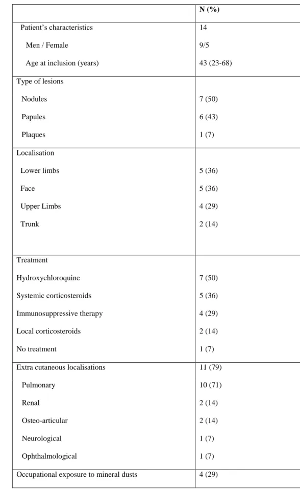
Table 1 Demographic, clinical and histopathological data of cases
N (%)
Patient’s characteristics Men / Female
Age at inclusion (years)
14 9/5 43 (23-68) Type of lesions Nodules Papules Plaques 7 (50) 6 (43) 1 (7) Localisation Lower limbs Face Upper Limbs Trunk 5 (36) 5 (36) 4 (29) 2 (14) Treatment Hydroxychloroquine Systemic corticosteroids Immunosuppressive therapy Local corticosteroids No treatment 7 (50) 5 (36) 4 (29) 2 (14) 1 (7) Extra cutaneous localisations 11 (79)
Pulmonary Renal Osteo-articular Neurological Ophthalmological 10 (71) 2 (14) 2 (14) 1 (7) 1 (7) Occupational exposure to mineral dusts 4 (29)
Accepted
Article
Biology
Hypercalcemia * 2 (14)
Elevated angiotensin converting enzyme level 4 (29) Number of granulomas per section
≤ 10 10 to 50 > 50 5 (33) 6 (40) 4 (26) Localisation of the granulomas
Superficial dermis Deep dermis Hypodermis 11 (73) 10 (67) 5 (33)
Presence of refractive material 5 (33)
Accepted
Article
Figure legends
Figure 1 Optic microscopy, PLE and SEM of granulomas in cutaneous sarcoidosis. (a) Optic
microscopy of a characteristic cutaneous granuloma (HES x 200). (b) PLE of the same
granuloma, showing birefringent particles. (c) Optical microscopy of another granuloma
(HES x 400); (d) PLE, showing birefringent particles. (e-f) SEM photographs showing an
intragranuloma inorganic deposit at two magnifications.
Figure 2 Examples of physicochemical analysis of the mineral silica (SiO2) deposits observed
within a granuloma. (a) µFT-IR microspectroscopy: IR spectrum showing the characteristic
signals of silica in the protein matrix of a granuloma. (b) FE-SEM/EDX the EDAX spectrum
identifies the presence of a significant signal related to silicon (Si) in the deposit (1.740 keV
cm-1)
Figure 3 Detection and physicochemical analysis of calcite (CaCO3)deposits at the periphery of
granulomas. (a) SEM photograph showing aggregates of submicrometer spherical deposits
localized between the collagen fibers (arrow). (b) µFT-IR microspectroscopy: IR spectrum
showing the characteristic peaks of calcite (CaCO3). (c) FE-SEM/EDX: the EDAX spectrum
identifies the presence of a significant signal related to calcium (Ca; 3.7 keV) and the absence
of a signal from phosphore (P; 2.01 keV) in line with the FTIR spectrum identifying calcite
Accepted
Article
Table 1 Demographic, clinical and histopathological data of cases
N (%)
Patient’s characteristics Men / Female
Age at inclusion (years)
14 9/5 43 (23-68) Type of lesions Nodules Papules Plaques 7 (50) 6 (43) 1 (7) Localisation Lower limbs Face Upper Limbs Trunk 5 (36) 5 (36) 4 (29) 2 (14) Treatment Hydroxychloroquine Systemic corticosteroids Immunosuppressive therapy Local corticosteroids No treatment 7 (50) 5 (36) 4 (29) 2 (14) 1 (7) Extra cutaneous localisations 11 (79) Pulmonary Renal Osteo-articular Neurological Ophthalmological 10 (71) 2 (14) 2 (14) 1 (7) 1 (7) Occupational exposure to mineral dusts 4 (29) Biology
Hypercalcemia * 2 (14)
Elevated angiotensin converting enzyme level 4 (29) Number of granulomas per section
≤ 10 10 to 50 > 50 5 (33) 6 (40) 4 (26) Localisation of the granulomas
Superficial dermis Deep dermis Hypodermis 11 (73) 10 (67) 5 (33) Presence of refractive material 5 (33)
Accepted
Article
Patients Clinical presentation Histopathology Calcemia
and ACE3 level PLE4 Physicochemical characterization of the deposits Gender1 age Skin
lesions location Type of skin lesion Other organs involved Location of the biopsy Number of granulomas2 Granulomas location 1 M 47 Upper and lower limbs
Nodules None Lower limb
10 Subcutis Both
normal
Positive Calcium carbonate
2 M 31 Face Papules Lung,
joint
Temple 21 Superficial and deep dermis
Hypercal-cemia Elevated ACE
Negative Calcium carbonate
3 M 55 Lower
limbs
Nodules Lung Lower limb
76 Deep dermis and
subcutis
NA5 Positive Calcium carbonate
4 F 61 Upper
limbs
Nodules Lung Arm 33 Deep dermis and
subcutis
Both normal
Negative -
5 M 68 Toes Plaques Lung Toe 10 Superficial and
deep dermis
NA5 Negative -
6 F 43 Face,
upper limbs
Nodules Lung Elbow 48 Superficial and deep dermis
Normal calcemia Elevated ACE
Accepted
Article
7 M 42 Upper
limbs
Nodules Lung Arm 115 Deep dermis Both
normal
Positive Crystalline silica
8 F 23 Fore Head Nodule NA5 Fore Head 70 Superficial and deep dermis
NA5 Positive Crystalline silica
9 M 41 Back Papules Kidney Back 73 Superficial and
deep dermis
Hypercal-cemia Normal ACE
Positive Calcium carbonate
10 F 51 Back Papules Lung Back 7 Superficial
dermis Both normal Negative - 11 M 38 Genitals, lower limbs Nodules Lung, Central nervous system Lower limb
33 Deep dermis and
subcutis NA5 Negative - 12 M 45 Lower limbs Papules Lung, kidney, eye Lower limb 22 Superficial dermis, deep dermis and subcutis Normal calcemia Elevated ACE Negative -
13 F 35 Scalp Papules None Scalp 9 Superficial
dermis
Both normal
Negative -
14 M 28 Face Papules Lung Face 19 Superficial and
deep dermis Normal calcemia Elevated ACE Negative -
Accepted
Article
Table 2: Patients, histopathology, biology and deposits characterisation in the 14 skin biopsies for cutaneous sarcoidosis.
1
M: male; F: female
2
Number of granulomas screened per biopsy
3
ACE: angiotensin conversing enzyme
4
PLE: polarized light examination
5