Review
Jérôme Lugrin, Nathalie Rosenblatt-Velin, Roumen Parapanov and Lucas Liaudet*
The role of oxidative stress during inflammatory
processes
Abstract: The production of various reactive oxidant species in excess of endogenous antioxidant defense mechanisms promotes the development of a state of oxi-dative stress, with significant biological consequences. In recent years, evidence has emerged that oxidative stress plays a crucial role in the development and per-petuation of inflammation, and thus contributes to the pathophysiology of a number of debilitating illnesses, such as cardiovascular diseases, diabetes, cancer, or neurodegenerative processes. Oxidants affect all stages of the inflammatory response, including the release by damaged tissues of molecules acting as endogenous danger signals, their sensing by innate immune recep-tors from the Toll-like (TLRs) and the NOD-like (NLRs) families, and the activation of signaling pathways ini-tiating the adaptive cellular response to such signals. In this article, after summarizing the basic aspects of redox biology and inflammation, we review in detail the current knowledge on the fundamental connections between oxidative stress and inflammatory processes, with a special emphasis on the danger molecule high-mobility group box-1, the TLRs, the NLRP-3 receptor, and the inflammasome, as well as the transcription factor nuclear factor-κB.
Keywords: cell signaling; danger signals; inflammasome; inflammation; innate immunity; oxidative stress; Toll-like receptors.
*Corresponding author: Lucas Liaudet, Department of Intensive Care Medicine, University Hospital Medical Center and Faculty of Biology and Medicine, CH-1010 Lausanne, Switzerland; and Division of Clinical Pathophysiology, University Hospital Medical Center and Faculty of Biology and Medicine, CH-1010 Lausanne, Switzerland, e-mail: [email protected]
Jérôme Lugrin and Roumen Parapanov: Department of Intensive Care Medicine, University Hospital Medical Center and Faculty of Biology and Medicine, CH-1010 Lausanne, Switzerland
Nathalie Rosenblatt-Velin: Division of Clinical Pathophysiology, University Hospital Medical Center and Faculty of Biology and Medicine, CH-1010 Lausanne, Switzerland
Introduction
Inflammation represents a fundamental biological process that stands at the foreground of a large number of acute and chronic pathological conditions. Complex inflamma-tory networks, involving countless cellular and humoral components, orchestrate critical aspects of diseases as various as myocardial infarction, diabetes, rheumatoid arthritis, sepsis, cancer, or Alzheimer’s disease to name only a few. As a direct corollary, clinical and basic research in the field of inflammation has been steadily growing, illustrated by the 415 000 references retrieved from a Medline search with the term ‘inflammation’, among which > 215 000 are < 10 years old (as of August 2013).
Inflammation occurs in response to any alteration of tissue integrity, in order to restore tissue homeosta-sis through the induction of various repair mechanisms. Proper regulation of these mechanisms is essential to prevent uncontrolled amplification of the initial inflam-matory response and a shift from tissue repair toward collateral damage and disease development (Goldszmid and Trinchieri, 2012). In recent years, evidence has been obtained that chemical processes involving redox reac-tions triggering cellular oxidative stress play critical roles in the pathophysiology of inflammation (Liaudet et al., 2009; Nathan and Cunningham-Bussel, 2013), a concept that had been already proposed almost 30 years ago in a milestone review article on the biochemistry of oxidative stress (Sies, 1986). The aim of this article is to provide an overview of the major mechanisms underlying this link between oxidative stress and inflammation.
Oxidative stress and biology of
free radicals
Many cellular processes are directed by reactions involv-ing the transfer of electrons between molecules, whose redox state (the balance between oxidized and reduced forms of electrons donors and acceptors) becomes thereby
modified (Schafer and Buettner, 2001). Redox homeostasis is maintained by cellular and extracellular redox buffer-ing systems, which include small molecule- and protein-based buffers such as the redox couples GSH/GSSG (glutathione-glutathione disulfide), cysteine/cystine, and oxidized/reduced thioredoxin (Banerjee, 2012). The balance of these buffering systems is maintained by key antioxidant enzymes, including superoxide dismutase, catalase, and the selenoproteins glutathione peroxidase and thioredoxin reductase, as well as non-enzymatic antioxidants such as α-tocopherol (vitamin E), ascorbate (vitamin C), β-carotene, and flavonoids (Steinbrenner and Sies, 2009; Chen et al., 2012).
Disruption of redox homeostasis occurs whenever an imbalance between reductants (electron donors) and oxidants (electron acceptors) develops, resulting either in a reductive stress (redox potential becomes more nega-tive) or an oxidative stress (redox potential becomes more positive), the latter being by far the most common form of redox imbalance in biological systems (Shao et al., 2012). Oxidative stress therefore results from the genera-tion of various oxidizing chemical species in excess of the
cellular reducing capacities, and is best defined as ‘an imbalance between oxidants and antioxidants in favor of the oxidants, leading to a disruption of redox signaling and control and/or molecular damage’ (Sies and Jones, 2007). Oxidant species include primarily free radicals, which are molecules or fragments of molecules containing one or more unpaired electrons in their molecular orbitals, and which stabilize by removing electrons from neighboring molecules. Certain non-radical species, such as hydrogen peroxide or peroxynitrite, also act as strong electron accep-tors due to their highly positive redox potential. The two main families of relevant oxidants in biology are the reac-tive oxygen species (ROS) and the reacreac-tive nitrogen species (RNS) (Pacher et al., 2007), as summarized in Figure 1.
The family of ROS
The superoxide anion radical (O2.-) is the primary ROS molecule formed in biological systems through the uni-valent reduction of molecular oxygen. Secondary ROS arise through the addition of a second and third electron,
L-arg METC ER NOX eNOS nNOS iNOS O2 XO Uncoupled NOS O2 .-NO. SOD Diffusion limited Reduction H2O H2O2 ONOO -NO3 -Catalase isomerization (detoxication) (detoxication) ONOOH Fe2+ Equilibrium CO2 OH. NO2. CO3.- Homolytic fission Oxidatio n ROS RNS
Figure 1 Molecular pathways of RNS and ROS generation. Nitric oxide (NO.) is formed from l-arginine and molecular oxygen (O
2) by the activity of various isoforms of NO synthase (endothelial,
neu-ronal, and inducible NOS). The superoxide radical (O2.-) is formed during cellular metabolism in the mitochondrial electron transport chain
(METC) and in the ER, or as a product of the enzymatic activities of NADPH oxidase (NOX), xanthine oxidase (XO), and uncoupled NOS. O2.- is
dismutated by superoxide dismutase (SOD) enzymes to hydrogen peroxide (H2O2), which can either be detoxified to water by catalase or
be converted to the hydroxyl radical (OH.) in the presence of metal (iron-mediated Fenton reaction). NO. and O
2.- spontaneously and rapidly
react to form the strong oxidant peroxynitrite (ONOO-). ONOO- can be detoxified by isomerization to nitrate (NO
3-), or may form secondary
radicals through homolytic fission (rupture of a covalent bond) or through reaction with carbon dioxide (CO2) of its conjugated acid
yielding, respectively, hydrogen peroxide (H2O2) and the hydroxyl radical OH.), through enzymatic- and metal-cat-alyzed (Fenton) reactions (Jomova et al., 2010). Further-more, O2.- promotes the formation of the potent oxidant and nitrating species peroxynitrite, following its sponta-neous reaction with nitric oxide (see below).
A continuous generation of O2.- occurs in the mito-chondria, due to the ‘leakage’ of electrons from the elec-tron transport chain, mainly at the ubiquinone binding sites of complex I and complex III, resulting in the one-electron reduction of oxygen (Drose and Brandt, 2012). Under physiological conditions, approximately 2% of oxygen consumed is thus diverted to O2.- as a by-product of mitochondrial respiration. Oxidative damage is pre-vented by the rapid scavenging of O2.- by the mitochon-drial enzyme manganese superoxide dismutase (Dhar and St Clair, 2012). The production of ROS may, however, sig-nificantly increase under various pathological conditions, for example, hypoxia or inflammation, which promote damage to electron transfer within the mitochondrial respiratory chain (Alfadda and Sallam, 2012). Besides mitochondria, the endoplasmic reticulum (ER) is another organelle contributing to cellular ROS generation. ER pro-motes protein folding through the formation of disulfide bonds within proteins, mediated by the enzymes protein disulfide isomerase and oxidoreductin 1, and coupled to the transfer of electrons to molecular oxygen, yielding H2O2. This process becomes significant under conditions of ER stress (triggered, for instance, by inflammatory cytokines or high glucose concentration), during which accumulation of misfolded proteins within the ER trig-gers a protective program termed the unfolded protein response, associated with an increased ER-dependent ROS generation (Laurindo et al., 2012; Popov, 2012).
The production of O2.- also depends on the activity of several enzyme systems, which include primarily the various isoforms of nicotinamide adenine dinucleotide phosphate (NADPH) oxidase (NOX), present in virtu-ally every cell type. They exist as seven distinct isoforms (NOX1–NOX5 and the dual oxidases DUOX1 and DUOX2), structurally composed of a core catalytic subunit and various regulatory subunits (p40phox, p47phox, p67phox, p22phox, NOXA1, NOXO1, DUOXA1, and DUOXA2) determin-ing spatial organization, membrane location, subcellular expression, and activation of the enzyme (Drummond et al., 2011; Segal et al., 2012). Activation of NOX catalyzes the transfer of electrons from cytosolic NADPH to molecu-lar oxygen to form O2.-. For NOX4 and DUOX1-2, the primary ROS produced is H2O2 instead of O2.-.
ROS produced by NOXs regulate multiple cellular (e.g., differentiation, proliferation, and migration) and
physiological (e.g., vascular tone, oxygen sensing) pro-cesses (Drummond et al., 2011; Segal et al., 2012). Excess ROS production and oxidative stress occurs when overac-tivation of NOX occurs in response to stimuli as diverse as hyperglycemia, angiotensin II, growth factors, hormones (Schramm et al., 2012), and most significantly inflamma-tory cytokines such as interleukin (IL)-1β (Ginnan et al., 2013) and TNFα (Frey et al., 2002). In addition, activated neutrophils and macrophages produce large amounts of ROS through the activation of NOX2 during the so-called oxidative burst, which is essential for eliminating invad-ing pathogens but which may also become a significant pathway of tissue injury under sterile inflammatory condi-tions associated with the activation of phagoyctes (Segal et al., 2012).
A second important enzymatic source of ROS is the xanthine dehydrogenase (XDH)/xanthine oxidase (XO) system. XDH is a metalloflavoenzyme converting xanthine and hypoxanthine into uric acid during the catabolism of purine nucleotides, using NAD+ as an electron accep-tor. Under inflammatory conditions and during tissue ischemia, XDH is converted to XO by oxidation of cysteine residues or by limited proteolysis (Engerson et al., 1987; Pritsos, 2000). XO does not reduce NAD+ but uses O
2 as an electron acceptor, thereby producing large amounts of O2 .-and H2O2 during purine catabolism (Nishino et al., 2008). The crucial role of XO-dependent oxidant generation is underscored by the great therapeutic potential of various pharmacological XO inhibitors, as recently reviewed (Pacher et al., 2006). Finally, additional enzymes may contribute to the formation of ROS, including cytochrome P450, cyclooxygenase, and uncoupled NO synthase, as detailed below.
The family of RNS
The parent molecule of all RNS is the free radical NO., gen-erated during the conversion of l-arginine to l-citrulline by the enzyme NO synthase (NOS) in a five-electron oxida-tive reaction. Three distinct isoforms of NOS exist, includ-ing neuronal (type I, nNOS), endothelial (type III, eNOS), and inducible (iNOS, type II) NOS. Whereas eNOS and nNOS are constitutively expressed, iNOS is expressed de
novo upon cell activation by inflammatory cytokines and
microbial products (Forstermann and Sessa, 2012). Under physiological conditions, NO. functions as a key cellular messenger and cytoprotective species by interacting with metals and other free radicals, and by modulating the bio-logical activity of a myriad of proteins through S-nitrosyla-tion of cysteine residues (Liaudet et al., 2000).
Alternatively, NO. may become a potent cytotoxic effector following its extremely fast reaction with O2.- (rate constant k = 0.38–1.6 × 10¹⁰ m-1 s-1) to generate peroxynitrite, the prototype of toxic RNS (Pacher et al., 2007). The oxida-tive chemistry of peroxynitrite depends in large part on the secondary formation of free radicals (Figure 1), including the hydroxyl radical (.OH), the carbonate radical (CO
3.-), and the nitrogen dioxide radical (.NO
2). The latter is notably responsible for the nitration of tyrosine residues within proteins, forming nitrotyrosine, which is used as a biochemical marker (‘footprint’) of peroxynitrite forma-tion in a given situaforma-tion (Pacher et al., 2007; Szabo et al., 2007; Ferrer-Sueta and Radi, 2009). By analogy with the term ‘oxidative stress’ associated with excessive ROS for-mation, the terms ‘nitroxidative stress’ (Calcerrada et al., 2011) and ‘nitrative stress’ (Sugiura and Ichinose, 2011) are frequently used to describe the consequences of exces-sive peroxynitrite formation in a biological system, while the term ‘nitrosative stress’ refers to the consequences of increased NO production (Pacher et al., 2005).
The generation of peroxynitrite may be significant under inflammatory conditions, due to the simultaneous generation of increased amounts of NO. and O
2.- (Pacher et al., 2007). It is also noteworthy that reduced availability of l-arginine (the NOS substrate) or tetrahydrobiopterin
(an essential NOS cofactor) can result in dysfunction of NO synthase, by ‘uncoupling’ its reductase and oxygenase domains (Kietadisorn et al., 2012). Uncoupled NOS gener-ates O2.- instead of NO., and thereby favors the formation of peroxynitrite. This process has been particularly well described in the context of inflammatory cardiovascu-lar pathologies, most significantly atherosclerosis, as reviewed recently (Forstermann and Li, 2011; Alkaitis and Crabtree, 2012).
Biological chemistry of ROS/RNS
ROS and RNS introduce various oxidative insults to lipids, proteins, and nucleic acids, with consequences ranging from subtle modulation of cell signal transduction pro-cesses to overt biomolecular damage and cell death, as illustrated in Figure 2. The one-electron oxidation of poly-unsaturated fatty acid (PUFA) residues may trigger a chain reaction of lipid peroxidation in biomembranes, causing significant alterations of membrane permeability (Valko et al., 2007). Oxidized PUFAs also form various cytotoxic aldehydes such as malondialdehyde and 4-hydroxynon-enal (Devasagayam et al., 2003). The reaction of peroxy-nitrite with PUFAs can generate nitrated lipids, which may
SH-oxidation (Cys)
Carbonylation (Pro, Thr, Lys, Arg)
Phenolic nitration (Tyr)
S-nitrosylation (Cys)
Proteins
• Sulfenic (-onic,-inic) acid • Mixed disulfide • S-glutathiolation • Ketones • Aldehydes Nitrated proteins (NO2adjonction) Nitrosylated proteins (NO adjonction) • Protein degradation
• Protein dysfunction • Protein degradation• Protein dysfunction • Protein dysfunction• Modulation of signaling • Protein dysfunction• Modulation of signaling
ROS RNS
PUFAs oxidation Lipoprotein oxidation Lipid nitration
• Lipid peroxidation • Toxic aldehydes Nitrated lipids (NO2 adjonction) Oxidized LDL Lipid s • Membrane alterations • Cytotoxicity • Pro-inflammatory • Atherosclerosis • NO transport • Cell signaling Nucleotide oxidation Formation of 8-oxoguanine (Deoxy)ribose oxidation DNA strand breakge
Nucleic
acids
• Mutagenicity • Mutagenicity
• PARP activation
Figure 2 Major molecular targets and biological consequences of oxidative stress.
ROS/RNS attack proteins, lipids, and nucleic acids, promoting various molecular modifications responsible for disturbed biological func-tions. Abbreviations: Cys, cysteine; Pro, proline; Thr, threonine; Lys, lysine; Tyr, tyrosine; PUFAs, poly-unsaturated fatty acids; LDL, low-density lipoproteins; PARP, poly(ADP-ribose) polymerase.
act as endogenous donors of .NO and as signaling mole-cules (Baker et al., 2009; Rubbo et al., 2009).
Several oxidative modifications of proteins may be introduced by ROS-RNS. The thiol (-SH) group of cysteine residues is a particularly sensitive target of oxidant species, forming sulfenic acid, mixed disulfide, S-glu-tathiolated derivatives, as well as sulfinic and sulfonic acid (Shao et al., 2012). Cysteine-bound thiol may also be nitrosylated through the addition of an NO group. Such
S-nitrosylation is a reversible modification playing
essen-tial roles in modulating the function of a great number of cellular proteins (Murray and Van Eyk, 2012; Haldar and Stamler, 2013). Tyrosine residues may be affected by per-oxynitrite-mediated nitration, that is, the addition of an NO2 group to the phenolic ring of tyrosine (Castro et al., 2011). A further oxidative modification of proteins is the addition of a carbonyl group such as ketone or aldehyde groups (carbonylation) to the side chain of amino acids, mainly proline, threonine, lysine, and arginine (Dalle-Donne et al., 2003). Oxidized proteins may be subject to accelerated degradation and loss of function, with poten-tially significant cytotoxic consequences (Dalle-Donne et al., 2003). For instance, oxidative alterations of mito-chondrial proteins (mainly induced by peroxynitrite) may precipitate apoptotic and necrotic cell death by triggering bioenergetic failure (Brown, 2007) and opening of the per-meability transition pore (Radi et al., 2002). An important aspect of redox modifications within proteins relies in the modulation of multiple cell signal transduction pathways (Burgoyne et al., 2012). While physiological variations of the cellular redox state play a crucial role in cellular homeostasis (concept of redox signaling), oxidative stress may result in pathological alterations of cell signaling, which may notably confer an inflammatory phenotype to the cell (Liaudet et al., 2009).
In DNA, oxidants can damage nucleobases, especially guanine, resulting in the formation of 8-oxoguanine, with potential mutagenic and carcinogenic consequences (Valko et al., 2007). Oxidants can also abstract hydro-gen atoms from the sugar phosphate backbone of DNA, promoting the generation of DNA strand breaks (Cadet et al., 2012). A striking consequence is the activation of the nuclear enzyme poly(ADP-ribose) polymerase (PARP). The latter is a family of 17 enzymes (primarily PARP-1 and PARP-2), which sense DNA strand breaks to initi-ate a program of DNA repair through the poly(ADP-ribo-sylation) of multiple nuclear protein substrates (Bai and Virag, 2012; Burkle and Virag, 2013). The catalytic activity of PARP consumes substantial amounts of cellular NAD+, initiating a secondary depletion of ATP. This may precipi-tate cell death through the necrotic pathway in conditions
of overt oxidative stress and PARP overactivation (Bai and Canto, 2012). In turn, necrotic cells release various intracellular components within the extracellular milieu, which trigger inflammatory responses through the activa-tion of specific innate immune receptors (Kono and Rock, 2008). It is also noteworthy that PARP enzymes recently emerged as important modulators of inflammatory dis-eases, by affecting immune cell maturation and differ-entiation and by regulating the expression of multiple inflammatory mediators, as extensively reviewed recently (Bai and Virag, 2012). These roles of PARP activation in promoting necrotic cell death and inflammation have prompted the development of a series of pharmacological inhibitors with significant therapeutic potential in many disease processes (Virag and Szabo, 2002).
Besides PARP, it is also worth to mention the role of another DNA repair enzyme in the regulation of inflamma-tion, namely 8-oxoguanine DNA glycosylase (OGG-1). The latter is a base excision repair enzyme that initiates the repair of oxidatively modified guanine (Hajas et al., 2013). Recent findings have provided evidence that this process is paralleled by enhanced immune responses (Mabley et al., 2005), as demonstrated by significant reduction of inflam-mation in OGG-1 knockout mice in conditions as diverse as endotoxic shock, diabetes, contact hypersensitivity (Mabley et al., 2005), and airway allergy (Li et al., 2012; Bacsi et al., 2013). The generation of DNA single-strand breaks during OGG-1-mediated DNA repair could represent the major underlying mechanism, by triggering the activa-tion of inflammatory transcripactiva-tion factors and the expres-sion of multiple cytokines (Li et al., 2012; Bacsi et al., 2013).
Inflammation in diseases: the role
of innate immune mechanisms
Inflammation represents an adaptive response to any con-dition perceived as potentially dangerous to the host, and which aims at the removal of the danger, the induction of tissue repair, and the restoration of tissue homeostasis (Okin and Medzhitov, 2012). Conceptually, inflammation can be viewed as a four-stage process, including a trig-gering system (the danger), a sensor mechanism (danger receptors), the transmission of the signal, and the pro-duction of mediators, and the activation of cellular effectors (Medzhitov, 2008), which may all be affected, at various degrees, by reactive oxidants (Figure 3), as will be detailed in the next sections. The prototypi-cal acute inflammatory response, characterized by loprototypi-cal vasodilation, extravasation of leukocytes, and release of
multiple plasma components, has been particularly well worked up in the field of invasion by microorganisms. In such infectious conditions, signature molecules released by the microbes, termed pathogen-associated molecu-lar patterns (PAMPs), activate innate immune defenses through the activation of the so-called pattern recognition receptors (PRRs) and downstream cellular pro-inflamma-tory signaling, with the ultimate goal of eradicating the pathogen (Barton, 2008; Kono and Rock, 2008; Medzhitov, 2010b). Over the past decade, evidence has been obtained that a comparable scenario is set in motion by a wide variety of non-infectious stimuli, from exogenous or endogenous origin. Thus, the innate immune system operates not only to sense ‘self’ vs. ‘non-self’ and to safe-guard against invading pathogens, but more generally, to identify and provide an adapted response to any form of danger (Matzinger, 2002) (Figure 3).
Innate immune defense and inflammation:
the danger model
A multiplicity of danger signals can be sensed by the host. Exogenous signals include primarily microbial molecules, for example, lipopolysaccharide (LPS) and flagellin from
negative bacteria, lipoteichoic acid from gram-positive bacteria, and viral RNA (Kawai and Akira, 2006), as well as non-infectious molecules, including allergens (Holgate, 2012) and airborne pollutants such as silica and asbestos (Dostert et al., 2008). Multiple endogenous signals are also able to elicit an inflammatory response through the activation of innate immune defense mecha-nisms. These are collectively termed ‘damage-associated molecular patterns’ or DAMPS (Kono and Rock, 2008), sometimes also referred as to ‘alarmins’ (Chan et al., 2012). DAMPs are released in conditions of tissue injury, and originate either from dying (necrotic) cells or from the breakdown of the extracellular matrix (Medzhitov, 2010a). Examples of intracellular DAMPs include heat shock proteins, S-100 proteins, high mobility group box-1 (HMGB1), ATP, and DNA (Kono and Rock, 2008). A specific subgroup of DAMPs originating from mitochondria (mito-DAMPs, MTDs) has been also recently described, compris-ing formyl peptides and mitochondrial DNA (Zhang et al., 2010). Extracellular DAMPs comprise fibronectin, hyalu-ronic acid, as well as peptides derived from collagen or elastin (Kono and Rock, 2008). Finally, various crystals acting as endogenous danger signals can promote strong inflammatory response, including monosodium urate (Martinon et al., 2009), calcium pyrophosphate dihydrate
Danger
signal Nonself, exogenous(microorganisms) Self, endogenous(tissue damage)
Molecular ROS RNS Molecular signature Pathogen-associated
molecular patterns (PAMPs) molecular patterns (DAMPs)Damage-associated
Sensor mechanisms
Pattern recognition receptors (PRRs)
TLRs NLRs
Transmission
of signal NF-κB Inflammasome
Inflammatory
response Inflammatory mediators
ROS RNS
ROS RNS
Figure 3 Molecular mechanisms of innate immune response.
The presence of danger signals (either exogenous or endogenous) alert the immune system through PRRs belonging to the Toll-like (TLRs) and the NOD-like (NLRs) families, triggering intracellular signaling through the transcription factor NF-κB and the molecular platform termed the inflammasome, fostering the production of multiple inflammatory mediators. Oxidative stress affects these processes at several levels, including the release of danger molecules, their sensing by PRRs, and their downstream signal transduction systems.
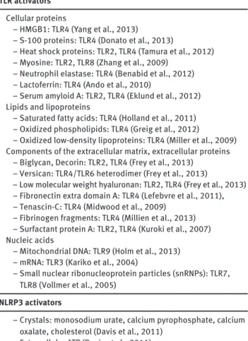
(Martinon et al., 2009), and cholesterol ester (Duewell et al., 2010). The main endogenous DAMPs and their receptors are presented in Table 1.
The appropriate recognition of the danger by the host is primordial for the elaboration of proper antimicrobial and adaptive responses. Sensing of PAMPs and DAMPs is ensured by a complex set-up of PRRs, which include pri-marily the Toll-like receptors (TLRs) (Trinchieri and Sher, 2007) and the nucleotide-binding oligomerization domain (NOD)-like receptors (NLRs) (Martinon et al., 2009). Other PRRs include C-type lectins, the receptor for advanced glycation end-products (RAGE), the retinoid acid-induci-ble gene I-like receptors, and the AIM2-like receptors (see Kingeter and Lin, 2012; Miyake and Yamasaki, 2012; Tang et al., 2012b for extensive recent reviews). PRR activa-tion triggers a wealth of intracellular signaling pathways, Table 1 Major endogenous activators of TLRs and NLRP3.
TLR activators Cellular proteins
– HMGB1: TLR4 (Yang et al., 2013) – S-100 proteins: TLR4 (Donato et al., 2013)
– Heat shock proteins: TLR2, TLR4 (Tamura et al., 2012) – Myosine: TLR2, TLR8 (Zhang et al., 2009)
– Neutrophil elastase: TLR4 (Benabid et al., 2012) – Lactoferrin: TLR4 (Ando et al., 2010)
– Serum amyloid A: TLR2, TLR4 (Eklund et al., 2012) Lipids and lipoproteins
– Saturated fatty acids: TLR4 (Holland et al., 2011) – Oxidized phospholipids: TLR4 (Greig et al., 2012)
– Oxidized low-density lipoproteins: TLR4 (Miller et al., 2009) Components of the extracellular matrix, extracellular proteins – Biglycan, Decorin: TLR2, TLR4 (Frey et al., 2013)
– Versican: TLR4/TLR6 heterodimer (Frey et al., 2013)
– Low molecular weight hyaluronan: TLR2, TLR4 (Frey et al., 2013) – Fibronectin extra domain A: TLR4 (Lefebvre et al., 2011), – Tenascin-C: TLR4 (Midwood et al., 2009)
– Fibrinogen fragments: TLR4 (Millien et al., 2013) – Surfactant protein A: TLR2, TLR4 (Kuroki et al., 2007) Nucleic acids
– Mitochondrial DNA: TLR9 (Holm et al., 2013) – mRNA: TLR3 (Kariko et al., 2004)
– Small nuclear ribonucleoprotein particles (snRNPs): TLR7, TLR8 (Vollmer et al., 2005)
NLRP3 activators
– Crystals: monosodium urate, calcium pyrophosphate, calcium oxalate, cholesterol (Davis et al., 2011)
– Extracellular ATP (Davis et al., 2011) – Amyloid β (Heneka et al., 2013)
– Oxidized mitochondrial DNA (Shimada et al., 2012) – Hyaluronan (Yamasaki et al., 2009)
– Pancreatic islet-derived amyloid polypeptide (Masters et al., 2010)
– High glucose (Tack et al., 2012)
– Fatty acids: palmitate, ceramide (Tack et al., 2012)
including kinases [for instance, mitogen-activated protein kinases (MAPKs), PI3 kinase], adaptors [such as myeloid differentiation primary response protein 88 (MyD88)], transcription factors [mainly nuclear factor-κB (NF-κB), activator protein-1 (AP-1), and interferon regulatory factors], as well as the inflammasome in the case of NLR activation (see below). Such signaling cascades foster the expression of cytokines, chemokines, enzymes, growth factors, and additional molecules that are required for antimicrobial resistance and tissue repair (Medzhitov and Horng, 2009; Tang et al., 2012b).
As stated previously, inflammation represents an essential adaptive response aimed at eradicating invad-ing pathogens and repairinvad-ing tissue damage elicited by noxious stimuli. Appropriate regulation of the mecha-nisms involved in such adaptation is essential to confine the inflammatory response in a localized compartment and to promote the switch between inflammation and repair, thereby allowing, in fine, the restoration of tissue homeostasis (Medzhitov, 2010a). However, there are situations in which such restoration may not adequately occur, resulting in persistent cellular stress perpetuat-ing and amplifyperpetuat-ing the inflammatory response. In these conditions, the process becomes maladaptive, leading to significant alterations of tissue functions, with systemic and persistent derangements of homeostasis (Okin and Medzhitov, 2012). Diabetes, atherosclerosis, chronic heart failure, neurodegenerative diseases, and cancer are typical examples of pathological processes associated with such chronic inflammatory changes (Pacher et al., 2007).
It is particularly noticeable that the release of ROS has long been recognized as a typical consequence of immune cell stimulation in vitro (Meier et al., 1989, 1990), and that both acute and chronic inflammatory states in vivo are coupled with significant alterations of redox equilibrium, due to the associated enhancement of oxidant generation (Pacher et al., 2007; Roberts et al., 2010; Li et al., 2013; Rochette et al., 2013). Accordingly, mitigating oxidative stress by the use of antioxidants has been evaluated as a potentially useful anti-inflammatory strategy in such con-ditions, as recently reviewed (Spychalowicz et al., 2012). Several distinct approaches to reduce oxidative stress have been used for this purpose, including natural (e.g., vitamin C, thiol compounds, flavonoids, and polyphenols) or synthetic (e.g., Trolox, dihydropyridines, edaravone) free radical scavengers and antioxidants (Augustyniak et al., 2010; Rahman and MacNee, 2012; Carocho and Ferreira, 2013), enzymatic inhibitors targeting NADPH oxidase (Drummond et al., 2011), NO synthase (Feihl et al., 2004), xanthine oxidase (Pacher et al., 2006), or PARP (Virag and Szabo, 2002), as well as catalytic antioxidants, including
superoxide dismutase mimetics (Muscoli et al., 2003) and peroxynitrite decomposition catalysts (Szabo et al., 2007). Overall, the results of these innumerable studies have clearly pointed out the strong association between oxidative stress and inflammation. Notwithstanding, the molecular mechanisms underlying the connection of these two funda-mental biological processes have often not been precisely examined. In the next sections of this review, we will there-fore attempt to fill this gap by presenting the current evi-dence supporting a mechanistic link binding redox stress, innate immunity, and inflammation, by focusing primarily on sterile (non-infectious) causes of inflammation.
Role of oxidative stress in
inflammation
Oxidative stress and inflammatory danger
signals: the illustrative case of HMGB1
As previously discussed, conditions promoting signifi-cant oxidative stress may precipitate cellular death and
extracellular matrix breakdown, due to biomolecular damage exceeding any capacity of repair. Necrotic cells and damaged ECM in turn release various intracellular and extracellular molecules, which act as ‘alarmins’ trigger-ing inflammatory cascades through recognition by PRRs (Chan et al., 2012). Furthermore, oxidative stress conditions may induce various modifications within lipids and pro-teins, generating the so-called oxidation-specific epitopes, which act as potent DAMPs able to trigger innate immune responses through binding to multiple PRRs. Examples of such oxidation-specific epitopes include oxidized phospho-lipids such as oxidized 1-palmytoyl-2-arachidonyl-sn-glyc-ero-3-phosphocholine (Imai et al., 2008; Kampfrath et al., 2011), oxidized cholesteryl esters (Choi et al., 2009), and oxidized low-density lipoporoteins (Lahoute et al., 2011), which are increasingly recognized as key mediators of the inflammation associated with the process of atherogenesis, as detailed in extensive recent reviews (Miller et al., 2011; Greig et al., 2012). As a detailed description of the multiple identified DAMPs is beyond the scope of this review, we will focus in the next section on the protein HMGB1 to illustrate the topics of the relations linking oxidative stress, DAMPs, and the development of sterile inflammation (Figure 4).
No
ROS ROS+ ROS+++
CXCR4 Rage TLR4 ROS RNS LPS TLR4 DNA LPT Inflammation TLR9 TLR2 HMGB1 IL-1 Necrosis IL-1R
All thiol Sulfonic
No
ROS ROS+ ROS+++
HMGB
Cys23 Cys23 Cys23
SH Cys45 Cys45 Cys
45
Cys106 Cys106 Cys106
SH HMGB Disulfide HMGB CXCR4 SH SH SH Rage SH S S TLR4 s O O OH Autophagy
Chemotaxis Inflammation Immune tolerance
Figure 4 Role of oxidative stress on the biology of HMGB1.
Excess ROS/RNS promote cell necrosis, leading to the passive release of intracellular HMGB1 into the extracellular milieu. HMGB1 can inter-act with multiple molecules, including LPS, DNA, lipoteichoic acid (LPT), and IL-1, to elicit inflammatory responses through distinct TLRs. Alternatively, HMGB1 can undergo various cysteine modifications depending on the local concentrations of oxidants. In the absence of ROS, HMGB1 is in the all thiol conformation, owing to reduced forms of Cys23, Cys45, and Cys106. All thiol HMGB1 interacts with the receptor for advanced glycation end-product (RAGE) to promote autophagic responses, and with the chemokine receptor CXCR4, triggering chemotac-tic responses. In the presence of increasing amounts of ROS, HMGB1 occurs either in the disulfide conformation (oxidation of Cys23 and Cys45), which targets TLR4 to initiate inflammatory responses, or in the fully oxidized conformation, due to oxidation to sulfonic acid of its three redox active cysteines. This latter form cannot activate inflammatory cells and thus promotes immune tolerance.
HMGB1 has been the first DAMP to be characterized (Scaffidi et al., 2002) and has been the subject of extensive literature over the past decade (Harris et al., 2012). HMGB1 is a ubiquitous nuclear non-histone protein that binds to DNA and participates in DNA replication, transcription, and repair (see Andersson and Tracey, 2011 for recent review). HMGB1 can be released extracellularly either through active secretion by activated monocytes (Wang et al., 1999) or by passive release by dying cells (Scaffidi et al., 2002), where it triggers a plethora of actions, including inflammation, chemotaxis, maturation of den-dritic cells, and endothelial cell activation to name a few (Andersson and Tracey, 2011). These effects are mediated following interactions of HMGB1 with several PRRs, pri-marily TLR4 and RAGE (Janko et al., 2013). HMGB1 also forms various complexes with many different molecules in the extracellular milieu, promoting additional pro-inflam-matory activities through interaction with TLR2, TLR3, TLR9, and the IL-1 receptor (Bianchi, 2009; Janko et al., 2013). HMGB1 plays significant roles in the pathophysiol-ogy of sterile inflammation (Andersson and Tracey, 2011) associated with diverse acute and chronic conditions, including myocardial infarction (Loukili et al., 2011), hepatic ischemia (Tsung et al., 2005), stroke (Schulze et al., 2013), circulatory shock and trauma (Andersson and Tracey, 2011), diabetes (Nogueira-Machado et al., 2011), cancer (Krysko et al., 2012), and arthritis (Harris et al., 2012), and it also acts as a key pro-inflammatory cytokine involved in the pathogenesis of sepsis (Yang et al., 2004). Thus, HMGB1 emerges as a central acting mediator at the intersection between sterile and infectious inflammation (Andersson and Tracey, 2011).
Several lines of experimental evidence support a key role played by oxidative stress in the process of HMGB1 release in the extracellular milieu. In a study using iso-lated cardiomyocytes in vitro, we reported that exposure of the cells to toxic concentrations of peroxynitrite induced necrotic cell death and was associated with the release of copious amounts of HMGB1 (Loukili et al., 2011). Similarly, Tang et al. (2007, 2011) reported that treatment of various mouse and human cell lines with H2O2 promoted significant extracellular HMGB1 release, whereas Tsung et al. (2007) showed that oxidants promoted the active release of HMGB1 by cultured hepatocytes through calmodulin-dependent kinase signaling. In vivo, using a rat model of myocardial infarction, we reported that a significant formation of per-oxynitrite occurred in the necrotic myocardium, together with HMGB1 accumulation. The elimination of peroxynitrite using peroxynitrite decomposition catalysts reduced myo-cardial infarct size and suppressed the build-up of HMGB1, providing direct evidence that peroxynitrite-mediated cell
death was the key trigger of cardiac HMGB1 accumulation during myocardial ischemia (Loukili et al., 2011). Thus, HMGB1 release represents a common response to the cel-lular stress imposed by free radicals and oxidants in vitro and in vivo, thereby representing an important mechanism linking redox stress and inflammation.
It is particularly noteworthy that HMGB1, while being released by oxidatively damaged cells, is also extremely redox sensitive, due to the presence of three critical cysteine residues. HMGB1 consists of two DNA binding motifs, box A and box B, and an acidic C-terminal tail. Box A possesses two vicinal cysteines (Cys23 and Cys45), whereas box B bears one single cysteine in position 106 (Cys106) (Tang et al., 2012a). Depending on the par-ticular redox environment, three distinct redox forms of HMGB1 can therefore be present: all thiol-HMGB1 (three cysteines in reduced SH form), disulfide HMGB1 (pres-ence of a disulfide bridge between Cys23 and Cys45 in box A), and fully oxidized HMGB1 (oxidation to sulfonate of Cys23, Cys45, and Cys106) (Tang et al., 2012a) (Figure 4). According to recent investigations, the recognition of the different redox conformations of HMGB1 may help devise specific pharmacological inhibitors, with potential thera-peutic activity against HMGB1-dependent inflammation (Gero et al., 2013).
Among these distinct redox forms, only disulfide HMGB1 has the ability to bind TLR4 and to promote innate immune responses (Tang et al., 2012a), as long as Cys106 is, at the same time, in a reduced conformation (Yang et al., 2013). In contrast, all thiol HMGB1 cannot bind to TLR4, but has been associated with the induction of autophagic responses in target cells through binding to the RAGE receptor (Kang et al., 2011). In addition, all thiol HMGB1 forms a complex with the chemokine CXCL12, to promote strong chemotactic responses on leukocytes through binding to the receptor CXCR4 (Venereau et al., 2012). These two distinct redox forms of HMGB1 are mutu-ally exclusive, which implies that, depending on the par-ticular microenvironment, HMGB1 functions either as a chemo attractant or as an inducer of cytokine release (Tang et al., 2012a; Venereau et al., 2012). In conditions of tissue injury in vivo, both redox forms of HMGB1 have been shown to be sequentially present (all thiol followed by disulfide), indicating that HMGB1 successively orches-trate leukocyte recruitment and the induction of cytokine secretion by adopting distinct redox conformations (Tang et al., 2012a; Venereau et al., 2012). Finally, fully oxidized HMGB1 loses both chemoattractant and pro-inflammatory activities, but instead triggers immunologic tolerance by preventing the activation of dendritic cells, which might explain the lack of inflammation associated with
apoptotic cell death (Kazama et al., 2008; Peter, 2008). Indeed, during apoptosis, mitochondrial production of ROS appears sufficient to promote the full oxidation and complete inhibition of HMGB1 (Kazama et al., 2008; Peter, 2008).
In addition to oxidative modifications, HMGB1 can also undergo acetylation of key lysine residues located within its nuclear localization sequence. This promotes the accumulation of HMGB1 within the cytoplasm, by pre-venting its nuclear re-entry, resulting in the active release of HMGB1 by monocytes/macrophages, particularly under conditions of inflammasome activation (Lu et al., 2012). The various posttranslational modifications of HMGB1 (oxidation and acetylation) have been the subject of an outstanding recent review by Yang et al. (2013).
To complete the picture of HMGB1-oxidant stress interactions, it is worth mentioning that HMGB1 itself may induce significant redox modifications by fostering the cellular generation of ROS and RNS (Janko et al., 2013). HMGB1-dependent activation of TLR4 triggers the upregu-lation of multiple genes, among which the genes encoding NOX and iNOS. Fan et al. (2007) showed that stimulation of ploymorphonuclear neutrophils (PMNs) in vitro with recombinant HMGB1 caused TLR4-dependent activation of NOX and subsequent increased ROS production. Using an experimental model of hemorrhagic shock in mice, these authors then reported that hemorrhagic shock was associated with significant increases of HMGB1 levels in most organs, together with a marked induction of NOX in PMNs, which could be abrogated both by TLR4 sup-pression and by neutralizing antibodies to HMGB1 (Fan et al., 2007). In another study by Sappington et al. (2002), incubation of Caco-2 human enterocytic monolayers with recombinant human HMGB1 resulted in enhanced expres-sion of iNOS, increased generation of NO and peroxyni-trite, together with an increased epithelial permeability, which could be abrogated by strategies removing NO and peroxynitrite (Sappington et al., 2002). Thus, it appears that oxidative stress represents both a cause and a conse-quence of HMGB1 release in multiple situations, and one may envision that such crosstalk might promote a vicious cycle of progressive inflammatory amplification.
Role of oxidative stress in the
biology of PRRs
Toll-like receptors
TLRs are key components of the innate immune system (Figure 5), whose primary function is to sense danger
signals released from pathogens (PAMPs). They are type I transmembrane proteins composed of an extracytoplas-mic, leucine-rich repeat domain for ligand recognition, a short transmembrane domain, and a cytoplasmic Toll/ IL-1 receptor (TIR) homology domain for signal transduc-tion (O’Neill and Bowie, 2007). Ten TLRs are expressed in humans, among which only TLR10 has no identified ligand thus far. TLR1, TLR2, TLR4, TLR5, and TLR6 are present in the plasma membrane to sense surface com-ponents of microbes. TLR2, which forms homodimers and heterodimers with TLR1 and TLR6, and which uses various coreceptors such as CD14, CD36, and Dectin-1, rec-ognizes bacterial lipopetides and peptidoglycans as well as mannans and glucans from fungal origin. TLR4, with the coreceptor MD-2, senses the complex formed by bac-terial LPS and CD14, and TLR5 detects bacbac-terial flagellin. TLR3, TLR7, TLR8, and TLR9 are expressed in intracellular compartments (ER, endosomes, lysosomes, and endolys-osomes) where they sense viral and bacterial nucleic acids (Lee et al., 2012; Song and Lee, 2012).
As previously discussed, TLRs also detect endoge-nous DAMPs released from damaged cells and extracellu-lar matrix, comprising proteins, fatty acids, lipoproteins, proteoglycans, and glycosaminoglycans (mainly detected by TLR4, and, to a lesser extent, TLR2), as well as nucleic acids and protein-nucleic acid complexes (detected by TLR3, TLR7, TLR8, and TLR9) (see Piccinini and Midwood, 2010 for extensive review). Several differences in the rec-ognition of PAMPs and DAMPs (different binding sites, different coreceptors and accessory molecules), and the engagement of distinct regulatory pathways may help discriminate exogenous from endogenous dangers. Most notably, DAMP recognition triggers negative feedback mechanisms to limit the potential damage to the host, and recent findings support an essential role for sialoside-based pattern recognition by members of the Siglec family in such negative regulation (Chen et al., 2009; Liu et al., 2011).
All TLRs, with the exception of TLR3, interact with the adaptor MyD88. The endosomal TLR7, TLR8, TLR9 and the cell surface TLR5 directly link MyD88, whereas TLR1, TLR2, TLR4, and TLR5 present on cell surface additionally recruit the linker protein TIR domain-containing adaptor protein (TIRAP also known as MAL) that connects TLRs and MyD88 TIR domains. Upon ligand binding, TLR3 and TLR4 recruit the protein TIR domain-containing adaptor inducing interferon-β (TRIF) either directly for TLR3 or by the intermediate of TRIF-related adaptor molecule (TRAM) for TLR4 (Gay and Gangloff, 2007; Song and Lee, 2012). MyD88 activates the IRAK-TRAF6-TAK1 axis that turns on inhibitor of κB (IκB) kinase (IKK) and MAPKs, which lead
to the activation of NF-κB and AP-1 transcription factors, respectively (Kawai and Akira, 2010; Song and Lee, 2012). Oxidative stress and Toll-like receptors: role of oxidants in TLR activation
It has been known for a long time that conditions asso-ciated with significant oxidative stress trigger enhanced responsiveness of cells from the innate immune system to pro-inflammatory stimuli (Botha et al., 1995; Fan et al., 1998). The underlying mechanism has remained elusive until recently, when it was proposed that oxidants may prime immune cells (‘reprogramming’) by upregulating TLR-dependent signaling (Figure 5). In a milestone study by Powers and coworkers (2006), exposure of rodent mac-rophages to oxidants, either in vivo (using an experimental model of hemorrhagic shock and resuscitation) or in vitro (using direct macrophage activation with H2O2), induced a significant increase in the surface levels of TLR4, as well as an increased responsiveness of cells to LPS. This effect was dependent on exocytosis of TLR4 from cytoplasmic compartments, as it could be suppressed by disruption of the cytoskeleton. Most important, the translocation of
TLR4 into the cell membrane colocalized with the lipid raft marker ganglioside GM1, implying the redistribution of TLR4 within lipid rafts (which are ‘floating’ microdo-mains within cell membranes, serving as signaling plat-forms). Finally, preventing TLR4 movement to lipid rafts using methyl-β-cyclodextrin suppressed the increased cel-lular responsiveness to LPS exposure to oxidants (Powers et al., 2006).
The study by Powers et al. emphasizes the crucial importance of receptor trafficking in the regulation of the ability of TLRs to sense their ligands, an issue that has been reviewed recently by McGettrick and O’Neill (2010). The essential role of oxidants in such process, as dem-onstrated in the above-discussed study, has been further confirmed by Nakahira et al. (2006), who showed that the trafficking of TLR4 to lipid rafts in response to LPS was entirely dependent on ROS produced secondary to the activation of NOX in RAW 264.7 murine macrophages. Similarly, Wong et al. (2009) demonstrated that a NOX-dependent generation of ROS was necessary for TLR4 recruitment and dimerization within membrane lipid rafts of cultured macrophages exposed to LPS, and additional studies have suggested comparable effects of oxidants in
TLR TLR Lipid raft
TIR TIR NOX
MyD88 TRIF p47phox
T ROS TLR NF-κB AP-1 IRF-3 ROS ↑NOX ↑NOS Phospholipids Sphingolipids RNS Inflammatory cytokines Cholesterol
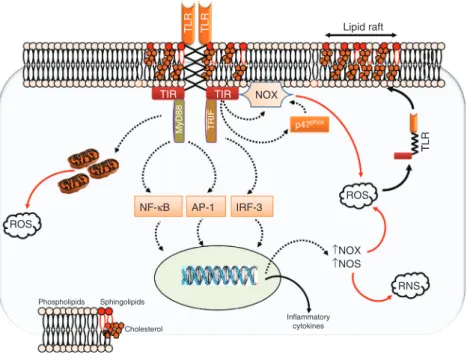
Figure 5 Interactions between oxidative stress and Toll-like receptors.
TLRs form active dimers within specific lipid rafts containing various amounts of phospholipids, cholesterol, and sphingolipids in the cell membrane. The extracellular domain connects through a transmembrane domain to the cytoplasmic TIR domain, which interacts with adaptor molecules such as MyD88 and TRIF to activate downstream signaling through NF-κB, AP-1, and interferon-regulatory factor-3 (IRF-3). Consequently, the expression of inflammatory mediators is upregulated, comprising notably pro-oxidant enzymes such as NOX and iNOS, producing high levels of ROS. TLR engagement also facilitates the generation of ROS within mitochondria, and promotes activation of NOX through direct interaction at the cell membrane or through enhanced phosphorylation of its p47phox subunit within the cytoplasm. The
resulting increase in intracellular ROS favor the mobilization and dimerization of TLRs within lipid rafts, creating a cycle of progressive amplification of the TLR response.
the membrane localization of TLR2 (Frantz et al., 2001; Dasu et al., 2008; Paul-Clark et al., 2009). The precise mechanisms of oxidant-mediated lipid raft modifications remain only partially elucidated. They involve, at least partly, alterations in lipid raft annexin VI content, activa-tion of calcium-dependent kinases, and the generaactiva-tion of ceramide. In turn, such modifications may result in modi-fications of lipid raft density and protein composition, with subsequent stimulation of TLR complex assembly (Cuschieri and Maier, 2007; de la Haba et al., 2013). Role of TLR activation in the generation of oxidants The previous paragraphs have highlighted the importance of oxidants in the activation of TLRs, by promoting their trafficking to the cell membrane. We will now focus on the opposite mechanism, that is, the induction of oxidant production in response to TLR activation (Figure 5). Increasing evidence is indeed accumulating showing that the formation of ROS represents an essential pathway of TLR-dependent signaling in cells from immune and non-immune origin, which occurs mainly through the activation of various NOX isoforms (Tsung et al., 2007; Ogier-Denis et al., 2008; Gill et al., 2010). In addition, the pro-inflammatory signaling cascades triggered by TLR engagement enhance the expression of iNOS, and thus promotes the generation of NO (Lewis et al., 2011; Shweash et al., 2011). In turn, the concomitant generation of O2.- (by NOXs) and NO. (by iNOS) results in the formation of peroxynitrite and other toxic RNS, implying that TLR activation may result both into oxidative and nitroxidative stress (Jozsef et al., 2006; Lucas and Maes, 2013).
While TLR-dependent oxidant formation is impor-tant to promote killing of invading pathogens (West et al., 2011), it may also result in significant cytotoxicity and col-lateral tissue injury in conditions of sterile inflammation. Additionally, oxidants may facilitate further TLR activa-tion, resulting in a cycle of progressive amplification of the initial inflammatory response. This process, which has been designed as the ‘TLR-radical cycle’, may represent an important mechanism in the maintenance of chronic inflammation in multiple human diseases, including car-diovascular diseases, diabetes, or neurodegenerative pro-cesses, as reviewed recently (Lucas and Maes, 2013).
The activation of NOX enzymes appears as a key process linking TLRs with secondary ROS generation. Such activation of NOX appears to result from several mechanisms, including (1) increased NOX protein expres-sion, (2) stimulated assembly of the NOX subunits, and (3) direct interactions between NOX and the TIR domain of TLRs. Increased NOX expression has been notably well
documented for NOX1 present in cells from gastrointes-tinal origin. NOX1 upregulation occurred in gastric epi-thelial cells in response to TLR4 activation by LPS from pathogenic Helicobacter pylori strains, and was shown to promote the induction of TNFα or cyclooxygenase 2 mRNA expression. This implies that NOX1 could be significantly involved in the pathogenesis of chronic gastric inflamma-tion induced by H. pylori infecinflamma-tion (Kawahara et al., 2005). Induction of NOX1 has also been evidenced in multiple colon cancer cell lines, including T84 (Kawahara et al., 2004), SW480, SW620, and CT-26 cells (O’Leary et al., 2012), as a result of TLR4 (O’Leary et al., 2012) and TLR5 (Kawahara et al., 2004) activation by LPS and flagellin, respectively. NOX1-dependent ROS production in these conditions promoted the release of the major chemokine IL-8 (Kawahara et al., 2004), and also greatly facilitated the adhesion of colon cancer cells to collagen I (O’Leary et al., 2012), implying a possible role in the establishment of intestinal inflammatory processes, as well as in increas-ing the metastatic potential of colon cancer cells.
The second important mechanism of TLR-mediated NOX activation is represented by stimulated assembly of the NOX subunits, particularly well documented in the case of NOX2 expressed by phagocytes. Various agonists of TLR2 (Huang et al., 2009), TLR4 (Bae et al., 2009; Kamp-frath et al., 2011), and TLR7/8 (Makni-Maalej et al., 2012) promoted NOX2 activation by triggering phosphorylation of its p47phox subunit. Deciphering the underlying
signal-ing mechanisms revealed a crucial role of MyD88 (Laroux et al., 2005), IRAK4 (Pacquelet et al., 2007; Picard et al., 2007), and phospho-p38 (Yang et al., 2008a; Makni-Maalej et al., 2012) signaling pathway in the upregulated p47phox phosphorylation. Finally, activation of NOX may
also result from direct interactions between TLRs and NOX, as indicated in a study by Park et al. (2004). Using yeast two-hybrid and GST pull-down assays, these authors showed that the carboxy-terminal region of NOX4 directly interacted with the cytoplasmic tail of TLR4 in HEK293T cells. Stimulation of TLR4 with LPS in this system induced ROS generation followed by NF-κB activation, pointing to direct interaction between TLR4 and NOX4 as a criti-cal mechanism regulating TLR-dependent innate immune responses (Park et al., 2004).
Generation of ROS secondary to TLR engagement does not only rely on the activation of NOX but also depends on the mitochondria (Figure 5). West and coworkers (2011) reported that the engagement of various TLRs (TLR1, TLR2, and TLR4) augmented mitochondrial ROS produc-tion by inducing the recruitment of mitochondria to mac-rophage phagosomes. This response occurred through the translocation of the adaptor TRAF6 to mitochondria,
leading to ubiquitination and enrichment at the mito-chondrial periphery of ECSIT (evolutionarily conserved signaling intermediate in Toll pathways), a protein impli-cated in mitochondrial respiratory chain assembly, with subsequent mitochondrial generation of ROS. The impor-tance of this process in antibacterial defense was critical, in view of the significant impaired capacity of ECSIT- and TRAF6-depleted macrophages to kill intracellular bacteria (West et al., 2011). However, it remains thus far unknown whether a similar mechanism accounts for TLR-depend-ent oxidative stress under sterile conditions.
To sum up, significant evidence has been obtained in recent years that TLR-ROS/RNS interactions are instru-mental in the induction and maintenance of innate immune responses. One may argue that targeting this ‘TLR-radical cycle’ might prove useful to prevent or treat many chronic inflammatory diseases (Lucas and Maes, 2013). Still, given the critical importance of TLR-depend-ent ROS generation in the elimination of invading patho-gens, impaired infectious control might well represent an important drawback of such strategy. Many issues need to be addressed to pinpoint how the organism discriminates between exogenous from endogenous danger signals and whether distinct signaling pathways modulate TLR-medi-ated responses during infectious and sterile inflamma-tion, in order to manipulate safely the TLR machinery for the therapy of chronic inflammatory diseases.
NOD-like receptors and inflammasomes
NLRs are located in the cytosol and sense a wide range of PAMPs and DAMPs. NLRs possess a common structure characterized by C-terminal leucine-rich repeat domain for ligand sensing, a central nucleotide-binding and oli-gomerization domain for activation, and an N-terminal domain for downstream signaling whose structure defines different subfamilies of NLRs (NLRA, NLRB, NLRC, NLRP, and NLRX) (Kersse et al., 2011). Upon activation, NLRs of the NLRP or NLRC families form multiprotein complexes termed inflammasomes, following assembly with the adapter protein ASC (apoptosis associated speck-like con-taining a CARD domain) and caspase-1. Once activated, the inflammasome platform promotes caspase-1-mediated cleavage and maturation of pro-IL-1β and pro-IL-18 into mature IL-1β and IL-18, which are critical to the develop-ment of inflammation (Martinon et al., 2009).
The NLRP3 inflammasome (also termed cryopirin or NALP3) has been thus far the best-studied member of this wide family (Figure 6). Two signals are required for full NLRP3 activation, including, first, a TLR-dependent
activation of NF-κB, resulting in the upregulation of NLRP3 and pro-IL-1β expression (‘priming’), and, second, an NLRP3 activating signal, which comprises multi-ple foreign and endogenous molecules (Franchi et al., 2012). Foreign signals comprise multiple pathogens and PAMPs that enter the cytosol, including bacteria (notably
Staphylococcus, Listeria, Clostridium, and Escherichia coli
species), fungi (mainly Candida and Aspergillus species), and viruses (for instance, adenovirus and influenza virus) (Davis et al., 2011). The activation of NLRP3 in response to invading pathogens mainly depends on the release of pore-forming toxins by bacteria (such as hemolysin from
Staphylococcus aureus and toxin A from Clostridium diffi-cile) (Koizumi et al., 2012), the release of double-stranded
RNA by viruses (Yu and Levine, 2011), and the release of hyphal fragments and the activation of the Syk tyros-ine kinase by fungi (Said-Sadier et al., 2010). Besides microbes, several non-infectious foreign molecules are robust activators of NLRP3, notably crystalline structures responsible for occupational lung inflammatory diseases (silica and asbestos) and particulate structures such as Co-Cr-Mo alloy metal particles used in prosthetic orthope-dic material (Caicedo et al., 2009) and metal nanoparticles such as titanium dioxide, used as a pigment in paint and cosmetics (Yazdi et al., 2010).
Multiple endogenous signals derived from the host itself have the ability to trigger NLRP3 activation, leading to a variety of acute and chronic inflammatory processes. Diverse crystals, including monosodium urate, calcium pyrophosphate dehydrate, and calcium oxalate, potently activate NLRP3 to induce joint inflammation in crystal-induced arthritis such as gout and pseudogout (Busso and Ea, 2012), as well as renal damage in crystalline nephro-pathy (nephrocalcinosis) (Mulay et al., 2013). Cholesterol crystals can also be sensed by NLRP3, promoting IL-1β-dependent inflammation in the vascular wall, a critical process involved in atherogenesis (Duewell et al., 2010). In the central nervous system, NLRP3 activation in the micro-glia due to sensing of extracellular insoluble β-amyloid peptide aggregates represents a key mechanism of neuro-inflammation involved in the pathogenesis of Alzheimer disease (Heneka et al., 2013). NLRP3 is also activated during tissue injury, mainly by hyaluronan released from damaged ECM (Yamasaki et al., 2009) and by ATP released by dying cells (Iyer et al., 2009; Silverman et al., 2009; Riteau et al., 2010; Davis et al., 2011), fostering the develo-pment of sterile inflammation in conditions as various as ischemia (Sandanger et al., 2013), cancer ( Zitvogel et al., 2012), circulatory shock (Xu et al., 2013), and toxic organ injury, e.g., acetaminophen-induced hepatotoxicity (Imaeda et al., 2009). Recent findings also indicated that
NLRP3 contributes to a large extent to the development of tissue fibrosis during chronic inflammatory processes (Xu et al., 2012; Negash et al., 2013), at least in part by aug-menting TGF-β signaling (Wang et al., 2013). Furthermore, inflammasome activation and IL-1β production also play an important role in the development and perpetuation of inflammation during metabolic syndrome, insulin resist-ance, and type 2 diabetes (De Nardo and Latz, 2011; Wen et al., 2012). Here, high glucose concentration, lipids (pal-mitate, ceramide), and islet-derived amyloid polypeptide represent the main triggers for NLRP3 activation (Menu and Vince, 2011; Zambetti et al., 2012).
Activation of NLRP3: the role of oxidative stress
The large number of known activators of NLRP3 makes it highly unlikely that NLRP3 directly senses all these dif-ferent triggers. Instead, it is now generally acknowledged
that NLRP3 activation occurs mainly as a consequence of a common form of cellular stress elicited by the different stimuli (Lamkanfi and Dixit, 2012). Three main putative mechanisms are currently considered for such activation (Figure 6). The first one, termed the channel model, is related to K+ efflux from the cell (Tschopp and Schroder, 2010). In this model, K+ efflux is consecutive to the activa-tion of the purinergic P2X7 receptor by extracellular ATP and the subsequent formation of pores by the recruitment of pannexin-1 (Pelegrin and Surprenant, 2006), or is elic-ited by microbial pore-forming toxins such as nigericin and hemolysin (Lamkanfi and Dixit, 2012). The second one, termed the lysosome rupture model (Tschopp and Schroder, 2010), depends on the destabilization and the rupture of the phagolysosome compartment, especially following the digestion of particulate material, and which results in the release of the lysosomal protein cathepsin B as an activator of NLRP3 (Hornung et al., 2008). Recent
PAMPs DAMPs
ATP
TLR TLR •K + channel
•Pore forming toxin •P2x7 channel
TIR TIR MyD88 TRIF K + K + Low intracellular K + PAMPs DAMPs TXNIP TXNIP TRX ROS NLRP3 Priming Phagolysosome Ox mtDNA ↓NAD+ ASC mtb Pro-IL-1β NLRP3 Cathepsin B Casp-1 Mature IL-1β ER Pro-IL-18 Inflammasome Mature IL-18
Figure 6 Role of oxidative stress on the process of NLRP3 activation.
The activation of TLRs by pathogen- or damage-associated molecular patterns (PAMPs, DAMPs) results in an increased expression of NLRP3, pro-IL-1β, and pro-IL-18 (priming stage). Activation of NLRP3 occurs through three distinct mechanisms: (A) K+ efflux in response to the
opening of K+ channels, the presence of bacterial pore-forming toxins, or the activation of the purinergic receptor channel P2X7 by
extracel-lular adenosine. (B) The release of cathepsin B by activated phagolysosomes. (C) Mitochondrial damage and ROS production. ROS can either separate thioredoxin-interacting protein (TXNIP) from its inhibitor thioredoxin (TRX), and TXNIP directly activates NLRP3. Damaged mitochondria also release oxidized DNA (Ox mtDNA) that activates NLRP3, or may be transported by microtubules (mtb) to the ER due to NAD+ shortage, bringing in close proximity NLRP3 with the adaptor ASC (apoptosis associated speck-like containing a CARD domain).
Ulti-mately, activated NLRP3 associates with ASC and caspase-1 in a multiprotein platform termed the inflammasome, which converts pro-IL-1β and pro-IL-18 into mature cytokines.
data have indicated that a similar mechanism triggers NLRP3 activation in myeloid cells exposed to chemother-apeutic agents such as gemcitabine and 5-fluorouracil (Bruchard et al., 2013).
The third model, termed the ROS model (Tschopp and Schroder, 2010), implicates the formation of ROS, deregu-lation of cellular redox status, and mitochondrial stress as key mechanisms in the process of NLRP3 activation (Rubartelli et al., 2011; Zhou et al., 2011; Rubartelli, 2012). Two main arguments support this model. First, it is partic-ularly noteworthy that all NLRP3 activators are capable of inducing intracellular ROS generation, and second, treat-ment with various ROS scavengers can block NLRP3 acti-vation by multiple agonists (Tschopp and Schroder, 2010). This later argument remains, however, controversial, owing to the fact that ROS inhibitors also inhibit the upreg-ulation of NLRP3 and pro-IL-1β (mechanism of ‘priming’), which may occur independently from the mechanism of NLRP3 activation (Bauernfeind et al., 2011). A possible molecular link connecting oxidative stress with NLRP3 activation has been proposed by Zhou et al. (2010) to be a protein termed TXNIP (thioredoxin-interacting protein). TXNIP is a member of the α-arrestin protein superfamily, which binds to the antioxidant protein thioredoxin (TRX), acting as a negative regulator of the TRX reductase activ-ity (Yoshioka et al., 2012). In their study, Zhou et al. (2010) first showed that TXNIP has the ability to bind to NLRP3 in human embryonic kidney T cells. They then showed that various NLRP3 agonists, including monosodium urate (MSU) and the imidazoquinoline imiquimod (R-837), pro-moted the association of NLRP3 with TXNIP, followed by inflammasome assembly and secretion of IL-1β by THP-1 cells. The interaction between TXNIP and NLRP3 was dependent on the secondary generation of ROS induced by MSU and R-837, which prompted the dissociation of TXNIP from TRX and its subsequent binding to NLRP3 (Zhou et al., 2010). This role of TXNIP has been debated, as it could not be reproduced in a further study by Masters et al. (2010), who used islet amyloid polypeptide and ATP as NLRP3 activators in mouse bone marrow-derived mac-rophages (BMDMs). When comparing IL-1β secretion acti-vation by BMDMs from TXNIP- or NLRP3-deficient mice, they found a decrease IL-1β in Nlrp3-/-, but not in TXNIP -/-cells, ruling out a significant role of TXNIP, at least in their experimental model (Masters et al., 2010).
Verification of the hypothesis that ROS act as second-ary mediators responsible for NLRP3 activation requires the identification of the source of ROS responsible for such role. In a report by Dostert et al. (2008), stimula-tion of THP-1 cells with asbestos, MSU, and ATP resulted in a ROS-dependent activation of NLRP3 and IL-1β
production, which was inhibited by NOX inhibitors and by knock down of its p22phox subunit. The possible role
of NOX-derived ROS in NLRP3 activation has, however, not been uniformly verified. Thus, in a study using mac-rophages obtained from mice lacking gp91phox (NOX2),
IL-1β response to multiple NLRP3 agonists was not modi-fied (Hornung et al., 2008). Furthermore, a study by van Bruggen et al. (2010) reported that primary peripheral blood mononuclear cells obtained from patients with chronic granulomatous disease, who lack the expression of p22phox and thus disclose impaired NOX activity, were
able to secrete normal amounts of IL-1β.
Recent observations have implicated mitochondria as the primary source of ROS ultimately responsible for the activation of NLRP3 (Zhou et al., 2011; Martinon, 2012). Many activators of NLRP3 have been shown to disrupt the inner mitochondrial membrane potential, possibly as a consequence of cellular potassium efflux and resulting alterations of mitochondrial matrix volume (Martinon, 2012). The ensuing mitochondrial dysfunction promotes the accelerated generation of ROS from the electron trans-port chain, leading to the oxidation of mitochondrial DNA (mtDNA) and its release into the cytosol (Figure 6), where it binds to NLRP3 to trigger its activation, as recently dem-onstrated by three independent groups (Nakahira et al., 2011; Zhou et al., 2011; Shimada et al., 2012). Oxidized mtDNA is thus emerging as a key ‘secondary’ danger molecule linking many forms of cellular stress to inflam-masome activation (Martinon, 2012). The crucial role of mitochondria in this scheme of event has been further reinforced by a recent report by Misawa et al. (2013), who showed that inducers of NLRP3 promoted the relocaliza-tion of mitochondria near the ER, allowing the apposirelocaliza-tion of the adaptor protein ASC (on mitochondria) to NLRP3 (on the ER) with consecutive assembly of the inflamma some. These processes were driven by a series of integrated signals involving a decrease of mitochondrial NAD+, fol-lowed by the accumulation of acetylated microtubules with subsequent, dynein-dependent, spatial rearrange-ment of mitochondria (Misawa et al., 2013).
The latest developments in the molecular biology of NLRP3 provide essential information to understand the mechanisms governing the initiation and perpetuation of inflammation in many pathological conditions. Mitochon-dria are vulnerable targets of multiple cellular stressors, and as such are particularly well positioned to perceive and signal the presence of noxious stimuli. These stimuli influence normal mitochondrial metabolism, resulting in a decrease of cellular NAD+, the production of ROS, and the release of oxidized mtDNA. Whereas such mecha-nisms have long been recognized as the key triggers of cell