Université de Montréal
Eph kinases and their ligands ephrins act in concert with sex hormones in regulating blood pressure
par Yujia Wang
Département de Sciences Biomédicales Faculté de Médecine
Thèse présentée à la Faculté des études supérieures en vue de l’obtention du grade de Philosophiae Doctor (Ph.D.)
en sciences biomédicales
Décembre 2015
Résumé
Les Erythropoietin-producing hepatocyte (EPH) sont la plus grande famille de récepteurs tyrosine kinase. Leurs ligands, les éphrines (EFNs), sont aussi des molécules exprimées à la surface cellulaire. Les EPH/EFNs sont impliqués dans de nombreux processus biologiques. L'hypertension artérielle (PA) est une maladie chronique qui, aujourd'hui, est devenue un problème médical critique dans le monde entier et un enjeu de santé publique. La découverte de nouvelles thérapeutiques de l'hypertension sont d'une grande importance pour la santé publique. Jusqu’à tout récemment, il existe seulement quelques études concernant le rôle de l’axe EPH/EFNs sur la fonction des cellules musculaires lisses vasculaires (CMLV). Dans nos études précédentes, nous avons montré qu'EPHB6 et EFNB1, de concert avec les hormones sexuelles, régulent la PA.
Dans la présente étude, nous avons constaté que les différents membres de la famille EPH/EFN peuvent réguler soit positivement, soit négativement, la contractilité des CMLV et la PA: tandis que EPHB4 et EFNB2 appartiennent à la première catégorie, EFNB1, EFNB3 et EPHB6 appartiennent à la deuxième.
In vivo, des souris males, mais non pas des femelles, porteuses d’une mutation EPHB4 (KO) spécifique du muscle lisse présentent une PA diminuée, comparée aux souris témoins (WT). Les CMLV de souris EPHB4 KO, en présence de testostérone, ont montré une contractilité réduite lors de la stimulation par la phényléphrine (PE). Au niveau moléculaire, la phosphorylation de la protéine kinase II dépendante de Ca2+/calmoduline et de la kinase de la chaine légère de la myosine (CLM) est augmentée, tandis que la phosphorylation de la kinase de la CLM est réduite dans les CMLV KO lors de la stimulation par PE, par rapport au WT CMLV. Cela fournit une base moléculaire à la réduction de la PA et de la contractilité des CMLV chez les souris EPHB4 KO.
EFNB2 est le ligand majeur de l’EPHB4. Comme attendu, les souris EFNB2 KO spécifique du muscle lisse avaient un phénotype de PA semblable, quoique non identique, aux souris EPHB4 KO. Les souris mâles EFNB2 KO, mais pas femelles, sous régime régulier ou riche en sel, présentent une PA réduite, par rapport à leurs homologues WT. Au niveau cellulaire, les CMLV des souris KO ont montré une contractilité réduite lors de la stimulation par PE par rapport aux témoins WT. Une région de l’acide aminé (aa) 313 à l’aa 331 dans la partie intracellulaire
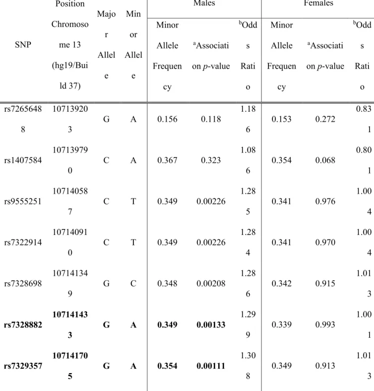
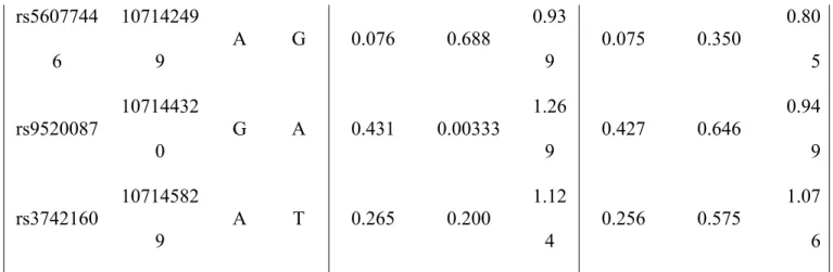
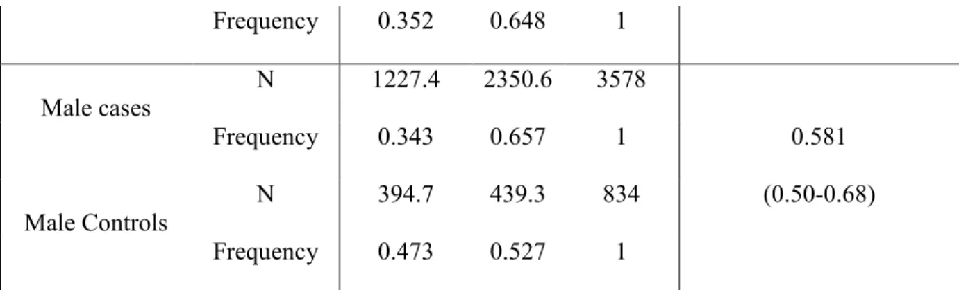
d’EFNB2 est essentielle pour la signalisation inverse qui régule la contractilité des CMLV, selon des études de mutation-délétion. Dans une étude de génétique humaine, nous avons identifié, dans le gène EFNB2, six SNP qui étaient associées significativement au risque d'hypertension artérielle, de façon dépendante du sexe, ce qui corrobore nos résultats chez les souris.
En revanche, la délétion du gène EFNB3 (KO) chez les souris femelles aboutit à une PA élevée et à une augmentation des résistances des petites artères in vivo, améliore la contractilité des petites artères ex-vivo et augmente la contractilité des CMLV in vitro. Les souris mâles KO ont une PA normale, mais la castration conduit à une augmentation significative de la PA dans les souris KO, mais pas dans les souris WT. Les CMLV des souris KO femelles ont montré une phosphorylation accrue de la CLM et une phosphorylation réduite de la kinase de la CLM, ce qui fournit à nouveau une base moléculaire aux phénotypes de PA et de contractilité des CMLV observés. Ce changement de signalisation est attribuable à une protéine adaptatrice Grip1. En effet, dans une étude d'association pan génomique par le Consortium International pour la Pression Sanguine, un SNP dans le gène GRIP1 a approché le seuil de significativité de la valeur p pour son association avec la pression diastolique.
Nos recherches, pour la première fois, ont révélé que EPH/EFNs sont de nouveaux composants dans le système de régulation de la PA. Les membres de la famille EPH/EFN peuvent agir comme des forces Yin et Yang pour régler finement le tonus des vaisseaux pour assurer l'homéostasie de la PA et de sa régulation. Ces effets de EPH/EFNs dépendent du sexe et des niveaux d’hormones sexuelles. À partir de ces nouvelles connaissances, nous pourrions développer une nouvelle thérapie personnalisée pour l’hypertension artérielle, utilisant des antagonistes d'hormones sexuelles ou des thérapies de remplacement d'hormones sexuelles, selon les niveaux d'hormones sexuelles des patients et les mutations dans les gènes de l'EPH/EFN.
Mots-clés: Erythropoietin-producing hepatocyte (EPH), éphrines, cellules musculaires lisses vasculaires, hypertension artérielle, hormones sexuelles
Abstract
Erythropoietin-producing hepatocyte (EPH) kinases are the largest family of receptor tyrosine kinases. Their ligands, ephrins (EFNs), are also cell surface molecules. Ephs/EFNs are implicated in many biological processes.
Hypertension is a chronic medical condition of high arterial blood pressure (BP). New hypertension therapeutic treatments are of great importance for public health. Until recently, there are only a few studies related to the role of EPHs/EFNs in vascular smooth muscle cell (VSMC) function. In our previous studies, we have found that EPHB6 and EFNB1 function in concert with sex hormones to regulate BP.
In the present investigation, we found that different EPH/EFN family members can either positively or negatively regulate the VSMC contractility and BP: while EPHB4 and EFNB2 belong to the former category, EFNB1, EFNB3 and EPHB6, the latter.
In vivo, male but not female smooth muscle-specific EPHB4 knockout (KO) mice presented decreased BP, compared to WT controls. VSMCs from EPHB4 KO mice in the presence of testosterone showed reduced contractility.
EFNB2 is the major ligand of EPHB4. As expected, smooth muscle-specific EFNB2 KO mice had a similar although not identical BP phenotype as EPHB4 KO mice. Male but not female EFNB2 KO mice on regular or high-salt diet presented reduced BP, compared to WT counterparts. At the cellular level, the KO VSMCs showed reduced contractility compared to WT controls. In a human genetic study, we identified in the EFNB2 gene six SNPs that were significantly associated with hypertension risk in a sex-dependent way, corroborating our findings in mice.
On the other hand, EFNB3 gene KO in female mice resulted in elevated BP and small artery resistance in vivo, enhanced small arterial contractility ex vivo, and augmented VSMC contractility in vitro. Male KO mice had normal BP, but castration led to significant BP elevation in KO but not in WT mice. VSMCs from female KO mice showed heightened MLC phosphorylation and reduced MLC kinase phosphorylation. This signaling change was mediated through an adaptor protein Grip1. Indeed, in a genome-wide association study by the International Consortium for Blood Pressure, an SNP in the GRIP1 gene approached the significant threshold p-value for its association with diastolic BP.
Our research for the first time revealed that EPHs/EFNs are novel components in the BP regulation system. Members of the EPH/EFN family may act as Yin and Yang forces to finely tune the vessel tone for BP homeostasis and regulation. Such effects of EPHs/EFNs depend on sex and sex-hormone levels. Based on this new knowledge, we could develop novel personalized hypertension therapy using sex hormone antagonists or sex hormone replacement therapy, depending on the sex hormone levels of the patients and mutations in EPH/EFN genes. Keywords: Erythropoietin-producing hepatocyte (EPH), ephrins, vascular smooth muscle cells, hypertension, sex hormones
Table des matières
Résumé ... i
Abstract...iii
Table des matières ... v
Liste des tableaux ... ix
Liste des figures ... x
Liste des sigles ... xii
Liste des abréviations ... xvi
Remerciements ...xviii
STATEMENT OF AUTHORSHIP ... 19
CHAPTER 1 INTRODUCTION ... 21
Part I. Function and signal pathways of EPH/Ephrins ... 21
Structure of EPH/ephrin family members ... 21
Functions of EPH/ephrin family members ... 23
Signaling pathways ... 23
The function of EPHs/ephrins in different biological processes and systems ... 27
EPHs/ephrins in the nervous system ... 27
EPHs/ephrins in cancer... 29
EPHs/ephrins in the immune system ... 32
EPHs/ephrins in the Glucose Homeostasis ... 34
EPHs/ephrins in bone maintenance and homeostasis ... 35
EPHs/ephrins in cardiovascular system... 37
EPHs/ephrins in angiogenesis ... 37
EPHs/ephrins in vascular smooth cells... 39
Part II. Hypertension ... 39
Symptoms and pathogenesis of hypertension... 40
Pathology of hypertension ... 42
Pathophysiology ... 42
Hypertension-related pathological conditions ... 47
Genetic disorders ... 48
Vascular Function ... 49
Vascular smooth muscle tone ... 49
Vascular smooth cell function ... 49
Main signaling pathways in VSMCs ... 49
Other molecules ... 53
Complications of hypertension ... 54
Complications affecting the heart ... 55
Complications affecting the brain ... 55
Complications affecting the eye ... 55
Complications affecting the kidneys ... 56
Complications associated with diabetes and hypertension ... 56
Hypertension treatment ... 56
Diuretics ... 57
β Blockers ... 57
Angiotensin-converting enzyme inhibitor (ACE Inhibitors) ... 58
Angiotensin II receptor blockers ... 58
Calcium channel blockers... 58
Nervous system inhibitors ... 58
Vasodilators ... 59
Treatment of hypertension during pregnancy... 59
Children and Teens ... 59
Part III. Sex hormones and blood pressure ... 60
Estrogen ... 60
Estrogen in the cardiovascular system ... 61
Estrogen Receptor ... 62
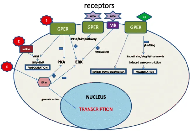
GPER in vascular contraction ... 62
Testosterone ... 64
CHAPTER 2 ARTICLE-1 ... 66
TITLE: EPHB4 expression in vascular smooth muscle cells regulates their contractility and EPHB4 deletion leads to hypotension in mice ... 66
Capsule ... 67
Abstract... 67
Introduction ... 68
Materials and Methods ... 70
Results ... 75 Discussion... 80 References ... 84 Figure legends ... 90 Tables ... 96 Figures ... 97 CHAPTER 3 ARTICLE-2 ... 102
TITLE: Reduced blood pressure after smooth muscle EFNB2 deletion and the potential association of EFNB2 mutation with human hypertension risk ... 102
Abstract... 103
Introduction ... 104
Materials and Methods ... 105
Results ... 109
Discussion... 116
Acknowledgements ... 121
References ... 122
Titles and Legends of Figures ... 128
Tables ... 132
Figures ... 136
CHAPTER 4 ARTICLE-3 ... 155
TITLE: Estrogen and testosterone in concert with EFNB3 regulate vascular smooth muscle cell contractility and blood pressure ... 155
New and noteworthy... 156
Introduction ... 157
Materials and Methods ... 160
Results ... 166
Discussion... 171
Reference ... 178
Tables ... 187
Figures and Legends ... 188
CHAPTER 5 ARTICLE-4 ... 200
TITLE: The role GRIP1 and EFNB3 signaling in blood pressure control and vascular smooth muscle cell contractility ... 200
Abstract... 201
Introduction ... 202
Materials and Methods ... 204
Results ... 208
Discussion... 220
Acknowledgments ... 224
REFERENCES ... 226
CHAPTER 6 General Discussion ... 237
Yin and Yang of the novel BP regulation system ... 237
EPH/EFN-related downstream signaling pathways ... 239
The mutations identified in human genetics studies could be loss-of-function, or gain-of-function or alternation-of-gain-of-function mutations ... 242
Sex hormones differentially influence the BP of males and females ... 243
Genetic diagnosis and hormone therapy ... 244
Contribution to sciences and future research directions ... 245
Liste des tableaux Chapter 2 Table 1 ... 96 Chapter 3 Table 1 ... 132 Table 2 ... 133 Table 3 ... 134 Chapter 4 Table 1. ... 187 Chapter 5 Table 1 ... 209 Table 2 ... 211 Chapter 6 Table 1. ... 237
Liste des figures Chapter 1
Figure 1. Features of Eph/ephrin signaling ... 22
Figure 2. Eph/Ephrin bidirectional pathways ... 26
Figure 3. The renin–angiotensin system ... 44
Figure 4. The mechanism of nitric oxide function in vessels ... 46
Figure 5. Regulation of smooth muscle contraction(Webb, 2003) ... 53
Figure 6. Effects of membrane estrogen receptor activation by estradiol, G1 agonist and aldosterone in vascular cells ... 64
Chapter 2 Figure 1. Generation of mice with smooth muscle cell-specific Ephb4 null mutation ... 90
Figure 2. Male EPHB4 KO mice were hypotensive ... 91
Figure 3. Reduced contractility of mesenteric arteries from male EPHB4 KO mice ... 91
Figure 4. VSMCs stimulated by both EPHB4 forward and reverse signalings show increased contractility ... 92
Figure 5. MLC, MLCK, MYPT and CamKII phosphorylation of VSMC from WT and KO mice ... 93
Figure 6. GRIP1 mediates EPHB4 reverse signaling in controlling VSMC contractility ... 94
Chapter 3 Figure 1. Generation of mice with smooth muscle cell-specific Efnb2 null mutation ... 128
Figure 2. BP and heart rate of EFNB2 KO mice ... 129
Figure 3. Both forward and reverse signaling by EFNB2 in VSMCs results in increased contractility ... 129
Figure 4. Critical regions in EFNB2 intracellular tail for regulating VSMC contractility ... 130
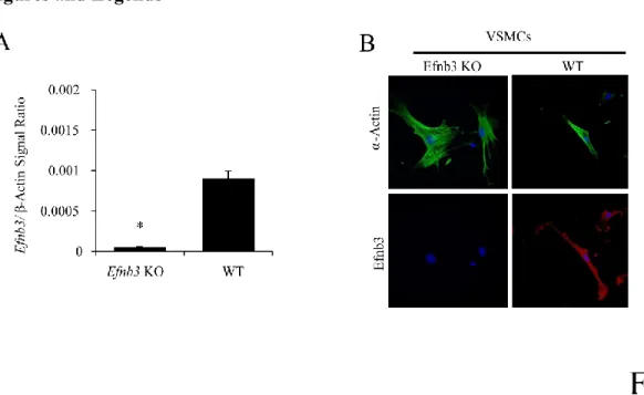
Chapter 4 Figure 1. Efnb3 deletion in vascular cells from Efnb3 KO mice ... 188
Figure 2. Increased BP in female Efnb3 KO mice ... 190
Figure 4. Signalling events related to contractility of KO and WT VSMCs ... 192 Figure 5. Estrogen enhances but testosterone suppresses and VSMC contractility and hence BP in the absence of EFNB3 ... 195 Figure 6. EFNB3 deletion enhances GPR30 expression and results in increased VSMC contractility in the presence of estrogen ... 197 Figure 7. Normal 24-hour urinary catecholamine levels in Efnb3 KO mice ... 199 Chapter 5
Figure 1. Crosslinking EFNB3 on VSMCs results in decreased contractility ... 214 Figure 2. GRIP1 in the EFNB3 reverse signaling pathway in VSMCs... 217 Figure 3. DISHEVELLED AND PDZ-RGS3 ARE NOT in the EFNB3 reverse signaling pathway in VSMCs... 219 Chapter 6
Figure 1. Possible signaling pathways downstream of EPHBs/EFNBs ... 240 Figure 2. GRIP1 is in the EPH/EFN signaling pathways leading to VSMC contractility ... 242
Liste des sigles
11β-HSD2 11b-hydroxysteroid Dehydrogenase Type 2
ABL Abelson Murine Leukemia Viral Oncogene Homolog ACE Angiotensin Converting Enzyme
ACTH Adrenocorticotropic Hormone
ADAM A Disintegrin and Metalloprotease AICD Activation-induced Cell Death
ARG Arginine
BP Blood Pressure
CAMKII Ca2+/calmodulin-dependent Protein Kinase II CAMP 3'5'-Cyclic Adenosine monophosphate
CBL Casitas B-lineage Lymphoma
CC Colon Carcinoma
CGMP 3'5'-Cyclic Guanosine Monophosphate
CKD Chronic Kidney Disease
CV Cerebral Vasospasm
DP Diastolic Pressure
EGF Epidermal Growth Factor
EMAX Maximal Tension
EPHS Erythropoietin Hepatoma Kinases ERK Extracellular-signal Regulated Kinase
GDI Gdp Dissociation Inhibitors
GEF Gdp Exchange Factors
GPER Membrane-associated G-protein
GRA Glucocorticoid-remediable Aldosteronism HCSCS Human Cardiac Stem Cells
HIC1 Hypermethylated In Cancer 1
HSP Heat Shock Proteins
IP3 Inositol 145-trisphosphate
JNC Joint National Committee
JNK Jun N-terminus Kinase
LRRK2 Leucine-rich Repeat Kinase 2 Gene MAPK Mitogen-activated Protein Kinase
MAPKK Mapk Kinase
MLC Myosin Light Chain
MLCK Myosin Light Chain Kinase
MLCP Myosin Light Chain Phosphatase MMP2 Matrix Metalloproteinase 2
MVD Microvessel Density
NHANES Us National Health And Nutrition Examination Survey
NHIGG Normal Human Igg
NO Nitric Oxide
PHAII Pseudohypoaldosteronism Type II
PTEN Phosphatase and Tensin Homolog PUFAS Polyunsaturated Fatty Acids PYK2 Proline-rich Tyrosine Kinase 2
RAAS Renin - Angiotensin - Aldosterone System
RAS Renin-angiotensin System
RHOGEF Rhoa Guanine Nucleotide Exchange Factor
ROS Reactive Oxygen Species
RTKS Receptor Tyrosine Kinases
SAH Subarachnoid Hemorrhage
SAM Sterile Alpha Motif
SDF-1Α Stromal Cell-derived Factor
SGN Spiral Ganglion Neurons
SH Src Homology
SHR Spontaneously Hypertensive Rats SNPS Single Nucleotide Polymorphisms SOCS Suppressors Of Cytokine Signaling
SP Systolic Pressure
TCR T-cell Receptor
TEC Thymic Epithelial Cell
TP Thromboxane Prostanoid
VEGFR Vascular Endothelial Growth Factor Receptors VSMC Vascular Smooth Muscle Cell
WNK With No Lysine
Liste des abréviations AB Antibody ARG Arginine CAM Calmodulin E2 17β-estradiol EFNs Ephrins KO Knockout PE Phenylephrine
Remerciements
I would like to express my special appreciation and thanks to my supervisor Dr. Jiangping Wu. It is my honor to be your student. I would like to thank you for encouraging me when I was at the nadir at times, so I can always stick with my dreams and faith. Your advice on both research as well as on my career has been priceless. I would also like to thank my co-supervisor Dr. Hongyu Luo. Thank you for giving me many advices and a great help in my research work. Your serious scientific attitude has deeply inspired me.
I appreciate my committee members, Professor Edward Bradley, Professor Christian F. Deschepper, Professor Tomoko Takano, for serving as my committee members. Thank you for your brilliant comments and suggestions.
I would especially like to thank my colleagues and friends. All of you had been there to support me whenever I need.
Finally, special thanks to my family, for the support of my work and my life. Thank you for your dedication and perseverance.
Thanks everyone from the bottom of my heart!
STATEMENT OF AUTHORSHIP
Here is a statement regarding the contribution of co-authors and myself to the four papers included in this thesis.
Chapter 2:
Wang Y, Thorin E, Luo H, Tremblay J, Lavoie JL, Wu Z, Peng J, Qi S1, Wu J. EPHB4 Protein Expression in Vascular Smooth Muscle Cells Regulates Their Contractility, and EPHB4 Deletion Leads to Hypotension in Mice. J Biol Chem. 2015; 290(22):14235-44
Y.W., Z.W., J.W., conceived and designed the experiments. Y.W., Z.W., H.L, S.Q., performed the experiments. Y.W., Z.W., J.T., JL.L., J.P., analyzed the data. J.W., initiated and guided the project, and participated in experimental design and manuscript writing.
Chapter 3:
Wang Y, Hamet P,Thorin E,Tremblay J,Raelson J, Wu Z,Luo H, Jin W, Lavoie J, Peng J, Marois-Blanchet F, Chalmers J, Woodward M; Harrap S,Qi S, Li C, Wu J. Reduced blood pressure after smooth muscle EFNB2 deletion and the potential association of EFNB2 mutation with human hypertension risk. Eur. J. Hum. Genet. (Revised)
Y.W., J.R., Z.W. H.L., W.J., conducted experiments; E.T. conducted vessel contraction studies; P.H. contributed to experiment design and manuscript writing; J. T., J. L., and J. P. performed telemetry; S.Q. carried out castration and ovariectomy; F.M., J.C., M.W., S.H., C.L. carried out human genetic studies; J.W. initiated and guided the project, and participated in experimental design and manuscript writing.
Chapter 4:
Wang Y, Wu Z, Thorin E, Tremblay J, Lavoie J, Luo H, Peng J, Qi S, Wu T, Chen F, Shen J, Hu S, Wu J. Estrogen and testosterone in concert with EFNB3 regulate vascular smooth muscle cell contractility and blood pressure. Am J Physiol Heart Circ Physiol. 2016 Apr 1;310(7):H861-72.
Y.W., and Z.W. conducted experiments in VSMC and contributed to experimental design and manuscript writing; E.T. conducted vessel contraction studies and contributed to manuscript writing; J. T., J. L., and J. P. performed telemetry; H. L. participated in KO characterization; S.Q. carried out castration and ovariectomy; T.W., F.C. J.S., and S.H. carried out human vessel studies; J.W. initiated and guided the project, and participated in experimental design and manuscript writing.
Appendix-1: Wang Y, Wu Z, Luo H, Peng J, Raelson J, Ehret GB, Munroe PB, Stoyanova E, Qin Z, Cloutier G, Bradley WE, Wu J. The role GRIP1 and EFNB3 signaling in blood
pressure control and vascular smooth muscle cell contractility. Plos One (submitted after revision)
Y.W., Z.W. H.L., J. P. conducted experiments; J.R., G.E., P.M., E.S., Z.Q., G.C., conducted human genetic experiments; W.B., contributed to experiment design; S.Q. carried out castration and ovariectomy. J.W., initiated and guided the project, and participated in experimental design and manuscript writing.
CHAPTER 1 INTRODUCTION
Part I. Function and signal pathways of Eph/Ephrins
Receptor tyrosine kinases (RTKs) are a large group of cell surface molecules with an intracellular tail that contains tyrosine kinase activity. They function as a sensor to detect extracellular signals and transmit them to the cells (Yarden & Ullrich, 1988). The effects of many growth factors are known to be mediated by high-affinity RTKs (Fantl, Johnson, & Williams, 1993).
Erythropoietin-producing hepatocyte (EPHs) form the largest family of RTKs, their corresponding ligands being named ephrins (Efn). About 20 years ago, the first member of the Eph kinase family was identified by Hirai et al.(Hirai, Maru, Hagiwara, Nishida, & Takaku, 1987) and was called Eph. Multiple other members were subsequently identified (Beckmann et al., 1994; Bennett et al., 1995; Davis et al., 1994; Drescher et al., 1995; Lackmann et al., 1996; Shao, Lou, Pandey, Pasquale, & Dixit, 1994; Winslow et al., 1995). Within a few years of the identification of EPHs, their ligands ephrins were identified (Bartley et al., 1994; Beckmann MP, 1994). Since then, active research has been carried out to understand their functions.
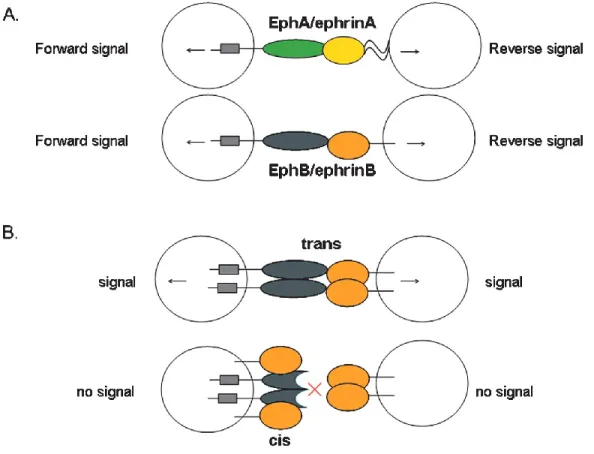
Structure of Eph/ephrin family members
Both EPHs and ephrins are membrane-bound proteins. According to sequence similarity, Eph receptors and the ligands are classified into A and B subfamilies. There are nine members of the EphA subfamily, EphA1-8 and EphA10, and five members of the EphB subfamily, EphB1-4 and EphB6. EphrinAs (A1–A6) anchor into the cell membrane by a glycosylphosphatidylinositol moiety, while ephrinBs (B1–B3) pass through the plasma
membrane by a short cytoplasmic tail. There is a PDZ-binding domain at ephrinBs’ C-terminus in the cytoplasmic tail (Figure 1) (Arvanitis & Davy, 2008).
Figure 1. Features of Eph/ephrin signaling
Both types of Eph receptors (EphAs and EphBs) contain an extracellular region, a single transmembrane spanning domain, and an intracellular region which comprises a tyrosine kinase domain, a sterile alpha motif (SAM), and PDZ-binding motif (Surawska, Ma, & Salgia, 2004). The extracellular domain comprises a ligand-binding domain, including a highly conserved N-terminal globular domain, a cysteine-rich domain, and two fibronectin type III repeats. The A-type receptors preferentially bind A-A-type ligands while the B-class receptors bind B-A-type ligands, but there are exceptions. For example, EphA4 can bind both A and B-type ephrins. One of the unique features of Eph/ephrin signaling is the fact that both receptors and ligands are competent
to transduce signals into the cells upon interaction (Lisle, Mertens-Walker, Rutkowski, Herington, & Stephenson, 2013).
Functions of Eph/ephrin family members Signaling pathways
Both EPHs and their corresponding ephrin ligands are membrane-bound proteins, they require direct cell-cell interactions for Eph receptor activation. The interactions between Eph receptors and ephrins can happen in trans (between two opposing cells) or in cis (within the same cell), and both can result in either cell-to-cell adhesion or de-adhesion. The Eph receptors are activated by ephrin ligands though forward signaling while the ephrin ligands are activated by Eph receptors through reverse signaling. Such bidirectional signalings are the characteristics of Eph/Ephrin signaling.
The interaction of the extracellular domains of Eph receptors and ephrins was described by Himanen et al.(Himanen et al., 2001a) on EphB2 and ephrinB2. This interaction forms a crystal structure on the cell surface. In the crystals, there are two different structures of bi-directional signaling presenting through a multiple-step process: dimeric and tetrameric. First, pre-clustered unbound ephrins form low-affinity ephrin-ephrin homodimers. Then Eph receptors bind to the ephrins in 1:1 ratio, forming high affinityand specificity heterodimers. Next, the dimer pairs of Eph-ephrin join into tetrameric complexes. The tetrameric complex contains two ligands interacting with two receptors and forming a circular structure (Himanen, Henkemeyer, & Nikolov, 1998). This ring-like tetrameric structure is necessarily required by the functional bidirectional signaling (Himanen & Nikolov, 2003). It allows the membrane-associated ephrins and Eph receptors to interact with the surfaces of adjacent cells (Drescher, 2002).
In solution, the range of EPHs and ephrins concentration can vary widely from 10 nM to 20 mM. At moderate concentration, EPHs and ephrins usually form heterodimers at a 1:1 stoichiometry, while, throughout higher concentration, those dimer pairs can form high-order tetramers (Himanen et al., 2001b). The Eph tyrosine kinase can then trans-phosphorylate each other on the tyrosine kinase domain and initiate forward and signalings via the tetramers, which can also mediate the phosphorylation of other proteins and associate with the receptor of various effector proteins. Importantly, the formation of the heterotetramers not only modulates the forward signaling but also repositions the ephrin transmembrane and cytoplasmic domains, converting them to a signaling-competent configuration. Tyrosine phosphorylation of the ephrin cytoplasmic tail follows, initiating reverse signaling. These reverse signaling acts through two types of residuals: the tyrosine/serine phosphorylation sites and the intracellular PDZ domain-binding motif (Himanen, Saha, & Nikolov, 2007). Besides, the extracellular cysteine-rich domain and the intracellular SAM domain of the EphB receptors also play a significant role in Eph–ephrin interaction by binding to the Ephrin carboxy-terminal PDZ domain binding motif (Himanen et al., 2001a; Holder & Klein, 1999; Stapleton, Balan, Pawson, & Sicheri, 1999). The main downstream signaling pathway of Eph/Ephrin is mediated by G-proteins (Noren & Pasquale, 2004). There is a multiple, critical connection between these cell surface proteins and small GTPases of the Ras and Rho families. Eph/Ephrin work like a switch to regulate the GTPases cycled between the inactive state and active state (Van Aelst & D'Souza-Schorey, 1997). These components are modulated by some adhesion molecules and growth factor receptors (Noren & Pasquale, 2004), resulting in cell morphologic and cell behavioral regulation.
Rho proteins are key regulators of actin cytoskeleton dynamics in cells (Ramachandran, Patil, Combrink, Sharif, & Srinivas, 2011). EphrinB reverse signaling also activates Rho GTPases. It
performs in a phosphotyrosine-independent docking mechanisms by the Src family tyrosine kinases (Van Aelst & D'Souza-Schorey, 1997).
The Eph/ephrin signaling pathway is also connected to the Ras/ mitogen-activated protein kinase (MAPK) cascade. Once the Eph receptors are activated, adaptor molecules associated with them will transmit signals into the cell. Then the extracellular-signal regulated kinase (ERK) / MAPK pathway will be suppressed (Figure 2)(Coulthard et al., 2012). Consequently, the communication from the cell surface to the DNA in the nucleus of the cell and the activity of the transcription factors are suppressed (Chang et al., 2003; Miao et al., 2001).
Figure 2. Eph/Ephrin bidirectional pathways
Beside the classical bidirectional signal pathway, Eph receptor and ephrins could also interact with other cell-surface communication systems, independent of receptor-ligand association. For example, recent studies have shown that members of the epidermal growth factor (EGF) receptor family can cooperate with EphA2 and promote cell motility and proliferation(Brantley-Sieders et al., 2008); ephrinBs could be phosphorylated on their intracellular tyrosine residues in response to platelet-derived growth factor receptor activation(Arvanitis & Davy, 2008).
Moreover, there are still many other proteins working in concert with Eph/Ephrins, such as Src Homology 2(SH2) and SH3 adaptor proteins(C. A. Cowan & Henkemeyer, 2001; Holland et al., 1997); phosphatidylinositol 3-kinase(Guan et al., 2010); focal adhesion kinase(Krupke & Burke, 2014); low-molecular-weight protein tyrosine phosphatase(Park, Warner, Mood, Pawson, & Daar, 2002); Abelson murine leukemia viral oncogene homolog (Abl) and Arginine (Arg) kinases(H. H. Yu, Zisch, Dodelet, & Pasquale, 2001); chemokine receptors(Zlotnik & Yoshie, 2000) and some adhesion molecules(Huynh-Do et al., 1999).
The function of EPHs/ephrins in different biological processes and systems
A great number of studies about Eph/ephrin functions have been carried out. EPHs/ephrins are implicated in many biological processes such as embryonic development, tissue boundary formation and cell migration. Additionally, they also play critical roles in angiogenesis (Cheng, Brantley, & Chen, 2002) and stem cell differentiation (Genander & Frisen, 2010).
EPHs and EPHRINs are expressed in many tissues and organs. They play important roles in the central nervous system(Dumas et al., 2000; Xin, Deng, Rishniw, Ji, & Kotlikoff, 2002), immune system(H. Luo, Charpentier, et al., 2011; H. Luo, Wan, Wu, & Wu, 2001; H. Luo, Wu, et al., 2011; H. Luo, Yu, Tremblay, & Wu, 2004; Hongyu Luo, Yu, Wu, & Wu, 2002; J. Wu & Luo, 2005; G. Yu, Luo, Wu, & Wu, 2003a, 2003b, 2004; G. Yu, Mao, Wu, Luo, & Wu, 2006), digestive system(Batlle et al., 2002), bone metabolism (Davy, Bush, & Soriano, 2006; C. Zhao et al., 2006), cardiovascular system (H. U. Wang, Chen, & Anderson, 1998) and other systems (Dravis et al., 2007; Hashimoto et al., 2007; Konstantinova et al., 2007).
The first described function of EPHs and ephrins was to regulate cell-cell communication in the central nervous system (Himanen et al., 2007). Later, more and more important functions of EPHs/Ephrins in the nervous system were found.
During neuron development, axons first need to find their appropriate target regions, and then have to recognize the correct area to form specific connections. During this process, many pieces of evidence showed that the EPHs/ephrins are critical in axon guidance (Flanagan & Vanderhaeghen, 1998).
GPI-anchored ephrinAs are involved in axon repulsion. Before cell–cell contact, A-Disintegrin-And-Metalloprotease (ADAM) will associate with ephrinA2 on the same cell. When encountering a second cell expressing EphA3 receptors, the interaction between ephrinA2 and EphA3 leads to the formation of higher-order ligand–receptor clusters, which help the ADAM10 to cleavage ephrin ectodomain (Egea & Klein, 2007), terminating the ephrinA2/EphA4 interaction, hence the repulsion.
Another report demonstrated that EPHs/ephrins cause axon guidance through the Rho family GEF Vav. During axon guidance, Vav-dependent endocytosis triggers EPHs and ephrins to form a ligand-receptor complex, leading to the generation of growth cone collapses, which is necessary for axon guidance. This endocytosis is blocked when Vav protein is absent, suggesting that Rho family GEF Vav can be a key molecular in Eph/ephrin signaling pathways relevant to axon guidance (C. W. Cowan et al., 2005).
Neural crest cells come from the neural epithelium. They can migrate to particular destinations, and differentiate into many cell types, including most neurons and glia cell of the peripheral nervous system, and most of the connective and skeletal tissue in the head. These cells migrate
following a particular pattern, either repulsive or attractive. EphrinB ligands are one set of repulsive cues, controlling the segmental migration of trunk and branchial neural crest cells (Wilkinson, 2001).
EphA4 is required for typical fasciculation of spiral ganglion neurons (SGN) axons in their fringe directions. In vivo study in mice demonstrated that a lack of EphA4 in SGN peripheral axons causes significantly smaller fascicles. A similar phenotype also happens when ephrinB2 is mutated (Cramer & Gabriele, 2014).
EPHs/Ephrins also help to maintain the functional plasticity in neuronal circuits. Some members of Eph receptor family, such as EphB1–3 and EphA4, are expressed in the subventricular zone while their ligands ephrinBs are expressed in astrocytes. Their interaction will disrupt the migration of neuroblasts and increase cell proliferation (Conover et al., 2000; Lledo, Alonso, & Grubb, 2006).
EPHs/ephrins in cancer
As EPHs/Ephrins play key roles in the regulation of cell migration and adhesion, they are strongly related to tumorigenesis. They are upregulated essentially in all types of cancer cells (Garber, 2010). Eph signaling in tumor suppressor activities has been reported in many different types of tumors, including colorectal, breast, prostate, and skin tumors (Pasquale, 2008).
Both ephrinBs and their receptors play a role in the growth and development of colon carcinoma (CC). In the intestinal epithelium, the activation of EphBs will enhance cell sorting and compartmentalization of CC cells (Cortina et al., 2007), and control the cell differentiation through E-cadherin–mediated adhesion (Herath et al., 2006). One particular pair of
EPHs/ephrins in CC is EphB4 and ephrin B2. Both of them are overexpressed in the epithelium of the intestinal luminal surface in CCs, regulating the tumor angiogenesis and vasculogenesis (Stephenson, Slomka, Douglas, Hewett, & Hardingham, 2001).
EPHs/Ephrins are overexpressed in ovarian cancers (Herath et al., 2006). EphrinA2 in tumor cells or endothelial cells has been observed in >75% of ovarian cancers. It is associated with increased microvascular density and increased expression of ovarian cancer special markers such as matrix metalloproteinases 2, 9, and 14 (Y. G. Lin et al., 2007). Overexpression of EphA2 is correlated to increased microvessel density (MVD), which is strongly associated with critical factors involved in angiogenesis and tumor invasion (Y. G. Lin et al., 2007).
EphA2 inhibitors are especially effective in treating ovarian cancers when used in combination with taxanes (a type of antimicrotubule agent), indicating that EphA2 is an attractive target for ovarian cancer therapy (Landen, Kinch, & Sood, 2005).
There are different types of EPHs expressed in breast cancers. EphB4 functions as a tumor suppressor in breast cancer. In breast cancer cells, EphB4 activates an anti-oncogenic pathway involving Abl family tyrosine kinases and the Crk adaptor protein. This Abl-Crk pathway downregulates the pro-invasive MMP2, not only inhibits breast cancer cell viability and proliferation, but also affect the motility and invasion,(Noren, Foos, Hauser, & Pasquale, 2006). In some breast cancers, the tumor suppressor gene HIC1 (hypermethylated in cancer 1) restricts ephrinA1 expression, the default function of which is to reduce tumor growth (W. Zhang et al., 2010).
The existing data show that EphA2 receptor overexpression and phosphorylation play a role in the progression of malignant prostate cancer, involving processes of cell adhesion, motility, invasion, and formation of metastases. EphA2 kinase deficient in the prostate tumor cells are correlated to cell rounding, retraction fiber formation, de-adhesion from the extracellular matrix, RhoA and Rac1 GTPase regulation, three-dimensional matrix invasion, and in vivo metastasis(Taddei et al., 2009).
Beside EphA2, EphB4 protein overexpression is also found in most prostate tumors. Such overexpression is induced by a loss of phosphatase and tensin homolog (PTEN) or p53, or induced by epidermal growth factor/epidermal growth factor and insulin-like growth factor-I. One report indicates that knockdown of the EphB4 protein by siRNA or antisense significantly inhibits cell viability, migration and invasion, and induces apoptosis in prostate cancer cell lines(Xia et al., 2005).
There seems to be a competition amongst Eph kinase family members in prostate cancer cells, in regulating tumor cell locomotion and invasiveness. Many members, such as EphB3, EphB4 and ephrin-B2, join in this competition. The balance between the different members leads to the switch of the migration of cancer cells from restrained to invasive (Astin et al., 2010).
Increased EphB4 and ephrin-B2 expression may reflect increased potential for growth and tumorigenicity in endometrial carcinomas. Furthermore, the EphB4 receptor kinases may modulate the biological behavior of endometrial carcinomas through autocrine and paracrine activation. That’s caused by Efnb2 ligand which expresses on the same or adjacent cells surface (Takai, Miyazaki, Fujisawa, Nasu, & Miyakawa, 2001).
The expression of EphA1 in nonmelanoma skin cancers is augmented. It seems to be a marker of the normal differentiated epidermis. The downregulation of EphA1 in nonmelanoma skin cancer may contribute to carcinogenesis of this cancer (Hafner, Becker, Landthaler, & Vogt, 2006).
The expression of EphA2 and its ligand, ephrinA1, is significantly increased in carcinomas of the urinary bladder (Abraham et al., 2006).
EPHs/ephrins in the immune system
Eph/ephrin signaling is incorporated with inflammation disorders actuated by infection, injury, and inflammation.
First, EPHs/ephrins are involved in the activation of T cells. A prominent Eph function in T cells relates to T-cell costimulation. With the solid-phase antibody against EphB6 or solid-phase EPHRINB1-3 Fc, T-cell responses to suboptimal T-cell receptor (TCR) ligation could increase(H. Luo et al., 2004; G. Yu et al., 2003a, 2003b).
Normally, the TCR signaling strength could be positively or negatively modulated by certain EPHs, leading to different thymocyte selection results (Freywald, Sharfe, Rashotte, Grunberger, & Roifman, 2003).
Stimulation of Eph receptors on human CD4+ T cells by ephrinA1 enhances their migration (Hjorthaug & Aasheim, 2007). EphA receptor activation induces rapid tyrosine phosphorylation of the Casitas B-lineage Lymphoma (c-Cbl) proto-oncogene. In T cells that express EphA1 and EphA4 receptors, Cbl phosphorylation is observed. The following signaling events involve adaptors Crk-L and Crk-II (Sharfe, Freywald, Toro, & Roifman, 2003), resulting regulation of T cell migration into inflamed tissues (Y. Huang et al., 2015).
Another pathway through which ephrinA1 regulates T cell function is via proline-rich tyrosine kinase 2 (PYK2). After the binding of ephrinA1 to its receptors, the phosphorylation and kinase activity of the Src kinase family member Lck will be induced. This activates focal adhesion-like kinase Pyk2 (Aasheim, Delabie, & Finne, 2005), which has an essential role in T cell activation and polarized secretion of cytokines (Sancho et al., 2002).
Chemotaxis is a necessary process in T cell migration. Ephrin-A1 strongly inhibits chemotaxis by altering the balance of activities of different small G proteins. In the same time, Ephrin-A1 also prevented Rho activation, lead to stromal cell-derived factor (SDF)-1α induced activation of cdc42. The cdc42 activation can further increase calcium flux and activation of MAPK (Sharfe, Freywald, Toro, Dadi, & Roifman, 2002). As we mentioned before, EPHs/Ephrins activate MAPK strongly, downstream of which are Jun N-terminus kinase (JNK), p38 and Erk. All those molecules play crucial roles in cell migration (C. Huang, Jacobson, & Schaller, 2004). Secondly, Eph/ephrin signals are involved in thymus development. During thymus development, different signals drive precursors to mature into functional T lymphocytes. The Eph family members are implicated in this process by regulating cytoskeleton function and cell adhesion (Munoz et al., 2002). For example, mice deficient of EphB in thymocytes experience a significant reduction of thymocyte subsets (Alfaro et al., 2008). The reduced EphA4 expression results in decreased numbers of double-positive (CD4+ CD8+) thymocytes (Munoz et al., 2006). Besides, the development and organization of thymic epithelial cell (TEC) was also influenced by EphB2 and EphB3 (Munoz et al., 2011). The migration of the thymic primordium into the thoracic cavity requires ephrinB2 expression (Foster et al., 2010).
Thirdly, EPHs’s function is not restricted to the enhancement of the strength of TCR signaling. They might modulate T-cell survival and death by modulating immune cell apoptosis,
depending on their activation status (J. Wu & Luo, 2005). After soluble EphrinA1 stimulation of CD4+ T cells, the IL-2 expression is reduced. So is IL-4 but not IFN-γ. As activation-induced cell death (AICD) depends on IL-2 and IL-4 cytokines and their receptors (Zubiaga, Munoz, & Huber, 1992), EphrinA1 could thus prevent AICD (Wohlfahrt et al., 2004).
EPHs/ephrins in Glucose Homeostasis
In order to maintain glucose homeostasis in the body, the glucose levels in the blood are controlled by insulin from β cells in the pancreas. The underlying molecular mechanisms of how β cells modulate insulin secretion is not entirely understood. Recently, some studies demonstrated that both Eph/ephrins A and B family members are expressed in mouse and human islets, and their signaling is strongly related to the β cell function.
A recent study shows that certain communication between β cells is via EphA receptors and ephrinA ligands both in vitro and in vivo. EphA forward signaling will inhibit insulin secretion while ephrinA reverse signaling could enhance insulin secretion. Those functions lead to a two-way autoregulation in pancreatic cells.When blood sugar level is low, EphA forward signaling can be activated, leading to increasing insulin secretion; when blood glucose level is high, EphA receptor is dephosphorylated, leading to downregulation of insulin secretion (Kulkarni & Kahn, 2007).
A new hypothesis is that EphA receptors not only exist on the cell surface but also in the intracellular insulin secretory granules. This suggests that EphA levels on the plasma membrane, and, therefore, EphA-ephrinA complexes, increases upon insulin release. This causes a feedback loop that negatively limits insulin secretion through increased EphA signaling. On the other hand, there is a positive feedback loop: when glucose levels are low, the ephrinA signaling will upregulate to increase insulin secretion (Konstantinova et al., 2007).
One animal experiment showed that ephrinA5 KO mice are mildly glucose intolerant. When subjected to the same concentration of glucose stimulation, compared with the control group, the significant reduction in their insulin secretion. In contrast, the insulin content of these islets was not significantly changed. Also, knockdown of ephrinA5 in MIN6 cells reduces glucose-stimulated insulin secretion (Konstantinova et al., 2007; Pasquale, 2008).
Another relevant peptide for glucose homeostasis is glucagon. When the inhibition of glucagon secretion is out of control, types 1 and 2 diabetes happen. However, the molecular mechanisms that regulate glucagon secretion in normal and diabetic states are not fully understood. A new study shows that the tonic stimulation of α-cell EphA receptors between neighboring islet cells could negatively regulate glucagon secretion. The effect of EphA is mediated by juxtacrine. Sorted α-cells lacking endogenous stimulation of EphA forward signaling from neighboring cells will result in enhanced basal glucagon secretion and the elimination of glucose-induced inhibition of glucagon secretion. In these cells, both normal basal glucagon secretion and glucose-induced inhibition of glucagon secretion can be recapitulated by restoration of EphA forward signaling. In vivo. α-cell-specific EphA4-/- mice exhibit abnormal glucagon dynamics (Hutchens & Piston, 2015).
EPHs/ephrins in bone maintenance and homeostasis
Bone homeostasis strictly maintains balance, this maintenance in the cell largely depends on the cellular communication between osteoclasts and osteoblasts, and closely related to the coupling between bone resorption and bone formation. A range of pathologic disorders, including osteoporosis and cancer-induced bone diseases, disrupt this coupling and cause subsequent alterations in bone homeostasis.
Eph receptors and their associated ligands, ephrins, are also expressed in the bone marrow microenvironment, including osteoclasts and osteoblasts. More and more evidence suggests that this receptor family is closely related to regulating normal and pathological bone remodeling (Edwards & Mundy, 2008).
Some skeletal malformations are caused by the developmental deficiencies in EphB/ephrinB signaling, including cleft palate, defective development of the skull vault, craniosynostosis, and other bone abnormalities. The mutations of EphA2, EphA3 or ephrinB1 in mice may cause the X-linked developmental disorder of craniofrontonasal syndrome (Davy & Soriano, 2005; Pasquale, 2008).
Genetic and another evidence supports a model in which EphB/ephrinB1 bidirectional signaling at the ectopic boundaries that form between ephrinB1-positive and negative osteoblast precursors leads to impaired gap junction communication, which inhibits osteoblast differentiation and delays ossification of developing calvarial bones(Pasquale, 2008).
EphB4 receptors are expressed on osteoblasts, whereas osteoclasts express the ligand ephrinB2. Forward signaling through EphB4 into osteoblasts enhances osteogenic differentiation. Reverse signaling through ephrinB2 into osteoclast precursors suppresses osteoclast differentiation. Cytokines produced by osteoblasts activate the transcription factors c-Fos and NFATc1 in osteoclast precursors, and then promote osteoclast differentiation and increase ephrinB2 expression. EphB4 / EphrinB2 protein signaling complex mediated a significant bone remodeling process: activating bone formation while inhibiting bone resorption (Pasquale, 2008; Raggatt & Partridge, 2010).
Besides these developmental roles, EphB/ephrinB bidirectional signaling between osteoblasts and osteoclasts has been implicated in the regulation of bone homeostasis in the adult (C. Zhao et al., 2006).
EPHs/ephrins in the cardiovascular system
The studies about EPHs/ephrins in the cardiovascular system are limited, and those studies are mainly focused on EphrinAs.
In the adult human myocardium, cardiomyocytes preferentially express ephrinA1 ligand. Human cardiac stem cells (hCSCs) express EphA2 receptor (Goichberg et al., 2010). EphrinA1 promotes the motility of EphA2-positive hCSCs, enhancing cardiac repair. Thus, in stem cell therapy, ephrinA1 stimulation to hCSCs before their myocardial delivery can improve cell targeting to the sites of injury (Goichberg et al., 2011).
EphrinA1is also a regulatory factor in cardiac valve formation. A loss of ephrinA1 results in thickened aortic and mitral valves in newborn and adult animals. Animal study shows that deficient of ephrinA1 may cause congenital heart defects (Frieden et al., 2010).
Decreased ephrinB1 is associated with molecular/functional cardiac defects. EphirnB1 acts as a stabilizer of the cardiomyocyte morphology and the overall cardiac tissue cohesion, and may represent a new factor in the pathogenesis of heart failure (Genet et al., 2012).
EPHs/ephrins in angiogenesis
Angiogenesis is a highly complex process, through which new blood vessels form from pre-existing vessels (Birbrair et al., 2014). It plays a critical role in normal development and the pathophysiology of multiple disease processes, including tumor neovascularization, ischaemic recovery, and wound healing (Makrilia, Lappa, Xyla, Nikolaidis, & Syrigos, 2009). More
recently, there have been many research on the participation of EPHs/Ephrins in angiogenesis. It has been revealed that EPHs/Ephrins play pivotal roles in modulating the angiogenic process in the cardiovascular system and tumor vascularization (J. Zhang & Hughes, 2006).
Most studies focused on the EphB/ephrinB sub-families, as they can induce capillary sprouting in vitro with a similar efficiency as VEGF, and contribute to the development and demarcation of the boundaries between arterial and venous vessels in the murine embryonic cardiovascular system (Adams et al., 1999; Kubis & Levy, 2003).
The interaction between EphrinB2 and EphB4 has been studied extensively with respect to angiogenesis. Recent reports show that the ephrinB2 ligand is an important artery endothelial marker in the initial stages of embryo angiogenesis, and its receptor EphB4 marks the embryo vein endothelium. These findings suggest that ephrinB2 and EphB4 are involved in establishing arterial versus venous identity and perhaps in anastomosing arterial and venous vessels at their junctions (Gale et al., 2001). One report demonstrates that ephrinB2 functions in the early embryonic stage as a typical instructive ligand to stimulate EphB4 receptor forward signaling during angiogenic remodeling(Yancopoulos, Klagsbrun, & Folkman, 1998). Because of defects in the embryonic vascular system reconstruction, mice lacking ephrinB2 die in utero before embryonic day 11.5(Adams et al., 1999; Y. Lin, Jiang, Ng, Jina, & Wang, 2014). However, the ephrinB2 not only express on the vascular endothelial cell surface, but also capable of playing a role in the adjacent mesenchymal cells. Furthermore, ephrinB2 expression in perivascular mesenchyme is not sufficient to compensate for the loss of ephrinB2 in these vascular cells (Gerety & Anderson, 2002).
The understanding of molecular and cellular mechanisms of EPHs/Ephrins governing vascular development is still poor. Some researchers claim that cell-bound ephrin ligands are the key
target molecules for vascular endothelial growth factor receptors (1 and VEGFR-2)(Adams & Klein, 2000). The EphB4 receptor binds to the membrane-anchored ligand ephrinB2(Gerety & Anderson, 2002), the ephrinb2 ligand binds to VEGF, Tie-1 and Tie-2 receptors, and the multimolecular complex then further bind to the angiopoietins, regulating angiogenesis.
EPHs/ephrins in vascular smooth cells
Until recently, there are only a few studies related to the role of EPHs/EPHRINs in vascular smooth muscle cell (VSMC) function. In our recent studies, we have found that EphB6, EphB4 and EphrinB1 in concert with sex hormones regulate blood pressure (BP). The interesting fact is that although they are all Eph/EphrinB family members, their functions in BP regulation are different. While EphB6 and EphrinB1 deletion positively regulate the VSMC contractility and blood pressure(H. Luo et al., 2012; Z. Wu et al., 2012), EphB4 deletion has an opposite effect in these regards(Y. Wang et al., 2015).
Part II. Hypertension
Hypertension is a chronic medical condition of high arterial blood pressure. In the arterial system, blood pressure is measured by two parameters, the systolic and diastolic pressures. The most recent diagnosis recommendation was established by the Eighth Report of the Joint National Committee (JNC): for the person aged 30 to 59 years old, normal blood pressure should be below 120/80mmHg; blood pressure between 120/80mmHg and 139/89mmHg is called "pre-hypertension; blood pressure staying at 140/90 mmHg or above means hypertension. For the
hypertensive persons aged 60 years or older, a BP goal of less than 150/90 mmHg will be suitable; while for hypertensive adults with diabetes or nondiabetic chronic kidney disease (CKD), the goal is recommended as for the general hypertensive population younger than 60 years(140/90 mmHg)(James et al., 2014).
Nowadays, hypertension has become a worldwide critical medical and public health issue. It was the third largest disease burden (4.5%) according to disability-adjusted-life-years (64 million) (Ezzati et al., 2002). Approximately more than one billion people have hypertension in the world. Hypertension is estimated to cause 7.1 million premature deathsper year (Whitworth & World Health Organization, 2003). Based on large-scale population-based studies of prevalence, the incidence of resistant hypertension, and associated risk factors carried out by US National Health and Nutrition Examination Survey (NHANES), the prevalence of resistant hypertension is 8–12% of adult patients with high blood pressure (6–9 million people in US). The increasing prevalence of resistant hypertension contrasts to the improvement in blood pressure control rates during the same period (Sarafidis, Georgianos, & Bakris, 2013).
With such high disease incidence, and very high attendant risks for cardiovascular and kidney diseases, hypertension always ends up with a high morbidity and mortality rate. So, hypertension treatment is of great importance to public health.
Symptoms and pathogenesis of hypertension
There is no common symptom of hypertension. Some patients may suffer from different degrees of a headache or vertigo, but these are not always serious. Hypertension is usually detected by regular health check (Kaplan & Opie, 2006).
There are two different types of hypertension, primary and secondary. Primary hypertension is also named essential, or idiopathic hypertension. It is defined as high BP without any known
causes. Primary hypertension is a polygenic disorder, accounting for 95% of all cases of hypertension. The diagnosis is made when no other causes for increased BP are found (Carretero & Oparil, 2000). Secondary hypertension is a type of hypertension with an underlying, potentially correctable cause, such as renovascular disease, aldosteronism, or other causes (Viera & Neutze, 2010).
Both environmental and genetic factors may contribute to regional and racial variations in BP and hypertension prevalence. Social studies show that urbanization, and the immigrants who were assimilated by the environment, will have some effect on blood pressure ("Hypertension: uncontrolled and conquering the world," 2007). Obesity and weight gain are strong, independent risk factors for hypertension. It has been estimated that 60% of hypertensives are >20% overweight (Messerli, Williams, & Ritz, 2007). Population studies show that the diet is important in the prevalence of hypertension, especially sodium intake. Age is also a risk factor for high blood pressure, when the elderly and high-salt diet coexist, the risk of developing hypertension could significantly increase. Diet low in calcium and potassium content also increases the risk of hypertension. The urine sodium-to-potassium ratio has a stronger correlation to blood pressure than is either sodium or potassium alone. Alcohol consumption, psychosocial stress, and low levels of physical activity may also contribute to hypertension. A number of factors increase BP: e.g., obesity(Parati, Liu, & Ochoa, 2014), insulin resistance(Cutler, 1996; Grassi, 2005), alcohol and substance abuse (Chaturvedi, 2004), high salt intake (in salt-sensitive patients)(Gong & Hubner, 2006; Zozaya, 2000), aging, perhaps sedentary lifestyle, risky sexual behavior(Scott & Happell, 2011; Shimbo et al., 2013) stress(Oehme et al., 1987), low potassium intake(Elliott, 1991), or low calcium intake(Cormick,
Ciapponi, Cafferata, & Belizan, 2015). Furthermore, many of these factors are additive, such as obesity and alcohol intake (Oparil, Zaman, & Calhoun, 2003).
Pathology of hypertension
Pathology of primary hypertension Pathophysiology
Cardiac output and peripheral resistance. The role of the circulatory system is to pump blood to various body systems. Blood flow arises because the heart muscle as a pumping organ of power, can generate a predetermined pressure difference. The relation between the pressure difference and flow can be described by the Ohm’s Law for electrical current (Mayet & Hughes, 2003). The formula is as follows:
ΔP = Q×R
(Where ΔP=pressure difference; Q=bulk flow; R=resistance)
The relation between mean arterial pressure, cardiac output, and peripheral resistance (Mayet & Hughes, 2003), The formula is as follows:
MAP=CO×PVR
(Where MAP=mean arterial pressure, CO=cardiac output (=stroke volume × heart rate), PVR=total peripheral vascular resistance)
An average blood pressure is maintained by the balance of the cardiac output and peripheral vascular resistance. The majority of essential hypertension are caused by raised peripheral resistance, which is determined by small arterioles.
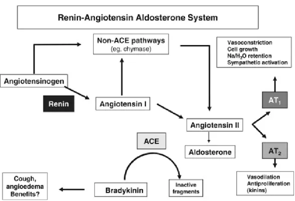
Renin-angiotensin system. The most important players in the endocrine system for blood pressure controlling are in the renin-angiotensin system(Engeli, Negrel, & Sharma,
2000).(Figure 2) The renin-angiotensin-aldosterone concatenation, via two effector components, angiotensin II and aldosterone, simultaneously regulates body sodium and water content, arterial blood pressure and potassium balance(Laragh et al., 1972). Renin is responsible for converting angiotensinogen to angiotensin I, which is converted to angiotensin II in the lungs by angiotensin converting enzyme (ACE). Angiotensin II is a potent vasoconstrictor and thus causes a rise in blood pressure. The circulating renin/angiotensin are now thought to be directly responsible for the increase in BP in essential hypertension (Lavoie & Sigmund, 2003). They are usually regulated in response to glomerular under-perfusion or a reduced salt intake (G. Beevers, G. Y. Lip, & E. O'Brien, 2001).
Angiotensin II is crucial in maintaining the structural and functional integrity of the vessel wall (Touyz & Schiffrin, 2000), and it plays a significant role in vascular smooth muscle cell contraction. Also, Angiotensin II induces vascular wall adhesion molecule-1(Kranzhofer, Browatzki, Schmidt, & Kubler, 1999).
In addition to the effect on arterial pressure, angiotensin II also significantly contributes to the development and progression of hypertensive heart disease (Figure 3)(Ibrahim, 2006; Kobori, Ichihara, Miyashita, Hayashi, & Saruta, 1999).
Figure 3. The renin–angiotensin system
Autonomic nervous system. Arteriolar constriction can also be caused by sympathetic nervous system stimulation, so the latter plays an important role in maintaining a normal blood pressure. Apparently, hemodynamics is changed in hypertension. The central nervous system seeks to maintain systemic blood pressure at a higher level (Julius, 1991). When vascular over-responsiveness sets in, the less sympathetic drive is needed to maintain neurogenic hypertension (Julius, 1990).
There is also an interaction between the autonomic nervous system and the renin-angiotensin system, both of which function together with sodium, circulating volume, and some of the more recently described hormones (Julius, Esler, & Randall, 1975).
Vascular endothelial cells. The vascular endothelium is a layer of squamous epithelial cells which directly contact with the blood. It tweaks the reactivity of vascular smooth muscles. The
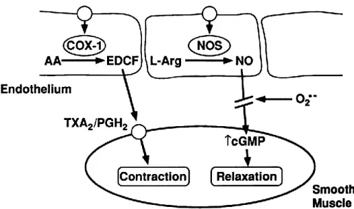
function of vascular endothelium includes the follows. It intervenes a physical obstruction between the vascular smooth muscle and hormones and other vasoactive substances circling in blood. It concentrates or metabolically degrades vasoactive substances, such as norepinephrine, serotonin, and kinins, and subsequently averts or reduces their movement in the vascular smooth muscle. It secretes vasoactive substances, such as nitric oxide (NO)(Forstermann & Munzel, 2006; Moncada & Higgs, 2006), prostacyclin(Moller & Grande, 1999) and endothelin-1(Bohm & Pernow, 2007). It releases other so-far unidentified vasoactive inhibitory and excitatory factors (De Mey, Claeys, & Vanhoutte, 1982).
Nitric oxide is a vasodilative molecule (Nathan & Xie, 1994). It has been found to play a significant role in many parts of the body (Figure 4(Faraci & Heistad, 1998)). It, acts not only as a physiological intercellular messenger but also displays cytotoxic activity in vivo (Beckman & Koppenol, 1996; Kroncke, Fehsel, & Kolb-Bachofen, 1997). NO serves as biological agents in regulating BP. Abnormal increase of nitric oxide synthase could reduce acetylcholine levels, and leads to a rise in 3'5'-cyclic adenosine monophosphate (cAMP) and 3'5'-cyclic guanosine monophosphate (cGMP) levels, and ultimately vessel relaxation (Bredt & Snyder, 1994; Hirsch et al., 1993; Lowenstein, Dinerman, & Snyder, 1994).
Figure 4. The mechanism of nitric oxide function in vessels
Endothelin is a powerful cytokine, which is a vasoconstrictive peptide, and may lead to a salt-sensitive rise in BP. It mainly modulates pulmonary BP (Galie, Manes, & Branzi, 2004). A functional isomer of endothelin, endothelin-1, is involved in essential hypertension. It modulates the autocrine feedback loops in vascular smooth muscle cells in vitro. It can down-regulate platelet-derived growth factor and transforming growth factor-beta (Hahn, Resink, Mackie, Scottburden, & Buhler, 1993). The endogenously generated endothelin-1 can contribute to the maintenance of peripheral vascular tone by regulating phosphoramidon and inhibitor of endothelin converting enzyme (Haynes & Webb, 1994). Besides, it can also activate local renin-angiotensin systems, and act as a mediator in cyclosporine A (CsA)-related renal vasoconstriction (Lanese & Conger, 1993).
Vasoactive substances. There are many other vasoactive systems and mechanisms. Some could affect sodium transport while some could maintain the vascular tone.
Atrial natriuretic peptide (ANP) is a hormone secreted from the atria of the heart. It is released by cardiac muscle cells in response to high blood volume (Song, Wang, & Wu, 2015). The role
of ANP is to reduce the retention of water, sodium and fat in the circulation, thus reducing blood pressure (Jeppesen et al., 2015).
Sodium-dependent glucose co-transporters are a family of glucose transporters. They contribute to renal glucose reabsorption (Wright, 2001). In renovascular hypertension, it may be upregulated by angiotensin II via the AT1 receptor, and then contribute to increased absorption of Na+ and therefore to the development or maintenance of hypertension (Bautista et al., 2004; Ismael-Badarneh et al., 2015). Some reports showed that it also can interrelate the calcium transport across of the vascular smooth muscle cell walls (McCarty & O'Neil, 1992).
Hypertension-related pathological conditions
Hypercoagulability: Patients with hypertension demonstrate vascular endothelial dysfunction. At the same time, the levels of hemostatic factors, platelet activation, and fibrinolysis increase abnormally, and the blood flow reduces. All those conditions suggest that hypertension confers a prothrombotic or hypercoagulable state (G. Beevers, G. Y. H. Lip, & E. O'Brien, 2001; Lip & Li-Saw-Hee, 1998).
Insulin sensitivity: Insulin resistance syndrome (IRS) has the potential to explain a large group of common metabolic and cardiovascular disorders (DeFronzo, 1997). The insulin resistance plays a role in the pathogenesis of essential hypertension, through an antidiabetic agent such as Troglitazone (Ogihara, Rakugi, Ikegami, Mikami, & Masuo, 1995), sympathetic nerve activity, or increasing muscle blood flow stimulated by insulin (Scherrer & Sartori, 1997).
Diastolic dysfunction: cardiac diastolic dysfunction has been found to be common in patients with hypertension. It will slow the maximal rate of left ventricular filling in hypertension (Fouad, Slominski, & Tarazi, 1984). Isolated diastolic dysfunction very often accompanies hypertensive
heart disease, and is also associated with myocardial ischemia and fibrosis in hypertension(Slama, Susic, Varagic, & Frohlich, 2002).
Genetic disorders
Liddle's syndrome: Liddle's syndrome is an autosomal dominant genetic disorder. It leads to high blood pressure associated with low plasma renin activity, increased resorption of sodium, metabolic alkalosis, low blood potassium, normal to low levels of aldosterone and water in the renal collecting tubules (Palmer & Alpern, 1998).
Syndrome of apparent mineralocorticoid excess (AME): AME is an autosomal recessive disorder causing hypertension and hypokalemia (Levtchenko et al., 2007). It results from defective 11b-hydroxysteroid dehydrogenase type 2 (11b-HSD2), which is critical for mineralocorticoid synthesis. The deficiency of mineralocorticoid allows the unmetabolized cortisol to bind to the mineralocorticoid receptor, inducing sodium retention, hypokalemia, suppression hypertension (Palermo, Quinkler, & Stewart, 2004).
Glucocorticoid-remediable aldosteronism (GRA): GRA is an autosomal dominant disorder in which the increase in aldosterone secretion produced by adrenocorticotropic hormone (ACTH) is no longer transient. It is one of the most common monogenic causes of hypertension (Halperin & Dluhy, 2011). The syndrome is characterized by high levels of aldosterone, suppressed plasma renin activity, high incidence of intracranial hemorrhage, and paradoxical sensitivity to glucocorticoid therapy (Kamrath, Maser-Gluth, Haag, & Schulze, 2011; Pizzolo et al., 2005). Pseudohypoaldosteronism type II (PHAII): PHAII is a group of genetic disorders, characterized by hypertension and hyperkalemia despite normal glomerular filtration rates. There are four types of PHAII, which are determined by the following gene mutations: WNK4 (PHA type IIB),
WNK1 (PHA type IIC), KLHL3 (PHA type IID), and CUL3 (PHA type IIE)(F. H. Wilson & Kahle, 1993).
Vascular Function
Vascular smooth muscle tone
The vessel tone is mediated by vascular smooth muscle cells signaling pathways. Generate tension leads to vasoconstriction and the release of tension is causing blood vessels to relax.(Woodrum & Brophy, 2001). The intrinsic tone of vascular smooth muscle is determined by the balance between contraction and relaxation signals, which in turn regulates the dynamic caliber of the blood vessel. This balance is regulated by some extracellular signals, including neural, humoral, ionic, and mechanical forces, which induce contraction or relaxation of the vascular smooth muscle. All those factors are thought to lead to an alteration in intracellular calcium concentration, resulting changes in vessel tone (Hill-Eubanks, Werner, Heppner, & Nelson, 2011).
Vascular smooth cell function
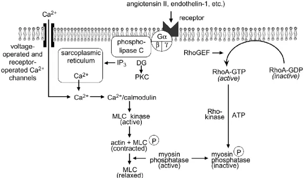
The function of the vascular smooth muscle cell (VSMC) is contraction/relaxation and the regulation of blood vessel tone-diameter, blood pressure, and blood flow distribution. The control of contraction and relaxation is dependent upon intracellular and extracellular signals(Clark & Pyne-Geithman, 2005). The contractile function of smooth muscle is modulated by the Ca2+/calmodulin interaction to stimulate phosphorylation of the light chain of myosin. In order to maintain the force generated, the dephosphorylation of the myosin light chain by myosin phosphatase is inhibited by the RhoA/Rho kinase pathway (Jono et al., 2000).