HAL Id: hal-00508557
https://hal.archives-ouvertes.fr/hal-00508557
Submitted on 30 May 2020
HAL is a multi-disciplinary open access
archive for the deposit and dissemination of
sci-entific research documents, whether they are
pub-lished or not. The documents may come from
teaching and research institutions in France or
abroad, or from public or private research centers.
L’archive ouverte pluridisciplinaire HAL, est
destinée au dépôt et à la diffusion de documents
scientifiques de niveau recherche, publiés ou non,
émanant des établissements d’enseignement et de
recherche français ou étrangers, des laboratoires
publics ou privés.
plant/fungal-lide machinery and effected by a
metazoan-like argonaute in the single-cell human
parasite Toxoplasma gondii.
L. Braun, D. Cannella, P. Ortet, M. Barakat, C.F. Sautel, S. Kieffer, J. Garin,
O. Bastien, O. Voinnet, M.H. Hakimi
To cite this version:
L. Braun, D. Cannella, P. Ortet, M. Barakat, C.F. Sautel, et al.. A complex small RNA repertoire is
genereted by a plant/fungal-lide machinery and effected by a metazoan-like argonaute in the single-cell
human parasite Toxoplasma gondii.. PLoS Pathogens, Public Library of Science, 2010, 6, pp.1-20.
�10.1371/journal.ppat.1000920�. �hal-00508557�
Plant/Fungal-Like Machinery and Effected by a
Metazoan-Like Argonaute in the Single-Cell Human
Parasite
Toxoplasma gondii
Laurence Braun1., Dominique Cannella1., Philippe Ortet2, Mohamed Barakat2, Ce´line F. Sautel1, Sylvie Kieffer3, Je´roˆme Garin3, Olivier Bastien4, Olivier Voinnet5*, Mohamed-Ali Hakimi1*
1 Laboratoire Adaptation et Pathoge´nie des Micro-organismes, CNRS UMR 5163 – ATIP+ group, Universite´ Joseph Fourier, Grenoble, France, 2 CEA, DSV, IBEB, LEMiRE, CNRS, Universite´ Aix-Marseille II, CEA Cadarache, Saint-Paul-lez-Durance, France,3 DSV/IRTSV Laboratoire EdyP, CEA, Grenoble, France, 4 UMR 5168 CNRS-CEA-INRA-Joseph Fourier University; CEA Grenoble, France,5 Institut de Biologie Mole´culaire des Plantes du CNRS, Universite´ de Strasbourg, Strasbourg, France
Abstract
In RNA silencing, small RNAs produced by the RNase-III Dicer guide Argonaute-like proteins as part of RNA-induced silencing complexes (RISC) to regulate gene expression transcriptionally or post-transcriptionally. Here, we have characterized the RNA silencing machinery and exhaustive small RNAome of Toxoplasma gondii, member of the Apicomplexa, a phylum of animal- and human-infecting parasites that cause extensive health and economic damages to human populations worldwide. Remarkably, the small RNA-generating machinery of Toxoplasma is phylogenetically and functionally related to that of plants and fungi, and accounts for an exceptionally diverse array of small RNAs. This array includes conspicuous populations of repeat-associated small interfering RNA (siRNA), which, as in plants, likely generate and maintain heterochromatin at DNA repeats and satellites. Toxoplasma small RNAs also include many microRNAs with clear metazoan-like features whose accumulation is sometimes extremely high and dynamic, an unexpected finding given that Toxoplasma is a unicellular protist. Both plant-like heterochromatic small RNAs and metazoan-like microRNAs bind to a single Argonaute protein, Tg-AGO. Toxoplasma miRNAs co-sediment with polyribosomes, and thus, are likely to act as translational regulators, consistent with the lack of catalytic residues in Tg-AGO. Mass spectrometric analyses of the Tg-AGO protein complex revealed a common set of virtually all known RISC components so far characterized in human and Drosophila, as well as novel proteins involved in RNA metabolism. In agreement with its loading with heterochromatic small RNAs, Tg-AGO also associates substoichiometrically with components of known chromatin-repressing complexes. Thus, a puzzling patchwork of silencing processor and effector proteins from plant, fungal and metazoan origin accounts for the production and action of an unsuspected variety of small RNAs in the single-cell parasite Toxoplasma and possibly in other apicomplexans. This study establishes Toxoplasma as a unique model system for studying the evolution and molecular mechanisms of RNA silencing among eukaryotes.
Citation: Braun L, Cannella D, Ortet P, Barakat M, Sautel CF, et al. (2010) A Complex Small RNA Repertoire Is Generated by a Plant/Fungal-Like Machinery and Effected by a Metazoan-Like Argonaute in the Single-Cell Human Parasite Toxoplasma gondii. PLoS Pathog 6(5): e1000920. doi:10.1371/journal.ppat.1000920 Editor: Shou-Wei Ding, University of California Riverside, United States of America
Received January 27, 2010; Accepted April 23, 2010; Published May 27, 2010
Copyright: ß 2010 Braun et al. This is an open-access article distributed under the terms of the Creative Commons Attribution License, which permits unrestricted use, distribution, and reproduction in any medium, provided the original author and source are credited.
Funding: M.-A. Hakimi and O. Voinnet were supported by grants from the Centre National de la Recherche Scientifique (CNRS, ATIP+), the Agence National de la Recherche (ANR blanche, AGO-hook) and the Institut National de la Sante et de la Recherche Medicale for M.-A. Hakimi (Contrat d’Interface CHU). The funders had no role in study design, data collection and analysis, decision to publish, or preparation of the manuscript.
Competing Interests: The authors have declared that no competing interests exist. * E-mail: Mohamed-Ali.Hakimi@ujf-grenoble.fr (MAH); olivier.voinnet@ibmp-ulp.u-strasbg.fr (OV) .These authors contributed equally to this work.
Introduction
Apicomplexa are unicellular eukaryotes that multiply intracel-lularly in their mammalian hosts. They include parasites of major medical importance like Plasmodium species, the causative agent of malaria, and Toxoplasma gondii, the most widespread apicomplexan parasite, present virtually everywhere on earth. Although usually causing only mild symptoms in the adult, Toxoplasma can cause severe and life-threatening diseases in developing fetuses and in immunocompromised individuals, especially AIDS and transplant patients [1,2]. Toxoplasma has a complex life cycle that includes infections of more than one host organism, differentiation through
several morphologically distinct forms, and both sexual and asexual replication [3]. Changes in gene expression is expected as (i) parasites progress through the cell cycle, (ii) parasites differentiate in specific stages, and (iii) parasites are exposed to the host immune system during infection [4]. How these changes are regulated at the molecular level remains to a large extent unknown. A puzzling feature is the apparent lack, in apicom-plexan parasites, of large families of recognizable specific transcription factors (TFs) operating in other eukaryotes [5]. Despite the paucity of recognizable TFs, apicomplexans are endowed with a rich repertoire of enzymes associated with epigenetics and chromatin remodeling, and this observation has
fueled the idea that epigenetics could play an important role in the control of gene expression [6,7].
Small regulatory RNAs are linked to epigenetic regulation of gene expression in several organisms but these are presently understudied in the Apicomplexa. The defining features of small silencing RNAs are their short length (,20–30 nucleotides) and their association with members of the Piwi/Argonaute (AGO) family of proteins, which they guide to their regulatory targets [8,9]. Many, albeit not all, small RNAs (sRNA) are produced by the RNase III-related enzyme Dicer. Small interfering RNAs (siRNA) are generated as populations from multiple Dicer cleavages along long dsRNA precursors, whereas microRNAs (miRNA) are discrete species generated from a single Dicer cleavage event of noncoding primary precursor transcripts containing small, imperfect stem–loop structures [10]. These distinct small RNA pathways compete and collaborate as they regulate genes and protect genome integrity from invading nucleic acids including viruses and transposons. They function as guides for effector complexes (RNA-induced silencing complexes, RISCs) that regulate gene expression by degrading mRNA, repressing its translation, or modifying chromatin. RNA silencing is an evolutionary ancient regulatory mechanism, and small RNA pathways in unicellular organisms appear, so far, to be relatively simple. In fission yeast, a single class of endogenous siRNAs has demonstrated roles in epigenetic silencing at centromeres and the initiation of heterochromatin assembly at the mat locus [11]. In the ciliated protozoan Tetrahymena thermophila, small RNAs are involved in developmentally regulated DNA elimination [12,13] and post-transcriptional gene regulation [14]. Particularly surprising is the recent finding that the unicellular green alga Chlamydomonas produces microRNAs that had been previously associated with developmental regulation and multi-cellularity [15,16].
Here, we show that the T. gondii genome, unlike in Plasmodium species [17], encodes all core components of an elaborate RNA silencing machinery that has been evolutionary shaped as a patchwork of factors of plant and fungal origin. We establish a comprehensive sRNA landscape of T. gondii through deep sequencing, and unravel that the most abundant sRNA classes
are formed by metazoan-like miRNAs as well as plant-like repeat-and-satellite-associated sRNAs coined rdsRNA and satRNA, respectively. Beyond the surprising complexity of the small RNAome, we provide a thorough biochemical characterization of the proteins that associate with the single T. gondii Argonaute protein, Tg-AGO. Unexpectedly these proteins constitute the near-entire cohort of previously identified human and fly miRNA-RISC components. Our data indicate that miRNA-loaded T. gondii argonaute associates with polysome probably to regulate translation of Tg-miRNA predicted targets, many of which include mRNA with perfect or near perfect complementarity. Tg-AGO also co-purifies with chromatin-repressing complexes, suggesting a role in transcriptional silencing, most likely through it demon-strated association with rdsRNAs or satRNAs.
Results/Discussion
The Toxoplasma RNA silencing machinery
Previous analyses have suggested a monophyletic origin for plant and animal Dicer proteins [18]. Sequence analyses show that the Toxoplasma genome (TOXODB release v6.0) [19] encodes only one Dicer-like protein, Tg-Dicer, which displays significant variability in primary sequence and domain organization com-pared to the Dicer consensus (Figure 1A): Tg-Dicer possesses an RNA helicase domain and two RNaseIII catalytic domains (RNaseIIIa and RNaseIIIb), but it lacks recognizable domains for dsRNA binding (DSRM) and PIWI-ARGONAUTE-ZWILLE (PAZ) functions. This organization is strikingly reminiscent of the DCL1 protein of the single cell algae C. reinhardtii (Figure 1A). Toxoplasma and Chlamydomonas Dicer-like sequences seem, indeed, orthologous, as they form a specific clade supported by a strong bootstrap score (Figure 1B), a consequence of a Drosha-like signature polypeptide that is more related (albeit weakly) to eubacterial RNaseIII enzymes and known to form an out-group with respect to higher plant and animal Dicers [18].
Ago-related proteins are divided into the Ago-like and Piwi-like subfamilies (Figure 2A); a third clade, termed ‘Group 3 Argonautes’, is worm-specific [20]. Toxoplasma Argonaute (Tg-AGO) is represent-ed at a single genomic locus; there is no evidence for Toxoplasma-encoded Piwi proteins. Tg-AGO belongs to the Ago-like family but only with weak bootstrap support, suggesting that the protein diverges significantly from its metazoan and plant counterparts. Nonetheless, the two key signature domains of the AGO family — the PAZ domain and the C-terminal PIWI domain— are conserved in Tg-AGO (Figure 2B). Overall, the PIWI domain shows the highest degree of conservation, but the Asp-Asp-Glu/Asp catalytic triad required for slicer activity is not found, suggesting that the protein lacks endonucleolytic cleavage capacity (Figure 2B). The second signature motif, the PAZ domain, contains only a few residues that are strictly conserved, while the middle (MID) domain of Tg-AGO harbors the residues (Y596, K600, Q610 and K644; Figure 2C) required to bind the characteristic 59 phosphate group of guide small RNA strands [21,22]. We also noted the presence, in the amino terminal part of Tg-AGO, of a stretch of repeated RGG residues (amino acids 1–68), in which the arginines have the potential to undergo methylation (Figure 2B). This feature is found in metazoan and plant AGO-related proteins and was shown to alter their stability and/or sub-cellular distribution, ultimately impacting their function [23,24,25].
A phylogenetic tree constructed by aligning the stereotypical RNA-dependent RNA polymerase (RDR) domain supports the monophyletic origin of the proteins found in C. elegans, fungi and Arabidopsis [26]. Inspection of the Toxoplasma genome showed the presence of a single RDR-like gene, suggesting the existence of an
Author Summary
Toxoplasma gondii is an important human parasite that causes life-threatening diseases in developing fetuses and in immunocompromised individuals, especially AIDS and transplant patients. Curiously, the Toxoplasma genome is deprived of most of the basic transcription factors that regulate gene expression in other eukaryotic cells. Therefore, alternative strategies must exist to modulate the many phases of the Toxoplasma complex life cycle that includes invasion of several hosts. Here, we investigate one of these strategies, by studying the repertoire of Toxo-plasma silencing small RNAs (sRNAs). In eukaryotes, most of these regulatory molecules, 20–30nt-long, are produced by members of the Dicer RNase-III family, and exert their various functions through ubiquitous proteins called Argonaute (Ago). The surprising diversity of the Toxoplas-ma sRNAome uncovered in our study is consistent with those molecules exerting key functions during the parasite’s life cycle, including, possibly, during virulent infection. The study also unravels an unsuspected level of complexity in the origin and mechanisms of action of the factors that generate and affect Toxoplasma sRNA, prompting a re-evaluation of our current views on RNA silencing in eukaryotes.
amplified RNA silencing machinery in this organism. Tg-RDR is closely related to Neurospora crassa RDR, QDE1, and forms a specific clade with plant RDRs, which itself constitutes an out-group from the RDRs of metazoans and from the fission yeast S. pombe (Figure 1C). We conclude from this analysis that a patchwork of factors of plant and fungal origin form the core processor components of the Toxoplasma RNA silencing machin-ery. This finding can be rationalized partly by the fact that the apicomplexa ancestor is a presumed endosymbiont of red algae [27]. We note, however, the moderate or poor phylogenetic relationship observed between Tg-Dicer, Tg-AGO and the corresponding paralogous proteins of its mammalian hosts.
Complexity of the Toxoplasma small RNAome
Having established that the Toxoplasma genome encodes all core components of an elaborate RNA silencing machinery, we sought to determine the small RNA landscape of this organism. To this aim, we prepared total RNA from freshly released, filtered parasites. Ethidium bromide staining revealed a relatively abundant class of small (s)RNAs, ,30 nucleotides (nt) in length (Figure S1A). The 20–40nt sRNA fraction was recovered by gel excision, cloned and subjected to deep sequencing using the Illumina technology. The sRNA library was constructed so as to represent only those sRNAs with a 59 monophosphate and a 39 hydroxyl group, the termini expected of miRNAs and siRNAs [28]. About 75% of a total of 5,701,506 reads,
Figure 1. Domain organization and phylogenetic analysis of Dicer and RDR proteins. (A) Dicer proteins cleave dsRNA precursors into characteristic lengths through the action of two RNase III domains (in yellow). The domain arrangement of several Dicer enzymes is compared to Toxoplasma Tg-Dicer (TgTwinScan_3226, TOXODB v4.3). Evolutionary relationships between Dicer (B) and RDR (C) proteins from the following species: AT: Arabidopsis thaliana, GL: Giardia lambdia, SP: Schizosaccharomyces pombe, DM: Drosophila melanogaster, HS: Homo sapiens, CE: Caenorhabditis elegans, CR: Chlamydomonas reinhardtii, NC: Neurospora crassa, TT: Tetrahymena thermophila, TG: Toxoplasma gondii. Tg-Dicer and Tg-RDR are shown by a green and black arrows respectively and their branch family are shaded in green and gray. Numbers above the nodes indicate neighbor-joining bootstrap percentages. doi:10.1371/journal.ppat.1000920.g001
represented tRNA and rRNA turnover products, as previously reported for other organisms (Figure S1B) [29]. After filtering low quality reads, 39, 59 adapters, and reads shorter than 17 nucleotides, a remaining 1,555,290 reads (,30% of total reads) matched the Toxoplasma genome (ToxoDB, version 4.3) (Figure S1B). Comparing mRNA and sRNA data for highly expressed genes suggested that the sRNA fraction contained only a very low level of degradation
products from longer mRNAs (data not shown). Most sRNA sequences were found to be 25–27nt in length, with 25nt representing the dominant size class (Figure 3A). Of the 1,222,203 total reads, 94,170 corresponded to non-redundant sRNAs, and 92% of these were single reads, thus unraveling a highly complex sRNA population. Plotting Toxoplasma sRNAs (Tg-sRNAs) species with 100% match on the reference genome (in 10-kb sliding windows)
Figure 2. Domain organization and phylogenetic analysis of Argonaute and PIWI proteins. (A) Evolutionary relationships between Argonaute and Piwi proteins from the following species: AT: Arabidopsis thaliana, GL: Giardia lambdia, SP: Schizosaccharomyces pombe, DM: Drosophila melanogaster, HS: Homo sapiens, CE: Caenorhabditis elegans, CR: Chlamydomonas reinhardtii, NC: Neospora caninum, TT: Tetrahymena thermophila, TG: Toxoplasma gondii. Tg-AGO (accession number: GU046561) is pointed out by a pink arrow. Argonaute (in pink), Piwi (in blue) and group 3 (in red) branch family are represented as shaded boxes. Numbers above the nodes indicate neighbor-joining bootstrap percentages. (B) Schematic depiction of Toxoplasma Tg-AGO and Arabidopsis At-AGO1 (GeneID: 841262) domain according to archaeon Aquifex aeolicus X-ray crystal structure. The amino-terminal domain (grey or blue) is linked to the PAZ domain (red). The MID domain (green) connects the PAZ domain with the PIWI domain (pink) at the carboxy-terminal end of the protein. In the PAZ domain, residues important for binding of small RNA 39 ends are indicated. In the MID domain, the residues required for 59 end binding to small RNAs and binding to the 7-methylguanine (m7G) cap of target mRNAs are shown. The conserved residues for RNase H cleavage activity (DDH) in the PIWI domain are also shown. RGG indicates the domain of Tg-AGO containing arginine-glycine-glycine repeats. Poly Q, polyglutamine-containing domain in Arabidopsis Ago1. (C) Sequence alignment of conserved 59– phosphate binding residues (MID domain) across various species of Argonaute proteins. Prefix Hs, Homo sapiens; Dm, Drosophila melanogaster; Aa, Aquifex aeolicus. Conserved residues are shaded in red.
showed that non-redundant Tg-sRNA with high read numbers (.1000) originate predominantly from non-coding intergenic regions or are embedded within introns of protein-coding transcriptional units (TUs) (Figure 3B). Other, medium-to-low abundance Tg-sRNA, by contrast, mapped to protein-coding TUs and a variety of DNA repeats and satellites (Figure 3B). These two classes of Tg-sRNA were detailed further, as described in the following sections.
Toxoplasma miRNAs
In a search for putative Toxoplasma miRNA candidates, we evaluated sRNA reads exhibiting the following features: (1) high abundance of sequence reads sharing the same 59 terminus; (2) exact match to one or several genomic loci displaying a characteristic fold-back structure typical of MIRNA precursors; and (3), when applicable, low abundant sequence reads
corre-Figure 3. A repertoire ofToxoplasmaendogenous small RNAs. (A) Size distribution of Toxoplasma sRNAs. The sets of redundant (grey) and unique (black) small RNAs were used to generate a histogram quantifying the number of sequences obtained for each size class. nt., nucleotides. (B) Distribution of small RNAs across the Toxoplasma genome. TU: Transcriptional unit, CDS: CoDing Sequence, UTRs: UnTranslated Regions. (C) A miR-60b production hot spot in chromosome VIIb is shown. Small RNAs with perfect matches were plotted within a 800-bp sliding window. Short thin lines above the long bars represent small RNAs derived from the antisense strands, and lines below the bars represent small RNAs from the sense strands. Vertical bars represent the consensus positions of sequencing reads that mapped to the predicted precursors, and the values indicate the total number of these reads. The fold-back structure of the precursor predicted with mfold is provided below. The mature region is shown in red and the passenger strand (microRNA*) in blue. The miR-60b–containing locus for the three canonical strains of Toxoplasma and Neospora caninum is depicted by sequence and structure. (D) A miR-4 production hot spot in chromosome VIII is shown along with the predicted structure and the sequence conservation across parasite species.
sponding to the labile (miRNA*) passenger strand of miRNA/ miRNA* duplexes, as predicted within the fold back structures. Fourteen sRNA families cloned at a high frequency met these MIRNA features and were thus annotated with high confidence as T. gondii miRNAs (Tg-miRNAs) (Figures 3, S2-S13 and Table S1). Genome browser views of some of these Tg-MIRNA indeed indicated the existence of a low frequency, single miRNA* (passenger strand) corresponding to the opposite strand of the duplex within the fold back-structure (e.g. Tg-miR-60b; Figure 3C). Moreover, the reconstituted duplexes sometimes had small 39 overhangs characteristic of Dicer processing (Figures 3C, S8-S10). Of the 14 annotated Tg-miRNA families, 7 gave detectable hybridization signals in Northern analysis carried out with 59 end-labeled antisense oligonucleotides (Figures 4A, 4C and S14). No signal was detected with RNA extracted from host cells, confirming that these sRNAs are Toxoplasma-specific. Hybridization often unraveled discrete sRNA species that were heterogeneous in size, reminiscent of the sRNA signals observed in S. pombe [30] and other organisms in which Dicer lacks a PAZ domain, required for the precise sizing of processed sRNAs [31,32,33]. In all cases, hybridization with antisense probes from precursor sequences flanking the mature miRNA gave no signal (data not shown), confirming the excision, by Tg-Dicer, of a single sRNA species, a landmark of plant and metazoan miRNA biogenesis. The members of the remaining 7 miRNA families were below detection levels, in agreement with their much lower cloning/sequencing frequencies (Figures S8-S13). In all cases -and as expected from their poor read counts due to their intrinsic instability- cloned miRNA* were also below detection levels of Northern analysis.
Sequencing and Northern analyses showed that the miR-60 family largely dominates the Toxoplasma miRNA landscape, accounting for 335,014 reads, of which 61% (280,723 reads) were contributed by miR-60a alone (Figure S2B). MIR-60, together with MIR-4, also constitute the two most diversified Tg-MIRNA gene families (with 8 distinct members in each) among the 14 families identified with high confidence (Figure S2B and S3B). In most cases of Tg-miRNAs with multiple precursors (6 families, Table S1), the mature miRNAs were not located on the same fold-back arms, which, furthermore, were also found to vary in sequence, suggesting that these genes do not share a common ancestor and, thus, have evolved separately. The 14 high-confidence Tg-miRNAs showed no significant homology to any of the known miRNAs of plants and metazoans, as assessed in the central miRBase depositary (release 14). Nearly all Tg-miRNAs and Tg-miRNA* (when available) had, however, directly identifiable orthologs in the genome of the apicomplexan Neospora caninum (dog parasite), when up to 3-nucleotide polymorphism was tolerated (Figures 3, S2-S8 and S13). Moreover, the size and abundance of these orthologous N. caninum (Nc)-miRNAs was confirmed by Northern blot analysis using the same antisense oligonucleotide probes employed for detection of Tg-miRNAs (Figures 4A and S14). The notable exception to sequence conservation in N. caninum was observed with Tg-miR-62, -64, -65 or -66, although this could be attributed to the incomplete N. caninum genome annotation (Figures S9-S12). Taking into account recent observations made in the single cell algae Chlamydomonas, apicomplexans thus provide the second reported example of unicellular organisms that produce miRNAs. Unlike in Chlamydomonas, nonetheless, and despite the relatedness of the Dicer proteins found in both organisms (Figure 1A and 1B), Tg-MIRNA fold-backs have length and thermodynamic features that are much closer to those of mammalian hosts than those of Chlamydomonas or higher plants [10]. Consistent with this idea, most Tg-miRNAs display a clear 59- nucleotide bias towards A, as also observed for most mammalian miRNAs [34]. Unlike many
mammalian MIRNAs, however, Tg-MIRNAs were not found to form genomic clusters. These findings further emphasize the surprising mosaic nature of the Toxoplasma RNA silencing machinery and small RNA loci.
The above 14 miRNA families were identified through deep-sequencing of small RNAs isolated from freshly egressed parasites, and so other miRNAs might exist that were simply too low in abundance to be cloned under these specific growth conditions. In addition, several Tg-sRNAs cloned at moderate to low frequency mapped to imperfect fold-backs scattered along the genome, with relatively low free energy (Figure S15). These hairpins are much more heterogeneous in size and structure than cognate Tg-MIRNA precursors, yet their processing produces discrete sRNA species. Although their relatively modest cloning frequencies precludes their detection by Northern analysis, including in N. caninum, the corresponding sRNA might represent recently-evolved miRNAs that may engage into miRNA-like regulatory activities. In plants, a model for MIRNA gene evolution, termed ‘‘spontaneous evolu-tion’’ stems from the high density of small-to-medium sized fold-back sequences scattered throughout the Arabidopsis genome. It has been proposed that following the capture of transcriptional regulatory sequences, some of these random fold-backs could occasionally give rise to new MIR genes. Stabilization through co-evolution with targets initially found by chance could then lead to the fixation of these genes in the genome [35].
Biochemical properties of Toxoplasma miRNA
Unlike metazoan miRNAs, plant miRNAs are methylated at the 29 hydroxy positions of their 39-last nucleotides [36]. This modification, mediated by the methyl-transferase HEN1, protects miRNA from 39 end uridylation and subsequent degradation [37]. However, Tg-miRNA species were found sensitive to b-elimination by periodate, which causes a diagnostic shift in sRNA mobility (Figure 4F). Thus, unlike their plant counterparts, but similar to metazoan miRNAs, Tg-miRNAs do not carry 39-end modifica-tions, a result also consistent with our failure to identify a HEN1 homolog in the Toxoplasma genome (TOXODB, release v5.2). Nonetheless, the 39 end of several cloned Tg-miRNAs was often found to contain untemplated adenine residues, which must be added, therefore, after processing by an as yet unidentified terminal adenyl-transferase (Figure S3B). It was shown recently that addition of adenylic acid residues on the 39-end apparently slows down miRNA turnover in Populus trichocarpa [38].
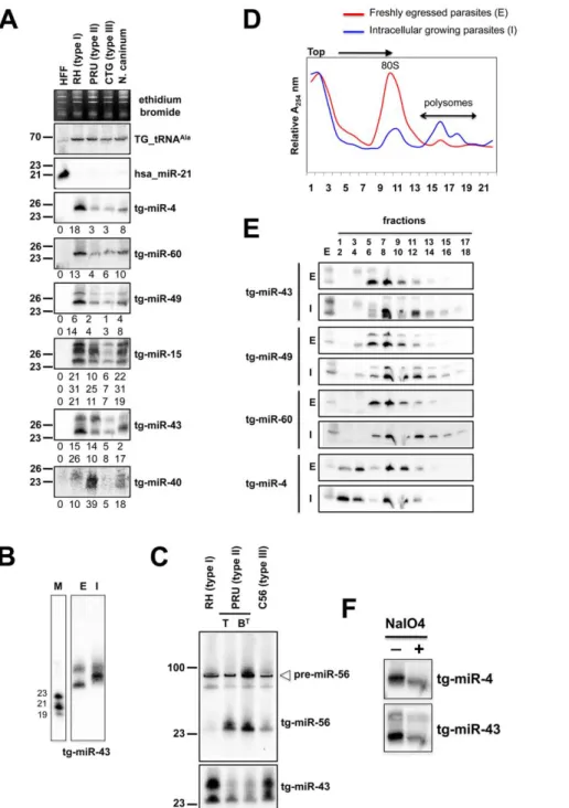
In plants and animals, miRNA are recruited by AGO proteins to enhance the turnover, or inhibit the translation of cognate mRNA targets. Consequently fractions of most plant and metazoan miRNAs associate with polysomes, the sites of active translation. The existence of miRNAs in Toxoplasma together with the absence of detectable slicer residues in the Tg-AGO predicted that Tg-miRNA would also associate, at least partly, with polysomes. To test this idea, protein extracts from freshly egressed parasites (E) ready to invade, or from fast-growing intracellular parasites (I), were fractionated and resolved on sucrose density gradients (see Methods). For the former (I), the absorbance profiles at 254 nM reflected the ribosome pattern expected from rapidly growing cells: there were few monosomes (80S) and the bulk of the ribosomes sedimented in the polysomal fractions (Figure 4D). By contrast, the amount of polyribosomes in invading parasites (E) was substantially reduced, and this was accompanied by a concomitant increase in 80S monosomes (Figure 4D). As previously observed in plants and metazoans, Tg-miRNAs distribution was found to span a wide range of molecular weights across the gradient (Figure 4E) [39,40,41]. Nonetheless, a fraction of several Tg-miRNAs co-sedimented with polysomes (Figure 4E,
Figure 4. Expression patterns and characteristics of representative microRNAs inToxoplasma.(A) Expression profile analyses of six parasite microRNA candidates by small-RNA Northern hybridization in three canonical strains of Toxoplasma and its close relative, Neospora caninum. To control for loading, blots were stripped and rehybridized with the indicated miRNA and the control probes (tRNAAlaand hsa_miR-21). Ethidium
bromide staining of rRNA was used as loading control. Hybridization signals were quantified with the ImageJ software, normalized to the amount of Toxoplasma tRNAAlasignal present in each sample, and shown as digit below each Northern blot. For miR-15, -43 and -49, each individual signal was
separately quantified. The number next to each panel represents the position of RNA markers. Tg, Toxoplasma gondii; hsa, Homo sapiens. (B) Total RNA from freshly egressed (E) and intracellular growing parasites (I) were analyzed by Northern hybridization with complementary probes for Tg-miR-43. RNA markers (left lane) are 19, 21 and 23 nucleotides. (C) Total RNA from types I, II (tachyzoite and pH-converted bradyzoite) and III were analyzed by Northern hybridization with complementary probes for Tg-miR-43 and -miR-56. Pre-miR indicates the foldback RNA precursor form; Tg-miR refers to the mature, predominant form. The number next to each panel represents the position of RNA markers. (D) Total RNA from freshly egressed (E, in red) and intracellular growing parasites (I, in blue) were subjected to sedimentation on 5%–45% sucrose gradients in the presence of cycloheximide (to preserve the association of translating ribosomes with mRNAs). Measurements of the total RNA in the gradient fractions reveal signature patterns of peaks corresponding to messenger-ribonucleoprotein complexes (mRNP), followed by the 80S ribosomal fractions and the polyribosomal fraction. As can be appreciated from the A254 profiles, the rapid proliferation of tachyzoites inside of the host cell (in blue) is diagnosed by the high level of translating polyribosomes. (E) A proportion of Tg-miRNAs cosediment with polyribosomes. Total RNA was extracted from each fraction and analyzed by Northern blots with complementary probes for 6 Tg-miRNAs [miR-43, -49, and 60 (shown) and mir-4, -15, and 40, (not shown)]. All of the Tg-miRNAs tested are predominantly localized to the mRNP with a clear shift to polyribosome fractions in intracellular growing parasites. (F) The 39 end of Tg-miR-4 and -43 are not modified. Gel-fractionated small RNAs were examined by b-elimination assay (see methods).
fractions 13–18). Moreover, the association with translating ribosomes was more pronounced in exponentially growing parasites (I), as expected. Not all Tg-miRNA, however, were found associated to polysomes, and this was notably the case of Tg-miR-4 (Figure 4E). Tg-Tg-miR-4 and other non-polysomal miRNA may regulate target mRNAs at later stages of parasite differenti-ation (e. g. bradyzoite); alternatively, they might not be involved in translation control (data not shown), as has been recently shown for a class of Arabidopsis miRNA [41] that use cleavage-competent and/or cleavage-resistant target sites found in specific non-coding RNAs to initiate the production of trans-acting (tasi)RNAs via the action of RDR6 [42]. Whether tasiRNA exist in Toxoplasma is an interesting question for future experiments. In any case, our findings demonstrate that several Tg-miRNAs are present in the form of miRNPs in polyribosome-containing fractions where they are likely to negatively regulate translation of target transcripts.
Complex regulation of Toxoplasma MIRNA gene expression
miRNAs orchestrate many biological functions and are notably involved in cell fate determination and/or integration of developmental or external stimuli. Plant and metazoan miRNA expression may thus vary greatly depending on growth conditions, changes in developmental stages or, in the case of parasites, changes in virulence. To investigate if, similarly, Tg-miRNA accumulation/processing is regulated differentially in Toxoplasma, we sampled miRNA from freshly egressed (E) or intracellular (I) parasites, as well as from three classical Toxoplasma isotypes. These isotypes are representative of the European and North American parasite population and correspond to three clonal lineages, designated type I, II and III, corresponding to reference strains RH, PRU and CTG, respectively [43]. These genotypes display contrasted virulence in mice: type I strains are lethal, whereas type II and III strains are hypo-virulent and typically establish chronic infections. There are additional phenotypic differences in migration, growth rate, and ability to convert from tachyzoite to the cyst-forming bradyzoite stage, notably [43].
For several of the Tg-miR detected by Northern analysis, we observed differential abundances between the Toxoplasma strains (Figure 4A and 4C). Thus, normalized to the Tg-tRNAAlasignal, the miR-4 signal was six fold greater in type I than it was in types II and III (Figure 4A). Likewise Tg-miR-4, -49 and -60 were more abundant in type I strain, whereas Tg-miR-40 and -56 were clearly more abundant in type II. Further investigation of these variations in miR-56 levels showed that they were attributable to differences in miRNA processing rather than transcription, because similar levels of pre-miR-56 were observed among the three isotypes, in Northern analyses (Figure 4C). This result reinforces the growing view that MIRNA genes can undergo extensive post-transcriptional regulation through mechanisms that selectively affect pri-miRNA processing and/or pre-miRNA stabilization [44], as uncovered recently with interactions involving murine pre-Let-7, lin-28 [45,46] and the RNA-binding protein KSRP [47], a homolog of which was indeed found associated with Tg-AGO (see following sections). These observations thus extend this concept to a single cell parasite; given the overall low genetic diversity among Toxoplasma isotypes [43], they further suggest that differential regulations of pathogen’s miRNA repertoires might, indeed, influence virulence. Analyses of Tg-miR accumulation between freshly egressed (E) and intracellular (I) Toxoplasma revealed additional scope for modulation of mature miRNA levels between the two parasitic states. For instance, there was a clear mobility shift with miR-43, which is unlikely explained by changes in pre-miR-43 steady levels, but rather, by alternative Dicer-mediated
processing events producing small RNA length variants or with modified termini (Figure 4B). Collectively, these observations unravel highly complex regulations of Tg-MIRNA gene expression, which might be used to refine the amplitude or regulatory outputs of target gene regulation during the parasite’s multiple biological states.
Toxoplasma miRNA exhibit perfect or near-perfect matches to many target transcripts
We then attempted to identify putative targets for representative members of the 14 unambiguous Tg-MIRNA families retrieved in this study. A bioinformatics approach was used to scan Toxoplasma transcripts for Tg-miRNA complementarity sites. Despite the resemblance of Tg-MIRNA and mammalian MIRNA genes, and the association of both types of molecules to polysomes, the absence of slicer residues in the Tg-AGO protein strongly suggests that the parasites’ miRNA engage into a distinct type of pairing to their targets (Figure 2B). Hence, in the mammalian host, most AGO2-bound miRNAs exhibit only moderate pairing to their targets, notably through a stretch of 6–7 contiguous 59 nucleotides known as ‘seed’, which is usually followed by several central mismatches that sterically hinder the RNAseH activity of AGO2 [48,49]. This loose miRNA:target pairing, which is thought to favor translational repression over slicing, makes it difficult to predict mammalian miRNA targets using computer algorithms. These algorithms, moreover, are often biased towards 39 UTRs, because these regions evolve much more rapidly than coding regions, and are, therefore, more prone to the identification of contiguous, 6–7nt seed-complementary sequences [50].
We found that most of the 14 Tg-miRNA analyzed have readily identifiable target sites in a variety of cellular transcripts (Table S2 and data not shown). Interestingly, these sites exhibit complete to near-complete complementarity to miRNAs -a feature of plant but not of metazoan miRNAs- and they are found in 59-UTR, coding region and 39-UTR, although there is a clear bias towards the latter region for most miRNA analyzed (Figure S16A and Table S2). Allowing up to 3 mismatches, more than 80 putative target transcripts were identified for miR-60a alone, the most abundantly sequenced Tg-miRNA. Using the same stringent parameters, an average of 25 cellular targets could be retrieved for each of the 14 Tg-miRNA (Figure S16A and Table S2). GO-term analysis of the putative Tg-miRNA target transcripts showed that they encompass virtually all known biological functions, with a somewhat stronger emphasis on translational control and cell cycle regulation, which might be expected for a single-celled, highly dividing parasite (Figure S16B). These predicted Toxoplasma miRNA:target interac-tions thus constitute an unprecedented situation in all eukaryotes studied so far, whereby a miRNA-loaded, slicer-deficient Ago (see later in the text) might regulate target gene expression, presumably at the translational level, through perfect or near-perfect binding sites that are predominantly –albeit not exclusively- located in 39-UTRs. To test the possibility of target cleavage and degradation mediated by Tg-AGO, we examined the levels of mRNA predicted as strong targets of isotype-specific Tg-miRNAs (Table S2). Real time PCR analyses revealed little, if any changes in mRNA levels between type I and II isotypes, contrasting with the differential abundances of the corresponding Tg-miRNAs. This result corroborates partially the suggestion that Tg-AGO acts mainly as a translational regulator, which is also in agreement with the cellular factors found in association with Tg-AGO (see later in the text). Owing to the lack of available antibodies for predicted targets and our current inability to generate Tg-Dicer or Tg-AGO mutants, experimental validation of the above hypothesis will be part of future experiments.
To date, the use of RNAi for specific gene silencing has remained largely inconclusive in Toxoplasma. Many laboratories have attempted to use this tool to down-regulate gene expression but very few reports showed successful double-stranded RNA induced gene silencing and there is currently no evidence for the production of specific siRNA [51]. We note that the use of RNAi is normally expected to result in mRNA turnover, as in metazoans or plants. The nature of the Tg-AGO (slicer deficient) and its possible mode of operation through translational repression (with a usually modest output on gene expression in metazoans) is obviously one parameter that could explain the lack of significant levels of mRNA degradation upon RNAi treatments in this organism.
Toxoplasma repeat-and satellite-associated sRNA
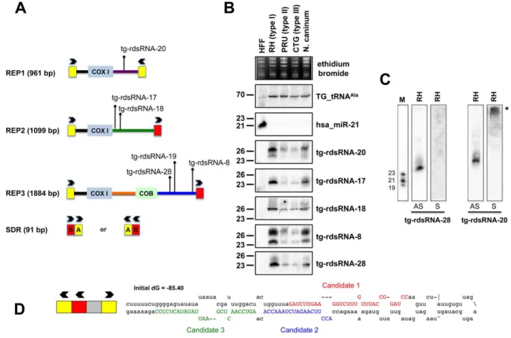
The largest bulk of medium-to-low abundance Tg-sRNAs does not meet the criteria of miRNA annotation and appears to match repetitive elements REP1, REP2 and REP3 (Figure 5A) [52]. REP elements are mitochondrial-like sequences dispersed throughout the nuclear genome of Toxoplasma. They are typically composed of mitochondrial-like genes, including COX1 (cytochrome oxidase subunit 1) and COB (apocytochrome b) that are flanked by a 91 bp short-dispersed repetitive sequence (SDR) organized as a
direct or inverted repeat (Figure 5A) that might play roles in generation or dispersal of the REP elements. Nonetheless, there is no sequence similarity between SDRs and other terminal repeats such as those of retroviral LTRs. Moreover, REP elements do not seem to be highly mobile [52].
Genome mapping showed that Toxoplasma REP-derived sRNAs (rdsRNAs) form discrete species that are exclusively generated from regions located downstream of the COX1 and COB sequences, (Figure 5A), with read counts typically ranging from .10,000 (rdsRNA-17) to a few hundred reads (rdsRNA-28). This fairly high abundance might be explained by the fact that the estimated number of REP elements is .500 copies per genome [52]. While their size range (21–27nt, Table S3) and sensitivity to periodate (not shown) was similar to that of Tg-miRNAs, about half of the rdsRNAs had a 59 terminal U instead of the prevalent A found in miRNAs. All tested Tg-rdsRNAs were readily detected by Northern blotting using 59end-labeled antisense oligonucleotides but, unlike Tg-miRNAs, they were consistently much more abundant in the highly virulent Toxoplasma isotype-I (Figure 5B). Interestingly, sense probes generated no signal for the Tg-rdsRNAs tested, consistent with the absence of Illumina reads corresponding to opposite-strand sRNA species (Figure 5C and data not shown). A sense probe for
Figure 5. Expression patterns and characteristics of repeat-associatedTg-rdsRNA inToxoplasma. (A) Schematic representation of REP elements. The three REP fragments and the short disperses repeat (SDR) are displayed from 59 to 39 with respect to the COX1 (cytochrome oxidase subunit 1) gene fragment. Blocks A and B of the SDR are labeled in yellow and red, respectively. Cloned Tg-rdsRNA are represented above: Tg-rdsRNA-8 (1155 reads), tg-rdsRNA-17 (464 reads), Tg-rdsRNA-1Tg-rdsRNA-8 (1134 reads), Tg-rdsRNA-19 (1326 reads), Tg-rdsRNA-20 (1066 reads) and Tg-rdsRNA-2Tg-rdsRNA-8 (223 reads). (B) Expression profile analyses of five parasite Tg-rdsRNA candidates by small RNA Northern hybridization in three canonical strains of Toxoplasma and its close relative, N. caninum. Controls and quantification method are as in Fig. 2A. The number next to each panel represents the position of RNA markers. (C) Small RNA blots were stripped and re-hybridized with antisense probes of selected Tg-rdsRNA sequence. Antisense and sense oligonucleotides (indicated by AS and S, respectively) were used to confirm the polarity of Tg-rdsRNA-20 and -28. (D) SDR-containing chromosomic region (TGME49_chrX:233424-233609) forms a stereotypic hairpin precursor predicted by mfold that harbor cloned small RNA candidates.
Tg-rdsRNA-20, however, gave a high molecular weight signal potentially resulting from hybridization of a double-stranded RNA precursor, although RNA folding algorithms did not reveal any significant secondary structure at, or in the vicinity of Tg-rdsRNA sequence matches (Figure 5C). Nonetheless, the detection of identical rdsRNA species in the related apicomplexan N. caninum and their isotype-specific accumulation (Figure 5B) together with their abundant loading into Tg-AGO (see below) make it unlikely that these species are simply random degradation products. This rather suggests the existence of a conserved mechanism that accounts for REP-dependent production of precursor molecules required for rdsRNA synthesis. A second class of low abundant Tg-rdsRNAs mapped directly to a long, imperfect stem-loop structure resulting from annealing of an individual ‘solo SDR’ unit. This structure is depicted in Figure 5D, together with the cloned sequences of contiguous or overlapping sRNAs that are likely produced via stepwise processing by the Tg-Dicer. Such imperfect structures might well represent the equivalent of the plant proto-MIRNA genes that arise from DNA-type non-autonomous elements known as miniature inverted-repeat transposable elements (MITEs). MITEs readily fold into imperfect stem-loops typical of miRNA precursors [53,54] and often generate multiple sRNA species, including heterochromatic siRNA that dampen MITE expression transcriptionally, as well as recently-evolved (or young) miRNAs that may not have yet undergone positive selection for host transcript targeting, and tend to accumulate at low levels, as seen here with the SDR-derived Tg-rdsRNAs.
Sequence analysis also revealed the existence of a third class of repeat-associated sRNAs in Toxoplasma, which map perfectly to high-copy-number (.800 copies per genome) satellite DNA Sat350 (ABGTg/TGR family, Figure 6A) and Sat529a (Figure 6B) [55]. Although these satellite-associated (Tg-sat)RNAs had very low read numbers, they formed near-contiguous stretches of sequence along the corresponding SAT loci. These two features (low read-number, accumulation as populations rather than discrete species) are highly reminiscent of plant heterochromatic siRNAs found at DNA repeats and transposon loci with no intrinsic potential to form fold-back structures. In Arabidopsis, heterochromatic siRNAs are typically synthesized thought the conversion of aberrant RNA molecules into long-dsRNA, via the action of RDR2 [56]. Upon its processing by DCL3, the resulting siRNA population engages into AGO4 or AGO6 to mediate cytosine methylation and histone modifications at the sites of its production, resulting in heterochromatin formation [57]. We speculate that, similarly, Tg-satRNAs originate from the action of Tg-RDR using SAT-derived aberrant transcripts as templates, and contribute to maintain the heterochromatic state found at both SAT350 and SAT529, which, indeed, are enriched in silent chromatin marks including H4K20 and/or H3K9 mono-methylations, as assessed by chromatin immunoprecipitation (Figure 6C). Similarly these silencing marks are also poorly but clearly enriched at REP- and MITE-derived sRNA loci (data not shown). We acknowledge that ChIP experiments for histone modifications only provide merely correlative evidence for a functional link between heterochromatin formation/spread and small RNA in Toxoplasma although some of the Tg-AGO-associated factors also support this idea (see later in the text). Assessing the formal contribution of Tg-AGO in DNA-based heterochromatic processes will require further experiments.
sRNA loading, sub-cellular localization and polysome association of Tg-AGO
In the absence of obvious, additional Ago-like proteins (including PIWI proteins) in the Toxoplasma genome (TOXODB, release v6.0), both transcriptional and post-transcriptional gene
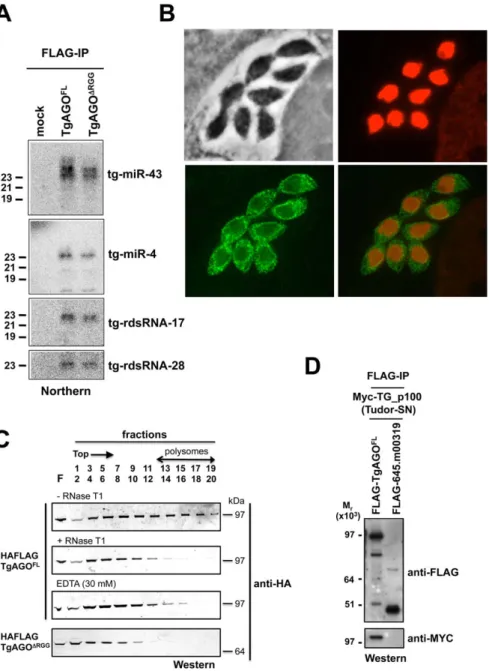
silencing events must, therefore, be operated via the same and unique Tg-AGO. To address this issue, we generated transgenic parasites expressing ectopically HAFlag-tagged, full-length Tg-AGO. RNP complexes were immuno-affinity purified (see next section), co-precipitated RNAs were extracted from the beads and analyzed by Northern using oligonucleotide probes specific to some of the highly abundant Tg-miRNAs and Tg-rdsRNAs studied above. Tg-miR-4 and -43, and as well Tg-rdsRNA-17 and -28 were indeed detected in the HaFlag-Tg-AGO immuno-precipitates but not in control immuno-precipitates (Figure 7A), indicating that Tg-AGO is a common effector of both types of sRNAs. This likely entails both cytoplasmic and nuclear distribution of the protein. Immunofluorescence and confocal microscopy revealed that Tg-AGO accumulates in tachyzoites mostly as granules of unidentified nature, but this labeling was superimposed over a diffuse cytoplasmic signal (Figure 7B and data not shown). Using acetylated histone H4 as a marker, confocal analyses also revealed a faint nuclear staining indicating that a minor portion of Tg-AGO localizes to the nucleus. Nuclear localization of Tg-AGO could be transient or highly dynamic, and under steady-state conditions. Alternatively, nuclear Tg-AGO could be incorporated into large protein complexes that prevent its optimal accessibility to antibodies.
The fact that a fraction of several Tg-miRNAs co-sediments with polysomes (Figure 4E, fractions 13–18) suggests that a portion of Tg-AGO should also be associated with polysomes, as has been shown for plant and metazoan miRNA-loaded AGOs [41,58–62]. We thus examined the distribution of HaFlag-Tg-AGO using polysome gradients: cytoplasmic extracts from intracellular parasites were prepared and fractionated on sucrose gradients (Figure 7C). The absorbance profiles at 254 nm showed a pattern of ribosomes with ribosomal subunits, monosomes, and polysomes. Consistent with previous findings in metazoans [61,62], most HAFlag-Tg-AGO was found near the top of the gradient, where soluble material and small ribonucleoprotein particles sediment. Some HAFlag-Tg-AGO was also heterodispersed throughout the gradient fractions, where polyribosomes and Tg-miRNAs co-sediment (Figures 4E and 7C). Treatments of cellular extracts with 30 mM EDTA or RNase T1, known to dissociate polysomes into ribosomal subunits and monosomes, caused a shift in HAFlag-Tg-AGO distribution from the denser fractions to the lighter fractions of the gradient (Figure 7C). This result suggests that a portion of miRNA-loaded Tg-AGO associates with polysomes to regulate translation of Tg-miRNA target mRNAs, perhaps in the cytoplasmic granules observed by immunofluorescence.
As noted previously, a characteristic feature of Tg-AGO is the presence, at the amino terminus, of a repeated RGG-rich region (amino acids 1–68), in which the arginine residues have the potential to undergo methylation (Figure 2B). This post-transla-tional modification is known to influence the stability, activity and/or sub-cellular distribution of some metazoan AGO-like proteins [23–25]. Tudor-domain proteins specifically recognize symmetrically dimethylated arginines (sDMA) such as those found in AGO-like proteins [63,64]. Accordingly, the immunopurified HAFlag-Tg-AGO complex (see below; Tables 1 and S4) was found to contain the Tudor-SN (tudor staphylococcal nuclease)/p100 homolog. Tudor-SN has five staphylococcal/micrococcal nuclease domains as well as Tudor domain, and it was described as a component of RISC in Caenorhabditis elegans, Drosophila and mammals [65]; more recently, the Tudor domain of the fly Tudor-SN was characterized as a specific sDMA-binding protein [64]. Reciprocal immunoprecipitation experiments further con-firmed the specific binding of Tg-AGO to Tg-Tudor/SN (Figure 7D). Furthermore, HAFlag-Tg-AGO was also found to
co-purify with Tg-PRMT1, which belongs to the family of arginine methyltransferases that use RGG motifs as substrates (Table 1). These results suggest that Tg-AGO is arginine-methylated, and that this modification might be specifically read by Tg-Tudor/SN, possibly to engage Tg-AGO into distinct modes of RNA silencing.
In particular, the RGG-rich region of the Trypanosoma brucei Tb-AGO1 was found critical to its association with polysomes [66]. To test if the same was true of Tg-AGO, we engineered HAFlag-Tg-AGODRGG, which carries a deletion of the RGG domain (amino acids 1 to 68). While HAFlag-Tg-AGODRGGwas loaded
Figure 6. Satellite-associatedTg-satRNA localized to heterochromatin inToxoplasma. New cloned Tg-satRNA species were assigned to repetitive satellite elements Sat350 and Sat529a, which are embedded in heterochromatin domains. Tg-satRNAs with perfect matches were plotted to satellite elements Sat350 (A) and Sat529a (B). The short thin lines below the long bars represent small RNAs derived from the sense strands with the reads indicated at the left. (C) Analyses of selected modified histones ratio at satellite Sat350 and Sat529a by quantitative chromatin immunoprecipitation PCR. Ratios of DNA precipitated with target modifications over DNA precipitated with core histone H3 were used to calculate relative precipitated fold enrichment shown on the y-axis. Experiments were triplicated and the data sets are concordant. Relative intensity is shown with standard deviations.
normally with Tg-miRNA and Tg-rdsRNA (Figure 7A), the majority of the mutant protein was found near the top of polysome gradients, and was notably absent in fractions where polyribo-somes sediment (Figure 7C). In addition, mass spectrometry analysis of the HAFlag-Tg-AGODRGGcomplex showed that it was no longer associated to Tg-Tudor/SN (data not shown). Thus, the proposed AGO arginine-methylation and association with Tg-Tudor/SN might allow post-loading sorting of distinct
Tg-AGO-containing RNP complexes towards specific silencing modes. We note that its association to Tg-Tudor/SN through an RGG domain together with its predominant cytoplasmic localization evoke the as yet unexplored possibility that Tg-AGO may serve as a PIWI protein. Thus, in addition to its possible role in heterochromatin formation, Tg-AGO might contribute to post-transcriptional gene silencing of repeats and transposons via Tg-rdsRNAs, as is seen with metazoan PIWI proteins.
Figure 7. Small RNA loading, sub-cellular localization and polysome association ofTg-AGO. (A) Northern blot analyses of Tg-miRNAs and Tg -rdsRNAs associated with Tg-AGO. Immuno-precipitation using anti-Flag antibody was performed from RH strains expressing ectopically HAFlag-tagged either full-length or delta-RGG truncated AGO. RNAs isolated from the immunoprecipitates were probed for miR-4, miR-43, Tg-rdsRNA-17 and Tg-rdsRNA-28. Mock: non-immune IgG (negative control). (B) Stably expressed recombinant protein HAFlag-TgAGOFLwas detected by
immunofluorescence assay using an HA antibody (in green) and compared to nuclear localization of acetylated histone H4 (in red). (C) To evaluate the sedimentation characteristics of Tg-AGO complexes, protein extracts containing HAFlag-TgAGOFLor HAFlag-TgAGODRGG were subjected to
sedimentation on 5%–45% sucrose gradients in the presence of cycloheximide (to preserve polyribosomes) or 30 mM EDTA (to disrupt polyribosomes). Aliquots of total extracts, either untreated (-RNase T1) or digested with RNase (+ RNase T1) were centrifuged through sucrose gradients and fractionated as described in Methods. Aliquots (equal volume) from indicated fractions were analyzed by Western blots with antibody against HA tag. (D) HAFlag-TgAGOFL and HAFlag-645.m00319 (negative control) were transiently co-expressed with Myc-Tg-p100 (Tudor/SN).
Following Flag affinity purification, the bound proteins were analyzed by Western blot using the anti-HA and the anti-myc antibodies. Molecular weight markers are indicated on the left.
Tg-AGO-associated proteins constitute the near-entire cohort of previously identified human and fly miRNA-RISC components
To characterize the molecular composition of Tg-AGO-containing complexes, HAFlag-Tg-AGO was affinity purified from total cell extracts of intracellular tachyzoite by incubation with anti-FLAG agarose beads. The immunoprecipitated FLAG– protein complexes were eluted using the FLAG peptide. The immunoprecipitated proteins were then separated by SDS-PAGE, excised, and identified by mass spectrometry (Figure 8A). Tables 1 and S4 list the names of the identified proteins, which, remarkably, were direct orthologs of nearly all of the previously identified components of human and Drosophila miRNA-RISC [39,40,67–69]. These co-purified proteins fell within several functional groups. The largest group encompasses mRNA-binding proteins, in particular the heterologous nuclear ribonucleopro-teins: HNRNPA3, HNRNPH1, HNRNPL, and HNRNPM
[39,40]. Several mRNA-binding proteins with putative functions in mRNA transport, stabilization and translation were also identified, including homologs of FUBP2/KSRP, nucleolin and FXR-related proteins, which are well known human and Drosophila Argonaute interactors [39,40,68]. Among the DEAD/DEAH box helicases, we found DDX17/DDX5, an ortholog of Drosophila p68, which has been shown to associate with Drosophila Ago2 [9], and DDX3X/Belle or DDX6/p54, which are all required for miRNA function (Table 1) [67,68]. Consistent with the hypothesis that Tg-AGO associates with mRNPs, a homolog of the polyadenylation binding protein PABPC [70] was identified in the immuno-precipitate, indicating that mRNAs were present in the purifications. Accordingly, treatment of the lysate with RNase T1 prior to immuno-precipitation abolished the integrity of the Tg-AGO1 complex, indicating that the interactions between HAFlag-Tg-AGO and several proteins were RNA-mediated (Figure 8B). Identification of Table 1. Summary of Tg-AGO-associated proteins identified by mass spectrometry.
ToxoDB v4.3 ID Band (peptides) Homolog (H.s) References RNA helicases 42.m00117 11 (18) DDX3X/Bel (D.m) 40, 68 46.m00027 14 (9) DDX17 39, 40 35.m00026 16 (20) DDX39 39 583.m00676 15 (3) DDX6/RCK 67 55.m04649 18 (5) DDX48 39 46.m00023 12b (15) FMR/FXR-like 39, 67 25.m02940 5 (9) FMR/FXR-like 39, 67
RNA binding proteins
49.m00009 20 (7) p100 (Tudor/SN) 65 35.m00901 9a (53) FUBP2/KSRP 40, 47 42.m03384 10a (20) PABPC1 39, 40, 70 80.m02340 9a (17) Nucleolin 39 57.m01690 15 (7) HNRNPA3 40 46.m01699 15 (14) HNRPH1 39, 40 55.m00241 21b (34) HNRNPM 40 80.m00057 15 (5) HNRNPL 39, 40 50.m00035 13a (4) NOP56 39 Translational machinery 20.m03912 9c (39) EEF2 40 76.m00016 15 (42) EEF1A1 40 42.m03276 21b (6) EIF4E2 67 RPS and RPL 18–21c 40
Transcription and Chromatin-related
TGME49_1053 4 (14) TgCRC230 75
583.m00590 9c (5) TBL1 75
42.m00014 16 (4) HDAC1 75
583.m05395 3 (3) AP2 transcription factor
641.m01573 5 (4) SMARCA2 38.m00018 18 (4) PRMT1 55.m04747 8 (3) RPB2 72 Others 55.m00015 20 (25) 14.3.3 family 69, 71 doi:10.1371/journal.ppat.1000920.t001
translation initiation and elongation factors, together with various 40S and 60S ribosomal proteins (Table S4) provides further support to the idea that the miRNA-loaded Tg-AGO, which
associates with polysomes, might prevent translation of target mRNAs. Another noticeable partner of Tg-AGO was a ortholog of human FUBP2, also known as KHSRP/KSRP, which binds with
Figure 8. Proteomic analysis of Tg-AGO complexes. (A) Tg-AGO complexes were immunoprecipitated using Flag antibodies. The co-immunoprecipitated proteins were analysed using mass spectrometry (see Table 1 and Supplemental Table S1). Molecular weight markers are indicated on the left. (B) Transgenic parasites expressing ectopically HAFlag-TgAGOFLwere lysed and whole-cell-extracts prepared in the absence or
presence (+RNase T1) of ribonuclease T1. HAFlag-TgAGOFL
was immunoprecipitated using anti-FLAG beads and flag peptide-eluted fractions were separated by SDS-polyacrylamide gel electrophoresis (4% to 12%), and visualized by silver staining. Molecular weight markers are indicated on the left. (C) Flag-immunopurified fractions from HAFlag-TgAGOFLand HAFlag-TgAGODRGGRNase T1-treated and untreated samples were analyzed by Western blot using a home-made anti-Tg-KSRP antibody. (D) Sedimentation characteristics of Tg-KSRP compared to HAFlag-TgAGOFL.
high affinity to the terminal loop of some miRNA precursors and promotes their maturation [47]. Tg-KSRP might account, at least party, for the post-transcriptional regulation of some Toxoplasma MIRNA genes, as uncovered in this study (Figure 4B and 4C). Consistent with an effect of Tg-KSRP on miRNA maturation rather than activity, Tg-KSRP was found to bind Tg-AGO in an RNA-independent manner (Figure 8C). Accordingly, Tg-KSRP did not associate to polyribosomes (Figure 8D) and was also found in HAFlag-Tg-AGODRGGimmuno-precipitates (Figure 8C).
Additional immuno-purified proteins are not obviously related to translational control but have been previously implicated as RISC-associated factors including Tg-Tudor/SN and Tg-PRMT1, already evoked above. Notably, Tg-AGO also co-purified with a conserved 14-3-3 protein (Table 1): 14-3-3 proteins that bind S. pombe Ago1 and human Ago2 are probably required for AGO protein functions in cell cycle and/or gene silencing pathways [71]. 14-3-3 proteins may also act as major regulators for the sorting of AGOs between distinct classes of RNA granules [69], which may include the Tg-AGO foci detected by immunofluores-cence in the present study (Figure 7B). Collectively, these results provide compelling evidence that Tg-AGO is part of a functional RISC whose core components are nearly all orthologous to factors required for post-transcriptional gene silencing and its regulation in metazoans.
Consistent with additional, DNA-level silencing functions of Tg-AGO, the second largest subunit of Tg-RNA polymerase II (Rpb2) also co-purified in the HAFlag-Tg-AGOFL immuno-precipitates (Table 1). Interestingly, a mutation of Rpb2 in fission yeast, rpb2-m203, disrupts coupling between transcription and siRNA processing in RNAi-dependent heterochromatin formation [72]. Also reminiscent of the fission yeast heterochro-matic RNAi pathway, HAFlag-Tg-AGO was associated with the histone deacetylase TgHDAC3, a protein that may play similar roles to S. pombe Clr3 in the spread of heterochromatin [73,74]. Remarkably, HAFlag-Tg-AGO was associated with all known components of the major transcriptional co-repressor complex CRC [75,76], which contains the two repressor proteins Tg-CRC230 and Tg-TBL1, the catalytic subunit Tg-HDAC3 and a new plant-like AP2-domain transcription factor (Table 1). Moreover, peptide sequencing by tandem mass spectrometry indicated that the subunits of the complex are sub-stoichiomet-rically represented. This finding is consistent with the as yet unconfirmed idea that Tg-AGO-bound rdsRNA and possibly satRNAs may guide transcriptional gene silencing processes by recruiting histone deacetylases, and subsequently histone meth-ylases (i.e. Tg-SET8 and Tg-SET3, [77]), to heterochromatic regions of the genome.
Conclusions
The present analysis thus uncovers an unsuspected level of complexity in the RNA silencing pathways of the single cell parasite T. gondii. This complexity not only lies in the mere diversity of the sRNAs identified, but also in the apparent mix-and-matched nature of the silencing components found in this organism, both in terms of their evolution and function. In this respect, the T. gondii RNA silencing machinery and its usage by the parasite bewilder many accepted notions in the field. For instance, in no organism studied so far has a single Ago protein evolved to mediate both repeat-associated and miRNA-mediated gene silencing, two pathways usually considered drastically different. Likewise, the metazoan-like Tg-miRNAs have readily identifiable mRNA targets displaying perfect to near-perfect complementarity in both CDS and UTRs, which is unprecedented in animals. Further studies of the Toxoplasma RNA silencing pathways will
undoubtedly reveal other surprises and, more importantly, might shed light on the molecular bases of virulence in this important Human parasite.
Materials and Methods
Parasite strains, cell culture, and in vitro bradyzoite differentiation
The parasite strains used in this study are the following: the T. gondii type I RH strain that has lost the ability to complete the two-host life cycle, the T. gondii type II Prugniaud strain that is capable of robust bradyzoite differentiation, and the T. gondii type III CTG and C56 strains. All T. gondii and N. caninum strains were maintained by serial passage in HFF monolayer under tachyzoite conditions in DMEM (Invitrogen) supplemented with 10% (vol/vol) FBS (Invitrogen). T. gondii type II Prugniaud strain was maintained under tachyzoite conditions in DMEM supple-mented with 10% (vol/vol) FBS and 25 mM Hepes buffer, pH 7.2. To induce in vitro bradyzoite differentiation, extracellu-lar tachyzoites were allowed to invade HFF cells for 16 hours, and the culture medium was removed and replaced by RPMI-1640 supplemented with 1% FBS and 50 mM Hepes buffer, pH 8.2. After 2–3 days of culture in alkaline medium, bradyzoite induction was assessed for P36 and SUMO expression by IFA as described previously [78]. The RHhxgprt- strain used in these studies contains a deleted or defective HXGPRT gene, which allows for the selection of transfected tachyzoites using mycophenolic acid.
Antibodies
Antisera against Tg-FUBP2/KSRP (35.m00901 gene) were produced by Eurogentec using the ‘Super Speedy immunization’ protocol and the following peptides Tg-FUBP2-1 (H2N-MARKKRGSAAT-PEEGC-CONH2) and Tg-FUBP2-2 (H2N-GTDKREDRGVTPEE DC-CONH2). Specific antibodies were affinity purified against both peptides. For immunoblot analysis purified antibodies were used at 1:1000 dilutions. Primary antibodies for IFA, ChIP and Western blot included antibodies against haemagglutinin epitope tag (HA, Roche Diagnostic, dilution at 1:1000), Polyclonal anti-H4-K20-1me (Abcam ab9051), Polyclonal anti-H4-K20-3me (Abcam ab9053), Polyclonal anti-H4-K20-1me (gift from Rice JC,Sims et al., 2006), Polyclonal anti-H4 Acetylated (K5-K8-K12-K16) (upstate 06-866), Anti-H3-K9-1me (upstate 450), Anti-H3-K9-2me (upstate 07-441), Anti-H3-K9-3me (upstate 07-442), Monoclonal anti-Myc (9E10 - sc40X, Santa-Cruz Bio.).
Immunofluorescence microscopy
Infected HFFs grown on coverslips were washed in PBS and fixed/permeabilized for 20 min at room temperature with PBS containing 3% (vol/vol) formaldehyde and 0.2% Triton X-100 (vol/vol). Blocking was performed with PBS containing 5% FBS and 5% goat serum for 1 h at room temperature. Samples were incubated in PBS containing 1% FBS with the primary antibodies, followed by the secondary antibodies goat anti–mouse IgG coupled with Alexa Fluor 488 and goat anti–rabbit IgG coupled with Alexa Fluor 568 (Invitrogen) at a 1:1,000 dilution each in PBS–1% FBS. Nuclei of host cells and parasites were stained for 10 min at room temperature with Hoechst 33258 at 2mg/ml in PBS. After four washes in PBS, coverslips were mounted on a glass slide with Mowiol mounting medium (48 mM Tris-HCl [pH 8.5], 4.8% Mowiol 4–88 [wt/vol], 12% glycerol [vol/vol]), and images were acquired with a fluorescence microscope (Axioplan 2; Carl Zeiss, Inc.).
Quantitative chromatin immuno-precipitation (QChIP) assay
QChIP assays were performed based on a modification of previously published methods [77,79]. Immuno-precipitated DNA were purified through PCR Purification Kit columns (QIAGEN) and used as a template in semiquantitative QPCRs to detect specific targets. Specific primer pairs (melting temperature, 55 to 65uC) amplifying 200- to 450-bp fragments were used (supple-mental Table S1). PCR was performed with 1mL of DNA and 500 nM primers diluted to a final volume of 20mL in SYBR Green Reaction Mix (Roche). Accumulation of fluorescent products was monitored by real-time PCR using a LightCycler 2.0 (Roche). Each PCR reaction generated only the expected specific amplicon, as shown by the melting-temperature profiles of final products (dissociation curve, automatically measured by the LightCycler 2.0) and by gel electrophoresis of test PCR reactions. No PCR products were observed in the absence of template. The fold difference of a given target sequence precipitated by a specific antibody was determined by dividing the amount of target sequence in the immunoprecipitate fraction by the amount of target sequence in input DNA (S8, S13). Real-time PCR was carried out in triplicate on 2 ng of DNA at 50uC for 2 min and 95uC for 10 min, followed by 40 cycles of 95uC for 15 s and 60uC for 1 min. Data were collected at 60uC. The concentration of primers and Taqman probes used was determined by following the optimization procedure described in PE Applied Biosystem’s protocol. For each experiment, the threshold was set to cross a point at which real-time PCR amplification was linear. For the majority of the experiments, data were analyzed with a threshold of 0.05. Data collected was analyzed and plotted using Microsoft Excel.
Satellite 350B: OL17 (CGACTCGGACGTCAGGCCATGCA-GAG) and OL18 (GCGCCTGAACAATACGCCCAACC).
Satellite 529A: OL19 (CTGCAGGGAGGAAGACGAAAGTTG) and OL20 (CTGCAGACACAGTGCATCTGGATT).
Chromatographic purification of Tg-AGO-containing complexes
Whole-cell extract (WCE) from transgenic intracellular tachy-zoites expressing ectopically HAFlag-TgAGOFL and HAFlag-TgAGODRGG was incubated with 500 ml of anti-FLAG M2 affinity gel (Sigma) for 1 h at 4uC. Beads were washed with 10 column volumes of BC500 buffer [20 mM Tris (pH 8), 0.5 M KCl, 10% glycerol, 1 mM EDTA, 1 mM DTT, 0.1% NP40, 0.5 mM PMSF, aprotinin, leupeptide, pepstatin, 1 ug ml21each]. Bound peptides were eluted stepwise with 250 ug ml21 FLAG peptide (Sigma) diluted in BC500 buffer. Each preparation was sufficiently clean such that individual peptide bands could be excised and sequenced by mass spectrometry.
Mass spectrometry peptides sequencing
Protein bands were excised from colloidal blue-stained gels (Invitrogen), oxidized with 7% H2O2 and subjected to in-gel tryptic digestion. Peptides were extracted with 5% [v/v] formic acid solution and acetonitrile, and injected into an Ultimate 3000 (Dionex) nanoLC system that was directly coupled to a LTQ-Orbitrap mass spectrometer (Thermo Fisher Scientific). MS and MS/MS data were acquired using Xcalibur (Thermo Fischer Scientific) and processed automatically using Mascot Daemon software (Matrix Science). Tandem mass spectra were searched against a compiled T. gondii database using the MASCOT program (Matrix Sciences, London) available via intranet.
Small RNA library preparation
RNA was extracted into TRIzol (Invitrogen), and aliquots of total RNA from RH strain were subjected to small RNA library construction as follows. To avoid any contamination with the host cell small RNAs, freshly released parasites were harvested from the culture supernatant, washed by centrifugation, and filtered through a 3-mm filter before use. For each library, 50mg of total RNA was size fractionated on a 15% tris-borate-EDTA (TBE) urea polyacrylamide gel (Invitrogen) and a 19–40 base pair fraction was excised. RNA was eluted from the polyacrylamide gel slice in 300mL of 0.3 M NaCl overnight at 4uC. The resulting gel slurry was passed through a Spin-X cellulose acetate filter column (Corning Inc.) and precipitated by the addition of 750 mL of ethanol and 3mL of glycogen (5 mg/mL; Ambion). After washing with 75% ethanol, the pellets were allowed to air dry at 25uC and pooled in diethylpyrocarbonate (DEPC)-treated water. The 59 RNA adapter (59-GUUCAGAGUUCUACAGUCCGACGAUC-39) was ligated to the RNA pool with T4 RNA ligase (Promega) in the presence of RNase Out (Invitrogen) 6 hours at 20uC. The ligation reaction was stopped by the addition of 26 Gel Loading Buffer II (Ambion). The ligated RNA was size fractionated on a Novex 15% TBE urea polyacrylamide gel (Invitrogen), and a 40– 70 base pair fraction was excised. RNA was eluted from the polyacrylamide gel slice in 300mL of 0.3 M NaCl overnight at 4uC. The RNA was eluted from the gel and precipitated as described above followed by resuspension in DEPC-treated water. The 39 RNA adapter (59-pUCGUAUGCCGUCUUCUGCUU-GUidT-39; p, phosphate; idT, inverted deoxythymidine) was subsequently ligated to the precipitated RNA with T4 RNA ligase (Ambion) in the presence of RNase Out (Invitrogen) 6 hours at 20uC. The ligation reaction was stopped by the addition of 26Gel Loading Buffer II (Ambion). Ligated RNA was size fractionated on a Novex 10% TBE urea polyacrylamide gel (Invitrogen), and the 70–100 base pair fraction was excised. The RNA was eluted from the polyacrylamide gel and precipitated from the gel as described above and resuspended in 4.5mL of DEPC-treated water. The RNA was converted to single-stranded cDNA using Superscript II reverse transcriptase (Invitrogen) and Illumina’s small RNA RT-Primer (59-CAAGCAGAAGACGGCATACGA-39) following the manufacturer’s instructions. The resulting cDNA was PCR-amplified with PhusionTMHigh Fidelity DNA Polymerase (NEB) in 15 cycles using Illumina’s small RNA primer set (59-CAA-GCAGAAGACGGCATACGA-39; 59- AATGATACGGCGAC-CACCGACAGGTTCAGAGTTCTACAGTCCGA-39). PCR products were purified on a Novex 6% TBE PAGE gel (Invitrogen) and the 100 base pair fraction was excised. The DNA was eluted into 100mL of 1x NEBuffer 2 at room temperature for 2 hours. The resulting gel slurry was passed through a Spin-X filter (Corning) and precipitated by the addition of 325mL of ethanol, 10mL of 3 M sodium acetate, and 3mL of glycogen (5 mg/mL; Ambion). After washing with 75% ethanol, the pellet was allowed to air dry at 25uC and dissolved in 10mL of resuspension buffer (10 mM Tris-HCl, pH 8.5). The purified PCR products were quantified on the Agilent DNA 1000 chip and diluted to 10 nM for sequencing on the Illumina 1G (GATC BIOTECH, Konstanz, Germany).
Northerns
RNA from Toxoplasma and Neospora strains was extracted into TRIzol (Invitrogen), deproteinized with phenol chloroform/ isoamyl alcohol, and RNA was recovered by ethanol precipitation. For small RNA analyses, 30 ug of purified RNA were separated on a 15% polyacrylamide (w/v) 8 M urea gel and transferred to GeneScreen nylon membranes. DNA oligonucleotides