HAL Id: tel-01423913
https://tel.archives-ouvertes.fr/tel-01423913
Submitted on 1 Jan 2017HAL is a multi-disciplinary open access archive for the deposit and dissemination of sci-entific research documents, whether they are pub-lished or not. The documents may come from teaching and research institutions in France or abroad, or from public or private research centers.
L’archive ouverte pluridisciplinaire HAL, est destinée au dépôt et à la diffusion de documents scientifiques de niveau recherche, publiés ou non, émanant des établissements d’enseignement et de recherche français ou étrangers, des laboratoires publics ou privés.
Conditional truncation of the FUS protein in mice : a
new animal model of the ALS/FTD continuum
Jelena Scekic-Zahirovic
To cite this version:
Jelena Scekic-Zahirovic. Conditional truncation of the FUS protein in mice : a new animal model of the ALS/FTD continuum. Neurons and Cognition [q-bio.NC]. Université de Strasbourg, 2016. English. �NNT : 2016STRAJ002�. �tel-01423913�
UNIVERSITÉ DE STRASBOURG
ÉCOLE DOCTORALE 414
Sciences de la vie et de la santé
INSERM 1118
THÈSE
présentée par :Jelena SCEKIC-ZAHIROVIC
soutenue le : 11 Janvier 2016
pour obtenir le grade de :
!"#$%&'($')*%+,-$&.,#/'($'0#&1.2!%&3
Discipline/ Spécialité
:
NeurosciencesTroncation conditionnelle de la protéine FUS
chez la souris: un nouveau modèle animal du
continuum sclérose latérale
amyotrophique/démence fronto-temporale
DIRECTEURS DE THESE :
M DUPUIS Luc Directeur de recherches, Université de Strasbourg
M CARONI Pico Professeur, University of Basel
Mme BARTOS Marlene Professeur, University of Freiburg RAPPORTEURS :
Mme NEUMANN Manuela PR PUPH, University of Tubingen
M BUEE Luc Directeur de recherchés, Inserm UMR_S 1172
EXAMINATEURS :
M CHELLY Jamel PR PUPH, Université de Strasbourg/IGBMC
Jelena SCEKIC-ZAHIROVIC
Troncation conditionnelle de la protéine FUS chez la
souris: un nouveau modèle animal du continuum
sclérose latérale amyotrophique/démence
fronto-temporale
Résumé
La sclérose latérale amyotrophique (SLA) et la démence fronto-temporale (DFT) sont deux maladies qui constituent un continuum clinico-pathologique. La mutation de FUS, une protéine nucléaire à fonctions multiples, provoque des cas familaux de SLA, et ces mutations provoquent une redistribution sub-cellulaire de FUS, du noyau vers le cytoplasme. Certains cas de DFT présentent !"#$"%%"&#'(&$)(* $(+!#,!+)-,%"#"!#%.,*&"!/"#'"#- $,$(+!&#'"#012. 3%#!."&$#4,&#connu si la maladie est provoquée par une perte de la fonction nucléaire de FUS et/ou un gain de fonction cytoplasmique.
Nous avons généré et caractérisé une lignée de souris exprimant une forme cytoplasmique de FUS (Fus- !"#). La localisation exclusive de FUS dans le cytoplasme provoque la mort des motoneurones via un gain de fonction dans les motoneurones eux-mêmes. Une localisation cytoplasmique partielle de FUS est suffisante pour développer un phénotype de la SLA et de DFT.
Les mécanismes élucidés permettront de comprendre les bases des SLA/DFT et de Mots-clés : SLA/DFT, FUS, cytoplasmique localisation, gain de fonction
Résumé en anglais
Amyotrophic lateral sclerosis (ALS) and Frontotemporal dementia (FTLD) are now considered as a unique clinicopathological spectrum referred to as ALS/FTLD. Cytoplasmic aggregation of the physiologically nuclear FUS protein is a hallmark feature of a subset of ALS/FTLD. It remains unknonwn whether the critical pathogenic event relies on a loss of FUS normal nuclear functions, a toxic gain of function of FUS in the cytoplasm, or a combination of both.
To answer this question we have generated a conditional mouse model expressing truncated FUS without nuclear localization signal - Fus !"#. Our data showed that complete cytoplasmic mislocalization of truncated FUS protein within spinal motor neurons is a major determinant of motor neuron degeneration via toxic gain of function. A partial mislocalization of truncated FUS protein was sufficient to trigger key features of ALS and of FTLD.These studies allowed the elucidation of mechanisms underlying FUS role in ALS/FTLD, and will hopefully lead to development of therapies for these devastating diseases.
ACKNOWLEDGMENTS
This thesis could not have been completed without the great support that I have received from so many people over the years. I wish to offer my most heartfelt thanks to the following people.
Firstly, I would like to express my sincere gratitude and deepest thanks to my supervisor Dr Luc DUPUIS for the continuous support of my PhD study and related research, for his patience, motivation, and immense knowledge. His guidance helped me in all the time of research and writing of this thesis. I could not have imagined having a better advisor and mentor for my PhD study.
My sincere thanks also go to Director Dr Jean-Phillippe LOEFFLER who provided me an opportunity to join his team, and who gave access to the laboratory and research facilities. Without his precious support it would not be possible to conduct this research.
I would like to thank to my thesis co-supervisors Prof. Pico CARONI and Prof. Marlene BARTOS for their help and encouragement.
I also wish to express my sincere gratitude and appreciation to Dr Caroline ROUAUX, who has taught me many of the techniques used in this project, and for being a friend.
I am grateful to Dr Frédérique RENE and Dr Jose-Luis GONZALEZ DE AGUILAR, for their insightful comments but also for the hard question which incented me to widen my research from various perspectives.
I am thankful to other members of my committee, Pr Manuela NEUMANN, Dr Luc BUEE, Pr Jamel CHELLY and Prof. Stefan ROTTER for finding the time in their busy schedules to review my thesis.
I am indebted to Hajer ELOUSSINI, for her love and encouragement. Thank you for your support when I have needed it the most. You really let me feel like at home. You are a great friend and a rock of stability in my life and I hope our strong friendship will continue in the future. Thank you with all my heart!
My work has greatly benefited from suggestions and kind encouragement from Sylvie DIRRIG-GROSCH and Jérôme SINNIGER. Your technical depth and attention to the detail, which I experienced, are amazing.
I thank my fellow lab mates Lavinia PALAMIUC, Aurélia VERNAY and Alexandre HENRIQUES, Pauline VERCRUYSSE, Gina PICCHIARELLI, Stéphane DIETERLE, Christine MARQUES and Laura ROBELIN for the stimulating discussions, for the sleepless nights we were working together before deadlines, and for all the fun we have had in the last three years.
To Mme Brigitte KUENEMANN, thank you for keeping your office door open and available for all the times when I needed it.
Thank you goes to all the other members of the LOEFFLER lab Annie PICCHINENNA and Marie-Jo RUIVO for providing a pleasant and productive working atmosphere.
The work of this thesis was supported by the Erasmus Mundus Neurotime program. My greatest thank to wonderful people in this unique program Dr Paul PEVET and Dr Domitille BOUDARD.
Last but not the least, I would like to thank my family: my husband, my brother and to my mother for supporting me spiritually throughout writing this thesis and in my life in general.
TABLE OF CONTENT
ABBREVATION 1
FOREWORD 3
INTRODUCTION 4 I. ALS AND FTLD: clinical, genetic and pathological link 6
A. Clinical presentations 6 1. Amyotrophic lateral sclerosis (ALS): 6 1.1. Phenotypic heterogeneity: 7
1.1.1. Multiple sites of onset: 8
1.1.2. Variability in age of onset: 9
1.1.3. Diversity of disease progression and survival: 9
1.1.4. Heterogenous clinical presentation: 10
2. Frontotemporal lobar degeneration (FTLD): 13
2.1. Phenotypic heterogeneity: 14
3. Clinical link among ALS and FTLD: 15
B. Molecular basis 1. Heterogenous molecular basis of ALS: 16
2. Heterogenous molecular basis of FTLD: 19
3. Genes linked to ALS and FTLD: 20
3.1. Pure, ALS genes: 21
3.1.1. Superoxidedismutase 1 (SOD1): 21
3.2. Overlapping, genes: 22
3.2.1. TAR DNA-binding protein (TARDBP): 22
3.2.2. Fused in sarcoma (FUS): 24
3.2.3. Hexanucleotide repeat expansion in C9ORF72: 24
3.2.4. Valosin-containing protein (VCP): 25
3.2.5. Ubiquilin 2 (UBQLN2): 26
3.2.6. Charged multivesicular body protein 2B (CHMP2B): A 26
3.2.7. Optineurin (OPTN): 27
3.2.8. Sequestosome 1 (SQSTM1): 27
3.2.9. Other genes: 27
3.3. Pure, FTLD genes: 28
3.3.1. Microtubule-associatedprotein TAU (MAPT): 28
3.3.2. Progranulin (PGRN): 28
4. Genetic link among ALS and FTLD: 29
C. Pathological characteristics: 31
1. Pathological overlap between ALS and FTLD: 31
1.1. Protein deposits/inclusions/aggregates in ALS and FTLD: 32
1.1.1. Common cellular pathomechanisms among ALS and FTLD: 36
1.2. Degeneration of neurons in ALS and FTLD: 39
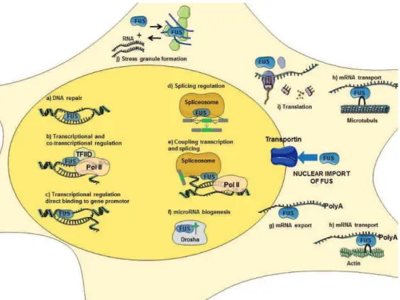
II. FUS 5 A MULTIFUNCTIONAL RNA/DNA BINDING PROTEIN 42
A. FUS structure and physiological role: 42
1. Gene structure of FUS: 42
2. Physiological role of FUS: 43
2.1. DNA repair: 44
2.2. Transcriptional regulation and gene expression: 44
2.3. mRNA Splicing: 45
2.4. RNA biogenesis and processing: 46
2.5. RNA transport and local translation: 46
2.6. Protein interactions: 47
B. Neurodegenerative disorders with FUS pathology: 48
1. FUS mutations related to FUS proteinopathies: 48
2. Similarities and differences related to FUS proteinopathies: 50
3. Possible mechanisms of FUS toxicity - Loss vs gain of function: 51
3.1. Alteration of gene expression and splicing: 51
3.2. Defective stress granule and protein aggregation: 52
3.3. Prion-like properties: 53
3.4. Post-translational modifications of FUS: 54
C. Animal models of FUS related protheinopathies ALS/FTLD 5 remaining questions: 56
1. Non-rodent models: 56
2. Rodent models: 57
2.1. FUS knockout and knockdown models: 57
2.2. Models with overexpression of wild type or mutant FUS: 60
2.3. Summary of current results in available Fus models: 64
III. STATE OF ART - AIMS OF THE THESIS: 66
RESULTS: 69
I. 617839:;3<=#=>? (submitted): 70
A. Summary 5 6 *%(/,$(+!#=>?: 70
B. Toxic gain of function from mutant FUS protein is crucial to trigger cell autonomous motor neuron loss: 74
II. 617839:;3<=#=>@ (in preparation): 131
A. Summary 5 Publication N>@: 132
B. Partial cytoplasmic mislocalization of FUS leads to motor neuron disease and behavioural symptoms relevant to FTLD: 133
1. Results: 134
2. Material and methods: 156
DISCUSSION: 168
I. A STUDY TO BE COMPLETED: 168
II. FUS !"#$%&MICE AS A GENETICALLY RELEVANT: 169 MODEL OF FUS-ALS
III. ARE FUS !"#$%& MICE A MODEL OF ALS USEFUL FOR PRECLINICAL
STUDIES ? 170
IV. ARE FUS !"#$%& MICE A MODEL OF FTLD? 171
V. GAIN VS. LOSS OF FUNCTION: 172
VI. WHY IS FUS !"#$% MICE PHENOTYPE SO MILD ?
THE SECOND HIT THEORY: 172
VII. GENERAL CONCLUSION: 173
BIBLIOGRAPHY: 175
ANNEX: 195
I. 617839:;3<=#=>A
VAPB/ALS8 MSP Ligands Regulate Striated Muscle Energy Metabolism Critical for Adult Survival in Caenorhabitis elegans
II. 617839:;3<=#=>B (submitted)
Alterations in the hypothalamic melanocortin pathway in amyotrophic lateral sclerosis
ABBREVIATIONS
AAVs ALS ALSbi ALSci AOS BIBD bvFTD CHCHD10 CHMP2B CLIP CNS CSF DPR ET EWSR1 FTD FTLD FUS GFAP GWAS HDAC1 IBMPFD LMN MND MAPT NCI NES NII NIFID NLS NMJ OPTN PGRN PLS PNFA Adeno-associated viruses Amyotrophic lateral sclerosis ALS with behavioural impairment ALS with cognitive impairment Apraxia of speechBasophilic inclusion body disease
Behavioural variant frontotemporal dementia
Coiled-coil-helix-coiled-coil-helix domain-containing protein 10 Charged multivesicular body protein 2B
Crosslinking and immunoprecipitation Central nervous system
Cerebrospinal fluid Essential tremor
Dipeptide repeat proteins Ewing RNA-binding protein Frontotemporal dementia
Frontotemporal lobar degeneration Fused in sarcoma
Glial Fibrillary Acidic Protein Genome wide associated studies Histone deacetylase 1
Inclusion body myopathy and Paget's disease of the bone Lower motor neuron
Motor neuron disease
Microtubule-associated protein tau Neuronal cytoplasmic inclusions Nuclear export signal
Neuronal nuclear inclusions
Neuronal intermediate filament inclusion disease Nuclear localization signal
Neuromuscular junction Optineurin
Progranulin
Primary lateral sclerosis Progressive nonfluent aphasia
PMA POMC PRMT1 RGG RIP RRM SBT SD SOD1 SQSTM1 TAF-15 TARDBP TBK1 TLS UBQLN2 UMN UPS VAPB VCP VE neurons WES WGS
Progressive muscular atrophy Pro-opiomelanocortin neurons
Protein arginine methyl transferases 1 Glycine rich region
RNA immunoprecipitation RNA recognition motif Somatic brain transgenesis Semantic dementia
Superoxidedismutase 1 Sequestosome 1
TATA-binding protein-associated factor 15 Transactive response DNA-binding protein TANK-binding kinase 1
Translocated in liposarcoma Ubiquilin 2
Upper motor neuron
Ubiquitin proteasome system
Vesicle-associated membrane protein-associated protein B Valosin-containing protein
Von Economo neurons Whole-exome sequencing Whole-genome sequencing
FOREWORD
The general topic of my PhD work was the understanding of the molecular mechanisms of amyotrophic lateral sclerosis (ALS). During these last 3 years, I pursued two major
objectives.
First, I sought to understand the consequences of loss of function of ALS8/VAPB, a gene that has been reported as mutated in a few familial ALS cases. When I arrived in the laboratory, it had already been shown that VAPB knock-out mice did not develop ALS, but showed a mild rotarod defects. I went further into the analysis of these mice and showed that they developed abnormal muscle response to fasting (Han et al, 2013, PLOS Genet). In collaboration with Dr Niels Decher, we also showed that VAPB knock-out mice suffered from heart defects (manuscript in preparation). While the analysis of VAPB knock-out mice led to interesting insights into the physiological function of VAPB, we concluded from our work that the loss of function of VAPB has likely little relevance to ALS. I have also participated in another study investigating a role of the hypothalamus in ALS (manuscript submitted). I included these papers in the Annex of the manuscript.
A second objective was initiated in parallel to understand the mechanisms of ALS mediated by FUS mutations. FUS mutations are a much more frequent cause of familial ALS than VAPB mutations, and lead to typical ALS. In the present manuscript, I describe our two step analysis of a novel knock-in model of FUS-ALS, with a first study focusing on the phenotype of homozygous mice (Publication N°1), and a second study in preparation describing the early characterization of heterozygous knock-in mice (Publication N°2).
5
INTRODUCTION
Neurodegenerative diseases are a group of diverse disorders characterized by progressive loss of specific populations of neurons. In the vast majority of cases, etiology is unknown. Although, these diseases affect different neuronal populations, lead to widely different clinical manifestations, and are characterized by disparate pathological findings, they share a common characteristic, the presence of misfolded and aggregated proteins in and/or around neurons.
Over the past few years, several clinical, genetic (molecular), and neuropathological features have been recognized as shared by different neurodegenerative diseases. As a typical example, an important scientific interest has been driven toward the DNA/RNA-binding protein FUS (fused in sarcoma). This growing interest in FUS arose after the discovery of variants in the FUS gene causing or contributing to multiple neurodegenerative diseases, including amyotrophic lateral sclerosis (ALS), rare forms of frontotemporal lobar degeneration (FTLD), polyglutamine diseases as well as essential tremor (ET). Additionally, abnormal aggregation of FUS protein has been reported in postmortem material of patients suffering from these neurodegenerative diseases in the absence of somatic FUS mutations. These findings suggest an important role of FUS in the pathogenesis of these diseases.
Although several lines of evidence indicate that cytoplasmic mislocalization of FUS, is a key event in disease pathogenesis, definitive in vivo evidence is lacking. The respective contributions of gain vs. loss of function, as well as the cell types in which the critical pathogenic events occur are still undefined.
More generally, the links between FUS dysregulation and pathophysiological processes leading to neurodegeneration in ALS/FTD are poorly understood.
Despite worldwide efforts in the research field, current understanding of the normal function of FUS, and its role in the pathology of ALS, FTLD and other neurodegenerative diseases, are still insufficient. Further understanding of the underlying pathogenic mechanisms of these FUS-related disorders might lead to improvements in the treatment and prevention of such disorders.
In this introduction, I will first describe ALS and FTLD as the major diseases involving FUS. I will then present the structure and physiological functions of FUS. The pathology of FUS, and the effects of FUS mutations, will be detailed to discuss the currently existing models of FUS diseases.
6
I. ALS AND FTLD: clinical, genetic and pathological link
ALS and FTLD were traditionally considered as two completely different neurological disorders with discordant clinical features. Over the years, however, several observations have suggested that there is a link connecting these disorders. Multiple lines of emerging evidence showed that these disorders share mutual clinical, pathological and genetic features and led to establishment of ALS/FTLD disease spectrum.
A. Clinical presentations
1.
Amyotrophic lateral sclerosis (ALS)Classical concept 4 a single disease vs. new concept 4 heterogeneous syndrome
Amyotrophic lateral sclerosis (ALS) is the most frequent adult onset degenerative disease of the motor neuron (motor neuron disease) with an incidence of 2.6/100 000.It is characterized by progressive loss of cortical-upper (UMN) and spinal-lower (LMN) motor neurons (153).The name describes the key features of the disease: muscle wasting (amyotrophic) due to the degeneration of the lower motor neurons and their axons and degenerationof the upper motor neurons and their corticospinal axonal tracts (lateral sclerosis) (4). Clinically, gradual loss of motor neurons leads to muscle weakness, fasciculation, spasticity and paralysis with a devastatingly rapid disease progression and respiratory failure as the cause of death for most patients (Figure 1) (3). Disease onset, generally in adulthood, peaks at 60570 years of age. Half of the affected individuals die within 3-5 years. The genetics of ALS comprises only 5-10% of cases due to identified genetic mutations while nearly 90-95% remains of unknown origin. Regardless of the genetics, ubiquitin-positive inclusions are found in dying motor neuronsin almost all ALS cases. More than a century after Jean-Martin Charcot first description of the disease in 1869, and in spite of many efforts towards the elucidation its etiology and the search for a treatment for ALS remains an incurable disease of unknown origin.
One possible reason for this failure could be due to the fact that for a long period, ALS has been identified as a single nosological entity, characterized by a pattern of progressive motor neuron degeneration.Textbook neurology addresses ALS as a degenerative disease that
selectively affects upper and lower motor neurons5 a description which suggests that the
clinical presentation of ALS is very homogenous. This may explain why the disease was expected to have a single cause, a single pathogenesis and a single phenotype. However, with growing knowledge of profound clinical, neuropathological and now genetic heterogeneity, the concept of ALS as one disease appears increasingly unsustainable.
7
1.1. Phenotypic heterogeneity: from motor neuron disease to neurodegenerative
disease
Clinical examination usually reveals atrophy and weakness of muscles, fasciculation, hyperreflexia and often a mild to severe hypertonia. Clinicians designate weakness, muscle atrophy and fasciculation as lower motor neuron signs, whereas hyperreflexia and hypertoniaindicate upper motor neuron involvement (Figure 1).
Figure 1. Different patterns of MN involvement in ALS
Schematic representation of LMN (red) and UMN (blue) degeneration with clinical signs (red and blue boxes) in ALS
The evidence of both upper and lower motor neuron involvement is required for the diagnosis of ALS, and have been incorporated in the so-called revised El Escorial criteriaor the newer Awaji criteria (Table 1) (5). Using these criteria ALS itself can be classified as definite,
8
Table 1. Diagnostic criteria for amyotrophic lateral sclerosis (ALS) derived from the revised El
Escorial and Awaji criteria
Reproduced from Costa et al., J. Arch. Neurol.,2012.
In spite of the precise clinical description of ALS, there is a considerable variability in its
phenotypic expressions, with regard to the site and age of onset, the rate of progression,
disease duration and clinical presentation, and the presence and degree of cognitive dysfunction. Among other parameters, phenotypic heterogeneity arises due to pattern of motor neuron involvement and extent of extra-motor involvement.
1.1.1. Multiple sites of onset
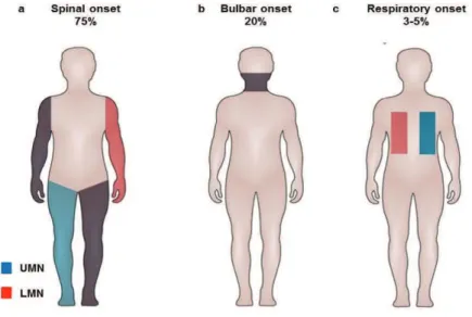
There are several classification systems in place for ALS. The first one relies on the site
of onset of the disease : spinal, bulbar or respiratory (Figure 2) (6). Most of the patients
present first with asymmetric and painless weakness in a limb, which is referred as spinal-onset ALS. In contrast, about 20% of patients present first with weakness in bulbar muscle causing, dysarthria, dysphagia and tongue fasciculations. Finally 355% of patients present first with a respiratory onset, characterized by orthopnea or dyspnea (7). Compared to spinal-onset
9 patients, with bulbar- and respiratory-onset patients have a worse prognosis and a decreased disease duration and long term survival (7), reviewed in(6).
Figure 2. Site of onset in ALS
Red indicates LMN involvement, blue indicates UMN involvement. Darker shading indicates more-severe involvement.
(a) In spinal-onset ALS, patchy UMN and LMN involvement is observed in all limbs. (b) In bulbar-onset ALS, UMN and LMN involvement is observed in the bulbar muscles. (c) In spinal-onset ALS, UMN and LMN involvement is observed in the respiratory muscles
Reproduced from : Swinnen & Robberecht, Nat. Rev.Neurol., 2014.
1.1.2. Variability in age of onset
The age of clinical onset of ALS is usually between the fourth or sixth decade of life.
However, onset at almost any age has been described. Juvenile ALS is rare and defined as ALS with age at onset before 25 years, and the course of progression is generally slower than in other forms of ALS. Onset after 80 years is associated with a particularly faster progression (8). Biological onset of the disease is unknown. In rodent models of ALS, which admittedly are overexpression models, abnormalities are present as early as embryonic development (9511). Nevertheless, these animals develop no clinical abnormalities until adulthood. Thus, biologically, the disease may start early in life and become clinically apparent much later.
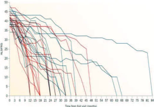
1.1.3. Diversity of disease progression and survival
The range of disease progression is wide. Although median survival in ALS is generally around 3 years from diagnosis, variability in survival is remarkable with some patients die within months after onset and others surviving for more than two decades (Figure 3). Less than 20% of patients survive over 5 years from the disease onset (10). Long survival is seen
10 more frequently in patients with juvenile ALS and upper motor neuron-predominant ALS (8). Large differences in survival and age at disease onset exist even between individuals from a same family, in whom ALS is caused by the same mutation, suggesting that other factors may modify the phenotype (12).
Figure 3. Variability of disease progression in ALS
Reproduced from: Swinnen & Robberecht, Nat. Rev.Neurol., 2014.
1.1.4. Heterogenous clinical presentation
Neurodegeneration of motor neuron
Depending on the overall pattern of upper or lower motor neuron involvement and the degree of asymmetry, first clinical presentation could vary in between two opposite extremes (Figure 4). On the one hand, patients in whom only the lower motor neurons are initially found affected, usually asymmetrically, are classified as progressive muscular atrophy (PMA). On the other hand, if only evidence of upper motor neuron degeneration is initially found, often symmetrically, a diagnosis of primary lateral sclerosis (PLS) is made. Both PMA and PLS progress over time to ALS in a significant proportion of patients (about 5% in each case). The main reason for distinguishing these phenotypes is the difference in prognosis. The patients with PMA fare slightly better than those with clinical evidence of both lower and upper motor neuron involvement while patients with PLS survive longer than both (13).
11 Further to this, a causal primacy of lower motor neuron over upper motor neuron degeneration remains an issue of debate. Many of the initial pathological changes in rodent models of ALS occur in the lower motor neurons, supporting a dying-back view of pathogenesis (14). However, ALS can also be viewed primarily as a disease of the upper motor neurons, which connect monosynaptically, only in humans and not rodents, with anterior horn cells. This is supported by the clinical observation that the oculomotor, abducens, and Onuf's motor nuclei, which all lack direct cortical motor neuron connections, are strikingly resistant to degeneration (15).
Figure 4. ALS, pattern of motor neuron involvement.
Red indicates LMN involvement, blue indicates UMN involvement. Darker shading indicates more-severe involvement. PMA (isolated LMN involvement) and PLS (isolated UMN involvement) constitute the ends of a spectrum of LMN and UMN involvement; intermediate phenotypes are considered to be different expressions of ALS
PMA- In progressive muscular atrophy, LMNs in arms and legs are involved, often proximally. PLS- In primary lateral sclerosis, UMNs of arms and legs are primarily involved, but later in the disease, discrete LMN involvement can be detected.
Reproduced from :Swinnen & Robberecht, Nat. Rev.Neurol., 2014.
Neurodegeneration of extramotor neurons
ALS phenotypic variability becomes more apparent when it combines different motor and extramotor involvement. In some patients, neurons in the prefrontal and temporal cortexare affected. This leads to cognitive and/or behavioural problems. ALS patients with mild behavioural dysfunction are classified as having ALS with behavioural impairment (ALSbi), whereas patients with mild executive and language dysfunction are said to have ALS with cognitive impairment (ALSci). In total, around 30% of ALS (16). However, in about 15% of ALS cases, patients who meet the Neary criteria for frontotemporal dementia (FTD), the main symptom of frontotemporal lobe degeneration (FTLD) are considered to have ALS-FTLD (17).
12 On the other hand, up to 50% of patients with a diagnosis of FTLD have some motor neuron involvement and are said to have FTLD5MND (16,17). Of note, 15% of FTLD patients show signs of motor neuron degeneration that meet criteria for ALS and are classified as ALS5FTLD (Figure 5) (17). This suggests that ALS and FTLD are at the ends of a same disease spectrum.
Although, there are extremes of relatively isolated involvement of each of these compartments PLS, PMA and FTLD, all of these overlap with classic ALS on post-mortem neuropathological examination, by showing protein inclusions.
Figure 5. ALS, a spectrum disorder.
ALS and FTD constitute the ends of a spectrum of motor neuron and frontotemporal neuron involvement. This spectrum includes patients with ALS who express isolated ALSci or ALSbi, and patients with ALS who meet the Neary criteria for FTLD and are, thus, diagnosed with ALS5 FTLD. Some patients with FTLD have insufficient motor neuron involvement for a diagnosis of ALS, and are classified as FTLD5MND
Reproduced from :Swinnen & Robberecht, Nat. Rev.Neurol., 2014.
Following examples of extramotor involvement comes from research that has been done in our laboratory. In ALS, spasticity is traditionally thought to be the result of degeneration of the upper motor neurons (18,19), although degeneration of other neuronal types, in particular serotonergic neurons, might also represent a cause of spasticity (20). A pathological study in ALS patients as well as in mice confirmed hypothesis that degeneration of serotonergic neurons could at least in part underlie spasticity (20). It was observed in patients that central serotonergic neurons suffer from a degenerative process with prominent neuritic degeneration, and sometimes loss of cell bodies. More recently, serotonergic degeneration has been observedin a
13 mouse model of ALS, further confirming the data from patients (H. Eloussini et al, Unpublished data). Another ongoing study from our laboratory, suggests that hypothalamic pro-opiomelanocortin neurons (POMC) are lost in different ALS animal models (SOD1 G86R, TDP43 :A?C;D# 012ENLS mice). The decrease of POMC caused abnormal food intake behavior, and could be implicated in ALS hypermetabolism problems, frequently observed in patients (P. Vercruysse et al, manuscript submitted).
2. Frontotemporal lobar degeneration (FTLD)
Frontotemporal lobar degeneration (FTLD) is a neurodegenerative disease first described by Arnold Pick in 1892, originally called Pick's disease. This initial description reported upon a patient with progressive aphasia and anterior temporal lobar atrophy (21). FTLD encompasses a group of heterogeneous diseases characterized by relatively selective atrophy of the frontal
and temporal lobes, (Figure 6) sometimes bilateral. Atrophy is due to progressive
degeneration of neurons particularly in the superficial layers of the frontotemporal cortex and in the dentate gyrus of the hippocampus (21).
(21)(21)FTLD is the second most common cause of early onset dementia, after Alzheimer's disease (22) and occurs with an incidence of 3.554.1/100 000 per year in individuals under 65 (23) and accounts for 20% of young onset dementia cases (24).
The term FTLD is often used to describe pathological conditions that are clinically predominantly or commonly presented by frontotemporal dementia (FTD). It is a syndrome that is characterized by progressive alteration in behaviour, language, sociability and personality, with relative preservation of memory at early disease stages (25,26). Signs and symptoms typically manifest in late adulthood, more commonly between the ages of 55 and 65, approximately equally affecting men and women.The median survival from the onset of symptoms is 6511 years, independent of age at onset or gender (27).
For FTLD, a stronger genetic contribution, compared to ALS, is reflected by the higher percentage (up to 30-50%) of patients with a familial history, while etiology of the rest remains unknown.
Like in other neurodegenerative diseases, detailed neuropathological studies have elicited proteinopathies defined by ubiquitin positive inclusions and/or aggregates (28531).
14
Figure 6. Brain atrophy in patients with FTLD
Reproduced from: Presentation for RNA Virtual Institute VH-VI-510 reevaluation: Dysmetabolism in Amyotrophic Lateral Sclerosis and Frontotemporal Dementia. Berlin, 201
2.1. Phenotypic heterogeneity
FTD is traditionally difficult to diagnose due to the heterogeneity of the associated symptoms. Clinical subtypes of FTD include the behavioural variant (bvFTD) and two forms of primary progressive aphasia (PPA): progressive nonfluent aphasia (PNFA) and semantic dementia (SD) (32). Signs and symptoms are classified into three groups based on the functions of the frontal and temporal lobes (33):
The behavioural variant frontotemporal dementia (bvFTD) is characterized by changes
in social behaviour and conduct, with loss of social awareness and poor impulse control (24), such as disinhibition, apathy, loss of empathy, or stereotypic behavior, leading to a loss of social competence (26,34). When executive functions are impaired, patients become unable to perform skills that require complex planning or sequencing.
Semantic dementia (SD) is characterized by the loss of semantic understanding,
conceptual knowledge resulting in impaired word comprehension, although speech remains fluent and grammatically faultless (24). Concomitant development of anomia is frequent (26).
Progressive nonfluent aphasia (PNFA) is characterized by progressive difficulties in
speech production and grammatical error-making, with relatively preserved language comprehension (24). Apraxia of speech (AOS) or orofacial apraxia is frequently accompanying the aphasia (34).
15 At least in the initial stages of FTD, the following abilities are preserved: perception, spatial skills, memory and praxis (24). In the later stages of the disease, patients tend to struggle with binge eating and present compulsive behaviors including overeating, stuffing oneself with food, changes in food habits (35).
Recent studies over several years have developed new criteria for the diagnosis of behavioral variant frontotemporal dementia (bvFTD). Six distinct clinical features have been identified as symptoms of bvFTD (36). 1. Disinhibition 2. Apathy/Inertia 3. Loss of Sympathy/Empathy 4. Perseverative/compulsive behaviors 5. Hyperorality
6. Dysexecutive neuropsychological profile
Of the six features, three must be present in a patient to diagnose one with possible bvFTD.
bvFTD accounts for more than 50 % of the FTLD cases, while PNFA and SD correspond each to 25 % of the cases (37). Overlap between the clinical syndromes of bvFTD, PNFA and SD can occur during the progression of the disease and clinical distinction between them is often complicated in advanced disease stages.
The comorbidity of behavioural alterations, cognitive impairment or dementia with ALS has been noticed since the early 20th century (38). Additionally, FTD is often associated with an extrapyramidal movement disorder (parkinsonism or corticobasal syndrome) (32).
3. Clinical link among ALS and FTLD
The clinical overlap between ALS and FTLD is now well established and increasingly recognized. As already noted above 30-50% of ALS patients show at least some executive function deficits, and 15% meet the clinical criteria for FTLD(16,17,32); likewise, up to 50% of FTLD patients present with some forms of motor neuron dysfunction and 15% meet criteria of ALS (16,17,32). However, before the recent genetic revolution in ALS and FTLD, the background of this clinical overlap was completely ignored (Figure 7).
16
Figure 7. ALS-FTLD clinical overlap
Reproduced from :Presentation for RNA Virtual Institute VH-VI-510 reevaluation: Dysmetablism in Amyotrophic Lateral Sclerosis and Frontotemporal Dementia. Berlin, 2015.
ALS with behavioral impairment (ALSbi), ALS with cognitive impairment (ALSci). B. Molecular basis
1. Heterogenous molecular basis of ALS:
The increasingly blurred boundary between sporadic to familial forms
There are three recognized forms of ALS: sporadic, familial and the endemic ALS5 Parkinson dementia syndromes of Guam and the Kii peninsula (39).
Most ALS cases, about 90-95 % (40) are isolated in nature, and are referred to as F&4+),'(/G# :82# H&:82I with unknown etiology. A subset of ALS cases (remaining 5-10%) is
(!J")($"',!'# )"K"))"'# $+# ,&# FK,-(%(,%G# :82# HK:82IL#Most of the inherited cases rely on an
autosomal dominant way, but autosomal recessive and X-linked forms also exist (41).
It is believed however that fALS incidence is underestimated due to flaws in patient history and reduced genetic penetrance of certain mutations. FALS is clinically indistinguishable from sporadic cases (sALS), except the mean age of onset for fALS occurs about 10 years earlier.
The early substrate for ALS is likely to involve genetic, developmental, and
environmental factors. For most apparently sporadic cases, multiple genetic factors with small
individual effects might in part affect development and maturation of the nervous system. This process might result in a motor system architecture that is more permissive to pathological
17 changes later in life, for example. Environmental triggers might then operate on an already primed system (Figure 8). Head injuries, cigarette smoking, exposure to toxic substances (e.g. heavy metals, pesticides) and other including diet (42,43) have all been linked to detrimental effects on the human body, however only recently these factors have been associated with sALS (44).One of the most cited environmental risk factor is physical activity. An active sports-oriented life stile has been associated with ALS patients (45) and increased incidence amongst professional athletes has also been reported (46).
As with other neurodegenerative diseases, given the genetic predisposition and the proper environmental conditions, the probability of developing familial or sporadic form of ALS increases with age (Figure 8) (47). Gender has been documented to be a factor in the likelihood of developing ALS, with male:female ratio of 1.5:1 (47).
Figure 8. Early supstrat of ALS 5 factors to consider in aethiology of ALS
As seen in several other neurodegenerative diseases, ALS is believed to occur due to a convergence of factors: genetic variation in genes, late disease onset age, in the 50s, indicates that the risk of getting ALS increases with age and several environmental factors are known to increase the risk of getting this disease: head trauma, smoking and exposure to diets rich in heavy metals.
The most significant advances in understanding of the etiology of neurodegenerative disorders come from the identification of disease-causing genes. For almost two decades, only one gene was known to have a role in ALS pathogenesis, superoxide dismutase 1 (SOD1). Today, the pace of gene discovery has greatly accelerated, fuelled in large part by advances in
18 new technologies like genome wide associated studies (GWAS), whole-exome (WES) and whole-genome sequencing (WGS) in relation to previous linkage gene analysis (Figure 9).
Figure 9. Progress of genetic findings related to ALS etiology and pathogenesis
Mendelian ALS genes are shown, with the sizes of the circles indicating the relative contribution of each gene toward the explanation of ALS cases. The effect of TBK1 is likely to expand, as indicated by the dashed circle outline, in the near future as additional studies emerge.
Reproduced from :Bettencourt & Houlden.Nat. Neurosci., 2015.
Mutations in almost 40 genes have been identified to be implicated in or associated with ALS pathogenesis (48). In the last 3 years alone, six new ALS genes have been discovered (40). In parallel this indicates ALS as genetically heterogenous disease. Mutations of all these genes account for 68% of fALS and surprisingly 11% of sALS patients (Figure 10) (40), while the rest of the cases remain of unknown cause. Among these, the most common ones, representing over 50% of the fALS cases (4), are found in the four major genes encoding superoxide dismutase 1 (SOD1; ~20%) (49), fused in sarcoma (FUS; 1-5%) (50,51) and trans-active response (TAR) DNA-binding protein TDP-43 (TARDBP; 1-5%) (52). Recently, a
hexanucleotide repeat expansion (GGGGCC)n in the C9ORF72 gene was identified as the most
19
Figure 10. Frequency distribution of genes involved insALS and fALS. Blurred line between
sALS and fALS
Reproduced from : Renton, Chio & Traynor. Nat. Neurosci.,2014.
A rapid advance in knowledge of the genetic architecture of ALS, reveals missing links between the genetic subtypes with clinical subtypes and pathological phenotypes. Despite the fact that each new genetic discovery is broadening the phenotype associated with the clinical entity we know as ALS, it also underlies multiple common points which are nowadays emerging themes: 1) genetic convergence that unifies the etiology, 2) a final common pathway of diverse proposed pathogenic mechanisms, 3) an oligogenic rather then monogenic nature of disease, and 4) overlapping phenotypes of the disease spectrum.
The early studies of the C9ORF72 hexanucleotide repeat expansion suggest that it is also present in a sizeable minority of apparently sALS cases (53,54) outlining the genetic
convergence between fALS and sALS. Likewise, mutations of the other genes SOD1 (49),FUS
(50,51), TARDBP (52), optineurin (OPTN) (55) and newly discovered TANK-binding kinase 1(TBK1) (56,57) are as well detectable in a small but significant proportion of the 90595% of sALS reporting no family history (Figure 10). All together, the current macrogenetic landscape in ALS, highlights the increasingly blurred boundary between familial and apparently
sporadic disease.
2. Heterogenous molecular basis of FTLD:
Given this variability in phenotype, it is not surprising that the molecular basis of FTLD is also heterogeneous. Unlike ALS cases, about 30-50% of patients have a familial history of
20 disease, ,!'#)"K"))"'#$+#,&#FK,-(%(,%G#0;8M#HK0;8MIL#When considering clinical FTLD subtypes, family history is most prominent in bvFTD (45%), especially when concomitant symptoms of MND are present (60%), while SD appeared to be the least hereditary FTLD subtype (<20%) (58). Otherwise, the remaining cases are with unknown etiology ,!'#,)"#)"K"))"'#$+#,&#F&4+),'(/G# FTLD (sFTLD). Combinations of genetic variants and environmental factors are likely to be responsible for the disease in the majority of patients with sporadic FTLD.
In the FTLD, disease causative mutations were identified in 9 genes and represent 30-50% of fFTLD with two major genes firstly described: the microtubule-associated protein tau (MAPT) (59) and the progranulin (PGRN) (60). Together, they account for 10%520% of FTLD (Figure 11).
In the past few years, remarkable advances have been made in the molecular genetics of FTLD. Today, the genetic causes responsible for the majority of autosomal dominant cases have been determined (61).
Figure 11. Frequency distribution of genes involved in fFTLD
Data taken from: Ling, Polymenidou and Cleveland. Neuron., 2013.
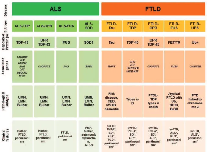
3. Genes linked to ALS and FTLD:
In this chapter causative genes linked to ALS and FTLD are presented divided in three groups (Table 2):
Pure ALS genes 5 found only associated with classical ALS and characterized by
21
Overlapping genes 5 involved in ALS and overlapping with other diseases,
characterized by the presence of overlapping motor and extramotor phenotypes and and with particular emphasis on genes found in both diseases ALS-FTLD.
Pure FTLD genes 5 found only associated with cognitive and behavioral phenotype 5
FTD
Table 2. Genes linked to ALS and FTLD
Reproduced from : Ling , Polymenidou and Cleveland. Neuron., 2013.
AD: autosomal dominant, AR: autosomal recessive, POAG: Primary open angle glaucoma, PDB: '()*+,-&./-*(-*&01&+2*&304*5&6789&/4:;<-/04&30.=&>=0-/+/-?
3.1. Pure ALS genes
3.1.1. Superoxidedismutase 1 (SOD1)
SOD1 mutations were found to cause familial ALS in 1993, representing the first
demonstration that linkage analysis could successfully pinpoint the underlying genetic cause of a rare neurodegenerative disease (49).
There are now more than 160 recorded mutations in this 153 amino-acid protein, mostly missense, all apparently resulting in fALS (20%) or sALS (1-2%) (40).
22 Considerable phenotypic heterogeneity occurs across the various SOD1mutations. For instance, the SOD1 A4V mutation can induce very aggressive disease while long survival has been reported in association with the SOD1 D90A mutation (40). In general, patients have almost exclusively lower motor neuron signs and almost never bulbar onset of disease (62). Although, cognitive impairment is not a prominent feature of SOD1 disease, patients with D90A manifest FTLD at the later stages of the disease (63).
SOD1 main physiological function is to protect cells from oxidative damage by metabolizing superoxide radicals (64).The molecular basis of the toxicity of mutant SOD1 is multifactorial. Because SOD1 detoxifies superoxide anion, it is likely that mutant SOD1
provokes oxidative stress (65). Whatever the molecular underpinnings of its cytotoxicity,
SOD1 can disrupt a wide set of cellular functions. While a complete recounting of these pathways is beyond the scope of this manuscript, some of the adverse effects include: provocation of cellular hyperexcitability (66), disruption of mitochondrial function (67), induction of the unfolded protein response (UPR) and endoplasmic reticulum (ER) stress (68), impairment of molecular motors and axonal transport (69), and early disruption of the neuromuscular synaptic structures (70).
Whatever the pathway is, it ends with misfolded proteins, having toxic effects on the cell's degradation machinery, impairing its two major components: the proteasomal pathway and the autophagy, and triggering its deposition in inclusion bodies within spinal motor neurons (71). Mutant SOD1 also spontaneously forms oligomers (72) as well as visible aggregates (73).
Neuropathologically, ALS patients with SOD1 mutation show loss of spinal cord neurons but while there is immunopositivity for ubiquitin and p62 inclusions in the spinal neurons and glia cells, there is usually negativity for TDP-43 (74).The protein appears to gain a novel, toxic
function that leads to motor neuron degeneration (75).
The SOD1 mutations discovery in ALS led directly to the development of the SOD1 transgenic mouse (76). Though important in elucidating the cellular mechanisms by which mutationof this gene predispose to motor neuron degeneration, the use of this model to select
agents for human trials has been increasingly called into question (77).
3.2. Overlapping genes
3.2.1. TAR DNA-binding protein (TARDBP)
In 2006 the protein TDP-43 was identified by Neumann and colleagues as major
component of the ubiquitin-positive neuronal inclusions that are the pathological hallmarks
23 FTLD and ALS are closely related conditions with overlapping molecular pathogenesis. Importantly, in 2008 it was shown that mutations in TARDBP caused a fraction of familial is causative gene mutated in ALS cases (52).
This presented a landmark event in our understanding of both diseases pathogenesis. There have now been more than 40 mutations found in TARDBP accounting for ~4% of fALS cases and a smaller percentage of sALS cases and rare FTLD (78). Most are missense mutations, but there are a few deletion mutations that give rise to a protein truncated at the very C-terminal (79).
Bulbar involvement was originally reported more frequently than might be expected, but
assimilation of all published phenotypes suggests that the proportion is about the same as for all ALS. Phenotype is usually ALS with or without FTLD but only rarely isolated FTLD (78).
Under normal conditions, TDP-43 is located in the nucleus, where it is involved as all DNA/RNA binding proteins in multiple steps of RNA metabolism (80). The data imply that TDP-43 causes pathogenesis in a two-step manner. The first step involves the exit of TDP-TDP-43 from the nucleus and the second step involves the irreversible formation of stress granule-based aggregates. Disease-associated mutations cause a shift of TDP-43 location from nucleus to cytoplasm and increase its aggregation propensity (81), but it is uncertain whether these changes are sufficient to initiate this vicious circle in vivo. TDP-435mediated toxicity may reflect either loss of its function in the nucleus, an acquired adverse effect of its pathological presence in the cytoplasm (gain-of-function), or both. Indeed, in a recently developed mouse model, the expression of mutant TDP-43 induced a phenotype even without formation of aggregates or abnormal processing of the cytoplasmic mutant protein and with no loss of TDP-43 from the nucleus which suggested a toxic gain of function (82). In line with this are findings
from the very recent study that, by comparing the pathological changes in TDP-43WTxQ331K (with
cytoplasmic aggregation and nuclear clearing) and TDP-43Q331K mice (with only cytoplasmic
aggregation) showed that the loss of nuclear TDP-43 may accelerate, but it was not essential
for the disease. Whereas cytoplasmic aggregation of TDP-43 seen in aged TDP-43Q331K and
TDP-43WTxQ331K mice, was sufficient to cause neurodegeneration (83). Thus, while nuclear
clearance of TDP-43 may accelerate disease it does not appear to be essential to cause neurodegeneration and loss of nuclear TDP-43 function may not be a primary or disease-critical event.
Neuropathology in cases of ALS with TARDBP mutations tends to reveal features similar to those of sALS, with neuronal loss and gliosis in the anterior horns of the spinal cord, and pallor of the corticospinal tracts. Immunohistochemically, there are TDP-43 positive
24
cytoplasmic inclusions in upper and lower motor neurons, but also in other regions of the
central nervous system including frontal and temporal cortex (84). In the brain, cytoplasmic TDP-43 undergoes secondary modifications such as hyperphosphorylation, ubiquitylation and processing into smaller fragments (28).
The discovery of the central role of TDP-43 in ALS pathogenesis has highlighted the importance of RNA processing. Additional support for this hypothesis comes from the discovery of FUS, another RNA-binding protein.
3.2.2. Fused in sarcoma (FUS)
In 2009, shortly after the identification of TDP-43, a second RNA-binding protein functionally homologous and structurally comparable with TDP-43, FUS was linked to familial ALS (50,51). FUS is discussed in more details afterwards (see below).
3.2.3. Hexanucleotide repeat expansion in C9ORF72
In 2011, a duo of teams made the discovery of a massive hexanucleotide repeat expansion in C9ORF72 as the cause of ALS and FTLD. The distinctive mutation in this gene is
an expansion of an intronichexanucleotide GGGGCC (G4C2) repeat motif (53,54). Normally
present in 30 or fewer copies, in C9ORF72-associated ALS, the repeat domain expands to encompass hundreds of tandem repeats (85). The expanded segment is transmitted as a dominant trait. Intriguingly, in several patients with C9ORF72 mutations, a second mutation has been found in another gene (TARDBP, for example) (86). The biological significance of this finding is unknown, but it suggests that in some families, ALS may be oligogenic in etiology (87).
It has reinvigorated the ALS and FTLD research field for a variety of reasons. First, the pathogenic expansion accounts for a remarkable percentage of both fALS (~37%) and familial FTLD (~21%) and genetically explains the majority of the overlap of these two disparate clinical syndromes (88). Second, the repeat expansion has been found to account for ~6% of apparently sALS cases and 6% of patients with supposed sFTLD (53,54,88).
C9ORF72 linked diseases clinically present with a widely variable phenotype including ALS or FTLD-ALS or FTLD (53). Bulbar onset is frequent in C9ORF72-associated ALS, as is
frontotemporal involvement, which is usually of the behavioural variant, but patients with primary progressive aphasia (PPA) have been described (53,54,89592).
The normal function of the presumably cytoplasmic protein C9ORF72 is unknown. However, it has recently been demonstrated that C9ORF72 regulates endosomal trafficking and
25 colocalizes with Rab proteins that are implicated in autophagy and endocytic transport. C9ORF72 also colocalizes with ubiquilin-2 and microtubule-associated protein light chain 3 (LC3) positive vesicles, and co-migrates with lysosome-stained vesicles in neuronal cell lines, providing further evidence that C9ORF72 regulates autophagy (93).
How repeat expansions in C9ORF72 cause ALS remains to be elucidated. In the ALS patients with a C9ORF72 expansion that have been studied, the levels of C9ORF72 mRNA were reduced by 50% (53), suggesting that the expanded allele does not generate mature mRNA. Thus, the C9ORF72 expansion may result ina loss-of-function. The expanded hexanucleotide repeats form nuclear RNA foci in neurons in the frontal cortex and spinal cord in patients with
C9ORF72 mutations (53). Pre-mRNA containing the expansion may thus exert a deleterious gain-of-function effect.These two mechanisms (haplo-insufficiency versus gain-of-function)
are not mutually exclusive and do not exclude other mechanisms for the repeat expansion to cause fALS.
Neuropathologically, patients with ALS who carry C9ORF72 hexanucleotide expansions present with typical features such as TDP-43-positive inclusions within the remaining motor neurons, as well as in the cortex and the hippocampus (91). Interestingly, two recent reports have indeed identified dipeptide repeat (DPR) proteins, which are most likely generated
through RAN translation from expanded G4C2 RNA, within aggregates of patients with
C9ORF72 expansion-associated ALS and/or FTLD (94,95). The pathogenic significance of these
DPR proteins remains to be demonstrated, but a study in Drosophila suggested that the DPR proteins could represent, by themselves the toxic agents (96).
3.2.4. Valosin-containing protein (VCP)
In the same year than C9ORF72, mutations in the valosin-containing protein (VCP) gene responsible for 152% of fALS cases were also reported (97). This is important given that these mutations provide another genetic link between motor neuron degeneration and FTLD, where
VCP mutations are rare and represent less than 1 % of the fFTLD cases (most frequently bvFTD
and SD) (98).
Today, 17 different mutations have been identified, and underlie an unusual clinical syndrome characterized by FTLD, inclusion body myopathy and Paget's disease of the bone (IBMPFD) (99). The existence of inclusion body myopathy in these patients was similarly interesting because it demonstrated that mutations in a single gene could result in pathology on both sides of the neuromuscular junction. This has given rise to the concept of multisystem
26 Physiologically, VCP interacts with a large number of ubiquitinated proteins to enable degradation or recycling and functions in multiple protein clearance pathways, including extracting misfolded proteins from the ER and sorting of endosomal proteins for proper trafficking. Depletion of VCP leads to accumulation of immature autophagosomes, similar to what is observed upon expression of IBMFD-linked mutations (100), suggesting that VCP is required for proper autophagy. Most intriguingly, TDP-43 is apparently mislocalized to the cytosol upon VCP-mediated autophagic dysfunction (100).
FTLD patients with a VCP mutation have TDP-43-positive inclusions (98).
The best supported hypotheses of the disease mechanism of VCP mutations are disturbed ubiquitin5proteasome mediated protein degradation, autophagy, or both (101).
3.2.5. Ubiquilin 2 (UBQLN2)
In 2011, missense mutations in UBQLN2, located on the short arm of chromosome X, were initially identified in autosomal dominant ALS (41). Nevertheless, ubiquilin 2 pathology has been observed in ALS patients who do not carry mutations in the gene.
Although, isolated ALS was the predominant phenotype, occasional patients had
concomitant symptoms of FTLD with abnormalities in both behaviour and executive functions;
however, none of these patients presented with FTLD alone.
Ubiquilin 2 is a member of the ubiquilin family, with unknown exact function. It has been implicated in ubiquitinated proteins degradation via both ubiquitin-proteasome system (UPS) and autophagy, in G-protein coupled receptor endocytosis , and may be an important component of the final common pathway mediating motor neuron degeneration (41).
In human spinal cord autopsy material of UBQLN2 ALS mutation carriers, inclusions were positive for UBQLN2, ubiquitin, p62, TDP-43, FUS and OPTN but not SOD1 (41,102). In cases with ALS-FTLD with or without UBQLN2 mutations, UBQLN2-positive inclusions are found in the hippocampus which are absent in ALS cases without dementia indicating that UBQLN2 aggregation and neurodegeneration are linked (41). Inclusions in spinal cord tissue from sALS and fALS patients with unknown mutations or mutations in SOD1, TDP-43 or FUS also stain positive for UBQLN2 (41,102).
3.2.6. Charged multivesicular body protein 2B (CHMP2B)
A mutation in the gene encoding charged multivesicular body protein 2B (CHMP2B), was found in a large Danish FTLD pedigree at chromosome 3 (103,104). Mutations affect the C-terminal end of the protein due to aberrant splicing.
27
CHMP2B encodes a protein with functions in the endosomal5lysosomal and the
autophagic protein degradation pathway.
Ubiquitin-immunoreactive inclusions do not stain for tau, TDP-43 or FUS antibodies (105) (106).
3.2.7. Optineurin (OPTN)
OPTN autosomal recessive mutations were described in a few ALS families in 2010 (55).
This has become interesting because of the intriguing phenotypic pleiotropy associated with such mutations. Indeed, the OPTN locus has been previously implicated in Paget's disease of bone (107). Dominant missense, recessive deletion and nonsense mutations of OPTN have been identified in both patients with fALS and with sALS.
Physiologically, OPTN functions as an inhibitor of NFNB-signaling (108), acts as an autophagy receptor(109) and participates in the regulation of vesicular trafficking and maintenance of the Golgi apparatus(110).
In sALS cases, OPTN is present in cytoplasmic inclusions and colocalizes with
ubiquitin, TDP-43, and possibly FUS (108,1115113).
3.2.8. Sequestosome 1 (SQSTM1)
SQSTM1 encodes p62 protein and mutations in this gene are known to cause Paget's
disease of bone (114). A candidate gene screening approach identified missense and deletion variants in ~1% of in familial and sporadic ALS patients (1155117).
Similar to ubiquilin, p62 has been shown to interact with polyubiquitinated proteins (118) and with LC3, allowing p62 to target polyubiquitinated proteins to the proteasome or autophagy. Therefore, both p62 and ubiquilin-2 link the ubiquitin-proteasome and autophagy pathways.
p62-positive inclusions have also been reported as deposited in neurons and glia of a
wide array of other neurodegenerative diseases (91). While the way these ALS-associated variants in p62 contribute to pathogenesis has not been established, autophagy/proteasome disturbance seems likely to play a role.
3.2.9. Other genes
Mutations in several other genes have been reported as rare causes of ALS or ALS-like syndromes. In 2014, whole-exome sequencing led to the identification of one mutation in the gene coiled-coil-helix-coiled-coil-helix domain-containing protein 10 (CHCHD10) in two families presenting with ALS/FTLD (119).
28 A large collaborative effort in 2015, by Cirulli et al., involving researchers from more than two dozen laboratories, recently led to the identification of a genome-wide association between rare, non-benign variants in TANK-binding kinase 1 (TBK1) and ALS (56). Most importantly, mutation in familial cases of ALS were recently identified in the TBK1 gene (57). Notably, the new ALS gene (TBK1) converges, together with previously known ALS genes (such as OPTN and SQSTM1) on autophagy pathway (56,57).
3.3. Pure FTLD genes
Two causal genes firstly identified: the microtubule-associated protein tau (MAPT) (59) and the progranulin (PGRN) (60) together account for 10%520% of FTLD cases (22).
3.3.1. Microtubule-associatedprotein TAU (MAPT)
In 1998, mutations in the MAPT gene located on chromosome 17 were identified in a number of families with FTLD and parkinsonism (59). Since then, 44 different MAPT mutations have been reported, accounting for 5520% of cases of familial FTLD (120).
MAPT encode the four microtubule-binding domains of TAU. In normal brain, the tau
protein occurs as six isoforms of which three contain three microtubule-binding domains (3R TAU) and three contain four microtubule-binding domains (4R TAU). A substantial number of missense mutations affecting the splicing of exon 10 result in aberrant ratios of 3R and 4R TAU. Consequently, the binding of TAU to tubulin is impaired either due to an increased expression of 4R tau relative to 3R TAU isoforms, or due to the altered binding properties of the mutant TAU protein (120). In addition, coding MAPT mutations increase the tendency of TAU to form
neurotoxic aggregates that are pathological characterictic of the more then 40% of FTLD
cases (121).
FTLD with tau-positive inclusions is mainly associated with bvFTD. However, forms of
PPA are also reported. Symptoms of associated motor neuron diseases are rare. On average,
FTLD-TAU is characterized by the earliest onset age in the FTLD syndromes (38).
3.3.2. Progranulin (PGRN)
A major breakthrough occurred in 2006 when the progranulin (PGRN) gene was identified as the second FTLD-related located gene on the same chromosome 17 (60). Indeed,
PGRN mutations account for an even larger proportion of FTLD families than do mutations in
29 the complete coding region and splice sites of the gene. They are loss-of-function mutations leading to reduced functional protein and resulting in haplo-insufficiency (123).
The characteristic pathological inclusions were tau-negative, ubiquitin-positive. It is now known that these inclusions are TDP43-positive.
Despite the fact that haplo-insufficiency is the common disease mechanism in all patients carrying a PGRNmutation, the associated clinical phenotype is variable, including bvFTD and
PNFA(124). Parkinsonism symptoms are often observed, but motor neuron symptoms are rare
(125).
4. Genetic link among ALS and FTLD
The description of mutations for several genes that are common to both ALS and FTLD provided a genetic link between them. Around six-seven genes that have been identified since nowadays represent a strong genetic proof of the continuum between these two pathologies (Figure 12).
The genetic link between ALS and FTLD was first established when TARDBP was found to be mutated in both diseases (52). This first gene was rapidly followed by a second one, FUS (50,51). A third, notable example was the identification of a hexanucleotide expansion in the
C9ORF72 gene, among both ALS and FTLD cases (53,54,126).The average mutation
frequencies reported in European populations are 37% for fALS, 6% for sALS, 21% for fFTLD, and 6% for sFTLD patients, which make C9ORF72 the strongest genetic link between these two diseases (61). Importantly, all these genes are involved in multiple steps of RNA metabolism (4).
30
Figure 12. ALS-FTLD genes
ALS-FTLD genes plotted to show phenotype, year of discovery and importance gauged by research outputs. The X axis is a score representing the involvement of each gene in ALS or FTLD. The Y axis represents year of mutation identification. The circle size represents the level of research on each gene.
Reproduced from: Al-Chalabi A et al., Acta Neuropathol.,2012
ALS, ALS/FTLD and/or FTLD causing mutations were also identified in genes involved in
protein clearance pathways or in maintaining proper protein homeostasis, including
ubiquilin-2 (UBQLN2) (41), vasolin-containing protein (VCP) (97,99), vesicle-associated membrane protein-associated protein B (VAPB) (127), p62/sequestosome (SQSTM1) (1155 117), optineurin (OPTN) (55), and charged multivesicular body protein 2B (CHMP2) (103,104).
Interestingly, these genes are associated with two major categories of cellular function: protein homeostasis and RNA metabolism. Together with the toxic protein aggregation that represent a classical pathological hallmark of both ALS and FTLD, these convergent genetic findings indicate that protein and RNA metabolisms are key cellular pathways systematically impaired in both diseases (Figure 13).