Computational Study of Actin Morphology and Rheology
byTaeyoon Kim
Master of Science in Mechanical Engineering Massachusetts Institute of Technology, 2007
Submitted to the Department of Mechanical Engineering in partial fulfillment of the requirements for the degree of
Doctor of Philosophy in Mechanical Engineering
at the
Massachusetts Institute of Technology
MASSACHUSETTS INSTlUTE OF TECHNOLOGY
MAY 18
2011
LIBRARIES
ARCHIVES
February 2011C Massachusetts Institute of Technology, 2011. All rights reserved.
Signature of Author
Department of Mechanical Engineering Oct 31, 2010
Certified by
Roger, D. Kamm Professor of Mechanical and Biological Engineering Thesis Supervisor
Accepted by
David E. Hardt Professor of Mechanical Engineering Chairman, Department Committee on Graduate Students
Computational Study of Actin Morphology and Rheology
byTaeyoon Kim
Submitted to the Department of Mechanical Engineering on Oct 31, 2010, in partial fulfillment of the requirements for the degree of
Doctor of Philosophy in Mechanical Engineering
Abstract
The cytoskeletal network consisting mainly of actin and actin binding proteins is
highly dynamic, provides structural integrity to cells, and plays a central role in a wide
range of mechanical and biological functions such as migration and the sensation of external forces. Thus, knowledge of actin cytoskeleton is indispensable for understanding the mechanics and many biological processes of cells. Although various theoretical, computational, and experimental investigations have been conducted, the underlying bases for these critical mechanical properties are still poorly understood. This thesis examines the morphology and rheology of actin networks through the development of a 3-D computational model.
First, the viscoelastic properties of actin networks irreversibly bound by actin crosslinking proteins (ACPs) were investigated. Relative contributions of the concentration and type of ACPs, the stiffnesses of actin filaments and ACPs, and thermal fluctuations were evaluated at various prestrain levels. These studies demonstrated for the first time that under typical biological conditions, extensional stiffnesses of both actin filaments and ACPs were surprisingly significant, but thermal fluctuations were relatively unimportant. At high tensions, only a small portion of networks supported a majority of the load.
Second, the relative importance of two mechanisms of ACPs which control dynamic properties of actin networks, unbinding and subdomain unfolding, was evaluated.
with unbinding and/or unfolding, it was found that despite the possibility of unfolding, ACP unbinding is the dominant mechanism governing actin rheology under typical experimental and physiological conditions. In addition, detailed processes by which unbinding plays such a role were investigated.
Lastly, roles that molecular motors play in the morphology and rheology of actin networks were studied. Motors enhanced elasticity of actin networks and led to heterogeneous networks to a degree that was highly dependent on how easily the motors unbind from actin filaments. ACPs helped the motors to make networks elastic and prevented the networks from being heterogeneous. Also, morphology of actin-motor networks was significantly affected by boundary conditions.
Thesis Supervisor: Roger D. Kamm
Acknowledgments
This work would not have been possible without contributions of numerous people.
First, I would like to thank my advisor, Prof. Roger D. Kamm, who has been a great mentor
as well as a friend. For the past 5 years, he has supported my research and inspired me with
his excellent intuition. I also thank my thesis committee members, Prof. Mary C. Boyce and
Prof. Gareth H. McKinley, who gave me valuable advices on my thesis research.
I would like to acknowledge Prof. Wonmuk Hwang for countless advices and helps
for developing my computational model and pursuing my research. I would also like to
express my gratitude to Dr. Hyungsuk Lee who was my colleague in Prof. Kamm's lab. I
could learn a wide variety of knowledge about my research topic from him, and he helped
to validate my computational model by performing an experiment. In addition, I am very
grateful to Dr. Jeenu Kim and Dr. Seung Eun Lee who were also my former colleagues. I
learned diverse computational techniques required for my research. I would also like to
thank Dr. Nathan Hammond and Philip Bransford. They were always open for discussion
about my research.
I thank all the past and current lab members for their friendship and supports
through 5 years: Cherry Wan, Tharathorn Rimchala, Vernella Vickerman, Ted Feldman, Anusuya Das, Ioannis Zervantonakis, William Polacheck, Kwang-Ho Lee, Aida Rahim, Peter Mack, Peyman Honarmandi, Ryo Sudo, Sid Chung, Young Kum Park, Helene
Kamm Lab members.
Most of my happiest time at MIT was spent with friends I met there. I thank Jooeun
Ahn, Hyun Jin In, Chung Jong Yu, Jaewon Cha, Yanghyo Kim, Yongkeun Park, Sung Joo
Bae, Minseung Ahn, Yongjin Sung, Yongdae Shin, Won-Yong Lee, Jongho Lee, Kimin
Jun, and all members in Korean Graduate Student Association Basketball Club and Korean
Graduate Student Association of Mechanical Engineering for the friendship.
I would never have completed this Ph.D. course if there had not been love and
support of my family. My parents, Bo Chin Kim and Jung Ja Kim, have never stopped
supporting me and have always believed in my potential and ability. I could be what I am
now via their endless love. Also, I would like to deeply thank my wife, Sunyoung Park, for
Contents
List of Figures ... 10
List of Tables ... 13
Chapter 1 Introduction... 14
1.1. G-actin, F-actin, and Polymerization... 15
1.2. Organization of Filaments via Actin Crosslinking Proteins (ACP)... 16
1.3. Molecular Motors ... 18
1.4. Previous Models for Cellular Mechanics...18
1.5. Previous Studies Investigating Actin Networks ... 20
Chapter 2 Investigating Rheology of Passive Actin Networks Crosslinked via Static Crosslinkers...23
2.1. Introduction ... 23
2.2. Methods ... 24
2.2.1. Dynamics and Mechanics...24
2.2.2. Coarse-graining Scheme Using Cylindrical Segments ... 25
2.2.3. Calculation of Steric Effects between Cylindrical Segments...27
2.2.4. Polymerization, Depolymerization, and Crosslinking ... ... 28
2.2.5. Preparation of a Network to Estimate Viscoelastic Properties ... 29
2.2.6. Adjusting Parameter Values for Coarse-graining ... 30
2.2.7. Bulk Rheology - Simulation ... 33
2.2.8. Bulk Rheology - Experiment ... 35
2.2.9. Segment-tracking Rheology ... 35
2.2.10. Computational Domain Size and Attainable Time Range... 36
2.3. Results...37
2.3.1. Comparison to Experiments ... 37
2.3.2. Effects of the Concentration and Type of ACPs on G' and G" ... 38
2.3.3. Effects of Prestrain on G' and G"... 41
2.3.4. Effects of Extensional Stiffness of Actin Filaments, Ks,A ... 43
2.3.5. Effects of Bending Stiffness and Thermal Fluctuation of Actin Filaments.. 45
2.3.6. Effects of Bending and Extensional Stiffnesses of ACPs... 48 7
2.3.7. Significance of Each Parameter at Various Prestrains... 49
2.3.8. The Supportive Framework Governing Viscoelastic Moduli... 51
2.4. Discussion...54
Chapter 3 Studying Effects of Dynamic Behaviors of Actin Crosslinking Proteins on Rheology of Passive Actin Networks... 59
3.1. Introduction ... 59
3.2. Methods ... 60
3.2.1. Preparation and Rheological Measurement of an Actin Network... 60
3.2.2. Dynamics and Mechanics... 61
3.2.3. Unfolding Event of ACP ... 61
3.2.4. Unbinding Event of ACP...63
3.2.5. Mimicry of Long-time Rheology ... 63
3.2.6. Simulation of a Micro-bead Experiment ... 64
3.2.7. Parallelization of Computational Codes...65
3.3. Results...67 3.3.1. Strain-stiffening ... 67 3.3.2 Stress R elaxation ... 71 3.3.3 Plastic Deformation...74 3.3.4 Micro-bead Rheology... 76 3.4. Discussion...76
Chapter 4 Investigating Roles of Molecular Motors in the Morphology and Rheology of Active Actin Networks... 79
4.1. Introduction ... 79
4.2. Methods ... 80
4.2.1. Preparation and Rheological Measurement of an Active Actin Network .... 80
4.2.2. Mechanics of Actin Filaments, ACPs, and Motors ... 81
4.2.3. Dynamic Behaviors of Motors ... 82
4.2.4. Boundary Conditions of the Computational Domain...85
4.3. Results...85
4.3.1. Effects of Concentrations of Motors and ACPs ... 85
4.3.1.1. Motor Concentration (RM)... 86
4.3.1.2. ACP Concentration (RACP)-...---..-..---...---.--... 88
4.3.2. Significance of Kinetic Param eters ... 89
4.3.3. Governing Factors of Network M orphology... 91
4.3.3.1. Effects of ACPs ... 91
4.3.3.2. Effects of M otors... 96
4.3.3.3. Effects of Adhesions to Boundaries and External Medium Stiffness. 97 4.4. Discussion...100
Chapter 5 Conclusions and Future W ork...105
List of Figures
Figure 1.1 Three primary constituent filaments of the cytoskeleton ... 14
Figure 1.2 Atomic structures of G-actin and F-actin... 15
Figure 1.3 Schematic representations of three models for cellular mechanics...19
Figure 2.1 A coarse-graining scheme using cylindrical segments with Nc = 5 ... 26
Figure 2.2 A schematic diagram showing the distribution of the repulsive force ... 28
Figure 2.3 Two representative networks used in this study ... 30
Figure 2.4 Comparison between computational and experimental results ... 37
Figure 2.5 Viscoelastic moduli of networks crosslinked by ACPC... 40
Figure 2.6 Viscoelastic moduli of networks bundled by ACPB ... 41
Figure 2.7 Behaviors of prestrained networks ... 42
Figure 2.8 Importance and effects of extensional stiffness of actin filaments in prestrained netw orks ... 44
Figure 2.9 Effects of bending stiffness and thermal fluctuation on G' and G"... 47
Figure 2.10 Effects of bending stiffnesses of ACPC, Kb,ACP,1 and Kb,ACP,2, on G' and G" ... ... 4 9 Figure 2.11 Relative decrease in G' at
fs
= 10 Hz due to 25-fold decrease of various stiffnesses at different prestrains... 51Figure 2.12 The supportive framework bearing most of stress ... 53
Figure 2.13 Values of shear storage modulus (G') at 10 Hz plotted against prestress ... 57
Figure 3.2 Invariance of rheological behaviors with ku0 'r fixed, which enables to mimic
long-tim e rheology ... 64
Figure 3.3 Parallelization of computational codes ... 66
Figure 3.4 Strain-stiffening behaviors with unbinding and unfolding...68
Figure 3.5 Displacement and turnover of ACPs during strain-stiffening with unbinding.... ... 7 0 Figure 3.6 Stress relaxation due to unbinding or unfolding... 73
Figure 3.7 Irreversible deformation of actin networks with unbinding or unfolding ... 75
Figure 3.8 Simulation of a micro-bead experiment ... 77
Figure 4.1 Walking of motors in our model... 83
Figure 4.2 G' and G" with different motor concentrations...87
Figure 4.3 G' with different ACP concentrations ... 88
Figure 4.4 Effects of kinetic parameters and the importance of motor unbinding ... 90
Figure 4.5 Morphology of networks consisting of actin filaments and motors with or w ithout A C P s ... 92-94 Figure 4.6 Morphology of networks employed for investigation of rheological effects of RACP ---... 95
Figure 4.7 Morphology of networks used to estimate the influences of RM on rheology .... ... 9 6 Figure 4.8 Morphology of networks whose G' was measured to probe the effects of 2
ubM
... 9 7
Figure 4.9 Network morphology under clamped boundary conditions with detachable
filam ents...9 8-99
Figure 4.10 Morphology of networks with moving boundaries following stress-strain
relation sh ip ... 10 1
List of Tables
Table 1.1 Examples of ACPs... 17 Table 2.1 List of adjusted parameters ... 32
Chapter 1
Introduction
Components of living organisms can be classified into organs, tissues, cells, and intra-cellular constituents in order of large to small length scale. Cells are fundamental functional constituents and play a significant role in multi-cellular organisms; they facilitate
reproduction, growth, and various chemical reactions [1]. Cells also exhibit a number of
interesting dynamic behaviors in response to external stimuli. Subjected to rapid deformation, cells tend to develop high levels of stress, whereas they easily deform under low strain rates with relatively little stress [2]. At fixed shear strain or stretch, marked stress relaxation has been observed both in vivo [2,3] and in vitro [4]. For most eukaryotic cells, these behaviors are attributable to the cytoskeleton, a highly dynamic structure mainly composed of three major constituents with distinct mechanical characteristics [5]:
Intermediate filaments Microtubules Microfilaments
Figure 1.1 Three primary constituent filaments of the cytoskeleton: intermediate
filaments, microtubules, and microfilaments (F-actin). They are distinctly distributed and play various roles in cells. (Adapted from [1].)
intermediate filaments, microtubules, and microfilaments (F-actin) (Figure 1.1). Actin is the most abundant intra-cellular protein in eukaryotic cells; typical concentrations of actin in the cytosol of nonmuscle cells range from 0.1 to 0.5 mM, but in some cellular structures, local concentrations can attain values as high as 5 mM [1]. It is widely believed that actin plays an important role in a wide range of biological and mechanical phenomena including
structural stability, migration, and intra-cellular processes [1].
1.1. G-actin, F-actin, and Polymerization
G-actin (monomeric actin) is a protein with molecular weight of 42 kDa whose dimension is about 5.5x5.5x3.5 nm [6] (Figure 1.2a). In various cells, G-actin is encoded
by a relatively well-conserved gene family [1]. G-actin self-assembles into F-actin,
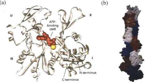
(a) (b) IV ATP-binding cleft N-terminus C-terminus
Figure 1.2 Atomic structures of G-actin and F-actin. (a)
p-actin
monomer contains a plate-like molecule divided by a central cleft. (Adapted from [1,6].) Each G-actin has molecular weight of 42 kDa. (b) F-actin consists of a tight double-stranded helix whose pitch is approximately 74 nm. (Adapted from [7].)a double helical filament with 7-9 nm in diameter and up to several microns in length [1]
(Figure 1.2b). F-actin is considered a semi-flexible polymer due to relatively large persistence length, ~10 pm [8,9]. Actin polymerization is governed mainly by a
nucleation-elongation process [10]. Three G-actins form a stable nucleus at low
probability [10,11], followed by quick elongation at both ends at significantly distinct rates: fast at barbed (plus) ends and slow at pointed (minus) ends. G-actin also depolymerizes at both ends at analogous rates. Thus, the dynamic behavior of F-actin occurs by elongation via assembly primarily at the barbed end and by shrinkage via disassembly primarily at the pointed end, in a polarized manner [12]. At a specific G-actin concentration, the
elongation rate at the barbed end is balanced by shortening rate at the pointed end, resulting in the net unidirectional movement of actin filaments called "treadmilling" [13].
1.2. Organization of Filaments via Actin Crosslinking Proteins (ACP)
Actin filaments are organized into orthogonal networks or bundles that are coupled to membrane proteins [5]. The organization of filaments is mediated by actin crosslinking
proteins (ACPs) such as a-actinin, fimbrin, fascin, and filamin [1,14]. Wide variations in
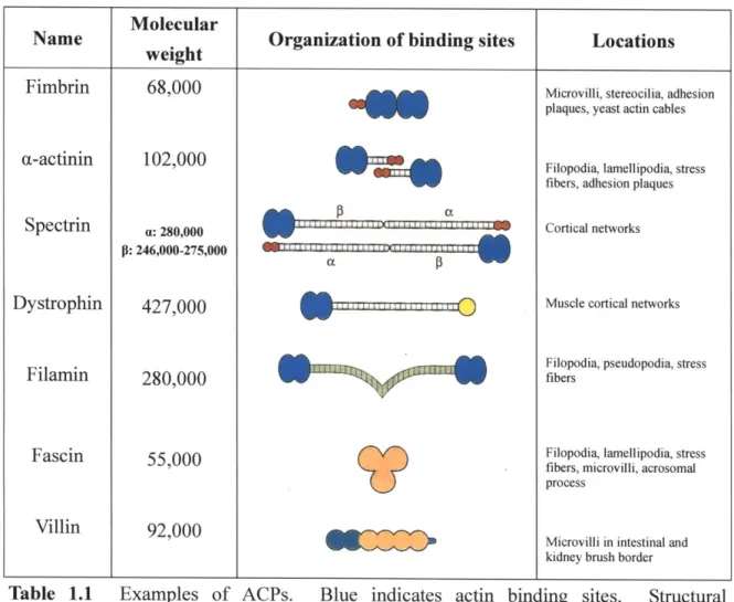
their function and structure in different organisms [15] indicate that ACPs, together with actin, evolved to satisfy various mechanical needs of cells (Table 1.1). Structural
arrangement of actin binding sites in ACPs is a main factor determining the organization of actin filaments [1]. If the binding sites are aligned in tandem, as is true for fimbrin and
fascin, ACPs tend to pack actin filaments into stress fibers as are prevalent in the extension
Molecular
Name . Organization of binding sites Locations
weight________ __
Fimbrin 68,000 Microvilli, stereocilia, adhesion
plaques, yeast actin cables
(1-actinin 102,000 Filopodia, lamellipodia, stress
fibers, adhesion plaques
Spectrin : 280,000 W Cortical networks
P: 246,000-275,000 1
Dystrophin 427,000 Muscle cortical networks
Filopodia, pseudopodia, stress
Filamin 280,000 fibers
Fascin 55,000 Filopodia, lamellipodia, stress
fibers, microvilli, acrosomal process
Villin 92,000
Microvilli in intestinal and kidney brush border
Table 1.1 Examples of ACPs. Blue indicates actin binding sites. Structural arrangement of the binding sites in ACPs determines whether they organize actin filaments into parallel bundles or orthogonal networks. Largely, large, long ACPs (e.g. filamin) tend to crosslink filaments at right angle, whereas short, small ACPs (e.g. fimbrin and fascin) are inclined to bundle filaments. (Adapted from [1].)
flexible arms and have tendency to organize the filaments into an orthogonal network found in cortical regions near a plasma membrane.
1.3. Molecular Motors
Unlike conventional materials, the cytoskeleton is not at thermal equilibrium; myosin, an active molecular motor walking on actin filaments in a polarized fashion via the consumption of ATP, generates internal stress, inducing high stiffness. This gives rise to a dynamic situation in which motors are under constant stress, stochastically unbind, reattach, and continue their motion along a new filament. Several types of myosin exist such as myosin I through XVIII. For instance, myosin V carries vesicles for transport and is a processive motor, meaning that a single myosin V alone is able to walk along filaments since it remains in a bound state for multiple steps [16]. By contrast, myosin II is a non-processive motor that binds to filaments for only a small fraction of lifetime, but self-assembles into a myosin minifilaments to increase processivity. These minifilaments are capable of producing contractile forces by pulling two actin filaments in opposite directions
[17,18].
1.4. Previous Models for Cellular Mechanics
(Most of this section is adapted from [19].)
Over the past several decades, three major models have emerged, each of which has been applied to the cytoskeleton (Figure 1.3). Each differs fundamentally, however, in terms of the primary mechanism(s) that determine the elastic or viscoelastic properties of the network. In the cellular solids model [20,21], the material is depicted as an athermal network of beams with rigid connections, and stiffness is determined primarily by the
bending of these individual members. The second model, based on the concepts of tensegrity [22], proposes that the network consists of a balance between elements in tension
and other elements in compression. In this model, the properties of the individual elements are relatively unimportant; network stiffness is determined instead by the level of tension in the system. The tensegrity model also neglects thermal effects. Finally, based on the assumption that crosslinks or entanglements occur over a length scale comparable to or smaller than the persistence length of thermally active filaments in the network, the cytoskeleton has been modeled as a semi-flexible polymer network [23]. The fourth model, based on the concepts of "soft glassy rheology" [24], will not be discussed here, in part because the fundamental physics giving rise to this behavior continues to be a source of some debate and uncertainty.
(a) (b) (c)
Figure 1.3 Schematic representations of three models for cellular mechanics. (a)
Biopolymer model. The network is comprised of thermally active filaments with varying degrees of crosslinks (light gray). (b) Cellular solids. The cytoskeleton is assumed to consist of unit cells constructed from elastic beams with rigid connections. (c) Tensegrity. The network is presumed to be composed of elements that are maintained in a state of tension either by interactions with other compressive members (as shown) or by tethering to an external matrix (Adapted from [19]).
1.5. Previous Studies Investigating Actin Networks
Investigating the morphological and rheological properties of actin networks is indispensable for elucidating the mechanics of cells as well as for understanding a wide variety of cellular processes. Thus, various experiments have been conducted to probe viscoelastic properties of cells and reconstituted actin gels using a variety of techniques such as microbead rheology, magnetic bead cytometry, and bulk rheology [25-44]. In experiments, discrepancies have been observed among measurements using dissimilar
methodologies, and many of the observed features are not well understood. For example, viscoelastic moduli measured by single-bead passive microbead rheology are much smaller
than those determined by 2-point microrheology or bulk rheology [25,33,43]. Also,
although distinct power law responses of the storage modulus have often been observed in
vivo and in vitro [29-32,44], their origin is not yet clearly understood.
In addition, recent experiments [45,46] demonstrated that the unbinding and
unfolding behaviors of a single ACP are rate-dependent, and can be described by several
models for bond rupture or domain unfolding including Bell's equation [47]. However, the link between such dynamic behaviors of single ACPs and the integrated properties of an actin network remains poorly understood due to difficulties in relating macroscopic
rheological measurements to underlying molecular events. Also, in vitro studies
incorporating myosin II showed a dramatic increase in elasticity of actin gels due to the contractile activities of molecular motors [18,48]. Nevertheless, the full understanding
about rheology of such actin-myosin networks is still lacking.
investigate characteristics of semi-flexible polymer networks including actin [49-59]. 2-D
[49,50] and 3-D computational models [51] studying affine and nonaffine deformations of
semi-flexible networks responding to large shear strain revealed two regimes dominated by
bending or stretching of filaments, respectively. Recently, using a microstructure-based
continuum mechanics approach, Palmer and Boyce reproduced many of the rheological
properties of actin networks observed in experiments [52]. The viscoelastic behavior of
semi-flexible networks was also investigated using dissipative particle dynamics and the
concept of microbead rheology [53,54]. Effects of unbinding or unfolding event of ACPs
on viscoelastic properties were investigated in several computational studies [60-62].
Recently, the rheology and morphology of actin-motor solutions have been studied in 2-D
and 3-D computational models [63,64].
To date, however, most of these computational models are too simplistic, or did not
either explicitly take into account ACP and motor mechanics or systematically account for
thermal fluctuations, all of which are potentially important factors governing matrix
viscoelasticity.
1.6. Thesis Overview
This thesis consists of three studies. In the first study, effects of prestrain, the
concentration and type of ACPs, the mechanical stiffnesses of actin filaments and ACPs, and the thermal fluctuation of actin filaments on viscoelastic properties of networks were
evaluated. The most important finding is that under conditions similar to cells, stretching
negligible. In the second study, we estimated the relative significance of two mechanisms
of ACPs governing dynamic behaviors of actin networks - unbinding and unfolding - by
probing the strain-stiffening, stress relaxation, and plastic deformation of the networks.
We found that under typical experimental and physiological conditions, the unbinding event
of ACPs is predominant, not unfolding. In the third study, we studied the roles of
molecular motors in the morphology and rheology of actin networks. The motors highly
Chapter 2
Investigating Rheology of Passive Actin Networks
Crosslinked via Static Crosslinkers
(Most of this section is adapted
from
[19,65].)2.1. Introduction
With the objective of extending previous computational works and providing new
insights into underlying mechanisms, we develop a Brownian dynamics model of the actin
network that includes features such as steric interaction among filaments, the usage of
explicit crosslinkers, a more realistic morphology, and the consideration of crosslinker
stiffness. By measuring stress in response to applied oscillatory shear strain ("bulk
rheology") and thermal fluctuations of individual segments in the polymeric chain
("segment-tracking rheology"), we investigate viscoelastic properties of actin-like networks
crosslinked by static crosslinkers showing neither unbinding nor unfolding event. It
should be noted, however, that some of the properties employed in our model, especially
for ACPs, were estimated since they are not well-known experimentally. Due to
simplifications in the model and parameter uncertainty, the results should therefore be
viewed as representative of a generic crosslinked network, but lack a quantitatively precise
correspondence to actin networks.
Nevertheless, we found features that semi-quantitatively capture experimentally
power laws as functions of the oscillation frequency. As the prestrain increased, the network became increasingly elastic. Bending and extensional stiffnesses of actin
filaments and ACPs played an important role depending on the degree of prestrain. We
found that the mechanical response of the network is dominated by a percolating
'supportive framework,' while other actin filaments contribute little to the viscoelastic moduli. Surprisingly, in typical physiological conditions where the distance between
crosslinking points along F-actin is much shorter than the actin persistence length, we
found that thermal fluctuation plays little role in viscoelasticity, so that the network
consisting of crosslinked F-actins can be viewed essentially as a deterministic overdamped
system in a viscous medium. In sum, our computational model elucidates how various
mechanical responses (thermal forces and the bending and stretching of actin filaments and
ACPs) govern viscoelastic properties of the network under different conditions.
2.2. Methods
2.2.1. Dynamics and Mechanics
In this simulation, actin monomers, filaments, and ACPs experience thermal
motion and interact with each other with defined potentials and binding probabilities.
Individual displacements are governed by the Langevin equation:
m, -- ' = F, - v +B(2.1)
dt' dt
where mi is the mass of ith element (actin or ACP), ri is the element's location, (i is the
deterministic force, and FB is a stochastic force satisfying the fluctuation-dissipation
theorem. Considering that inertia of all elements is negligible on the length and time
scales of interest, the Langevin equation is simplified by setting the acceleration term to
zero and cast in nondimensional form using kBT, (C,A, and LC,A as primary variables, where
C,A and LC,A are the friction coefficient and length of a segment of F-actin (as explained
later). Positions of all elements are updated using the Euler integration scheme:
1 _ 1 (fBj)
i, (i + Ai) =r 1(i) +
j
AT = (i)+ ) ± + Ai + iAI (2.2)(, dir,) {;
where dimensionless variables are indicated by the tilde "~". Two interaction potentials describe bond stretch and bending with stiffness kc, denoted by subscripts "s" and "b", respectively [66]:
Us = -c(F -r)2, Ub kb ( -60 )2 (2.3)
2 2
where 12 is the distance between two points constituting a chain, 0 is the bending angle,
and the subscript 0 denotes the equilibrium value.
2.2.2. Coarse-graining Scheme Using Cylindrical Segments
In our previous model [66], we treated a segment of F-actin as a spherical particle
representing two G-actins. To simulate larger length and time scales, we introduced
coarse-graining in which a cylindrical segment represents several monomers. We kept
thermal forces on ACPs and actin segments in a form similar to our previous model, but
incorporated a cylindrical geometry for calculating repulsive forces.
As seen in Figure 2.1, the points of two adjacent elements on a filament correspond
Figure 2.1 A coarse-graining scheme using cylindrical segments with NC = 5. Dashed lines show monomers and ACPs used to generate the network using the polymerization model [66]. Once the network is formed, it is coarse-grained by replacing the original spheres by cylinders as shown.
model. Accordingly, the diameter of the cylindrical segment, OrC,A, is the same as the
diameter of actin monomer of the previous model, uA= 7 nm, and its length, LC,A, is NC-uA. By letting each monomer of the polymerized network evolve into a cylindrical segment, a
larger network is created, albeit with a lower concentration of ~1-100 pM. In addition, one point on a filament axis and the other point representing the center of an ACP determine the two end points of each arm of the ACP, indicating that ACPs have a structure with two cylindrical arms whose length and diameter are LC,ACP and UC,ACP respectively. In this study, Nc was set to 10; this degree of coarse-graining is appropriate since the length of one rigid cylindrical segment, LCA = 70 nm, is still much shorter than the persistence
length of an actin filament, lp 10 pm.
One consequence of this coarse-graining technique is that it increases the arm C ACP
ACP
length of ACPs. That is, since the redundant volume of the original monomers is
neglected, the arm length of ACPs is extended by (LC,A - UC,A) / 2. This produces a network for which the shortest distance between two bundled filaments is (LC,A - UC,A) /2
(63 nm in this case) that exceeds the typical spacing formed by many of the bundling ACPs
(e.g. fascin, fimbrin, and a-actinin). However, it is not expected that this will significantly
alter the qualitative behavior of bundled networks since the change in the ACP arm length
is much shorter than the length of F-actin. Previous experiments where different bundling
ACPs led to distinct macroscopic behaviors [40,41] are more likely due to the different
stiffness and dissimilar binding affinities of the ACPs rather than their physical size.
2.2.3. Calculation of Steric Effects between Cylindrical Segments
Repulsive forces are computed according to the following harmonic potential
depending on the minimum distance, F12, between two cylindrical segments, 1 and 2:
S 6Ci + C,i,2 2 i - 2 UC,i,1 + C,i,2
U, 2 2 2 (2.4)
0 if F2 ;; '' 'i ' Ci2''
2
where 6c, 1 and &ci,2 are diameters of the cylindrical segments (i = A or ACP), and ic
is the strength of repulsive effects. Forces calculated from the potential (Equation 2.4),
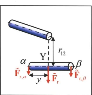
Fr, are distributed onto the two end points constituting the cylindrical segment, a and
p,
via the following equations:F, =Fr L F F (2.5)
L
l 12
a
Yi
Y,
Fra
y
Fr~p
Figure 2.2 A schematic diagram showing the distribution of the repulsive force acting on point Y, Fr onto two end points, a and
p.
The proportion of each force is determined by y, the distance between point a and point Y.where y is the distance between point a and point Y on the same segment (0 < y < Lc,,) in Figure 2.2.
2.2.4. Polymerization, Depolymerization, and Crosslinking
Monomer assembly and disassembly are important determinants of the morphology of actin networks. Given the rate constants obtained in recent in vitro experiments [67], it would take -100 s for an actin filament 1.5 pm in length to completely depolymerize, or
~1-10 s to polymerize the same filament with CA= 12.1 1iM. Though in vitro depolymerization of F-actin is very slow, various ACPs can accelerate it in vivo. However, we assume here for simplicity that neither polymerization nor depolymerization of actin
filaments occurs within the time scale of interest in this study, -1 s. Unlike our previous model [66], an ACP can bind to any point along an actin filament in any circumferential
direction. Neither unbinding nor unfolding of ACPs is permitted in this study.
2.2.5. Preparation of a Network to Estimate Viscoelastic Properties
The measurement of viscoelastic moduli can be influenced by the detailed
geometry and the extent of percolation, especially if the network is small. Therefore, the
use of a geometrically identical network for all simulations enables us to systematically
control and isolate the effect of a given parameter. In other studies, actin networks have
been generated by the random placement of equal-length filaments [49-51,54,57].
However, we generated more realistic networks using the previous model that incorporates
both polymerization and crosslinking. A somewhat heterogeneous network bundled by ACPB (Figure 2.3a) and a well-percolated, homogeneous network crosslinked by ACPc
(Figure 2.3b) were prepared. In Figure 2.3a, ladder-like structures consisting of two actin
filaments and multiple ACPB are evident, in contrast to thick bundles that are often
observed in experiments. The relative absence of thicker bundles in these simulations is
attributable to the small domain size compared to an average filament length. With Nc
-10, the filament length (Lf) is 1.5 pm ± 0.65 tm (average ± standard deviation), and the
actin concentration (CA) is 12.1 pM. We randomly removed ACPs to change RACP (Table
2.1), while maintaining the overall network geometry. In addition, actin filaments longer
than the width of the simulation domain (2.8 pm) were severed to minimize finite size
(a) (b)
Figure 2.3 Two representative networks used in this study. (a) A network bundled via ACPB and (b) crosslinked via ACPC, both of which consist of actin filaments of various
lengths (cyan) and ACPs (red). For visualization, VMD was used [68]. Two arms of each ACP drawn with partial red and cyan are connected to filaments, forming crosslinks. In (a), due to the small computational domain, ladder-like structures are predominant rather than long, thick bundles. The linear dimension of the simulation box is 2.8 pim. These two networks form the basis for all simulations; networks with low RACP were obtained from these by eliminating a portion of active ACPs. The inset of each plot shows the detailed geometry of bundled or crosslinked structures consisting of actin filaments and ACPs.
2.2.6. Adjusting Parameter Values for Coarse-graining
Upon coarse-graining, several parameters need to be adjusted. For the cylindrical
geometry of the segments, approximate forms for friction coefficients are [69]:
3+2Lci / oc, (2.6)
5
4+ Lc,i / ac,j
where r is the viscosity of the surrounding medium. However, since the mostly
crosslinked cylindrical segments move predominantly in the transverse direction, and
considering that (Cj is only 1.64 times higher than (C,A,j (LC,A /C,A 10), A
was used in all directions for simplicity. To test this assumption, we compared
simulations with and without the anisotropic friction coefficient at zero prestrain. At
fs
10 Hz, we obtained G' = 5.07 Pa (isotropic) and 5.00 Pa (anisotropic), and G" = 2.02 Pa
(isotropic) and 2.39 Pa (anisotropic). Since motion along the filament axis is more
suppressed with prestrain, the effect of anisotropic friction coefficient will be even less.
We also assumed a constant friction coefficient regardless of the filament length to which
the segment belongs. Hydrodynamic interactions between filaments are expected to play
little role and were ignored since the volume fraction of actin is low (~0. 1 %), and because
the filaments have high aspect ratio (long thin rod) [70].
Since the geometry of ACPs changes after coarse-graining, the following
equilibrium values for additional harmonic potentials were used for ACPB:
C,ACP 0.7, CACP =0.143, 01q = 0
eq /7 (2.7)
2
ksACP= 5, 000, b,ACP,1 = 250, kb,ACP,2
and for ACPc:
C,ACP =1.45, &CACP= 0'14 3 O 0.417,
q (8
s
=2
where LC,ACP and &C,ACP are the length and diameter of a cylindrical arm of ACP, 0,,q is
the equilibrium angle between two arms of ACP, and 02,eq is the angle formed by an arm
of ACP and the axis of the actin filament to which the ACP is bound. ks,ACP, Kb,ACP,1, and kb,ACP,2 are stiffness constants related to LC,ACP, O9,eq, and 0
2,eq , respectively (Figure 2.1).
Variable
Symbol
Value
Diameter of cylindrical actin segments
Length of cylindrical actin segments
Time step
Strength of repulsive force
Extensional stiffness of actin filament
Bending stiffness of actin filament
Concentration of actin
Ratio of CACP to CA
Extensional stiffness of ACP
*Bending stiffness 1 of ACP
*Bending stiffness 2 of ACP
Viscosity of medium (water, 300 K)
Boltzmann energy (300 K) UC,A LC,A At Kr KS,A Kb,A CA RACP Ks,ACP Kb,ACP,1 Kb,ACP,2 kBT 7-0X10- [in] (0.1) 7.0x10- [in] (1.0) 6.17 x10⁻⁹ [s] (2.0 x 10-') 1.69x10⁻³ [N / in] (2,000) 1.69x 10² [N / n] (20,000) 1.06x 18 [N m] (255.0) 0, 0.01, and 0.021 (ACPC) 0, 0.01, 0.02, and 0.04 (ACPB) 4.23 x03 [N / n] (5,000) 1.04x 10¹⁸ [N m] (250.0) 4.14 10-18 [N m] (1,000) 0.8599x10 [kg / in s]
4.142x10²¹
[j] (1.0)Table 2.1 List of adjusted parameters. Numbers in parentheses are corresponding dimensionless values as defined in the text. The value and notation of other parameters are the same as in [66].
* The same values are used for both ACPB and ACPC.
The value of 0
2,eq is the same as in the previous model, and 1,e of ACPc was adjusted to
maintain an equilibrium minimal distance of 70 nm between two crosslinked filaments.
ks,ACP was set to be one fortieth that of an actin filament due to computational efficiency.
kb,ACp,1 and b,ACP,2 of ACP were estimated to be similar to the bending stiffness of an
actin filament. Other parameters were also modulated according to the altered scale, as
listed in Table 2.1. In our previous model by which the network was generated [66], a
crosslink was allowed only if the torsional angle between two filaments was close to the
equilibrium value (0 for ACPB and 7t/2 for ACPc), and a finite stiffness was assigned to the
torsional angle between crosslinked filaments. However, because the other two bending
forces can preclude free torsional rotation, the torsional force is neglected here for
simplicity.
2.2.7. Bulk Rheology - Simulation
The concept of a strain-controlled bulk rheometer used in experiments was adopted
in order to measure the viscoelastic moduli of the generated networks. First, all actin
filaments were severed at the upper and lower boundaries, and the periodic boundary
condition on those surfaces was deactivated. Cylindrical actin segments within 70 nm
from the bottom surface were fixed, whereas those within the top 70 nm were forced to
move following an imposed strain. For the application of prestrain, the top boundary was
translated at a constant strain rate,
fX,
up to the desired strain. To measure differential viscoelastic moduli as in [27], a small sinusoidal strain (5%) was superposed on top of thefinite prestrain. The sum of forces on the ends of filaments attached to the top boundary
force component parallel to the surface was considered. In addition, due to the small
dimension of the system, the time scale for water diffusion through the computational
domain is of order 10 ps. Since this is smaller than the smallest period of oscillatory
strain, we assumed that the imposed shear strain immediately induces a linear velocity
profile within the fluid. Consequently, after calculating the stress due to filament forces, we added a shear stress expressed as:
rZ, = r/ dy" (2.9)
dt
where rzx is the shear stress exerted in the x-direction on a plane perpendicular to the
z-direction (pointing from the bottom to the top face), and yx is the shear strain applied in the
x-direction. The induced velocity of medium affects the movement of elements via the v
term of Equation 2.2.
Finally, dividing the measured stress (rzx) by the differential strain (yx), viscoelastic
moduli, G' and G", can be evaluated:
G*(fs)
-G'(fs)= G*(fs)l Cos#0 (2.10)
G"(f )= G*(fs) sin #
where
#
is phase delay between strain and stress, and|r.|
andlx
are the amplitudeof the differential stress and differential strain, respectively. Note that
#
is zero for a perfectly elastic material and equals rr /2 for a perfectly viscous one.2.2.8. Bulk Rheology - Experiment (performed with the help ofDr Hyungsuk Lee)
We also measured the mechanical properties of in vitro F-actin networks with a
rheometer (AR-G2, TA Instruments) using a 40 mm parallel plate geometry. Lyophilized
actin monomer from rabbit skeletal muscle was purchased from Cytoskeleton, Inc (Denver,
CO). To minimize artifacts caused by sample preparations [71], the actin was stored at high concentration (10 mg/ml) at -80'C and thawed rapidly at 37'C before each experiment.
Recombinant filamin A was purified from Sf9 cell lysates, and recombinant human gelsolin
was produced in Escherichia coli. Solutions of gelsolin, filamin, actin polymerization
buffer, and actin were gently mixed. The solutions were then loaded within 10 s into a
rheometer to form a crosslinked F-actin network. After 2 hr of polymerization at room
temperature, frequency-dependent shear moduli, G' and G", were measured in the range of
0.1-10 Hz. To obtain mechanical properties in the linear elastic regime, strain was maintained below 2%.
2.2.9. Segment-tracking Rheology
Many experiments have used microbead rheology to probe viscoelastic moduli of
actin gels based on the concept that the thermal motion of the bead is reflective of the gel's
viscoelastic properties. Here, we used a variation of this approach, and tracked the mean
square displacement (MSD) of individual actin segments. First, the domain was divided
into a cubic lattice comprised of NMSD cells (NMSD= 512) of equal volume. Then, one
cylindrical element was randomly selected per cell, and MSDs of the center of these
elements were recorded over time. Using a well-known approximate method [72], G' and
G" were calculated from the MSDs. Considering that this method was initially designed 35
for a spherical bead, it was appropriately modified for cylindrical elements:
G* (w) kBT
7rb (Ar2(1/))F[1
+X(w)]
G'(w) G* (w) cos(zX(w) /2) (2.11)
G "(o)= G* (co) sin(rX(w) /2)
where rb is the effective radius of an actin segment, which satisfies (C,A,1 62
Ifrb-F[1± (w)] is the gamma function, where y(co) is the power law exponent describing the
logarithmic slope of
(Ar2
(t)) at co =1lt, X(w)= d n (Ar.2 d in tt=1/0)
2.2.10. Computational Domain Size and Attainable Time Range
In spite of the coarse-graining using cylindrical segments, the length and time
scales that our model can attain are still much smaller than those of usual experiments.
For example, Lf in this computational model is a few microns at most, and the width of the
3-D domain is less than 5 pim. In such a small domain, it is difficult to investigate the
effects of a wide range of Lf or CA on viscoelastic moduli since Lf cannot be longer than the width of the domain to avoid artifacts associated with self-repulsion, and because an
increase in CA was achieved by a decrease in domain size with a constant number of molecules. If we increase the number of molecules, simulation time significantly
increases. On the other hand, many in vitro studies have used quite long actin filaments
(-20 pim) with relatively large systems of size -O(1 mm). It takes about 16 days to reach
1 s in typical conditions using an Intel@ Xeon@ 2.66GHz CPU, but experimental results
experimental results was not possible.
2.3. Results
2.3.1. Comparison to Experiments (with the help of Dr. Hyungsuk Lee)
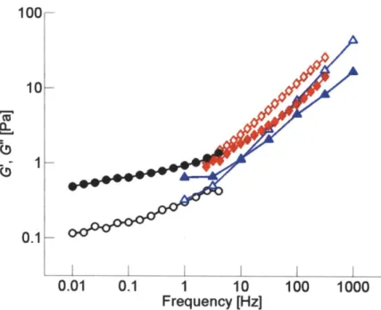
While recognizing the limitations of any direct quantitative comparisons between our model predictions and experiments, we conducted one set of experimental measurements under conditions similar to those of the simulation (mean filament length <Lf> = 1.5 pm., RACP 0.01, and CA = 12.1 gM) and compared viscoelastic moduli. In
0.01 0.1 1 10
Frequency [Hz] 100 1000
Figure 2.4 Comparison between computational and experimental results at (Lf> = 1.5 ptm, RACP = 0.01, and CA= 12.1 pM. Solid symbols: G', open symbols: G". For simulation, bulk rheology (blue triangles) and segment-tracking rheology (red diamonds) were employed, while the experiment was conducted only using a bulk rheometer (black circles). Overlap occurs in the frequency range of 1-10 Hz.
order to match <Lf>, gelsolin was added to the sample; the length distribution was determined by fluorescence imaging. One difficulty in matching simulation and
experimental conditions originates from the fact that RACP in the experiment corresponds to
the total amount of ACPs in the sample, including both those in active and inactive (partially bound or free) states, whereas in our simulation, it indicates the net amount of active ACPs that crosslink or bundle two filaments. In addition, due to computational
constraints, the oscillation frequency tested in the simulation overlaps that of the bulk rheology experiment only in a narrow range. Despite these difficulties, as seen in Figure
2.4, the values of G' and G" computed using the model are in reasonable agreement with the experiment, both qualitatively and quantitatively, perhaps better than one might have expected given the range of uncertainty of some of the parameters.
2.3.2. Effects of the Concentration and Type of ACPs on G' and G"
We computed G' and G" of structures crosslinked via ACPc (RACP = 0, 0.01, and
0.021) and those bundled via ACPB (RACP = 0, 0.01, 0.02, and 0.04) using both bulk
rheology and segment-tracking rheology. Networks without ACPs exhibit a slope of G'
close to 0.75, as indicated by the black solid line in Figures 2.5a and 2.6a. This value has been observed in various experiments [30,73-75], and it is known to originate from
transverse thermal undulations of actin filaments [55]. Interestingly, when repulsive
forces between the filaments are eliminated, the slope of G' estimated via segment-tracking
rheology approaches unity (data not shown), implying that the volume exclusion effect of neighboring filaments creates a tube-shaped space that hampers free translation and rotation
of the filament [56]. Although the filament confined in this way can perform reptation, it
is operative on long time scales that are beyond those attainable in these simulations, so the
MSD observed here primarily reflects transverse thermal motions, resulting in the 0.75
slope. On the other hand, the plateau in G' often exhibited by experiments [28,33,76] was
not observed within the frequency range of these simulations. Note that the plateau
modulus is induced by entanglement effects that become more pronounced at longer time
scales. The combination in these simulations of relatively short filaments leading to
longer entanglement time [28] and computational constraints precluding simulations for
longer times limited our ability to observe a plateau. Values of viscoelastic moduli attained
using bulk rheology and segment-tracking rheology exhibit surprisingly good agreement
(Figure 2.5) even though segment-tracking rheology (Equation 2.11) was originally
developed for a test particle much larger than the meshwork of filaments [72]. ACPC
elevates the magnitude of G' and reduces its slope (Figure 2.5a), implying that the
frequency dependence of G' is reduced as networks incorporate more ACPC. G' follows a
power law, G' ~fso3 (dashed line), forfs < 100 Hz at the highest crosslink density, RACP 0.021, which is within the range of powers observed in cells, 0.15-0.3 [29,32,44].
However, the magnitude of G' from these simulations was much lower than in vivo values.
This is likely due to many factors, notably the absence of prestrain. G" increases slightly
as the amount of ACPC is increased, but the slope remains similar (Figure 2.5b). In
addition, a decrease in the phase delay, tan1(G"/G') (Equation 2.10), accompanies the
increase in RACP, indicating that ACPC elevates the elasticity of the network. At
f
~ 103 Hz, the phase delay depends only weakly on RACP, but as frequency decreases, it decreasesmore quickly with higher RACP, implying a greater effect by crosslinking at lower
Networks bundled by ACPB exhibit a behavior distinctly different from that of
ACPc (Figure 2.6). Large differences in G' and G" were observed between
segment-tracking rheology and bulk rheology. This originates from the heterogeneity of the
bundled network attributable to the small computational domain, for which viscoelastic
moduli measured by bulk rheology depend strongly on whether or not there are bundles that
percolate between the top and bottom boundaries. We thus discuss results of only
B segment-tracking rheology for networks formed by ACP
ACPB increases G' but has little effect on its slope in contrast to ACPc (Figure
2.6a), whereas the phase delay is only slightly influenced by RACP. ACPC is able to form a well-percolated network even at relatively low RACP, so that the network gels more
efficiently [27] and acts like a single-body elastic object in response to shear stress or strain.
By contrast, ACPB bundles filaments together, resulting in the relatively low level of
(a) (b) 100 0.3 100 10 10
0.75
0.1 0.1 1 10 100 1000 1 10 100 1000 Frequency [Hz] Frequency [Hz]Figure 2.5 Viscoelastic moduli of networks crosslinked by ACPc (a) G' and (b) G". Open symbols: segment-tracking rheology, solid symbols: bulk rheology. RACP =0.021
(black circles), 0.01 (red triangles), and 0 (blue diamonds). With more ACPC, the magnitude of G' increases, and its slope decreases. G" is slightly larger for networks with higher RACP.
(a) (b) 100 100 11 1-0.75 0.11 1 10 100 1000 1 10 100 1000 Frequency [Hz] Frequency [HzJ B
Figure 2.6 Viscoelastic moduli of networks bundled by ACP . (a) G' and (b) G".
Open symbols: segment-tracking rheology, solid symbols: bulk rheology. RACP = 0.04 (black circles), 0.02 (magenta triangles), 0.01 (blue inverted triangles), and 0 (green diamonds). Large discrepancies exist between results obtained by segment-tracking rheology and by bulk rheology with nonzero RACP due to heterogeneity of the bundled network.
percolation for the same value of RACP as reflected by its low connectivity [66]. Although the diffusivity of bundled filaments is lower, the lack of connectivity leads to the absence of elastic behavior at long time scales. In order for ACPB to increase network elasticity to a similar extent by ACPc, its concentration may have to be much higher.
2.3.3. Effects of Prestrain on G' and G"
Only bulk rheology is employed here since the application of prestrain leads to a
highly nonuniform distribution of the load, with a small fraction of highly tensed filaments
and a larger number of filaments under little or no stress. Due to such heterogeneity, segment tracking rheology underestimates G' and G" as it randomly traces NMSD segments from the entire network. The effect of prestrain on G' is analogous to that of ACPc. As
seen in Figure 2.7a, G' increases and produces a weaker dependence on frequency at higher prestrain (y); at y = 0.55, G' is virtually independent of frequency and is nearly 100-fold
larger than that at y = 0 for
fs
< 10 Hz. This means that large prestrain transforms the network into a highly elastic one that exhibits a phase delay close to 0 at all frequencies and results in G' comparable to in vivo values [29,32,44], wherein it has been postulated that prestress or prestrain plays a significant role [27]. G" also exhibits interesting behavior; athigh prestrain, it increases slightly at low frequency, similar to in vitro observations using heavy meromyosin (HMM) [3 8,77], and a similar increase in G" was also observed in vivo
[39,74]. This suggests that at low frequencies, viscous effects play an important role. Tharmann and coworkers argued that this trend of G" may be due to the unbinding of HMM. However, since unbinding was not permitted in these simulations, the increase in
(a) 1000 (b) 10" S A A A A 1001 I. 2' -10 10LJ0.85 10 102 162 110 10- -100616161 Frequency [Hz] r [Pal
Figure 2.7 Behaviors of prestrained networks. (a) G' (solid symbols) and G" (open
symbols) of networks with RACP 0.021 at various prestrain: y= 0.55 (black circles), 0.4 (magenta triangles), 0.2 (blue inverted triangles), and 0 (green diamonds). At high prestrain, G' becomes nearly independent of frequency. G" with high prestrain slightly increases at low frequency. (b) G' at
fs
= 3.16 Hz versus prestress, ro. G' begins toincrease at about 0.1 Pa and follows a power law, G'~0..
the low-frequency G" with prestrain must originate from a different mechanism. Note that
such a tendency may have been more evident if the simulations were capable of reaching
even lower frequencies.
Also, the relation between G' at
fs
= 3.16 Hz and prestress (To) was investigated.It remains relatively constant until a threshold prestress (To ~ 0.1 Pa, Figure 2.7b) beyond
which it increases following a power law, G'~ ro0 8 5. The exponent of 0.85 is close to the
value of 1 found in in vitro experiments under similar conditions [27].
2.3.4. Effects of Extensional Stiffness of Actin Filaments, Ks,A
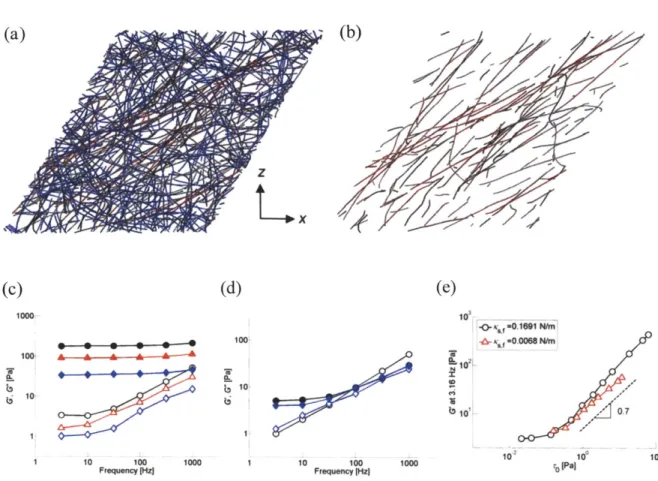
To illustrate how prestrain transforms a network into a more elastic one, we display
the network using a color scale depending on bond length averaged for duration of 0.1 ms.
Only a small number of actin filaments aligned in the x-z direction are highly stretched
(Figure 2.8a,b). As mentioned above, this heterogeneity precludes using segment-tracking
rheology which measures thermal motions of randomly selected segments, many of which
are not a part of the highly stretched filaments in prestrained networks. The mean
filament length of the entire network increased by only 0.5% with y= 0.55. However, due
to the large value of Ks,A (Table 2.1), this results in large spring forces that contribute
significantly to the high magnitude of G'.
We also performed simulations with different extensional stiffness (Ks,A 0.0338 and 0.0068 N/m) for networks with y = 0.55 (Figure 2.8c). G' and G" decreases with
lower Ks,A, but its phase delay is virtually unchanged (data not shown). When y = 0, however, variation in KSA has little or no effect since most actin filaments are not highly
(c) (d) (e)
1000 10.0M'f
100 A
1010
1 1 Fq n 1000 1 10 100 100 10 1
Frequency I~zj FRequen [HZI
Figure 2.8 Importance and effects of extensional stiffness of actin filaments in prestrained networks. (a) Color-map of the prestrained network with y = 0.55 (red: highly stretched bonds, gray: intermediately stretched bonds, blue: least stretched bonds. (b) Similar plot showing only actin filaments with bonds stretched by more than 0.5%. (c,d) Influence of the extensional stiffness of actin filaments, Ks,A, on G' and G" for a network crosslinked by ACPC (RACP =0.021) at (c) y = 0.55 and (d) y = 0. Solid symbols: G', and open symbols: G" with Ks,A 1.69x 10⁻² (black circles), 3.38x 10⁻³ (red triangles), and 6.76x10⁴ N/m (blue diamonds) (e) G' atfs=3.16 Hz as a function of prestress, ro, for Ks,A
= 6.764x 10⁴ (red triangles) and 0.01691 N/m (black circles). G' of both cases remains
nearly constant at low prestress, but starts to increase above -0.1 Pa. The behavior is similar for the two values of Ks,A except that lower Ks,A leads to a slight reduction in both the level and slope (-0.7, dashed line) of G' above the threshold stress level.


![Figure 2.1 A coarse-graining scheme using cylindrical segments with NC = 5. Dashed lines show monomers and ACPs used to generate the network using the polymerization model [66]](https://thumb-eu.123doks.com/thumbv2/123doknet/14440123.516762/26.918.217.710.137.447/figure-graining-cylindrical-segments-dashed-monomers-generate-polymerization.webp)