HAL Id: inserm-00663767
https://www.hal.inserm.fr/inserm-00663767
Submitted on 27 Jan 2012
HAL is a multi-disciplinary open access
archive for the deposit and dissemination of
sci-entific research documents, whether they are
pub-lished or not. The documents may come from
teaching and research institutions in France or
abroad, or from public or private research centers.
L’archive ouverte pluridisciplinaire HAL, est
destinée au dépôt et à la diffusion de documents
scientifiques de niveau recherche, publiés ou non,
émanant des établissements d’enseignement et de
recherche français ou étrangers, des laboratoires
publics ou privés.
implications for clinical trials.
Virginie Forest, Ashok Tirouvanziam, Christian Perigaud, Sarah Fernandes,
Marion Fusellier, Jean-Claude Desfontis, Claire Toquet, Marie-Françoise
Heymann, Dominique Crochet, Patricia Lemarchand
To cite this version:
Virginie Forest, Ashok Tirouvanziam, Christian Perigaud, Sarah Fernandes, Marion Fusellier, et al..
Cell distribution after intracoronary bone marrow stem cell delivery in damaged and undamaged
myocardium: implications for clinical trials.. Stem Cell Research and Therapy, BioMed Central, 2010,
1 (1), pp.4. �10.1186/scrt4�. �inserm-00663767�
Introduction
Th e possibility of tissue repair by autologous adult pro-genitor cells immediately captured the attention of clinicians confronted with the disabling, life-threatening circumstance of heart failure. During the last fi ve years, more than a dozen clinical studies using bone marrow cells have been published, ranging from case reports to
formal trials, deploying a range of diff ering cell-based therapies with the shared objective of improving cardiac repair [1].
Although most of these initial human trials suggest functional improvement, randomized clinical trials reported contradictory results [2, 3] suggesting that the ideal protocol design still needs to be defi ned, with the help of large animal models [4]. Several issues have been identifi ed by the European task force in translational research in the heart [5], including the type of cells to be used, the route of delivery, the number of cells and the volume to be given.
Abstract
Introduction: Early randomized clinical trials of autologous bone marrow cardiac stem cell therapy have reported
contradictory results highlighting the need for a better evaluation of protocol designs. This study was designed to quantify and compare whole body and heart cell distribution after intracoronary or peripheral intravenous injection of autologous bone marrow mononuclear cells in a porcine acute myocardial infarction model with late reperfusion.
Methods: Myocardial infarction was induced using balloon infl ation in the left coronary artery in domestic pigs.
At seven days post-myocardial infarction, 1x10(8) autologous bone marrow mononuclear cells were label ed with fl uorescent marker and/or 99mTc radiotracer, and delivered using intracoronary or peripheral intravenous injection
(leg vein).
Results: Scintigraphic analyses and Υ-emission radioactivity counting of harvested organs showed a signifi cant
cell fraction retained within the heart after intracoronary injection (6 ± 1.7% of injected radioactivity at 24 hours), whereas following peripheral intravenous cell injection, no cardiac homing was observed at 24 hours and cells were mainly detected within the lungs. Importantly, no diff erence was observed in the percentage of retained cells within the myocardium in the presence or absence of myocardial infarction. Histological evaluation did not show arterial occlusion in both animal groups and confi rmed the presence of bone marrow mononuclear cells within the injected myocardium area.
Conclusions: Intravenous bone marrow mononuclear cell injection was ineff ective to target myocardium. Myocardial
cell distribution following intracoronary injection did not depend on myocardial infarction presence, a factor that could be useful for cardiac cell therapy in patients with chronic heart failure of non-ischemic origin or with ischemic myocardium without myocardial infarction.
© 2010 BioMed Central Ltd
Cell distribution after intracoronary bone marrow
stem cell delivery in damaged and undamaged
myocardium: implications for clinical trials
Virginie F Forest
1,2, Ashok M Tirouvanziam
3, Christian Perigaud
2,3, Sarah Fernandes
1,2, Marion S Fusellier
4,
Jean-Claude Desfontis
4, Claire S Toquet
5, Marie-Françoise M Heymann
2,5,6, Dominique P Crochet
1,2,3;
Patricia F Lemarchand*
1,2,3R E S E A R C H Open
Access
*Correspondence: patricia.lemarchand@univ-nantes.fr
1INSERM UMR915, l’institut du thorax, IRT-Université de Nantes, 8 quai Moncousu,
BP 70721, Nantes, F-44007 Cedex 1, France
Full list of author information is available at the end of the article
© 2010 Forest et al.; licensee BioMed Central Ltd. This is an open access article distributed under the terms of the Creative Commons Attribution License (http://creativecommons.org/licenses/by/2.0), which permits unrestricted use, distribution, and reproduction in any medium, provided the original work is properly cited.
Our aim was, with an experimental design of acute myocardial infarction (MI) and autologous cardiac cell therapy that closely match clinical trials [2, 3] to analyze and quantify whole body and heart distribution of injected autologous bone marrow-mononuclear cells (BM-MNCs). We also evaluated the optimal route of delivery (intracoronary versus peripheral intravenous injection). Materials and methods
Animal procedures
Animal procedures were approved by the Institutional Committeefor Use and Care of Laboratory Animals at the Veterinary School ofNantes and conform to the Guide for
the Care and Use of Laboratory Animals (NIH Publication
No.85-23, revised 1996). Forty-six domestic pigs (35 to 40 kg) were included in the study. For MI induction and cell injection, anesthesia was induced by intramuscular injec-tion of ketamine (10 mg/kg)/xylazine (2 mg/kg) and main-tained with mechanical tracheal venti la tion using inhaled isofl urane (2%). All animals received subcutaneously morphine (0.2 mg/kg) prior and after all procedures. Experimental myocardial infarction
We used the MI experimental model initially described by Suzuki et al [6], with slight modifi cations. Five days prior MI induction, animals received β-blocker carazolol (20 μg/kg) twice per day. Propranolol (0.05 mg/kg) was injected intravenously just before MI induction procedure to prevent arrhythmia. After the placement of a 6F sheath in the femoral artery, an extra-back up 6F guiding catheter (Boston Scientifi c Scimed, Inc, Boston, USA) was placed under fl uoroscopy into the left main coronary artery. A 3 mm over the wire balloon catheter was placed either into the left anterior descending coronary artery distal to the fi rst diagonal branch or into the left circumfl ex. After balloon infl ation a coronary thrombus was induced by injections below the balloon of thrombin (900 UI, Sigma Aldrich Corporation, Saint Quentin Fallavier, France) and fi brinogen (5 mg, Sigma). Th e balloon was defl ated two to three minutes after injections. Coronary occlusion was checked by angio-gram, and MI was confi rmed by ECG alteration and by plasma creatine kinase elevation 24 hours after MI induc-tion (Refl otron, Roche Diagnostics, Meylan, France). Preparation and cryopreservation of autologous bone marrow mononuclear cells (BM-MNCs)
Five days after MI, a total of 100 mL of bone marrow was aspirated into heparin-treated Bone Marrow Aspiration/ Intraosseous Infusion needle (15Ga, 10mm, Cardinal Health France 205 S.A.S, Châteaubriand, France) from the iliac crests and shoulder blades under general anaesthesia. Five points of puncture were performed at each site of 5 mL each. Th e duration of the procedure was
15 to 20 minutes. BM-MNCs were isolated using Ficoll gradient (Eurobio Les Ulis, France), and a total of 244 x 106 ± 33 x 106 BM-MNCs was obtained.
BM-MNCs were cryopreserved in foetal calf serum 10% DMSO. Cryo-preservation for two days avoided two general anaesthesias the same day in seriously disabled animals and decreased the number of red blood cells to be injected.
Dual fl uorescence and radioactive labelling
Seven days after MI induction, BM-MNCs were quickly thawed and labeled immediately before injection using a green intracellular fl uorescent dye, Carboxy Fluorescein diacetate Succinimidyl Ester (CFSE, Molecular Probes Invitrogen, Cergy Pontoise, France) [7].
99mTechnetium (99mTc) labeling was performed after
CFSE labeling, using the IsolinkTM kit according to the
manufacturer’s instructions (Mallinckrodt Medical B.V., Petten, the Netherlands). CFSE + BM-MNCs were incubated with IsolinkTM/99mTc for 40 minutes at 37°C and
pH = 6. Before injection, BM-MNCs were centrifugated and resuspended in 10 mL NaCl to obtain a fi nal solution containing 300-400 MBq 99mTc-CFSE + BM-MNCs. Less
than 5% cell mortality was observed in all experiments using trypan blue exclusion method after thawing, after radiotracer and fl uorescence labeling, and immediately before injection.
99mTc cell leakage was evaluated before injection and
after four hours incubation at 37°C in vitro. Every hour, supernatant was collected. 99mTc activities were quantifi ed
on both supernatant and BM-MNC pellet using a gamma counter. Radio-labeling effi cacy, defi ned as the percentage of 99mTc cell activity in the fi nal cell solution, was 98 ± 5%
(n = 3) after four hours and was stable during the four hour experiment, suggesting no 99mTc cell leakage.
Autologous BM-MNC injection
At Day 7 after MI, a second catheterization was per-formed. A 3 mm over the wire balloon catheter was advanced to the site of MI induction. An angiogram was performed before completing the procedure. In the absence of coronary reperfusion, a recanalization was performed with the guide wire before its distal place ment. BM-MNCs were injected less than 30 minutes after preparation. Th e balloon was infl ated at low pressure (two bars) and 3.3 mL of labeled BM-MNCs were injected through the lumen. Th e balloon was defl ated two to three minutes later. Th e same procedure was repeated three times with a two minute interval between procedures. A total of 108 BM-MNCs were injected in 10 mL of NaCl at a
fi nal concentration of 107 BM-MNCs/mL.
For intravenous delivery, 108 BM-MNCs were injected
via the superfi cial vein of the left front leg, using a bolus injection of 15 seconds.
Nuclear imaging and gamma counting
Planar scintigraphic images of 99mTc-BM-MNC body
distri bution were obtained 1hour and 24 hours after tracerinjection, using a gamma camera fi tted with a low energy all purpose high resolution parallel hole collimator (DS7, Sopha Medical, GEMS, Buc, les Yvelines, France). Th e energy window was centred at 140 keV ± 20%. Using a dedicated image processing co mputer, a static image was recorded into a 64 × 64 matrix for 35 seconds. Th e camera was calibrated using a standard source measured with a gamma counter before each acquisition to convert image counts to activity in MBq. For each experiment a calibrated dose of technetium was used to quantify activity decay after 24 hours. Anesthetized animals were positioned in supine positions, at a constant distance of 30 cm from the collimator, and images were recorded in thoracic and abdominal projections. On each image, a region of interest was drawn around each organ to quantify counts within the drawn area. Organ activity in each region of interest was expressed as the percentage of the total initial injected activity (measured as the diff erence between the 99mTc-BM-MNCs syringe content
before and after injection) with correction for 99mTc
physical decay.
Animals were sacrifi ced, their organs were excised and imaged with the gamma camera, and activity was measured in weighted samples of each organ and used to quantify organ gamma counting radioactivity (ACAD activimeter, Lemer Pax, Nantes, France). All quantitative analyses were performed by two investigators blinded to animal treatment group.
Pathologic examination and immunohistology
Animals were sacrifi ced 1 hour or 24 hours after BM-MNC injection. Heart samples were cryopreserved and sliced into contiguous transverse 5 μm thick cryosections or were embedded in paraffin and stained with Hematoxylin-Eosin-Safran (HES). Immunolabeling was performed to detect VonWillebrand Factor antibody (DakoCytomation SA. France, Trappes France). Cell nuclei were labeled using propidium iodide. Histology analyses were performed by two pathologists blinded to animal treatment group.
Statistical analysis
Data were expressed as mean ± SD. Th e Kruskal-Wallis testwas used for comparison of independent samples and Spearman’s test for correlation coeffi cient. A valueof
P <0.05 was considered statistically signifi cant.
Results
Procedural results
Myocardial infarction was induced in 37/46 pigs (Figure 1A). MI induction was complicated by death
related to ventricular fi brillation during the procedure in eight cases (21.6%), and immediately after at the wake up time in four other cases (10.8%). Total death rate was 32% during and immediately after MI induction, probably due to extended MI [8].
Th e 25 surviving animals were randomly assigned to three diff erent groups: fi ve were used as controls (MI without BM-MNC injection), 16 received intracoronary BM-MNCs, four received intravenous BM-MNCs. Two deaths were observed during balloon infl ation for BM-MNC intracoronary injection (one from a ventricular fi brillation and one from a second MI, probably related to the injection site location being within a non-infarcted area). In the 9/47 animals without MI induction, seven received intracoronary BM-MNC injection and two were used as radioactivity controls (intracoronary injection of IsolinkTM/99mTc only).
Myocardial infarction model
MI was induced by complete obstruction of the left coronary artery using balloon infl ation and injection of thrombin and fi brinogen (n = 23; left anterior descending artery: n = 15; left circumfl ex artery: n = 8) (Figure 1B). Transient ST-segment elevation and isolated ventricular extrasystoles occurred during each coronary balloon occlusion, but abnormal ECG profi le (ST-segment eleva-tion) persisted only after injections of thrombin and fi brinogen. Plasma creatine kinase levels increased 24 hours after coronary occlusion (1,222 ± 604 UI/l before coronary occlusion, and 4060 ± 1961 UI/l at 24 hours, P <0.01), confi rming MI induction. Macro-scopic examination of the heart performed seven to eight days after coronary occlusion showed extensive trans-mural antero-inferior MI encompassing 41.25 ± 5% of left anterior ventricle area (n = 23), with a thinning of the injured wall. Statistical analysis did not show any diff erence in MI size between animal groups, nor between animals with left anterior descending artery or left circumfl ex coronary occlusion, nor between animals with or without spontaneous reperfusion at the time of cell injection (not shown). Histopathologic examination confi rmed MI, with associated infl ammatory reaction and loss of cardiomyocyte nuclei (Figure 1C). Coronary angiography at seven days after MI showed spontaneous reperfusion in 18/23 pigs. Histological analyses showed an adherent residual thrombus within the coronary artery without total occlusion of the vessel lumen. Coronary occlusion in the fi ve out of 23 remaining pigs required reperfusion before BM-MNC injection. Neither in pigs with spontaneous reperfusion nor in those with mechanical reperfusion, angiographic control showed abnomalities of blood fl ow distribution when compared to basal angiographic data obtained before MI induction.
Biodistribution and 99mTc-BM-MNC cardiac engraftment
after MI and intracoronary injection
Scintigraphic images were used to track 99mTc-BM-MNCs
after intracoronary injection. Radioactivity in the kidneys and the bladder due to free 99mTc clearance, was observed
15 minutes after injection (Figure 2A). At one hour after injection, whole pig body images revealed intense regional accumulation of radioactivity in the heart (Figure 2B). At
24 hours a persistence of heart and lung radioactivity was observed, with a decrease in signal intensity (Figure 2C).
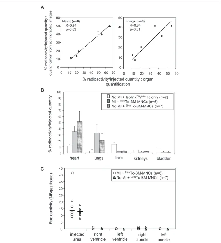
Radioactivity was expressed as the fraction of measured radioactivity, decay-corrected and divided by the injected radioactivity amount. In the fi rst set of experiments, radioactivity was quantifi ed one hour after 99m
Tc-BM-MNC intracoronary injection both in regions of interest drawn on the planar images and in isolated organs Figure 1. Pig model of acute myocardial infarction. A: Experimental design and animal groups. MI = myocardial infarction; IC = intracoronary
delivery; IV = peripheral intravenous delivery. B: Induction of myocardial infarction. Coronary angiography, before, during, immediately after and seven days after occlusion of left anterior descending artery and fi brinogen/thrombin injections. C: HES staining of myocardial tissue seven days after MI induction, characterized by a central necrotic zone and an infl ammatory response.
B C A 1h sacrifice (n=7) 1h sacrifice (n=1) 1h sacrifice (n=6) 1h sacrifice (n=2) 1h sacrifice (n=5) 24h sacrifice (n=8) 24h sacrifice (n=2) No MI (n=9) 12 deaths during procedure 46 pigs MI (n=37) IC injection of IsolinkTM/99mTc only (n=2) IC cell injection (n=16) No cell injection (n=5) IC cell injection (n=7) IV cell injection (n=4)
2 deaths during procedure
24h sacrifice (n=1) n=9
(Figure 3A). Th ere was no signifi cant diff erence between both quantifi cation methods (n = 6, R = 0.94 for heart, and R = 0.84 for lungs). Th erefore results from either method were used in following experiments. As controls, two pigs without MI but with intracoronary injection of IsolinkTM/99mTc only were used to quantify free IsolinkTM/99mTc
fi xation in each organ. At one hour radioactivity rate was 11.5 ± 2.1% in the heart, 3.5 ± 2.1% in the lungs and 14.5 ± 2.1% in the liver (Figure 3B), and at 24 hours radioactivity rate was null (not shown). Consistent with the in vivo scintigraphic images (Figure 2A), after 99m
Tc-BM-MNC intracoronary injection high levels of radioactivity were measured in the heart (34.8 ± 9.9% of injected radioactivity) and in the lungs (32.6 ± 13.9%) (n = 6, Figure 3B). Small quantities of radioactivity were ob-served in other organs (2 ± 1.4% in the liver, 1.7 ± 0.6% in the kidneys). Radioactivity was also quantifi ed in the infarcted site and in other cardiac tissues (right ventricle, healthy left ventricle and auricles, Figure 3C). We observed an accumulation of radioactivity in the infarcted area, Almost no radioactivity was detected remote from injection site (right ventricle, healthy left ventricle, auricles). 24 hours after BM-MNC injection, radioactivity persisted with lower intensity in the heart (6 ± 1.7%) and in the lungs (11 ± 2.6%) (n = 3, Figure 4B).
Importantly, there was no correlation between the infarction size and the heart radioactivity rate at one hour and 24 hours (not shown).
Myocardial infarction and BM-MNC cardiac engraftment To investigate the role of MI on BM-MNC homing and engraftment in the heart, 99mTc-BM-MNC were
intra-coronary injected in pigs with MI and in pigs without MI. In both animal groups, ECG alteration (ST-segment elevation) was observed during intracoronary BM-MNC injection. ST-segment elevation stopped immediately after the procedure, suggesting that no micro-infarction was induced by injection itself favouring cardiac homing. Importantly, no signifi cant diff erence in heart and lung BM-MNC
distribution was observed between both groups, with a high radioactivity ratio in the heart (34.8 ± 9.9% in MI group
versus 51.2 ± 17.2% in no MI group, Figure 3B).
Biodistribution of BM-MNCs and delivery route
Th e efficacy of intracoronary administration versus systemic intravenous administration was evaluated in pigs with MI. After intravenous cell delivery at 1 hour and 24 hours, radioactivity was detected mainly in the lungs both on scintigraphic images (n = 4, Figure 4A) and by radioactivity quantifi cation, in marked contrast with intracoronary injection (Figure 4B). After intravenous injection, cardiac radioactivity was almost null (0.16 ± 0.23%) as compared to pigs after intracoronary injection (34.8 ± 9.9%). After 24 hours, no cardiac radioactivity was observed in intravenously injected pigs, suggesting an absence of cardiac homing at 24 hours.
Histological analyses
For histological analyses, BM-MNCs were labeled with a fl uorescent marker (CFSE) prior to injection and animals were sacrifi ced one hour or 24 hours after injection. As noticed by others [9] microscopic fl uorescent artefacts were observed in the myocardium of control animals with MI and without BM-MNC injection, characterized by fl uorescent leukocytes and autofl uorescence within the infarcted tissue. However, these artefacts could be distinguished from BM-MNCs by their small size and their limited location within necrotic areas [10] (Figure 5A). BM-MNCs in non necrotic areas were detected in each animal that received CFSE+ BM-MNCs (Figure 5B-5C), and were not observed in any control animals (with MI and without BM-MNC injection, Figure 5D). CFSE+ BM-MNCs were also observed in heart tissue 24 hours after intracoronary injection in each animal that received CFSE+ BM-MNCs (Figure 5J). Th e presence of BM-MNCs was confi rmed in HES histo-logical analyses (Figure 5E-5F). BM-MNCs appeared as clusters of mononuclear cells, characterized by round Figure 2. 99mTc-BM-MNC biodistribution in animals with myocardial infarction after intracoronary injection. A-B: abdomen (A) and thorax
(B: anterior and lateral) scintigraphic images at 15 minutes and 1 hour. C: thorax scintigraphic image at 24 hours in the same animal.
lungs lungs heart
B
Anterior
Lateral
1hC
heart lungsAnterior
24hA
bladder kidneys lungs 15 minAnterior
and dense blue nuclei, in contrast with elongated and blue pale cardiomyocyte nuclei. Th ese round and dense blue nuclei were not observed in control animals in HES
histologic analyses (not shown). BM-MNCs were located in majority in the peri-infarcted myocardium and only a few BM-MNCs were detected in infarcted myocardium. Figure 3. 99mTc-BM-MNC biodistribution after intracoronary injection in animals with or without myocardial infarction. The percentage of
radioactivity in organs was calculated by dividing organ radioactivity with the total injected quantity after radioactive decay correction, one hour after injection. A: Comparison between two radioactivity quantifi cation methods in the same animal: values from measured radioactivity in isolated organs (x-axis), and values from regions of interest drawn in the scintigraphic whole body acquisition images (y-axis). B: Radioactivity quantifi cation in animals with (MI) or without (no MI) myocardial infarction, after intracoronary injection of IsolinkTM/99mTc only or 99mTc-BM-MNCs. Bars indicate
mean and SD (P >0.05, not signifi cant). C: Individual quantities of radioactivity in pigs with or without MI (P >0.05, not signifi cant). Radioactivity was quantifi ed in 1 g myocardium inside the infarcted area, in the left and right ventricles distant from infarcted area, and in the left and right auricles.
A % r adioac tiv ity /inj ec ted qua ntit y : quantif ic ation f ro m sc intigr aphic im ages 0 10 20 30 40 50 0 10 20 30 40 50 60 Lungs (n=6) R=0.84 p=0.61 0 10 20 30 40 50 60 0 10 20 30 40 50 60 70 Heart (n=6) R=0.94 p=0.63
% radioactivity/injected quantity : organ quantification 0 10 20 30 40 50 60 70 80 90 100 heart lungs B % r adioact ivit y/ inj ect ed quant
ity No MI + IsolinkTM/99mTc only (n=2)
MI + 99mTc-BM-MNCs (n=6) No MI + 99mTc-BM-MNCs (n=7)
liver kidneys bladder
C R adi oa cti vi ty (M B q /g ti ss ue ) injected area right ventricle left
ventricle auricleright auricleleft
0 5 10 15 20 25 30 35 40 45 MI + 99mTc-BM-MNCs (n=6) No MI + 99mTc-BM-MNCs (n=7)
No BM-MNCs were observed remote from the injection site (auricle, right ventricle, non infarcted myocardium).
Tissue sections were stained for endothelial cells and capillaries using anti-VonWillebrand antibody (Figure 5G). Neither thrombus nor arterial occlusion was observed in both groups. BM-MNCs were incorporated within myocardial tissue and were not observed within the lumen neither into wall blood vessel, and no cells were observed with double staining CFSE+ and endothelial marker. Double labeling for macrophages using anti-MAC-1 antibody was negative (not shown), suggesting that transplanted BM-MNCs had not been phagocyted.
We also investigated extracardiac BM-MNC biodistri-bution by histological analysis. CFSE+ BM-MNCs were disseminated in whole lungs (Figure 5H). Sections of other vital organs (liver, spleen, kidneys and thymus) demonstrated no signifi cant cell location, only cell debris (Figure 5I). Importantly, the presence of BM-MNCs was confi rmed one hour (Figure 5K-5L) and 24 hours (not shown) after intracoronary injection in every pig without
MI and again, neither thrombus nor arterial occlusion was observed. Th ese data were in accordance with analyses of scintigraphic data and radioactive organ quantifi cation (Figure 3B).
Th e absence of cardiac homing after BM-MNC intra-venous injection at 24 hours as observed on scintigraphic images was also confi rmed by histological analyses, showing an absence of fl uorescence in myocardium inside and outside MI area (not shown). In contrast, CFSE+ BM-MNCs were numerous and dispersed in whole lungs when injected intravenously (not shown). Discussion
In a porcine model of acute MI with late reperfusion and autologous cardiac cell therapy, we quantifi ed biodistri-bution using a radiolabeling technique. Most of the BM-MNCs delivered by the intracoronary route were distributed within the heart and lungs. Th e presence of MI did not modify BM-MNC cardiac homing and engraftment after intracoronary injection. Finally, Figure 4. 99mTc-BM-MNC biodistribution according to delivery route. A: Scintigraphic acquisition images, 1 hour and 24 hours after peripheral
intravenous 99mTc-BM-MNC injection in the same pig with myocardial infarction. B: Radioactivity quantifi cation, calculated by dividing organ
radioactivity with the total injected quantity after radioactive decay correction. Bars indicate mean and SD.
A heart lungs 1h 24h heart lungs liver liver B lungs kidneys MI + intracoronary99mTc-BM-MNC injection – 1h (n=6) MI + intravenous99mTc-BM-MNC injection – 1h (n=2) MI + intracoronary99mTc-BM-MNC injection – 24h (n=3) MI + intravenous99mTc-BM-MNC injection – 24h (n=2) 0 10 20 30 40 50 60 70 80 90 100
heart liver bladder
% r adioact ivit y/ inj ect ed quant ity
systemic intravenous BM-MNC injection promoted lung homing rather than cardiac engraftment.
Th e fi rst prerequisite for cell therapy success is the engraftment and thus, homing of transplanted cells to the Figure 5. Immunohistologic analyses. Representative organ sections, after intracoronary injection of BM-MNCs labeled prior to injection with
CFSE (green fl uorescence). Cell nuclei were labeled with propidium iodide (red fl uorescence). A: Fluorescent artefacts within the necrotic area of infarcted tissue. B-C: Myocardium sections of infarcted tissue, in absence of necrotic area, one hour after CSFE+ BM-MNC intracoronary injection.
D: Myocardium section of a control animal (with myocardial infarction, without BM-MNC injection). E-F: The following section of C was stained
with HES. Engrafted BM-MNCs within the infarcted zone appeared as round and dense blue points (arrow), in contrast to the elongated and blue pale cardiomyocyte nuclei (star). G: Myocardium section stained for endothelial cells and capillaries using anti-VonWillebrand antibody (red fl uorescence). H-I: Lung (H) and liver (I) section. Cell nuclei labeled with propidium iodide (red fl uorescence). J: Myocardium section of infarcted tissue, 24 hours after CFSE+ BM-MNC intracoronary injection. K-L: Myocardium section of an animal without myocardial infarction, one hour after CFSE+ BM-MNC intracoronary injection, using VonWillebrand antibody (K) or HES coloration (L).
X20 X20 B X40 X20 D X40 J X20 E X40 G I K X40
*
F X20 H X20 * X40 X40 X20 L A Ctarget area. Intracoronary delivery off ers the advantages of a non-surgical method that can be performed percu-taneously during angioplasty for acute MI. In the present study we showed that intracoronary BM-MNC injection can allow cell engraftment into myocardium tissue, whereas peripheral systemic intravenous adminis tration was not effi cient at 24 hours. Th is suggests that the cardiac niche eff ect favoured by MI was not suffi cient to attract intravenously injected BM-MNCs within 24 hours, thus limiting the effi cacy of systemic intravenous injection. Chemoattractant factors secreted by the infarcted heart might be too diluted in the body of large animals like pigs to attract BM-MNCs. If this is the case, similar results may be expected in humans. Another hypothesis is that the ability to trap intravenously injected BM-MNCs within the heart is limited, since only 4% to 5% of cardiac output is dedicated to supplying the coronary arteries [11]). Finally, cardiac engraftment following systemic intravenous BM-MNC delivery might be limited by lung entrapment prior to coronary artery access. Numerous studies have been performed in rodent [12] and large animal models [13, 14], using intravenous injection of mesenchymal stem cells, showing mesen-chymal stem cell engraftment after 24 hours. Several hypotheses can be discussed to explain these discrepan-cies with our results. First, our protocol closely matched clinical trial protocols, in which a seven-day delay between acute MI and cell injection seems to be the best time for effi cacy following intracoronary injection [3, 15]. However, this time may be too long after intravenous cell injection, in regards to local infl ammatory response and niche eff ect. In most studies mesenchymal stem cells were injected 1 to 72 hours after myocardial infarction [12-14]. Second, BM-MNCs were labeled with
99mTechnetium, a radioelement that has a short half life
and may not have been detected at 24 hours. However, we did not observe any fl uorescent cell within the heart 24 hours after intravenous injection, corroborating our radioactivity data. Finally, mesenchymal stem cells may have a better capability for cardiac homing than BM-MNCs, that contain a very small stem cell number. Interestingly, a recent study comparing intra-aortic, intravenous, and intramyocardial delivery of mesen chy-mal stem cells in rats observed 5% cell survival at 48 hours after intravenous delivery, mostly in the lungs [15].
Th e radioactive labeling using IsolinkTM kit did not
discriminate cell type, and was not species nor cell specifi c. In a pilot study we evaluated 99m
Tc-hexa-methyl-propylen-amine-oxime (HMPAO) labeling, often used in humans. However, 99mTc-HMPAO labeling of BM-MNCs
was totally unsuccessful (not shown). We then switched to the radioactive linker IsolinkTM, which allowed us to
inject a large quantity of 99mTc-labeled cells, signifi cantly
higher than in other studies [16, 17]. Although, similar to
previous studies [11, 18, 19], the viability of IsolinkTM/99m
Tc-labeled BM-MNCs was not altered, the adverse eff ects of labeling on the migratory and functional abilities of BM-MNCs cannot be entirely excluded. Th e majority of cardiac BM-MNC homing studies were performed in rodents, and only a few studies established the fraction of transplanted cells retained within the myocardium using direct radioactive labeling of the cells. In our study, radioactivity quantifi cation in each organ showed 34.8 ± 9.9% total radioactivity in the heart with an accumulation within the injection site, one hour after intracoronary BM-MNC injection, and 6.0 ± 1.7% at 24 hours. Importantly, histology results were in accordance with scintigraphic imaging data confi rming the presence of numerous BM-MNCs at one hour and 24 hours after intracoronary injection. As in our study, Hou et al [8], using a pig model of reperfused MI with intracoronary cell delivery and radioactive cell quantifi cation, observed a largely right-sided distribution of BM-MNC. However, only 2.6 ± 0.3% of BM-MNCs were detected in the heart, with some BM-MNCs being distributed to the right ventricle. Importantly, the experimental model in this study was xenogeneic, with human BM-MNCs being injected into pigs. Human cells may lack the correct membrane receptors to home into the pig myocardium or may undergo acute lysis due to a xenogeneic reaction. In a recent study, using a similar model of reperfused MI in pigs with autologous BM-MNC transplantation [20] 6.5% of autologous transplanted BM-MNCs were detected in the heart four days after injection, a result close to ours at 24 hours. Finally, cardiac homing cell rate at one hour might be overestimated by including a possible leakage of radioactive label from the cells, although this appears unlikely as no radioactive cell leakage was observed in vitro in the next four hours following radioactive labeling.
Although phase I-II clinical trials have been completed using coronary delivery of BM cells, only one study used coronary delivery of 99mTc-labeled BM-MNCs (in a single
patient), and observed intense cardiac cell engraftment [17]. Th e study of Hofmann et al [11] showed that in fi ve patients with MI, only 1.3 to 2.6% of 18F-FDG-labeled
BM-MNCs were detected in the infarcted myocardium one hour after intracoronary BM-MNC injection. However, the number of injected BM-MNCs was 30-fold higher than in our study, so the absolute number of retained BM-MNCs within the heart was 39 to 78 x 106
BM-MNCs, a number close to ours (34.8 ± 9.9 x 10⁶
retained BM-MNCs). Intracoronary injection of a very high cell number may saturate the binding sites for cardiac cell homing, raising questions about the ideal number of cells to be injected.
After intracoronary injection, we observed a similar biodistribution of BM-MNCs in animals with or without
MI, despite the large size of the infarcted area (45% of the left ventricle). Several studies have shown that acute myocardial infarction is followed by an acute local infl ammatory reaction involving upregulation of chemo-kines receptors and adhesion molecules, thereby facilitat-ing adhesion and infi ltration of cells involved in tissue repair, including stem cells [21-23]. Th e dynamic capa-bility of BM-MNCs to migrate and the niche eff ect are central in regenerative medicine [21-23]. In our model, to carry out intracoronary injection, blood fl ow was stopped three times for at least two minutes to prevent backfl ow and prolong contact time between BM-MNCs and myocardium. When BM-MNCs were injected without prior MI induction, each maneuver for intracoronary cell injection resulted in ST-segment elevation, as already described for repeated angioplasty balloon infl ations [24, 25]. Although we did not evaluate the intensity of the ischemia induced by balloon infl ation, this suggests that the injection technique created local downstream ischemic and preconditioning eff ects [25], thus rendering the local microenvironment more receptive to cell homing [24]. A recent study in a pig model of myocardial infarction and intracoronary injection of autologous BM cells, balloon occlusion was found ineff ective to increase cell homing [24]. In another study in a similar model, single-bolus delivery was as eff ective as three balloon-occlusion deliveries [26]. Importantly, both studies were performed only in pigs with myocardial infarction and not in healthy pigs, suggesting that the cardiac niche eff ect favoured by MI was suffi cient to attract injected BM-MNCs without any further eff ect of balloon occlusion. Th e fact that the presence and the size of MI did not infl uence cell engraftment could be useful for cardiac cell therapy in patients with chronic heart failure of non-ischemic origin or with ischemic myocardium without myocardial infarction. Two clinical trials have been published with patients who have had a myocardial infarction at least three months before BM-MNC coronary injection [1, 27, 28]. A clinical benefi t was observed in both studies, suggesting that BM-MNCs homed to the myocardium despite the absence of acute myocardial infarction. Th e hypothesis of cardiac homing in the absence of acute myocardial infarction was recently confi rmed in two Phase I cardiac cell therapy clinical trials on patients with chronic ischemic cardiomyopathy and receiving an intracoronary injection of radiolabeled CD133+ or CD34+ cells [16, 29].
Conclusions
Myocardial cell distribution following intracoronary injection did not depend on MI presence. Given these results, further eff orts to analyze the mechanisms of adhesion and transmigration through the vascular wall will be rewarding.
Abbreviations
99mTc = 99mTechnetium; BM-MNC(s) = bone marrow-mononuclear cells; CFSE =
carboxy fl uorescein diacetate succinimidyl ester; DMSO = dimethyl sulfoxide; ECG = electrocardiogram; HES = hematoxylin-eosin-safran; HMPAO = hexa-methyl-propylen-amine-oxime; MBq = Mega Becquerel; MI = myocardial infarction; SD = standard deviation.
Competing interests
The authors declare that they have no competing interests.
Authors’ contributions
VF carried out the cell therapy product preparation and drafted the manuscript. AT implemented the myocardium infarction animal model and carried out myocardial infarction inductions. CP implemented the cardiac cell therapy animal model and carried out bone marrow harvests. SF participated in the design of the study and helped to draft the manuscript. MF performed the analysis and interpretation of scintigraphic data. JCD carried out the radioactive experiments. CT participated to the conception and design of histological experiments. MFH carried out the analyses and interpretation of histological data. DC participated in carrying out in vivo experiments and helped to draft the manuscript. PL conceived of the study, and participated in its design and coordination, and drafted the manuscript. All authors read and approved the fi nal manuscript.
Acknowledgements
The authors gratefully acknowledge Stéphane Madec for technical expertise in imaging. This work was supported, in part, by an INSERM Avenir grant, the Association Française contre les Myopathies, the Agence de la Biomédecine, and by the Juvenile Diabetes Research Foundation.
Author details
1INSERM UMR915, l’institut du thorax, IRT-Université de Nantes, 8 quai
Moncousu, BP 70721, Nantes, F-44007 Cedex 1, France. 2Université de
Nantes, Faculté de Médecine, Institut Fédératif de Recherche Thérapeutique 26 (IFR26), 8 quai Moncousu, BP 70721, Nantes, F-44007 Cedex 1, France.
3CHU Nantes, l’institut du thorax, Hôpital Nord Laënnec, Boulevard Jacques
Monod, Nantes, F-44 093 Cedex 1, France. 4Animal Pathophysiology and
Functional Pharmacology Unit (UPSP 5304), Ecole Nationale Vétérinaire de Nantes, Atlanpole La Chantrerie, Nantes, F-44307 Cedex 3, France. 5CHU
Nantes, Service d’Anatomo-pathologie, Hôpital Nord Laënnec, Boulevard Jacques Monod, Nantes, F-44093 Cedex 1, France. 6INSERM U957, Laboratoire
de Physiopathologie de Résorption Osseuse et Thérapie des Tumeurs Osseuses Primitives, Faculté de Médecine 1, rue Gaston Veil, Nantes, F-44035 Cedex 1, France.
Received: 28 Jul 2009 Accepted: 15 Mar 2010 Published: 15 Mar 2010
References
1. Martin-Rendon E, Brunskill SJ, Hyde CJ, Stanworth SJ, Mathur A, Watt SM: Autologous bone marrow stem cells to treat acute myocardial infarction: a systematic review. Eur Heart J 2008, 29:1807-1818.
2. Lunde K, Solheim S, Aakhus S, Arnesen H, Abdelnoor M, Egeland T, Endresen K, Ilebekk A, Mangschau A, Fjeld JG, Smith HJ, Taraldsrud E, Grogaard HK, Bjornerheim R, Brekke M, Muller C, Hopp E, Ragnarsson A, Brinchmann JE, Forfang K: Intracoronary injection of mononuclear bone marrow cells in acute myocardial infarction. N Engl J Med 2006, 355:1199-1209.
3. Schachinger V, Erbs S, Elsasser A, Haberbosch W, Hambrecht R, Holschermann H, Yu J, Corti R, Mathey DG, Hamm CW, Suselbeck T, Assmus B, Tonn T, Dimmeler S, Zeiher AM: Intracoronary bone marrow-derived progenitor cells in acute myocardial infarction. N Engl J Med 2006, 355:1210-1221. 4. Arnesen H, Lunde K, Aakhus S, Forfang K: Cell therapy in myocardial
infarction. Lancet 2007, 369:2142-2143.
5. Bartunek J, Dimmeler S, Drexler H, Fernandez-Aviles F, Galinanes M, Janssens S, Martin J, Mathur A, Menasche P, Priori S, Strauer B, Tendera M, Wijns W, Zeiher A: The consensus of the task force of the European Society of Cardiology concerning the clinical investigation of the use of autologous adult stem cells for repair of the heart. Eur Heart J 2006, 27:1338-1340. 6. Suzuki M, Asano H, Tanaka H, Usuda S: Development and evaluation of a
new canine myocardial infarction model using a closed-chest injection of thrombogenic material. Jpn Circ J 1999, 63:900-905.
Lamirault G, Heudes D, Orsonneau JL, Heymann MF, Charpentier F, Lemarchand P: Autologous myoblast transplantation after myocardial infarction increases the inducibility of ventricular arrhythmias. Cardiovasc Res 2006, 69:348-358.
8. Hou D, Youssef EA, Brinton TJ, Zhang P, Rogers P, Price ET, Yeung AC, Johnstone BH, Yock PG, March KL: Radiolabeled cell distribution after intramyocardial, intracoronary, and interstitial retrograde coronary venous delivery: implications for current clinical trials. Circulation 2005,
112:I-150-I-156.
9. Lafl amme MA, Murry CE: Regenerating the heart. Nat Biotechnol 2005, 23:845-856.
10. Anversa P, Leri A, Rota M, Hosoda T, Bearzi C, Urbanek K, Kajstura J, Bolli R: Concise review: stem cells, myocardial regeneration, and methodological artifacts. Stem Cells 2007, 25:589-601.
11. Hofmann M, Wollert KC, Meyer GP, Menke A, Arseniev L, Hertenstein B, Ganser A, Knapp WH, Drexler H: Monitoring of bone marrow cell homing into the infarcted human myocardium. Circulation 2005, 111:2198-2202.
12. Nagaya N, Fujii T, Iwase T, Ohgushi H, Itoh T, Uematsu M, Yamagishi M, Mori H, Kangawa K, Kitamura S: Intravenous administration of mesenchymal stem cells improves cardiac function in rats with acute myocardial infarction through angiogenesis and myogenesis. Am J Physiol Heart Circ Physiol 2004, 287:H2670-H2676.
13. Kraitchman DL, Tatsumi M, Gilson WD, Ishimori T, Kedziorek D, Walczak P, Segars WP, Chen HH, Fritzges D, Izbudak I, Young RG, Marcelino M, Pittenger MF, Solaiyappan M, Boston RC, Tsui BM, Wahl RL, Bulte JW: Dynamic imaging of allogeneic mesenchymal stem cells traffi cking to myocardial infarction. Circulation 2005, 112:1451-1461.
14. Krause U, Harter C, Seckinger A, Wolf D, Reinhard A, Bea F, Dengler T, Hardt S, Ho A, Katus HA, Kuecherer H, Hansen A: Intravenous delivery of autologous mesenchymal stem cells limits infarct size and improves left ventricular function in the infarcted porcine heart. Stem Cells Dev 2007, 16:31-37. 15. Abdel-Latif A, Bolli R, Tleyjeh IM, Montori VM, Perin EC, Hornung CA,
Zuba-Surma EK, Al-Mallah M, Dawn B: Adult bone marrow-derived cells for cardiac repair: a systematic review and meta-analysis. Arch Intern Med 2007, 167:989-997.
16. Goussetis E, Manginas A, Koutelou M, Peristeri I, Theodosaki M, Kollaros N, Leontiadis E, Theodorakos A, Paterakis G, Karatasakis G, Cokkinos DV, Graphakos S: Intracoronary infusion of CD133+ and CD133-CD34+ selected autologous bone marrow progenitor cells in patients with chronic ischemic cardiomyopathy: cell isolation, adherence to the infarcted area, and body distribution. Stem Cells 2006, 24:2279-2283. 17. Mesquita CT, Correa PL, Felix RC, Azevedo JC, Alves S, Oliveira CC, Sousa AL,
Borojevic R, Dohmann HF: Autologous bone marrow mononuclear cells labeled with Tc-99m hexamethylpropylene amine oxime scintigraphy after intracoronary stem cell therapy in acute myocardial infarction. J Nucl Cardiol 2005, 12:610-612.
18. Brenner W, Aicher A, Eckey T, Massoudi S, Zuhayra M, Koehl U, Heeschen C, Kampen WU, Zeiher AM, Dimmeler S, Henze E: 111In-labeled CD34+ hematopoietic progenitor cells in a rat myocardial infarction model. J Nucl Med 2004, 45:512-518.
19. Penicka M, Lang O, Widimsky P, Kobylka P, Kozak T, Vanek T, Dvorak J, Tintera J, Bartunek J: One-day kinetics of myocardial engraftment after
intracoronary injection of bone marrow mononuclear cells in patients with acute and chronic myocardial infarction. Heart 2007; 93:837-841. 20. Moelker AD, Baks T, van den Bos EJ, van Geuns RJ, de Feyter PJ, Duncker DJ,
van der Giessen WJ: Reduction in infarct size, but no functional
improvement after bone marrow cell administration in a porcine model of reperfused myocardial infarction. Eur Heart J 2006, 27:3057-3064. 21. Orlic D, Hill JM, Arai AE: Stem cells for myocardial regeneration. Circ Res
2002, 91:1092-1102.
22. Askari AT, Unzek S, Popovic ZB, Goldman CK, Forudi F, Kiedrowski M, Rovner A, Ellis SG, Thomas JD, DiCorleto PE, Topol EJ, Penn MS: Eff ect of stromal-cell-derived factor 1 on stem-cell homing and tissue regeneration in ischaemic cardiomyopathy. Lancet 2003, 362:697-703.
23. Niessen HW, Lagrand WK, Visser CA, Meijer CJ, Hack CE: Upregulation of ICAM-1 on cardiomyocytes in jeopardized human myocardium during infarction. Cardiovasc Res 1999, 41:603-610.
24. Tossios P, Krausgrill B, Schmidt M, Fischer T, Halbach M, Fries JW, Fahnenstich S, Frommolt P, Heppelmann I, Schmidt A, Schomäcker K, Fischer JH, Bloch W, Mehlhorn U, Schwinger RH, Müller-Ehmsen J: Role of balloon occlusion for mononuclear bone marrow cell deposition after intracoronary injection in pigs with reperfused myocardial infarction. Eur Heart J 2008, 29:1911-1921. 25. Yellon DM, Downey JM: Preconditioning the myocardium: from cellular
physiology to clinical cardiology. Physiol Rev 2003, 83:1113-1151. 26. Doyle B, Kemp BJ, Chareonthaitawee P, Reed C, Schmeckpeper J, Sorajja P,
Russell S, Araoz P, Riederer SJ, Caplice NM: Dynamic tracking during intracoronary injection of 18F-FDG-labeled progenitor cell therapy for acute myocardial infarction. J Nucl Med 2007, 48:1708-1714.
27. Assmus B, Honold J, Schachinger V, Britten MB, Fischer-Rasokat U, Lehmann R, Teupe C, Pistorius K, Martin H, Abolmaali ND, Tonn T, Dimmeler S, Zeiher AM: Transcoronary transplantation of progenitor cells after myocardial infarction. N Engl J Med 2006, 355:1222-1232.
28. Strauer BE, Brehm M, Zeus T, Bartsch T, Schannwell C, Antke C, Sorg RV, Kogler G, Wernet P, Muller HW, Kostering M: Regeneration of human infarcted heart muscle by intracoronary autologous bone marrow cell transplantation in chronic coronary artery disease: the IACT Study. J Am Coll Cardiol 2005, 46:1651-1658.
29. Schots R, De Keulenaer G, Schoors D, Caveliers V, Dujardin M, Verheye S, Van Camp G, Franken PR, Roland J, Van Riet I, Everaert H: Evidence that intracoronary-injected CD133(+) peripheral blood progenitor cells home to the myocardium in chronic postinfarction heart failure. Exp Hematol 2007, 35:1884-1890.
doi:10.1186/scrt4
Cite this article as: Forest VF, et al.: Cell distribution after intracoronary
bone marrow stem cell delivery in damaged and undamaged myocardium: implications for clinical trials. Stem Cell Research & Therapy 2010, 1:4.