HAL Id: hal-02529318
https://hal-amu.archives-ouvertes.fr/hal-02529318
Submitted on 15 Mar 2021
HAL is a multi-disciplinary open access archive for the deposit and dissemination of sci-entific research documents, whether they are pub-lished or not. The documents may come from teaching and research institutions in France or abroad, or from public or private research centers.
L’archive ouverte pluridisciplinaire HAL, est destinée au dépôt et à la diffusion de documents scientifiques de niveau recherche, publiés ou non, émanant des établissements d’enseignement et de recherche français ou étrangers, des laboratoires publics ou privés.
wetland-derived streptomyces sp. actif450
Mabrouka Benhadj, Metrouh, Roumaisa, Taha Menasria, Djamila
Gacemi-Kirane, Slim Fatma Zohra, Stephane Ranque
To cite this version:
Mabrouka Benhadj, Metrouh, Roumaisa, Taha Menasria, Djamila Gacemi-Kirane, Slim Fatma Zohra, et al.. Broad-spectrum antimicrobial activity of wetland-derived streptomyces sp. actif450. EXCLI Journal, 2020, 19, pp.360-371. �10.17179/excli2020-1124�. �hal-02529318�
Original article:
BROAD-SPECTRUM ANTIMICROBIAL ACTIVITY OF
WETLAND-DERIVED
STREPTOMYCES
SP. ACTIF450
Mabrouka Benhadj1,2, Roumaisa Metrouh1, Taha Menasria1*, Djamila Gacemi-Kirane3,
Fatma Zohra Slim1, Stephane Ranque4
1 Department of Applied Biology, Faculty of Exact Sciences and Natural and Life
Sciences, Larbi Tebessi University, 12002 Tebessa, Algeria E-mail: mabrouka.benahdj@univ-tebessa.dz
2 Biomolecules and Application Laboratory, Faculty of Exact Sciences and Natural and
Life Sciences, Larbi Tebessi University, 12002 Tebessa, Algeria
3 Department of Biochemistry, Faculty of Science, University Badji Mokhtar Annaba,
Annaba, 23000, Algeria
4 Aix Marseille University, IRD, APHM, SSA, VITROME, IHU-Méditerranée Infection,
19-21 Boulevard Jean Moulin, 13005 Marseille, France
* Corresponding author: T. Menasria, Department of Applied Biology, Faculty of Exact Sciences and Natural and Life Sciences, Larbi Tebessi University, 12002 Tebessa, Algeria. E-mail: tahamenasria@hotmail.com; ORCID: https://orcid.org/0000-0003-4925-6165
http://dx.doi.org/10.17179/excli2020-1124
This is an Open Access article distributed under the terms of the Creative Commons Attribution License (http://creativecommons.org/licenses/by/4.0/).
ABSTRACT
The increased incidence of invasive infections and the emerging problem of drug resistance particularly for commonly used molecules have prompted investigations for new, safe and more effective microbial agents. Ac-tinomycetes from unexplored habitats appear as a promising source for novel bioactive compounds with a broad range of biological activities. Thus, the present study aimed to isolate effective wetland-derived actinomycetes against major pathogenic fungi and bacteria. Water samples were collected from various locations of Fetzara Lake, Algeria. Thereafter, an actinomycete designated ActiF450 was isolated using starch-casein-agar medium. The antimicrobial potential of the newly isolated actinomycete was screened using the conventional agar cylin-ders method on Potato Dextrose Agar (PDA) against various fungal and bacterial pathogens. A wetland-derived
Streptomyces sp. Actif450 was identified as Streptomyces malaysiensis based on its physiological properties,
morphological characteristics, and 16S rDNA gene sequence analysis. The antimicrobial activity of
Streptomy-ces sp. ActiF450 showed a potent and broad spectrum activity against a range of human fungal pathogens
in-cluding moulds and yeasts, such as Arthroderma vanbreuseghemii, Aspergillus fumigatus, A. niger, Candida
al-bicans, C. glabarta, C. krusei, C. parapsilosis, Fusarium oxysporum, F. solani, Microsporum canis, Rhodotorula mucilaginous and Scodapulariopsis candida. In addition, high antibacterial activity was recorded
against pathogenic staphylococci. The novel Streptomyces sp. ActiF450 may present a promising candidate for the production of new bioactive compounds with broad-spectrum antimicrobial activity.
Keywords: Coastal wetland, Streptomyces, antifungal activity, Candida spp.
INTRODUCTION
The last two decades have seen unprece-dented changes in the pattern of fungal
dis-eases (FDs) in humans. The rising preva-lence of FDs, such as candidiasis, aspergillo-sis, pneumocytosis and cryptococcoaspergillo-sis, have
become a major health problem worldwide (Casadevall, 2018; Richardson and Warnock, 2012).
Fungal diseases are difficult to manage because they tend to be chronic, hard to di-agnose, and more recalcitrant to therapy such that most mycoses require treatment courses lasting months or longer (Liu et al., 2018). These diseases have gained a much greater importance, largely because of their increas-ing incidence among transplant recipients and immune compromised patients, includ-ing those with acquired immunodeficiency syndrome (AIDS) (Richardson and Warnock 2012). In fact, the more widespread use of immunosuppressive therapies and increased movements of patients at risk are among the main acquired risk factors contributing to FDs (Liu et al., 2018).
The world is facing an ever-increasing problem of antimicrobial resistance (Menasria et al., 2015). In fact, new re-sistance mechanisms emerge and spread globally threatening our ability to treat common infectious diseases, resulting in death and disability of individuals (Bou-koucha et al., 2018). Different saprophyte and commensal fungi from both yeast and mold forms are recognized among agents that cause human mycosis (Benammar et al., 2017), including species of Candida spp.,
Aspergillus spp., Pneumocysti spp.,
dimor-phic (Coccidioides and Paracoccidioides), dermatophytes (Trichophyton spp.), and en-capsulated yeast Cryptococcus spp., which are present in the localized and disseminated forms of the disease (Liu et al., 2018; Rich-ardson and Warnock, 2012). Fungal infec-tions including superficial and invasive fun-gal infections ‘IFIs’ have increased signifi-cantly during the past last decades, which can be an economical burden and a substan-tial medical concern, particularly in immun-ocompromised population (Antinori et al., 2018). Also, the effectiveness of current anti-fungal therapies in the management of these infections is under discussion, due to several limitations, such as off-target toxicity and drug-resistant emergence (Liu et al., 2018).
The increased incidence of invasive my-coses and the emerging problem of drug re-sistance (Casadevall, 2018), particularly for the azole and polyene compounds or com-monly used molecules, have prompted inves-tigations for new, safe and more effective an-tifungal agents (Liu et al,. 2018). In addition, the incidence of S. aureus-linked infections has increased, with highly virulent strains being encountered (Menasria et al., 2015). The aforementioned pathogens can be ac-quired from hospitals and increasingly from non-clinical environments (i.e., community-associated infections) (Antinori et al., 2018; Merradi et al., 2019).
Microorganisms are known to produce various bioactive compounds with great po-tential to be developed as therapeutic drugs for humans and animal uses (Chaudhary et al., 2013). In fact, many of these compounds were derived from the genus Streptomyces (Benhadj and Gacemi-Kirane, 2016). So far, over 850 species of Streptomyces have been isolated and validly published (LPSN, 2019), and more than 600 species have been record-ed to be excellent sources of bioactive mole-cules (Supong et al., 2017).
Streptomyces are generally prevalent in
soils and diverse natural habitats (Seipke et al., 2012). It is known to be the largest anti-biotic-producing genus in the microbial world discovered so far (Benhadj et al., 2019), also having the ability to produce oth-er important bioactive secondary metabo-lites, such as antifungals, antivirals, anti-tumorals, anti-hypertensives, and especially immuno-suppressants (de Lima Procópio et al., 2012). Accumulated evidence indicates that the production of bioactive compounds from actinomycetes is associated with nonri-bosomal peptide synthetase (NRPS) and polyketide synthase (PKS) pathways (Al-bright et al., 2014), suggesting that nonribo-somal peptides, polyketides and their hybrid compounds are the major secondary metabo-lites of actinomycetes (Komaki et al., 2018). Sequencing of Streptomyces genomes has shown treasure troves of unsuspected and uncharacterized biosynthetic gene
clus-ters, referred as silent or cryptic pathways, for secondary metabolites and antibiotic-like substances than originally anticipated (Niu et al., 2016). These cryptic gene clusters are substantially tied to the environmental condi-tions in which secondary metabolites pro-duction may evolve (Onaka, 2017). General-ly, studies on actinobacteria are confined to the terrestrial ecosystems and less signifi-cance has given to marine or fresh water sys-tems. It has been shown that marine actino-mycetes and strains from unexplored habitats were found to represent a rich source for di-verse bioactive metabolites with potential applications (Benhadj et al., 2018).
Algeria harbors several wetlands of which fifty are classified as Ramsar sites of international importance in terms of biodi-versity and functional role (Menasria et al., 2019). However, aspects related to microbio-ta remain little known. It is suggested that the changes in salinity, light, temperature, nutrient availability and other physicochemi-cal and microbiologiphysicochemi-cal processes in such ecosystem turned out to be the driving forces for metabolic pathway adaptations that could result in the production of valuable metabo-lites (Menasria et al., 2018). In this study, a potent antimicrobial actinobacteria was iso-lated from a coastal wetland (Ramsar wet-land) within the West Mediterranean Basin (Fetzara Lake, northeastern Algeria) (www.ramsar.org). This strain was identified as Streptomyces sp. ActiF450 and exhibits broad antifungal spectrum activities against different medically important bacteria (Satphylococcus aureus), yeast and moulds species like Candida spp., Kluveromyces spp., Rhodotorula spp., Aspergillus spp.,
Fusarium spp., Microsporum spp., and
oth-ers. In addition, to evaluate the antimicrobial potency, various extractions of solid cultures using different organic solvents were per-formed.
MATERIAL AND METHODS
Sample collection and isolation
Water samples from Fetzara Lake (36°43’ and 36°50’N, 7°24’ and 7°39’E)
were collected during winter (January 2017). The samples were heat treated at 50 °C for 30 min and 10-fold serial dilutions were pre-pared. Aliquots of 0.1 ml were spread plated onto Casein Starch agar (CSA) (g/l) (soluble starch, 10; casein, 0.3; KNO3, 2.0;
MgSO4.7H2O, 0.05; K2HPO4, 2.0; NaCl, 2;
CaCO3, 0.02; FeSO4.7H2O, 0.01 agar 20)
supplemented with (2.5 μg/ml of rifampicin, 10 μg/ml of amphotericin B and 75 μg/ml of fuconazol). Plates were incubated at 30 °C for 7 days up to 4 weeks, and actinobacteria-like strain designated ActiF450 was isolated, subcultured and maintained on CSA slants at 4 °C. Spore suspensions were prepared in glycerol 20 % and stored at -80 °C for fur-ther use.
Morphological characteristics of the isolate Morphological characteristics such as aerial mass color and substrate mycelium were observed on soya flour mannitol (SFM) plates. The aerial mass and color of the sub-strate mycelium were recorded and classified according to Bergey’s Manual of Systematic Bacteriology and ISCC–NBS Color Charts standard (Kelly and Judd, 1964; Vos et al. 2009). The tolerance of NaCl, pH, and the effect of temperature was determined by cul-tivating on ISP2 medium. Nitrate reduction, hemolytic activity and production of amyl-ase, gelatinamyl-ase, protease and lipase were de-termined by cultivation on various media as described by Benhadj et al. (2019) using starch, gelatin, casein and Tween 80 as sub-strates, respectively.
DNA extraction, molecular identification and phylogenetic analyses
The genomic DNA of the strain ActiF450 was extracted as described by Kie-ser et al. (2000). A loopful of mycelium was scraped from colonies grown on CSA and suspended in 5 ml of saline-EDTA (5M NaCl, 0.5 M EDTA [pH 8.0], Tris-HCL [pH 7.5]) by vortexing. Lysozyme was added to a final concentration of 1 mg/ml, followed by incubation at 37 °C for 60 min. Later, 10 μl of 1 % (wt/vol) proteinase K and 200 μl of
10 % sodium dodecyl sulfate were added, and the mixed solution was incubated at 50 °C for 2 hours. Subsequently, 500 μl of 5M NaCL was added and the lysate was cen-trifuged (15,000 g, 5 min) before sequential extractions with phenol, followed by chloro-form. The aqueous phase was precipitated using 0.6 ml isopropanol and two volumes of 70 % ethanol. The DNA was suspended in 100 μl of TE 1X (10 mM Tris-HCl at pH 8, 1 mM EDTA) and stored at -20 °C for fur-ther use.
The 16S rRNA gene was amplified by PCR using the universal primers Fd1 (5’ AGAGTTTGATCCTGGCTCAG) and rP2 (5’-AAGGAGGTGATCCAGCC) (Weisburg et al., 1991). Sequence similarity calcula-tions were carried out using an alignment search program with the EzTaxon-server (Chun et al., 2007). Evolutionary trees based on the aligned sequences were inferred using the neighbor-joining method (Saitou and Nei, 1987) and topologies were evaluated by bootstrap sampling expressed as percentage of 1000 replicates. Phylogenetic analyses were conducted using MEGA software ver-sion 6 (Tamura et al., 2013).
Test microorganisms
For antifungal activity investigation, the following pathogenic yeasts and filamentous fungi were used: Candida krusei ATCC6258, Candida parapsilosis ATCC22019, clinical Candida isolates (ICF18, ICF19, ICF20, ICF21, ICF22, ICF23, ICF24, ICF35, ICF37 and ICF38),
Saccharomyces sp. ICF43, Kluveromyces sp.
ICF44, Rhodotorula mucilaginosa YA1,
As-pergillus fumigatus MA1, A. calidoustus A5, A. niger (IAF27 and MA2), Aspergillus sp.
ICF58V, Arthroderma vanbreuseghemii ICF62B, Scopulariopsis brevicaulis ICF57,
Fusarium oxysporum, F. solani, Scedospori-um apiospermScedospori-um, Lichtheimia corymbifera
ST87, Lomentospora prolificans ST67,
Mi-crosporum canis ICF58B, Pecilomyces vari-otii, Penicillium chrysogenum ICF 59, Rhi-zopus oryzae and Scodapulariopsis candida
ICF53. For antibacterial activity, seven
clini-cal Staphylococcus aureus isolates were used including two references strains
coccus aureus ATCC25293 and Staphylo-coccus aureus ATCC43300. All fungal and
bacterial cultures were maintained on Potato Dextrose Agar (PDA) and Luria Bertani slants respectively at 4 °C.
Antimicrobial activity screening
Antimicrobial activity was first screened using the conventional agar cylinders meth-od on Potato Dextrose Agar (PDA) and Lu-ria Bertani (LB) plates for fungi and bacteLu-ria respectively (Benhadj et al., 2019). Myceli-um plugs (7 mm diameter) of ActiF450 in-cubated at different time (3, 7, 10 and 14 days) were inoculated onto PDA and LB plates previously inoculated with target pathogens. Secondly, a double layer method was used for confirmation. The active strain ActiF450 was inoculated as a spot in the cen-ter of ISP2 plates at 30 °C for 7 days. Afcen-ter incubation, the plates were then covered by 10 ml of PDA and LB previously inoculated with target fungi and bacteria respectively (Badji et al., 2005). The inhibition zones around each spot were measured (mm) after 24 h at 37 °C for bacteria, 48 h and 7 days at 30 °C for yeast and moulds respectively. Two replicates were prepared for each test and plates with indicator strain were used as control.
Extraction of bioactive molecules
To extract antimicrobial compounds, strain ActiF450 was inoculated onto ISP2 plates at 30 °C for 10 days. After growth, the cultures were fragmented and extracted with an equal volume of different solvents (n-butanol, dichloromethane, ethanol, ethyl ace-tate, methanol and hexane). The mixtures were filtered through a Whatman No. 1 filter after a vigorous agitation for one hour, and the collected organic extracts were concen-trated using a rotary vacuum evaporator. The crude extracts were separately dissolved in 10 % dimethyl sulfoxide (DMSO) and abso-lute methanol with a final concentration of 20.0 mg/ml. After filtering through a
0.22 μm Millipore filter, different crude ex-tract solutions were used to analyze the inhi-bition ability using the disc diffusion method (Mehalaine et al., 2017).
RESULTS AND DISCUSSION
Phenotypic and molecular characterization of ActiF450
In the present study, an active actinobac-terium ActiF450 strain was isolated from a natural and unexplored wetland ecosystem (Fetzara Lake) located in Northeastern Alge-ria. The vegetative and aerial mycelia as well as soluble pigments of the strain ActiF450 were evaluated after cultures on different media for 10 days at 30 °C. Colors of myce-lia were determined using the ISCC-NBS centroid color chart (Kelly and Judd, 1964). ActiF450 can grow well on ISP1, ISP2, Bennett and Glucose Yeast Extract Agar (GYEA). The strain produced white grayish aerial and substrate mycelia on ISP2 agar. However, no diffusible pigments were pro-duced, after 10 days of incubation. The mi-croscopic analysis revealed that the strain ActiF450 produced well developed and branched aerial mycelium with spore chains in the top ends (Figure 1). The strain was moderately halo tolerant up to 5 % of NaCl concentration. Growth occurs at 25 to 40 °C (optimum, 37 °C) and pH 6.0–9.0 (optimum, pH 7.0). ActiF450 could degrade casein, starch, Tween 80 and liquefy gelatin indicat-ing the variety for their complex metabolites and genomic organization (Bentley et al., 2002). However, the strain was unable to produce urease and H2S.
The 16S rRNA partial sequence (1,417 bp) of the strain ActiF450 was amplified and sequenced. The data indicated that strain ActiF450 belongs to the genus Streptomyces and was referred to as Streptomyces sp. ActiF450. The sequence was subjected to alignment and the BLAST search showed high level of similarity values (99.65 %, 99.22 % and 99.15 %) with Streptomyces
malaysiensis NBRC16446 T (Al-Tai et al.,
1999), S. samsunensis M1463T (Sazak et al.,
2011) and S. solisilvae HNM0141T (Zhou et
al., 2017), respectively. The result was sup-ported by the phylogenetic tree constructed using the neighbor-joining method, where
Streptomyces sp. ActiF450 formed a
sub-clade and clustered with both S. malaysiensis and S. samsunensis (Figure 1).
Antimicrobial activity of ActiF450
The results presented in Table 1 showed that the strain Streptomyces sp. ActiF450 ex-hibited a broad-spectrum antimicrobial activ-ity against both indicator organisms (bacteria and fungi), from which highly activity were recorded. High inhibitory activities were found against the yeast Rhodotorula
muci-laginosa YA1 (inhibition zone diameter of
67.6±2.7 mm), and filamentous fungi
Asper-gillus niger MA2 (54.0 ± 6.0 mm), followed
by Microsporum canis ICF58B (55.0 mm),
Arthroderma vanbreuseghemii ICF62B
(49.2±2.8 mm), Fusarium oxysporum F15 (41.2±7.4 mm), Penicillium chrysogenum ICF59 (41.1±5.9 mm) and Scodapulariopsis
candida ICF53 (35.0±2.5 mm). Data clearly
indicate that strain ActiF450 exhibited a sig-nificant antimicrobial activity against
Can-dida spp. (15 to 27 mm), CanCan-dida-like
spe-cies Kluveromyces sp. ICF44
(25.6±6.2 mm), Saccharomyces sp. ICF43 (17.0±0.0 mm)as well as Gram-positive bac-teria (Staphylococcus aureus clinical iso-lates) (26.5-35.5 mm).
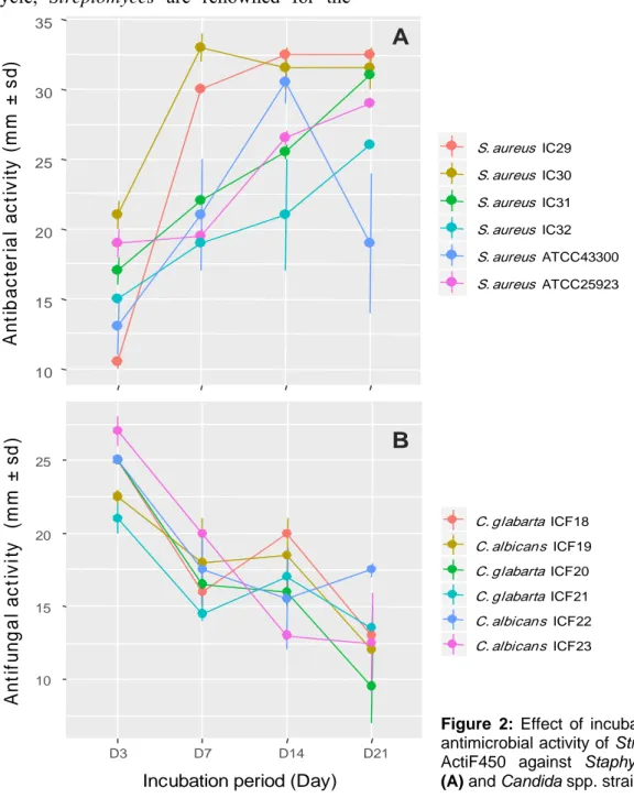
As shown in Figure 2a, the antimicrobial activity of the strain ActiF450 against select-ed S. aureus isolates startselect-ed after two days of incubation and reached a maximum after seven days of culture in ISP2. These activi-ties were persistent until the end of the incu-bation. Furthermore, the anticandidal activity was observed at the first four days of incuba-tion. However, the highest activity was rec-orded at the third day of incubation period. Thereafter, the activity dramatically declined after one week (Figure 2b).
Streptomyces castelarensis DSM40830T (AY508511)
Streptomyces mordarskii NRRL B-1346T (EF408735)
Streptomyces antimycoticus NBRC12839T(AB184185)
Streptomyces sporoclivatus NBRC100767T(AB249934)
Streptomyces geldanamycininus NRRLB3602T(DQ334781)
Streptomyces melanosporofaciens DSM40318T(FNST01000002)
Streptomyces yatensis NBRC101000T(AB249962)
Streptomyces solisilvae HNM0141T(KY366443) Streptomyces sp. ActiF450
Streptomyces malaysiensis NBRC16446T(AB249918)
Streptomyces samsunensis M1463T(EU077190)
Streptomyces indonesiensis DSM41759T(DQ334783)
Streptomyces rhizosphaericus NBRC100778T(AB249941)
Streptomyces griseiniger NRRLB1865T(AJ391818)
Streptomyces asiaticus A14P1T(AJ391830)
Streptomyces cangkringensis D13P3T(AJ391831)
Streptomyces hygroscopicus NBRC13472T(BBOX01000593)
Streptomyces demainii NRRLB-1478T(DQ334782)
Streptomyces albiflaviniger NRRLB-1356T(AJ391812)
Streptomyces yogyakartensis NBRC100779T(AB249942)
Streptomyces javensis NBRC100777T(AB249940)
Streptomyces violaceusniger NBRC13459T(AB184420)
Streptomyces iranensis HM35T(FJ472862)
Streptomyces rapamycinicus NRRLB-5491T(EF408733)
Streptomyces celluloflavus NRRLB-2493T(JOEL01000102)
Streptomyces kasugaensis BCRC12349T(NDXL01000004)
Streptomyces abikoensis NBRC13860T(AB184537)
Streptomyces lacticiproducens GIMN4.001T(GQ184344)
Streptomyces morookaense LMG20074T(AJ781349)
Streptomyces milbemycinicus NRRL5739T(EU170126)
Streptomyces aldersoniae NRRL18513T(EU170123)
Streptomyces cuspidosporus NBRC12378T(AB184090)
Streptomyces sparsogenes ATCC25498T(MAXF01000077)
Streptosporangium roseum DSM43021T(NR074558) 98 100 55 93 56 92 69 83 98 76 87 50 95 61 91 85 87 63 53 64 99 52 68 0.01 B C A
Figure 1: Neighbor-joining tree based on 16S RNA gene sequences of Streptomyces sp. ActiF450
and closely related species. Numbers at the nodes indicate levels of bootstrap support based on an analysis of 1000 re-sampled datasets. The scale bar corresponds to 0.01 substitutions per nucleotide position (A). Morphological characters of colony (B), and cell morphology (C) of the strain ActiF450. The strain ActiF450 was grown on SFM medium and the aerial mycelia morphology was viewed using optical microscopy (x100).
Table 1: Antimicrobial activity of Streptomyces sp. ActiF450
Test microorganism Activity mm (Day)
Yeast
Candida krusei ATCC6258 25.0±0.0 (D7)
Candida parapsilosis ATCC22019 20.0±0.0 (D7)
Candida albicans sp. ICF19 22.0±0.5 (D3)
Candida albicans sp. ICF22 25.0±0.0 (D3)
Candida albicans sp. ICF23 27.0±1.0 (D3)
Candida albicans sp. ICF24 15.0±0.0 (D7)
Candida albicans sp. ICF35 19.3±1.5 (D3)
Candida albicans sp. ICF37 18.0±2.6 (D3)
Candida albicans sp. ICF38 15.6±1.1 (D7)
Candida glabarta sp. ICF18 25.0±0.0 (D3)
Candida glabarta sp. ICF20 25.0±0.0 (D3)
Candida glabarta sp. ICF21 21.0±1.0 (D3)
Saccharomyces sp. ICF43 17.0±0.0 (D7)
Kluveromyces sp. ICF44 25.6±6.2 (D3)
Rhodotorula mucilaginosa YA1 67.6±2.7 (D7)
Mould
Arthroderma vanbreuseghemii ICF62B 49.2±2.8 (D7)
Aspergillus fumigatus MA1 14.9±2.0 (D7)
A. calidoustus A5 20.9±2.0 (D7) A. niger IAF27 45.0±6.0 (D7) A. niger MA2 54.0±6.0 (D7) Aspergillus sp. ICF58V 35.0±0.0 (D7) Fusarium oxysporum F15 41.2±7.4 (D7) F. solani 19.9±4.0 (D7)
Lichtheimia corymbifera ST87 Not detected
Lomentospora prolificans ST67 Not detected
Microsporum canis ICF58B 55.0±0.0 (D7)
Pecilomyces variotii 30.0±6.0 (D7)
Penicillium chrysogenum ICF 59 41.1±5.9 (D7)
Scedosporium apiospermum 28.0±0.0 (D7)
Scopulariopsis brevicaulis ICF57 15.3±1.1 (D7)
Scodapulariopsis candida ICF53 35.0±2.5 (D7)
Bac
teria
Staphylococcus aureus ATCC25293 35.5±3.5 (D10)
S. aureus ATCC43300 26.5±0.5 (D10) S. aureus IC13 30.0±0.0 (D7) S. aureus IC29 30.0±0.5 (D10) S. aureus IC30 32.5±0.5 (D10) S. aureus IC31 33.0±1.0 (D7) S. aureus IC32 31.0±0.0 (D14) S. aureus IC33 21.0±0.5 (D14) S. aureus IC34 31.5±4.0 (D10)
Several reports highlighted the produc-tion of various antimicrobial compounds by
Streptomyces strains with different and
vari-able activities depending on culture condi-tions including; media nature and/or compo-sition (nitrogen and carbon sources), time of incubation even the indicator organisms used in the confrontation test (Vijayakumar et al., 2012).
The differences in susceptibility among tested organisms are in concordance with previous studies by the fact that fungal or-ganisms are eukaryotic, and their structures are different from bacteria (Benhadj et al., 2019). The absence of acidic phospholipids and presence of sterols may reduce suscepti-bility of eukaryotic cells to lytic molecules (Alan and Earle, 2002). The maximum anti-bacterial activity after seven to ten days of
incubation may be attributed to the fact that ActiF450 reached the stationary phase of growth. It has been reported that the bioac-tive metabolites production by Streptomyces takes place in the stationary phase of the growth (Augustine et al., 2004) and the de-crease in the anticandidal activity after three days can be attributed to the decrease in the supply of nutrients or the accumulation of toxic substances.
Members of the genus Streptomyces have attracted extensive attention due to the tre-mendous success of their natural products in practical application. With their complex life cycle, Streptomyces are renowned for the
production of an outstandingly large number of bioactive metabolites and accounts for 80 % of the currently available antibiotic-like substances including antifungals and an-tibacterials (Niu et al., 2016). However, few studies related to the antimicrobial activities of Streptomyces malaysiensis, or the closest strains (S. samsunensis and S. solisilvae) were attempted so far. Our results indicate that ActiF450 could be a potential strain for the development of antimicrobial drugs against a wide range of human pathogenic bacteria and fungi including Candida-like species and dermatophytes (Figure 3).
10 15 20 25 30 35 D3 D7 D14 D21 Tested day A n ti b a c te ria l a c ti v it y ( m m ± s d) S. aureus IC29 S. aureus IC30 S. aureus IC31 S. aureus IC32 S. aureus ATCC43300 S. aureus ATCC25923 10 15 20 25 D3 D7 D14 D21
Incubation period (Day)
A n ti fu n g a l a c ti v it y (m m ± s d ) C. glabarta ICF18 C. albicans ICF19 C. glabarta ICF20 C. glabarta ICF21 C. albicans ICF22 C. albicans ICF23
B
A
Figure 2: Effect of incubation period on
antimicrobial activity of Streptomyces sp. ActiF450 against Staphylococcus spp.
Aspergillus fumigatus
Fusarium oxyporum Rodoturella mucilaginosa
Fusarium solani Arthroderma vanbreuseghemii Penicillium chrysogenum
Scopulariopsis brevicaulis Aspergillus niger Figure 3: Antifungal activity of Streptomyces sp. ActiF450 on the growth of different test organisms.
The active strain ActiF450 was inoculated as a spot in the center of ISP2 plates at 30 °C for 7 days. After, the plates were then covered by 10 ml of PDA pre-viously inoculated with target fungi.
Extraction of bioactive molecules
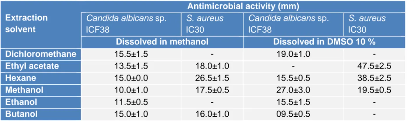
As an effort to study the antimicrobial potential of the strain, assays were conducted on the different extracts of ActiF450 against
Candida albicans sp. ICF23 and S. aureus
IC30 (Table 2). Traditionally, the discovery of bioactive molecules from microbial sources such as actinomycetes has generally involved, cultivation under different growth conditions, screening of biological activities, extraction of the metabolites, and analysis of the extract for bioactivity. The results re-flected that the ethyl acetate, hexane and methanol extracts dissolved in DMSO at 10 % exhibited potent activity against both
C. albicans ICF23 and S. aureus IC30. Other
extracts showed moderate activity. In addi-tion, significant differences in susceptibility were noted; where S. aureus was high sus-ceptible to ActiF450 extracts compared to C.
albicans.
This fact might indicate the existence of a synergistic effect among different metabo-lites produced by ActiF450 which is lost when they are separated during the extrac-tion (Genilloud, 2017). Otherwise, the anti-fungal activity might be related to the pres-ence of proteins or enzymes labile to hydro-philic solvents such as chitinases and glu-canases capable of degrading the cell wall of the fungi (Benhadj et al., 2019). Studies
re-viewed by Genilloud (2017) show that acti-nomycetes continue to be a valuable source of antibiotic activity as they produce a broad spectrum of different antifungal and antibac-terial compounds. Moreover, study results indicate a marked effect of the extraction solvents on the isolation of bioactive com-pounds. It has also been reported that organic solvents provides a higher efficiency in ex-tracting compounds for antimicrobial activi-ties (Lima-Filho et al., 2002).
Previous study on Streptomyces
malay-siensis MJM1968 reported a strong
inhibito-ry activity in vitro by dual-culture in vitro assay on several phytopathogenic fungi such as Alternaria mali, Cladosporium
cladospor-ioides, Colletotrichum gloeosporcladospor-ioides, Fusarium chlamydosporum, Fusarium ox-ysporum, Rhizoctonia solani (Cheng et al.,
2010). In addition, the compound, which ex-hibited antifungal activity, was identified as Azalomycin F complex.
Recently, Zhang et al. (2019) have re-ported the effect of different fermentation factors for Azalomycin F production from S.
malaysiensis strain ECO00002. Similarly, Li
et al. (2008) have also reported another promising antifungal activity of perhydro-furopyran C-nucleoside named Malayamycin isolated from Streptomyces malaysiensis ATB-11 (Al-Tai et al., 1999).
Table 2: Effect of extraction solvent on antimicrobial activity of ActiF450
Extraction solvent Antimicrobial activity (mm) Candida albicans sp. ICF38 S. aureus IC30 Candida albicans sp. ICF38 S. aureus IC30
Dissolved in methanol Dissolved in DMSO 10 %
Dichloromethane 15.5±1.5 - 19.0±1.0 - Ethyl acetate 13.5±1.5 18.0±1.0 - 47.5±2.5 Hexane 15.0±0.0 26.5±1.5 15.5±0.5 38.5±2.5 Methanol 10.0±1.0 17.5±0.5 27.0±3.0 19.5±0.5 Ethanol 11.5±0.5 - 15.5±1.5 - Butanol 15.0±1.0 16.0±1.0 09.5±0.5 -
CONCLUSION
The present study provides evidence that Algerian wetlands are a rich and valuable re-source of potentially active actinomycetes. Concisely, this study highlighted for the first time the antimicrobial potentials of wetland-associated actinobacteria with the ability to suppress major human fungal and bacterial pathogens in vitro. Further studies are need-ed to enhance isolation and selection of acti-nomycetes from unexplored ecosystems for antibiotic discovery. Hopefully, these new agents will meet the challenges as we at-tempt to manage serious underlying infection diseases. In addition, knowledge of the ac-tinobacteria gene clusters may provide im-portant answers toward understanding the metabolites biosynthetic pathway.
Disclosure of interest
The authors declare no conflicts of inter-est regarding the publication of this study. Funding sources
This research did not receive any specific grant from funding agencies in the public, commercial, or not-for-profit sectors.
REFERENCES
Alan AR, Earle ED. Sensitivity of bacterial and fun-gal plant pathogens to the lytic peptides, MSI-99, ma-gainin II, and cecropin B. Mol Plant Microbe Interact. 2002;15:701-8.
Albright JC, Goering AW, Doroghazi JR, Metcalf WW, Kelleher NL. Strain-specific proteogenomics accelerates the discovery of natural products via their biosynthetic pathways. J Ind Microbiol Biotechnol. 2014;41:451-9.
Al-Tai A, Kim B, Kim SB, Manfio GP, Goodfellow M. Streptomyces malaysiensis sp. nov., a new strep-tomycete species with rugose, ornamented spores. Int J Syst Evol Microbiol. 1999;49:1395-402.
Antinori S, Corbellino M, Parravicini C. Challenges in the diagnosis of invasive fungal infections in im-munocompromised hosts. Curr Fung Infect Rep. 2018;12: 12-22.
Augustine SK, Bhavsar SP, Baserisalehi M, Kapadnis BP. Isolation, characterization and optimization of an-tifungal activity of an actinomycete of soil origin. In-dian J Exp Biol. 2004;42:928-31.
Badji B, Riba A, Mathieu F, Lebrihi A, Sabaou N. Activité antifongique d'une souche d'Actinomadura d'origine saharienne sur divers champignons patho-gènes et toxinopatho-gènes. J Mycol Méd. 2005;15:211-9. Benammar L, Menasria T, Chergui A, Benfiala S, Ayachi A. Indoor fungal contamination of traditional public baths (Hammams). Int Biodeter Biodegrad. 2017;117C:115-22.
Benhadj M, Gacemi-Kirane D. Les actinomycètes: source de biomolécules d'intéret. Sarrebruck: Éditions universitaires européennes, 2016.
Benhadj M, Gacemi-Kirane D, Toussaint M, Hotel L, Bontemps C, Duval RE, et al. Diversity and antimi-crobial activities of Streptomyces isolates from Fet-zara Lake, north eastern Algeria. Ann Biol Clin. 2018; 76: 81-95.
Benhadj M, Gacemi-Kirane D, Menasria T, Guebla K, Ahmane Z. Screening of rare actinomycetes isolated from natural wetland ecosystem (Fetzara Lake, north-eastern Algeria) for hydrolytic enzymes and antimi-crobial activities. J King Saud Univ Sci. 2019;31:706-12.
Bentley SD, Chater KF, Cerdeño-Tárraga AM, Chal-lis GL, Thomson NR, James KD, et al. Complete ge-nome sequence of the model actinomycete
Streptomy-ces coelicolor A3(2). Nature. 2002;417(6885):141-7.
Boukoucha M, Menasria T, Bouguerra N. Phenotypic characterization and genotypic subtyping of
Salmo-nella enterica Serovars enteritidis and Gallinarum
iso-lated from human and poultry-reiso-lated samples. Food Biotechnol. 2018;32:206-21.
Casadevall A. Fungal diseases in the 21st century: the near and far horizons. Pathog Immun. 2018;3:183-96. Chaudhary HS, Yadav J, Shrivastava AR, Singh S, Singh AK, Gopalan N. Antibacterial activity of acti-nomycetes isolated from different soil samples of Sheopur (A city of central India). J Adv Pharm Tech-nol Res. 2013;4:118-23.
Cheng J, Yang SH, Palaniyandi SA, Han JS, Yoon TM, Kim TJ, et al. Azalomycin F complex is an anti-fungal substance produced by Streptomyces
malay-siensis MJM1968 isolated from agricultural soil. J
Korean Soc Appl Biol Chem. 2010;53:545-52. Chun J, Lee JH, Jung Y, Kim M, Kim S, Kim BK, et al. EzTaxon: a web-based tool for the identification of prokaryotes based on 16S ribosomal RNA gene se-quences. Int J Syst Evol Microbiol. 2007;57:2259-61. de Lima Procópio RE, da Silva IR, Martins MK, de Azevedo JL, de Araújo JM. Antibiotics produced by
Streptomyces. Braz J Infect Dis. 2012;16:466-71.
Genilloud O. Actinomycetes: still a source of novel antibiotics. Nat Prod Rep. 2017;34:1203-32.
Kelly KL, Judd DB. ISCC-NBS color-name charts il-lustrated with centroid colors. Washington, DC: U.S. National Bureau of Standards, 1964 (Supplement to National Bureau of Standards circular, Vol. 553). Kieser T, Bibb MJ, Buttner MJ, Chater KF, Hopwood DA. Practical Streptomyces genetics. Norwich: John Innes Foundation, 2000.
Komaki H, Sakurai K, Hosoyama A, Kimura A, Iga-rashi Y, Tamura T. Diversity of nonribosomal peptide synthetase and polyketide synthase gene clusters among taxonomically close Streptomyces strains. Sci Rep. 2018;8(1):1-11.
Li W, Csukai M, Corran A, Crowley P, Solomon PS, Oliver RP. Malayamycin, a new streptomycete anti-fungal compound, specifically inhibits sporulation of
Stagonospora nodorum (Berk) Castell and Germano,
the cause of wheat glume blotch disease. Pest Manag Sci. 2008;64:1294-302.
Lima-Filho JVM, Carvalho AF, Freitas SM, Melo VM. Antibacterial activity of extracts of six macroal-gae from the northeastern Brazilian coast. Braz J Mi-crobiol. 2002;33:311-4.
Liu N, Tu J, Dong G, Wang Y, Sheng C. Emerging new targets for the treatment of resistant fungal infec-tions. J Med Chem. 2018;61:5484-511.
LPSN. List of Prokaryotic names with Standing in Nomenclature. Founded in 1997 by Jean P. Euzéby. 2019. http://www.bacterio.net/.
Mehalaine S, Belfadel O, Menasria T, Messaili A. Chemical composition and antibacterial activity of es-sential oils of three medicinal plants from Algerian semi-arid climatic zone. Phytothérapie. 2017;15:1-9. Menasria T, Samir TINE, Mahcene D, Benammar L, Megri R, Boukoucha M, et al. External bacterial flora and antimicrobial susceptibility patterns of
Staphylo-coccus spp. and Pseudomonas spp. isolated from two
household cockroaches, Blattella germanica and
Blat-ta orienBlat-talis. Biomed Environ Sci. 2015;28:316-20.
Menasria T, Aguilera M, Hocine H, Benammar L, Ayachi A, Si Bachir A, et al. Diversity and bio-prospecting of extremely halophilic archaea isolated from Algerian arid and semi-arid wetland ecosystems for halophilic-active hydrolytic enzymes. Microbiol Res. 2018;207:289-98.
Menasria T, Monteoliva-Sánchez M, Benammar L, Benhadj M, Ayachi A, Hacène H, et al. Culturable halophilic bacteria inhabiting Algerian saline ecosys-tems: a source of promising features and potentiali-ties. World J Microbiol Biotechnol. 2019;35(9):132. Merradi M, Kassah-Laouar A, Ayachi A, Heleili N, Menasria T, Hocquet D, et al. Occurrence of VIM-4 metallo-β-lactamase-producing Pseudomonas
aeru-ginosa in an Algerian hospital. J Infect Dev Ctries.
2019;13:284-90.
Niu G, Chater KF, Tian Y, Zhang J, Tan H. Special-ised metabolites regulating antibiotic biosynthesis in
Streptomyces spp. FEMS Microbiol Rev. 2016;40:
554-73.
Onaka H. Novel antibiotic screening methods to awaken silent or cryptic secondary metabolic path-ways in actinomycetes. J Antibiot. 2017;70:865-70. Richardson MD, Warnock DW. Fungal infection: di-agnosis and management. 4th ed. Hoboken, NJ: Wiley-Blackwell, 2012.
Saitou N, Nei M. The neighbor-joining method: a new method for reconstructing phylogenetic trees. Mol Bi-ol EvBi-ol. 1987;4:406-25.
Sazak A, Şahin N, Güven K, Işık K, Goodfellow M.
Streptomyces samsunensis sp. nov., a member of the Streptomyces violaceusniger clade isolated from the
rhizosphere of Robinia pseudoacacia. Int J Syst Evol Microbiol. 2011;61:1309-14.
Seipke RF, Kaltenpoth M, Hutchings MI.
Streptomy-ces as symbionts: an emerging and widespread
theme? FEMS Microbiol Rev. 2012;36:862-76. Supong K, Sripreechasak P, Tanasupawat S, Dan-wisetkanjana K, Rachtawee P, Pittayakhajonwut P. Investigation on antimicrobial agents of the terrestrial
Streptomyces sp. BCC71188. Appl Microbiol
Bio-technol. 2017;101:533-43.
Tamura K, Stecher G, Peterson D, Filipski A, Kumar S. MEGA6: molecular evolutionary genetics analysis version 6.0. Mol Biol Evol. 2013;30:2725-9.
Vijayakumar R, Panneerselvam K, Muthukumar C, Thajuddin N, Panneerselvam A, Saravanamuthu R. Optimization of antimicrobial production by a marine actinomycete Streptomyces afghaniensis VPTS3-1 isolated from Palk Strait, East Coast of India. Indian J Microbiol. 2012;52:230-9.
Vos P, Garrity G, Jones D, Krieg NR, Ludwig W, Rainey FA, et al. Bergey's manual of systematic bac-teriology. New York, NY: Springer-Verlag, 2009. Weisburg WG, Barns SM, Pelletier DA, Lane DJ. 16S ribosomal DNA amplification for phylogenetic study. J Bacteriol. 1991;173:697-703.
Zhang Q, Bai T, Li M, Shi Z, Yang Q, Peng R, et al. Optimization of seed culture conditions of S. malay-siensis for producing azalomycin F. Southwest China J Agric Sci. 2019;32:316-21.
Zhou S, Yang X, Huang D, Huang X. Streptomyces
solisilvae sp. nov., isolated from tropical forest soil.