HAL Id: hal-02637508
https://hal.inrae.fr/hal-02637508
Submitted on 27 May 2020HAL is a multi-disciplinary open access archive for the deposit and dissemination of sci-entific research documents, whether they are pub-lished or not. The documents may come from teaching and research institutions in France or abroad, or from public or private research centers.
L’archive ouverte pluridisciplinaire HAL, est destinée au dépôt et à la diffusion de documents scientifiques de niveau recherche, publiés ou non, émanant des établissements d’enseignement et de recherche français ou étrangers, des laboratoires publics ou privés.
Distributed under a Creative Commons Attribution| 4.0 International License
New cyclolignans from Origanum glandulosum active
against beta-amyloid aggregation
Abdelkader Basli, Jean-Claude Delaunay, Eric Pedrot, Stéphane Bernillon,
Khodir Madani, Jean-Pierre Monti, Jean-Michel Merillon, Mohamed Chibane,
Tristan Richard
To cite this version:
Abdelkader Basli, Jean-Claude Delaunay, Eric Pedrot, Stéphane Bernillon, Khodir Madani, et al.. New cyclolignans from Origanum glandulosum active against beta-amyloid aggregation. Records of Natural Products, ACG Publications, 2014, 8 (3), pp.208-216. �hal-02637508�
The article was published by Academy of Chemistry of Globe Publications www.acgpubs.org/RNP © Published 05/01/2014 EISSN: 1307-6167
Rec. Nat. Prod. 8:3 (2014) 208-216
New Cyclolignans from Origanum glandulosum Active
Against
ββββ
-amyloid Aggregation
Abdelkader Basli
1,2, Jean-Claude Delaunay
1, Eric Pedrot
1,
Stéphane Bernillon
3, Khodir Madani
2, Jean-Pierre Monti
1,
Jean-Michel Mérillon
1, Mohamed Chibane
4and Tristan Richard
1*1
GESVAB (EA 3675), ISVV, Université de Bordeaux 2, 33882 Villenave d’Ornon, France. 2
Université de Bejaia, Laboratoire 3BS, Béjaia, Algérie. 3
INRA - UMR 1332 BFP, Centre INRA de Bordeaux, Villenave d’Ornon, France. 4
Université AMO Bouira, Faculté des Sciences, Lab. de Technologie Alimentaire, Bouira, Algérie. (Received January 10, 2013; Revised January 29, 2014; Accepted March 18, 2014)
Abstract: Origanum glandulosum Desf is an endemic flavoring herb widely distributed in North Africa that is
commonly used in traditional medicine. This oregano species is rich in essential oils but little is known about its phenolic composition. In the present study, a crude extract of O. glandulosum was prepared in order to isolate and investigate its neuroprotective potential to inhibit β-amyloid peptide (Aβ) aggregation. The three major compounds of the extract were isolated: rosmarinic acid and two cyclolignans in Origanum genus, globoidnan A and a new derivative named globoidnan B. Rosmarinic acid and globoidnan A showed significant anti-aggregative activity against β amyloid aggregation (IC50 7.0 and 12.0 µM, respectively). In contrast, globoidnan
B was found to be less active.
Keywords: Origanum glandulosum; oregano; cyclolignans; β-Amyloid aggregation; NMR. ©2014 ACG
Publications. All rights reserved.
1. Introduction
Origanum genus is widely distributed throughout the Mediterranean region and numerous species, subspecies and hybrids have been described [1,2]. Belonging to the Origanum genus, O. glandulosum Desf is an endemic widely distributed taxon in North Africa (Algeria and Tunisia) that is commonly used as a spice as well as in traditional medicine. O. glandulosum Desf is rich in essential oils, but little is known about its phenolic composition. To our best knowledge, phenolic constituents of O. glandulosum extract have not been previously investigated by modern analytical methods.
The genus Origanum is rich in essential oils and polyphenols that have antimicrobial, antioxidant and other bioactive properties [3-5]. Several studies indicate that polyphenols present in high amounts in natural products could play a preventive role in the incidence of age-related neurological disorders. These findings have been supported by epidemiological studies [6,7] and confirmed by in vitro and in vivo studies [8,9]. With regard to Alzheimer’s disease (AD), Hamaguchi et al. have shown that rosmarinic acid inhibits β-amyloid peptide (Aβ) aggregation in vitro and
*
Basli et.al., Rec. Nat. Prod. (2014) 8:3 208-216 209
in vivo [10]. Rosmarinic acid derivatives, which are plant secondary metabolites belonging to the class of hydroxycinnamic acid esters, are prominent constituents of the Origanum species [4,5,11]. Thus oregano extracts could be of therapeutic value in AD prevention. AD is the most common type of senile dementia and is characterized by the abundance of extracellular deposition of Aβ. Because Aβ aggregation promotes pro-inflammatory responses and activates neurotoxic pathways leading to dysfunction and death of brain cells [12], many researchers favor therapeutic approaches that target formation, deposition and clearance of Aβ fibrils [10,13-18].
The present study was undertaken to (i) identify and quantify the major phenolic constituents of O. glandulosum extract by high-performance liquid chromatography (HPLC), mass spectrometry (MS) and NMR analysis; (ii) evaluate in vitro activity of the extract on Aβ aggregation in AD prevention; (iii) evaluate the inhibitory potential of the pure compounds against Aβ fibril formation.
2. Materials and Methods
2.1. Reagents and standards
The Aβ (25-35) (Aβ25-35) peptide sequence corresponding to residues of the human wild type
sequence was purchased from Bachem California (Torrance, CA). Except for petroleum ether, which was of synthesis grade, all organic solvents used were of HPLC grade. Methanol and ethanol were purchased from Carlo Erba (Val de Reuil, France). Acetonitrile, ethyl acetate and petroleum ether were purchased from Scharlau Chemie (Sentmenat, Spain). Curcumin (Cur) and trifluoroacetic acid (TFA) were purchased from Sigma–Aldrich (St. Louis, MO, USA). Water was purified using the Elga (Bucks, UK) water purification system with a resistivity of no less than 18 MΩ/cm. NMR experiments were performed in deuterated acetone-d6 purchased from Euriso-top (Gif-sur-Yvette, France).
2.2 Plant materiel
The material consisted of the aerial part of Algerian Origanum glandulosum. Samples were collected from Boukhlifa in the city of Bejaia (north-eastern Algeria), when the plant is highly present. Botanical identification was made on a herbarium specimen by the Botany Laboratory of Bejaia University (Bejaia, Algeria). The samples were well dried in the shade and away from moisture, then ground into powder for analysis.
2.3 Extraction of plant samples
A portion of the finely powdered material from Origanum glandulosum (100 g) was first extracted three times with 70% methanol (MeOH, 2 L) during a 24-h period at ambient pressure and temperature. After removal of MeOH under reduced pressure at 35°C, the aqueous phase was defatted with petroleum ether (0.8 L) and then extracted with ethyl acetate (EtOAc). Extraction was carried out until a colorless extract was obtained. The EtOAc solutions were concentrated and lyophilized.
2.4 Instrumentation
The lyophilized extract was dissolved in 50% methanol and chromatographed using HPLC-DAD. The chromatography apparatus, an Agilent 1200 from Agilent Technologies (Santa Clara, CA, USA), was equipped with a degasser, a binary pump, an autosampler module, a column heater/selector and a UV–visible-DAD detector from the same provider. The Origanum extract was passed through a 0.45 µm Millipore membrane before chromatographic separation at 23°C with a 250 × 4 mm i.d., 5 µm, Prontosil 120-5-C18-AQ reverse-phase column, Bischoff (Leonberg, Germany). Water 0.1% TFA (solvent A) and acetonitrile 0.1% TFA (solvent B) were used as mobile phases. The gradient elution program was as follows (v/v): 0 min 10% B, 5 mn 10% B, 10 mn 15% B, 60 mn 22% B, 62 mn 100% B linear for 10 min, followed by 10 min for re-equilibration. The detection wavelengths were set at 260 and 320 nm. This HPLC was coupled to an Esquire 3000+ ion trap mass spectrometer using an ESI source from Bruker Daltonics (Billerica, MA, USA). The HPLC output
flow was split with a passive splitter with an average 1:100 ratio depending on the flow solvent, viscosity and rate. Nitrogen drying gas was set at 9.0 L/min and 350°C, nebulizer pressure was set at 27 psi. ESI–MS parameters (positive mode): HV capillary – 4100 V, end plate offset – 500 V, capillary exit 134.3 V, skimmer 40 V, trap drive 59.3, scan 25000 µs, rolling average 2 and trap averages 5.
Final purification was performed by semi-preparative HPLC (Varian, model ProStar 210 coupled with a diode array detector ProStar 335) with a Varian Microsorb 100 C18 Dynamax reverse-phase column (250 x 21.4 mm, 5 µm particle size) at room temperature. The flow rate was 15 mL/min. The solvent system and the gradient program used were the same as previously described. The eluted compounds were manually collected according to the visualization of the UV profile.
NMR spectra were recorded on a Bruker Avance 600 MHz spectrometer (600 MHz for 1H and 151 MHz for 13C experiments) at 300 K in the acetone-d6 deuterated solvent. Spectra were referenced
to the signal of acetone-d6 at δH 2.06 and δC 29.9 ppm.
Accurate masses were measured on a microTOF-Q mass spectrometer (Bruker, Bremen, Germany). Sample solutions were infused at 3µL/min. Electrospray ionization in negative mode was used. Each spectrum was calibrated with a 10 mM lithium formiate solution using enhanced cubic calibration model of microTOF control software (Bruker).
2.5 Measurement of inhibitory activity by UV–visible spectroscopy
Aβ25–35 peptide stock solution of 1 mM was prepared by solubilizing the lyophilized Aβ25–35
peptide in sterile water at 4°C, then by sonication for 1 min. The peptide stock solution was aliquoted and stored at – 20°C. All steps were carried out at 4°C to prevent Aβ polymerization.
The detailed method for measuring the inhibitory activity on Aβ aggregation was given in a previous report [19]. Briefly, UV–visible measurements were used to search for inhibitors of Aβ fibril formation. Stock solutions of Aβ25-35 at 1 mM were prepared by solubilizing the lyophilized Aβ by
vortexing briefly in sterile water at 4°C, then by sonicating for 10 min. Extract and pure polyphenols were solubilized in MeOH solution to a concentration of 1 mg/mL. Stock solutions were aliquoted and stored at – 20°C. UV–visible spectrometry was performed on a Cary 300 spectrophotometer (Varian, USA). To study Aβ fibril inhibition, experiments were carried out by using a reaction mixture containing 80 µL phosphate buffer (10 mM final concentration), 10 µL Aβ25-35 (100 µM final
concentration) and 10 µL tested compound, pH 7.2.
3. Results and Discussion
3.1. Structure elucidation
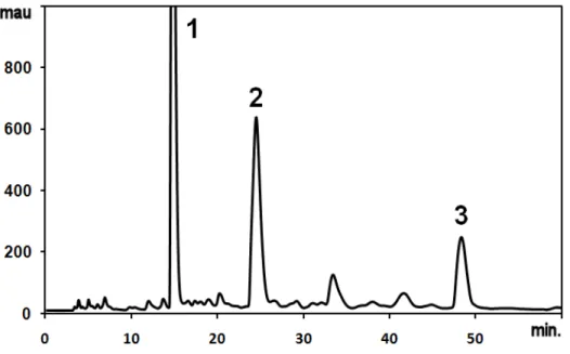
To assess whether the extracts contained a large number of different compounds, and in an attempt to identify these compounds, HPLC analysis was carried out with the EtOAc extract of O. glandulosum (Fig. 1). The analytical chromatogram of the extract presented three major peaks. The phenolic compounds corresponding to these peaks were isolated and their structures were elucidated by 1D- and 2D-NMR and MS data.
Basli et.al., Rec. Nat. Prod. (2014) 8:3 208-216 211
Figure 1. HPLC chromatogram of O. glandulosum extract.
Globoidnan B (1), peak tR = 15.1 min, 208 mg/100g fresh weight (FW), was obtained as a brown,
amorphous powder, UV(MeOH) λmax (log ε) 236 (3.23), 370 (4.26) nm (Fig. 2). The molecular
formula C27H22O12 was deduced from a quasi-molecular ion peak in the ESI-HRMS mass spectrum at
m/z 537.1033 [M–H]– (required 537.1038 for C27H21O12). The 1H- and 13C-NMR data (Table 1) are
consistent with a highly aromatized molecule, as the 13C-NMR chemical shifts suggested that 21 of the 27 carbons were aromatic. COSY and HMBC correlations identified resonances consistent with a 1,2,3-trisubstituted dihydronaphtalene-6,7-diol moiety (δC: 148.3, 144.7, 138.7, 131.5, 124.4, 122.6,
116.8, 116.7, 47.6, 45.9 ppm; δH: 7.61, 6.95, 6.67, 4.49, 3.92 ppm) as well as an alkylated 3,4-catechol
(δC: 145.3, 144.5, 128.6, 121.6, 117.2, 115.7, 74.0, 37.4 ppm; δH: 6.82, 6.73, 6.64, 5.15, 3.07, 3.05
ppm) and a mono-substituted 3,4-catechol (δC: 145.3, 145.0, 136.0, 119.3, 115.7, 115.2 ppm; δH: 6.68,
6.50, 6.45 ppm). These data, along with the observation of three carbonyl 13C resonances (δC: 173.2,
170.0, 166.5 ppm) consistent with the presence of ester and carboxylic acid parts, accounted for all of the available double bond equivalent.
Further analysis of the HMBC spectrum revealed several diagnostic correlations (Fig. 2). The HMBC correlations H-2′′/C-1 and H-6′′/C-1 as well as H-2/C-1′′ established that the mono-substituted 3,4-catechol was attached at the C-1 position of the 1,2,3-trisubstituted dihydronaphtalene-6,7-diol. The HMBC correlations H-2/C-9 and H-4/C-9 placed an ester carbonyl at C-3 of the 1,2,3-trisubstituted dihydronaphtalene-6,7-diol. The HMBC correlation H-1/C-10 was consistent with a carboxylic acid at the C-2 position, and hence established the substitution pattern of the 1,2,3-trisubstituted dihydronaphtalene-6,7-diol moiety as shown.
The point of attachment for the 3-(3,4-dihydroxyphenyl) lactic acid moiety to the 1,2,3-trisubstituted dihydronaphtalene-6,7-diol at C-3 was determined through the observed HMBC correlation H-8′/C-9 and confirmed by the NMR chemical shift data for C-8′ (δC: 74.0 ppm; δH: 5.15
ppm), which was consistent with an ester methine. Finally, the remaining carboxylic acid functionality have to be attached at position C-8′ as indicated by the HMBC correlation H-7′/C-9′, leading to the proposed structure.
The relative stereochemistry of compound 1 was determined by ROESY experiments and by coupling constant analysis. The relationship between H-1 and H-2 is trans on the basis of the small J value (2.1 Hz) between these two protons [20,21]. This relationship was confirmed by the NOE correlations between H-2/H-2′′ and H-2/H-6′′. The stereochemistry of C-8′ was deduced from comparison with literature [20,21].This compound is a novel ester composed of (3,4-dihydroxyphenyl) lactic acid and 1-(3,4-dihydroxyphenyl)-6,7-dihydroxy-1,2-dihydronaphthalene-2,3-dicarboxylic acid, named globoidnan B. Interestingly, this is the first report of this type of cyclolignan from Origanum genus.
Figure 2. Selective HMBC correlations of Globoidnan B (1).
Rosmarinic acid (RA) (2), peak tR = 24.4 min, 188 mg/100g fresh weight (FW), was obtained as a
white amorphous powder, UV(MeOH) λmax (log ε) 290 (3.32), 328 (3.42) nm (Fig. 3). The molecular
formula C18H16O8 was deduced from a quasi-molecular ion peak in the ESI-HRMS mass spectrum at
m/z 359.0768 [M–H]– (required 359.0772 for C18H15O8). Rosmarinic acid was identified by its
observed MS, HRMS, 1H- and 13C-NMR data (Table 1) and by comparison with the literature [21,22].
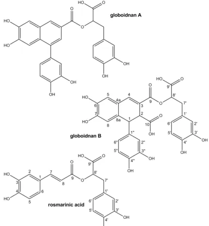
Figure 3. Structure of Globoidnan B (1), Rosmarinic acid (2) and Globoidnan A (3).
Globoidnan A (3), tR = 48.5 min, 120 mg/100g FW, was obtained as a brown amorphous powder,
UV(MeOH) λmax (log ε) 221 (4.12), 358 (3.56) nm. The molecular formula C26H20O10 was deduced
from a quasi-molecular ion peak in the ESI-HRMS mass spectrum at m/z 491.0983 [M–H]– (required 491.0984 for C26H19O10). The
1
H- and 13C-NMR data are summarized in Table 1. The NMR data were found to be very similar to Globoidnan B, except for the naphthalene moiety. COSY and HMBC correlations identified resonances consistent with a 1, 3-disubstituted naphthalene -6,7-diol moiety (δC:
Basli et.al., Rec. Nat. Prod. (2014) 8:3 208-216 213
149.3, 145.5, 139.5, 131.1, 128.9, 127.0, 124.9, 123.9, 112.1, 108.9 ppm; δH: 8.33, 7.70, 7.45, 7.36
ppm) as well as a mono-substituted 3,4-catechol and a 3-(3-4-dihydroxyphenyl) lactic acid moiety. HMBC correlations H-2′′/C-1 and H-6′′/C-1 as well as H-2/C-1′′ established that the monosubstituted 3,4-catechol was attached at the C-1 position, and the HMBC correlations H-2/C-9 and H-4/C-9 placed an ester carbonyl at C-3 of the 1,3-disubstituted naphthalene -6,7-diol. Finally, the HMBC correlation H-8′/C-9 indicated the attachment point between the 1,3-disubstituted naphthalene-6,7-diol and the 3-(3-4-dihydroxyphenyl) lactic acid moieties, leading to the proposed structure of globoidnan A . While this cyclolignan has been previously reported in Eucalyptus globoidea [23], this is the first report in the Origanum genus to our knowledge.
Table 1. 1H- and 13C-NMR data of Globoidnan B (1), Rosmarinic acid (2) and Globoidnan A (3). C 1, δC 1, δH, J(Hz) 2, δC 2, δH, J(Hz) 3, δC 3, δH, J(Hz) 1 45.9 4.49 d (2.1) 126.6 - 139.5 - 2 47.6 3.92 d (2.1) 114.1 7.17 d (2.1) 123.9 7.70 d (1.8) 3 122.6 - 145.3 - 127.0 - 4 138.7 7.61 s 147.8 - 128.9 8.33 d (1.8) 4a 124.4 - 124.9 - 5 116.7 6.95 s 115.1 6.86 d (8.1) 112.1 7.45 s 6 148.3 - 121.8 7.05 dd (2.1, 8.1) 149.3 - 7 144.7 - 145.8 7.56 d (16.0) 145.5 - 8 116.8 6.67 s 113.9 6.31 d (16.0) 108.9 7.36 s 8a 131.5 - 131.1 - 9 166.5 - 165.6 - 166.6 - 10 173.2 - 1′ 128.6 - 128.1 - 128.9 - 2′ 117.2 6.82 d (2.0) 116.3 6.85 d (2.1) 117.2 6.94 d (2.0) 3′ 145.3 - 144.6 - 145.5 - 4′ 144.5 - 143.7 - 144.6 - 5′ 115.7 6.73 d (8.1) 114.9 6.75 d (8.1) 115.8 6.79 d (8.1) 6′ 121.6 6.64 dd (2.0, 8.1) 120.6 6.67 dd (2.1, 8.1) 121.6 6.76 dd (2.0, 8.1) 7′ 37.4 3.05 dd (7.2, 14.4) 36.7 3.03 dd (8.5, 14.4) 37.5 3.17 dd (8.5, 14.4) 3.07 dd (5.2, 14.4) 3.13 dd (4.3, 14.4) 3.24 dd (4.3, 14.4) 8′ 74.0 5.15 dd (5.3, 7.2) 73.0 5.22 dd (4.3, 8.5) 73.9 5.39 dd (4.3, 8.5) 9′ 170.0 - 170.3 - 171.2 - 1′′ 136.0 - 133.0 - 2′′ 115.2 6.50 d (2.2) 117.6 6.99 d (2.1) 3′′ 145.3 - 145.4 - 4′′ 145.0 - 145.4 - 5′′ 115.7 6.68 d (8.1) 116.0 7.00 d (8.0) 6′′ 119.3 6.45 dd (2.2, 8.1) 128.9 6.82 dd (2.1, 8.0)
3.2. Neuroprotective activity of O. glandulosum extract
Given the central importance of Aβ aggregation in the pathogenesis of AD, particular interest is currently being shown in research aimed at therapeutic developments that target amyloid production, aggregation, clearance and toxicity. Reports that describe the effects of polyphenols on Alzheimer’s Aβ aggregation in vitro are already available. Using a routine in vitro assay based on UV–visible measurements and electron microscopy, we recently reported that resveratrol derivatives inhibit the aggregation of the peptide [19]. For this assay we used the Aβ25-35 peptide fragment that preserves the
properties of neurotoxicity and aggregation of the entire peptide [24]. Using this method, the activity of the EtOAc extract of Origanum glandulosum was examined. The O. glandulosum EtOAc was tested at concentrations between 0.01 to 5 µg/mL. The extract reduced Aβ fibril formation in a
dose-dependent manner (Fig. 4). The inhibitory properties of major phenolic constituents were evaluated to evaluate their influence in the anti-aggregative properties of the global extract.
Figure 4. Effect of ethyl acetate (EtOAc) extract of Origanum glandulosum on Aβ25–35 fibril inhibition. Reaction
mixtures containing 100 µM Aβ25–35, 10 mM phosphate buffer, pH 7.2, and various concentrations of EtOAc
extract were incubated at 15°C for 5 h. Means and SD of three independent experiments are shown. 3.3. Neuroprotective activity of major compounds
To evaluate the effect of the major phenolic compounds of the extract, a primary evaluation of the inhibition percentage of Aβ fibril formation was performed at a concentration of 10 µM. Its inhibitory effect was compared to that of curcumin as a standard [25]. The compounds exhibiting an inhibitory activity at least equal to that of curcumin were further analyzed to determine their IC50 values. The
inhibition percentages and the IC50 values are summarized in Table 2.
Table 2. Inhibition of Aβ25–35 fibrils formation.
Compound Inhibition (%) IC50 (µµµµM) Curcumin 43 ± 3 10 ± 2 Globoidnan A 40 ± 4 12 ± 2 Globoidnan B 20 ± 3 - Rosmarinic acid 53 ± 4 7 ± 2
Globoidnan A (IC50 12.0 µM) and RA (IC50 7.0 µM) exhibited efficient inhibition of Aβ
aggregation. Both exhibited inhibitory activity in the same order of magnitude as curcumin. These results are in agreement with those of Ono et al. indicating that curcumin, nordihydroguaiaretic acid and RA have similar anti-amyloidogenic activity [13]. In contrast, globoidnan B is less active. The activity of globoidnan B is two-fold lower than that of globoidnan A. Globoidnan A differs from B only by the presence of carboxylic acid, indicating a specific interaction between globoidnan A and Aβ.
RA is an ester of caffeic acid 3,4-dihydroxyphenyllactic acid and is commonly found in species of the Lamiaceae. It has several biological activities such as antioxidant, anti-inflammatory, antimutagenic, antibacterial, and antiviral [11]. In AD pathogenesis, numerous reports indicate that RA can modulate various processes. It reduces Aβ-induced neurotoxicity in PC12 cells including reactive oxygen species formation, lipid peroxidation, DNA fragmentation, caspase-3 activation, and tau (τ) protein hyperphosphorylation [26], and inhibits fibril formation [13, 18]. Globoidnan A is a cyclolignan found for the first time in the Origanum genus in this study. It was first isolated from Eucalyptus globoidea and reported to inhibit HIV integrase [23]. Our results show its ability to inhibit the progress of Aβ aggregation to a degree almost comparable to that of curcumin.
Basli et.al., Rec. Nat. Prod. (2014) 8:3 208-216 215
RA and globoidnan A are the main compounds responsible for the results obtained in this work. It is very difficult to compare the inhibition activities of the pure compounds with that of the whole extract. Taking HPLC analysis into account, a quantification of the compounds within the extract indicates that RA, globoidnans A and B represent 23, 15 and 26% respectively of the whole extract. The highest tested concentration of the extract is 5 µM (Inhibition 45%) which corresponds to 0.4, 0.4 and 0.7 µg/mL of RA, globoidnans A and B, respectively in the extract. Pure compound were tested at 10 µM. This concentration corresponds to 4.9 µg/mL of globoidnan A (Inhibition 40%). Therefore, the inhibitory activity of the extract is more pronounced than that of the pure compound. This finding could be attributed to other compounds not extracted or to an additive/synergistic effect.
4. Conclusions
To conclude, using 1D-and 2D NMR and MS analyses the major constituents of O. glandulosum extract were determined to be rosmarinic acid, a known lignan globoidnan A and a new lignan globoidnan B. Rosmarinic acid is prominent constituent of Origanum species. However, this is the first report of the presence of cyclolignans in this genus. Although globoidnan A has been previously identified in Eucalyptus globoidea, globoidnan B is a new cyclolignan derivative isolated from nature for the first time in this study. Interestingly the present findings suggest that O. glandulosum extract and constituents inhibit Aβ fibrils formation.The high concentration of RA and cyclolignan derivatives may at least partially explain the anti-aggregative potential of the extract as proved by our experimental results.
Acknowledgments
The authors thank the PROFAS program for financial support. NMR and MS experiments were undertaken at the Metabolome Facility of Bordeaux Functional Genomic Center (Bordeaux, France).
References
[1] J.H. Ietswaart (1980). A taxonomic revision of the genus Origanum (Labiatae), Le Hague: Leiden Botanical Series 4, Leiden University Press.
[2] M. Skoula and J.B. Harborne (2002). Taxonomy and Chemistry of Oreganum. In: Oregano: The genera Origanum and Lippia, ed: S.E. Kintzios, Taylor & Francis (CRC Press), New York, pp. 67-108.
[3] F.E. Babili, J. Bouajila, J.P. Souchard, C. Bertrand, F. Bellvert, I. Fouraste, C. Moulis and A. Valentin (2011). Oregano: Chemical analysis and evaluation of its antimalarial, antioxidant, and cytotoxic activities, J. Food Sci. 76, C512-C518.
[4] V. Exarchou, N. Nenadis, M. Tsimidou, I.P. Gerothanassis, A. Troganis and D. Boskou (2002). Antioxidant activities and phenolic composition of extracts from Greek Oregano, Greek Sage, and Summer Savory, J. Agric. Food Chem. 50: 5294-5299.
[5] V. Exarchou, M. Godejohann, T.A. van Beek, I.P. Gerothanassis and J. Vervoort (2003). LC UV-Solid-Phase Extraction-NMR-MS combined with a cryogenic flow probe and its application to the identification of compounds present in Greek Oregano, Anal. Chem. 75, 6288-6294.
[6] J.M. Orgogozo, J.F. Dartigues, S. Lafont, L. Letenneur, D. Commenges, R. Salamon, S. Renaud and M.B. Breteler (1997). Wine consumption and dementia in the elderly: A prospective community study in the Bordeaux area, Nat. Rev. Neurol. 153, 185-192.
[7] V. Deschamps, P. Barberger-Gateau, E. Peuchant and J.M. Orgogozo (2001). Nutritional factors in cerebral aging and dementia: epidemiological arguments for a role of oxidative stress, Neuroepidemiology
20, 7-15.
[8] C. Ramassamy (2006). Emerging role of polyphenolic compounds in the treatment of neurodegenerative diseases: A review of their intracellular targets, Eur. J. Pharmacol. 545, 51-64.
[9] A. Ebrahimi and H. Schluesener (2012). Natural polyphenols against neurodegenerative disorders: potentials and pitfalls, Ageing Res. Rev. 11, 329-345.
[10] T. Hamaguchi, K. Ono, A. Murase and M. Yamada (2009). Phenolic compounds prevent Alzheimer's pathology through different effects on the amyloid-β aggregation pathway, Am. J. Pathol. 175, 2557-2565.
[12] C. Pereira, P. Agostinho, P.I. Moreira, S.M. Cardoso and C.R. Oliveira (2005). Alzheimer's Disease-associated neurotoxic mechanisms and neuroprotective strategies. Curr. Drug Targets 4, 383-403. [13] K. Ono, K. Hasegawa, H. Naiki and M. Yamada (2004). Curcumin has potent anti-amyloidogenic effects
for Alzheimer's β-amyloid fibrils in vitro. J. Neurosci. Res. 75, 742-750.
[14] Y. Porat, A. Abramowitz and E. Gazit (2006). Inhibition of amyloid fibril formation by polyphenols: structural similarity and aromatic interactions as a common inhibition mechanism. Chem. Biol. Drug Des.
67, 27-37.
[15] C. Rivière, T. Richard, X. Vitrac, J.M. Mérillon, J. Valls and J.P. Monti (2008). New polyphenols active on β-amyloid aggregation. Bioorg. Med. Chem. Lett. 18, 828-831.
[16] S. Bastianetto, Y. Dumont, Y. Han and R. Quirion (2009). Comparative neuroprotective properties of stilbene and catechin analogs: Action via a plasma membrane receptor site CNS Neurosci. Ther. 15, 76-83.
[17] T. Richard, P. Poupard, M. Nassra, Y. Papastamoulis, M.L. Iglésias, S. Krisa, P. Waffo-Teguo, J.M. Mérillon and J.P. Monti (2011). Protective effect of ε-viniferin on β-amyloid peptide aggregation investigated by electrospray ionization mass spectrometry. Bioorg. Med. Chem. 19, 3152 3155.
[18] K. Ono, L. Li, Y. Takamura, Y. Yoshiike, L. Zhu, F. Han, X. Mao, T. Ikeda, J.I. Takasaki, H. Nishijo, A. Takashima, D.B. Teplow, M.G. Zagorski and M. Yamada (2012). Phenolic compounds prevent amyloid
β-protein oligomerization and synaptic dysfunction by site specific binding. J. Biol. Chem. 287, 14631-14643.
[19] C. Rivière, T. Richard, L. Quentin, S. Krisa, J.M. Mérillon and J.P. Monti (2007). Inhibitory activity of stilbenes on Alzheimer's β-amyloid fibrils in vitro. Bioorg. Med. Chem. 15, 1160-1167.
[20] H. Tazaki, D. Taguchi, T. Hayashida and K. Nabeta (2001). Stable isotope labeling studies on oxidative coupling of caffeic acid via o-quinone. Biosci. Biotechnol. Biochem. 65 , 2613-2621.
[21] M. Wang, J. Li, M. Rangarajan, Y. Shao, E.J. LaVoie, T.C. Huang and C.T. Ho (1998). Antioxidative Phenolic Compounds from Sage (Salvia officinalis). J. Agric. Food Chem. 46, 4869-4873.
[22] Y. Lu and L.Y. Foo (1999). Rosmarinic acid derivatives from Salvia officinalis. Phytochem. 51, 91-94. [23] S.P.B. Ovenden, J. Yu, S.S. San Wan, G. Sberna, R.M. Tait, D. Rhodes, S. Cox, J. Coates, N.G. Walsh
and B.M. Meurer-Grimes (2004). Globoidnan A: A lignan from Eucalyptus globoidea inhibits HIV integrase. Phytochem. 65, 3255-3259.
[24] C.J. Pike, A.J. Walencewicz-Wasserman, J. Kosmoski, D.H. Cribbs, C.G. Glabe and C.W. Cotman (1995). Structure activity analyses of β-amyloid peptides: contributions of the β25-35 region to aggregation and neurotoxicity. J. Neurochem. 64, 253-265.
[25] C. Rivière, Y. Papastamoulis,, P.Y. Fortin, N. Delchier, S. Andriamanarivo, P. Waffo-Teguo, G.D.W.F. Kapche, H. Amira-Guebalia, J.C. Delaunay, J.M. Mérillon, T. Richard and J.P. Monti (2010). New stilbene dimers against amyloid fibril formation. Bioorg. Med. Chem. Lett. 20, 3441-3443.
[26] T. Iuvone, D. De Filippis, G. Esposito, A. D'Amico and A.A. Izzo (2006). The spice sage and its active ingredient Rosmarinic Acid protect PC12 cells from amyloid-beta peptide-induced neurotoxicity, J. Pharmacol. Exp. Ther. 317, 1143-1149.
© 2014 ACG Publications +