ORIGINAL PAPER
A scanning electron microscopy study of Argulus vittatus
(Rafinesque-Schmaltz, 1814) (Crustacea: Branchiura)
from Algerian coast
Djamila Ider&Zouhir Ramdane&Lucie Courcot& Rachid Amara&Jean-Paul Trilles
Received: 19 February 2014 / Accepted: 24 March 2014 / Published online: 12 April 2014 # Springer-Verlag Berlin Heidelberg 2014
Abstract A study of the Algerian Branchiura, Argulus vittatus (Rafinesque-Schmaltz, 1814) was conducted using the scanning electron microscopy (SEM). New morphological features are reported for the first time (mouth con, first max-illa, second maxmax-illa, structures and ornamentation of thoracic segments, structure of semen papillae, etc.). The morphology of small and large female specimens was compared. Two new hosts, Pagellus erythrinus L. and Sparus aurata L., are re-ported for this species. Until now, six host species were reported for A. vittatus, and stenoxenic specificity for Sparid fishes was observed for Algerian specimens. The biogeo-graphical distribution of this species seems to be restricted to the eastern coast of Algeria. Additional information about the host specificity, ecology, and life cycle of this parasitic species were given.
Keywords Argulus vittatus . Morphology . SEM . Life cycle . Algerian coast
Introduction
Branchiura, or carp lice, is a worldwide (except Antarctica) group of crustaceans parasitizing fresh and brackish water fishes, occasionally marine fishes (Rushton-Mellor1994; Møller and Olesen2010).
This class includes four genera, Argulus O.F. Müller, 1785, Chonopeltis Thiele, 1900, Dipteropeltis Calman, 1912, and Dolops Audouin, 1837 (Gruner 1996). According to Poly (2009), Argulus species occur in marine, estuarine, and fresh-water habitats, whereas the other three genera are found only in freshwater. Few species, Argulus foliaceus Linnaeus, 1758, A. japonicus Thiele, 1900, and Dolops ranarum (Stuhlmann, 1891) are well-known enough. However, most species remain until now more or less uninvestigated (Møller2009).
Monod (1928) described numerous new species of Argulus from Africa and provided identification keys. The genus Argulus O.F. Müller, 1785, including nearly 150 nominal species (Rushton-Mellor 1994), is the largest genus of the class Branchiura. This last contains in reality about 129 valid species (Poly 2009). However, several difficulties related to taxonomic features persist until now. For example, Tam (2005) reported that specimens belonging to the genus Argulus are the only species with a preoral spine. Møller and Olesen (2010) clearly confirmed that an inverted preoral spine is present in Dipteropeltis. They noted that the distal spine itself is not extended in this specimen, which is similar to specimens of Argulus. According to a phylogenic study, the same authors reported that maybe, the genera Argulus and Dipteropeltis have the same ancestor.
Branchiuran species were well-known in marine ecosys-tems (Monod1928; Cressey1978; Poly2009). From coasts near Algeria, Wilson (1902) reported a single specimen of Argulus purpureus (Risso, 1827) now a junior synonym of
D. Ider
:
Z. Ramdane (*)Laboratoire de Zoologie Appliquée et D’écophysiologie Animale, Université A/Mira-Faculté des Sciences, Targa Ouzemour, Béjaïa 06000, Algeria
e-mail: zohir22000@yahoo.fr L. Courcot
:
R. AmaraLaboratoire D’océanologie et Géosciences, UMR 8187, Université du littoral, 62930 Wimereux, France
J.<P. Trilles
UMR 5119 (CNRS-UM2-IFREMER-IRD), Equipe Adaptation Ecophysiologique et Ontogenèse, Université de Montpellier 2, CC. 092, Place E. Bataillon, 34095 Montpellier Cedex 05, France e-mail: jp.trilles@cegetel.net
A. vittatus (Rafinesque-Schmaltz, 1814) (Rushton-Mellor
1994; Poly1998; Walter and Boxshall2010). Few accurate descriptions of this species were performed by the author; the dorsal view was only figured. Since the oldest inaccurate studies conducted on this species (Risso1816,1826; Thorell
1864; Wilson 1902) one century ago, no additional studies were performed particularly on the morphology of its ventral side and appendages. Wilson (1902), collecting a dried spec-imen near the Algerian coast, considered doubtfully that this specimen belongs to the species A. purpureus, and the author reported that this species needs more description and draw-ings. Recently, a description of A. vittatus (Rafinesque-Schmaltz, 1814) with more precise drawings on the general morphology and appendages was published by Ramdane (2009) and Ramdane and Trilles (2012). However, many features of these appendages remain still unknown.
Recently, some ultrastructural studies on the four genera of Branchiura were performed. However, until now, the mor-phology and appendages of A. vittatus were not studied using the scanning electron microscopy (SEM). In the present study, accurate investigations using SEM were conducted on this species. The general morphology and the structure of append-ages between the small and large specimens were compared. Several ecological details were also recorded: the site where the specimens were collected, the fish hosts, the parasitic specificity, the parasitologic indexes, and the distribution of the species. Additional information on the ecology and the life cycle of this species were also reported.
Materials and methods
This study was conducted from January to December 2013 in the gulf of Béjaïa (eastern coast of Algeria). Host fishes were collected from the Béjaïa fishing port, transferred in the lab-oratory, and examined for Argulids. Parasites were removed from the hosts and preserved in 70 % ethanol. They were later cleared in 85 % lactic acid for 1 to 2 h before dissection in a drop of lactic acid on a wooden slide under a dissecting microscope. According to Cressey (1978), it may be necessary to clear heavily pigmented specimens in a warm solution of 10 % KOH for 15 min or more in order to study them microscopically. The body parts and appendages were mounted on slides and examined under a compound micro-scope. Drawings were made using a microscope with a camera lucida. Measurements were made with a micrometer eyepiece and given as means followed by the range in brackets. The date, the sampling area, the name, and the size of the host fish and the position of the parasite were noted. Parasitologic indexes (prevalence (P) and mean intensity (Im)) were calcu-lated according to Margolis et al. (1982) and Bush et al. (1997). The geographical distribution and host fishes are
specified. Host nomenclature and fish taxonomy are accord-ing to FishBase (Froese and Pauly2011).
For the scanning electron microscopy (SEM), six large and one small specimens (stored in 70 % ethanol) were dehydrated in a graded ethanol (Carlo Erba, Absolute, PA) series 80 % (1/2 h), 90 % (1/2 h), and 100 % (1/2 h). The specimens were immersed in hexamethyldisilazane (HMDS), Molekula) for 1/2 h (twice). After removing the excess of HMDS, the specimen was allowed to evaporate overnight under a fume hood. Finally, samples were mounted on aluminum stubs (Agar Scientific) with double sticky carbon tabs (Agar Scientific) and sputter coated under Argon flow with Au/Pd Polaron SC 7620 during 90s. Afterward, the specimens were examined with a SEM LEO 438 VP. After an initial round of observations in the SEM, an additional dissection was under-taken to reveal more details. Specimens used in this study were conserved in the collection of the laboratory of oceanol-ogy and geosciences, ULCO (France).
Results
Number of collected specimens 19
Sex of collected specimens, female (male unknown) Site of attachment, the right pectoral fin of hosts Hosts:
Boops boops (L., 1758) Sparus aurata (L., 1758) Pagellus erythrinus (L., 1758) Prevalence:
Boops boops (number of host, n=369; P=4.61 %) Sparus aurata (number of host, n=50; P=2 %) Pagellus erythrinus (number of host, n=34; P=5.88 %) Description
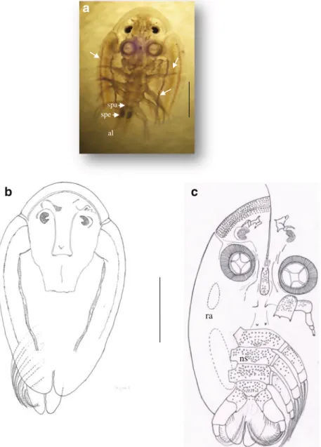
All collected specimens were females. Thoracic mean length =12.20 mm (3.9–17 mm). The abdomen size is 1/3 times of carapace with two lobes acuminate orbicular. The small and large specimens of A. vittatus show a general body shape elliptical with symmetrical sides (Figs. 1a, b, 2a, b, c, and
3c); cephalic region oval with spines observed ventrally and ciliary processes in the distal extremity (Fig.3a, b, c); and carapace longer than wide, usually 98 % of the total body length in adult specimens. In small specimens, the carapace is very translucent, and all thoracopods and compound eyes can be observed easily in dorsal view. The dorsal side of carapace of the small and large specimens is convex and white-blue with three violet lines, separated by three white lines in either lobe (Fig.2a); the posterior sinus reach half the total length of the carapace, and anterolateral sinuses are deep. Many single scales cover the thoracic ventral surface of small specimen (Figs.2cand3d). In large specimens, the scales are minute
and increase in number covering the entire ventral surface of thoracic segments and thoracopods (Fig.1a, i). The orbicular abdomen is covered by the carapace; the anal sinus reaches the middle of the abdomen (Fig.1a); the lobes are acuminate, orbicular. The spermathecae are well visible at the anterior part of the female abdomen (Fig.2a). The compound eyes are small and semi-lunar easily visible in dorsal view. A nauplius eye is also visible dorsally (Fig.2a, b). The ventral surface of the head is covered with many, small, regularly arranged spines of similar size (Fig.3a, b, c). Paired respiratory areas are visible on the ventral surface of the lobes; the anterior respiratory area is small and about 0.5 times size of the large
respiratory area (Fig.2c). The thorax of many specimens was distended with eggs and indistinctly four segmented. Small and large specimens of A. vittatus have the same structure of antennae, strong and well armed; the first antenna comprises two parts, stout proximal part and slender distal part, both two are segmented. A pair of small postantennal spines is visible on the ventral surface of the cephalothorax (Figs.3aand4i) (for more details, see Ramdane and Trilles 2012). Semen papillae well developed were observed in the anterior part of abdomen of small and large specimens (Figs.1d, e, h, and2a). A retractile preoral spine is located midway between max-illary suckers; it is so long (it goes beyond the anterior
b
c
d
e
f
g
h
i
j
a1 a2 sp mc mx1 mx2 th p1 p2 p3 p4 al l nl p sca
Fig. 1 A. vittatus female (large specimen): general morphology, ventral view (a), overlapping of abdomen (b pointed with arrows), dorsal view of thoracopods showing lack of flagella (c, pointed with arrows), semen papillae (f, pointed with a black arrow, d, e, h), position and structure of lamellae (f pointed with arrows, g), ornamentation of thoracic ventral side (i), and structure of thoracopod setae (j). a1 first antenna, a2 second antenna, al abdominal lobes, l lamella, mc mouth cone, mx1 first maxilla (sucker), mx2 second maxilla, nl natatory lobes, p pore, p1–p4 thoracopods, sc scales on the ventral body surface, sp pre-oral spine, th carapace. Scale bars: a (2 mm). b, c, f (1 mm). e, i (40μm). d (10 μm). h, j (20 μm). g (100μm)
extremity of carapace and measure five times the mouth cone) and situated inside the mouth cone (Fig.4a, b, i). The preoral spine comprises two different parts, an anterior part (related to the mouth cone) and a distal part (stout and retractile), maybe, the part used by the parasite to bite the skin of the host fish (the penetration part) (Fig.4i).
The mouth cone (Fig.4a, b, c) is 1/3 as broad as long, slender, bearing numerous simple scales and pores (Fig.4c, d) in its posterior ventral side (scales extending to half of the mouth cone) (Fig.4c), tube-like outgrowth from the body with the mouth opening distally. The mouth comprises the labrum, the central part, and the labium (Fig.4e). The labium, well integrated in the labrum part of the mouth, is U-shaped where-as the labrum (superior lip) is circular. The labrum shows two pairs of short setae close to the mouth opening, a ciliary process in the distal margin and a brush-like in the proximal
margin (Fig.4f, indicated by arrows). The labium is with two pairs and series of denticles along the inner margin (oral cavity) (Fig. 4e, pointed by black arrow), funnel-like in the posterior extremity, and it shows a pair of minute accessory spines at the posterior part of the mouth tube and one pair of fingerlike structure (digitiform) in the anterior part (Fig.4e, indicated by arrow). The mouth cone tip is covered with minute setae (Fig.4f). A pair of labial spines is well visible in the oral cavity (Fig.4e, f, g); the gnathal processes of the mandibles are flat, sickle-shaped, with spines of about equal length on their margin (Fig.4g, h). Two pairs of spines are visible in the bottom of the oral cavity, behind the mandibles (Fig. 4h). The small (3.9 mm) and the large specimens (17 mm) show the same mouth cone structure.
The first maxilla (sucking disks) is conical rounded; the disk shows a marginal membrane and a central muscle (break,
a
al spe spab
ra nsc
Fig. 2 A. vittatus female (small specimen): general morphology, ventral view (a, c), dorsal view (b). The three purple lines (pointed with arrows); al abdominal lobes, ns numerous scales on the ventral surface, ra respiratory area, spa semen papillae, spe spermathecae. Scale bars: a, b, c (1 mm)
structure, regular, 4 to 5 parts) (Figs.3cand4a). The marginal membrane comprises three different parts: the inner zone, the wide middle zone with sclerites, and a peripheral zone (Fig.5a, c). The inner zone shows a complex surface structure with a great number of parallel folds, and it is covered distally by plate on which the sclerites of the middle zone were probably deposited. The middle zone of the membrane shows parallel lines of 24–28 sclerites. The largest sclerites are located in the basal side, whereas the smallest sclerites are visible in the distal part of the middle zone. In the small specimens (3.9 mm), the inner zone contains tooth-shaped structure (Fig.6a, indicated by arrows) and a reduced number of sclerites in the middle zone (10–13) (Fig. 6a). A short ciliary structure is visible in the thin peripheral zone. The sucking disks of small specimens show a striate central muscle (Fig.6a, b) enhancing maybe the attachment of the parasite on its host. The second maxilla is uniramous and five-segmented (Figs.5band6e, f, g), small but robust and not armed; the
proximal part is two-segmented. The basal segment (the largest) shows three stout, equal, pointed spines; the basal plate is rectangular and armed with many scales and minute spines (Fig. 5f). The second segment is armed with one to three multi-digit scales close to the proximal margin (Figs. 5dand 6d). The third segment is cylindrical armed in its proximal part with one to three multi-digit scales and also with simple scales (Fig. 5d). The fourth segment, shorter and smaller than the previous segments, is rectangular and covered with numerous simple scales. The ventral surface of the segment shows simple stout setae continuous with the field of simple scales as well as a few pores (Fig. 5g). The distal part is three-segmented, the distal segment with two small claws and a large blunt claw (Fig. 5e). No differences were noted between the second maxilla of small and large specimens (Fig. 6e, f, g). Pair of small post-maxillary spines is visible behind the second maxilla (Fig. 2c).
a
b
c
Df
a1 a2 spi cp spi l sp mc mx2 mx1 p1 p2 p3 p4 ab thd
e
nl s1 s2 p4Fig. 3 A. vittatus female (small specimen). a, b Details of the ventral cephalic region. c General morphology, ventral view. d Ornamentation of the ventral surface and structure of the setae of thoracopods (long and ramified). e Details of the thoracopod 4 (c, pointed with arrows, f) reduced lamella, a1 first antenna, a2 second antenna, ab abdomen, cp ciliary processes, l lamella, mc mouth cone, mx1 first maxilla (sucker), mx2 second maxilla, nl natatory lobes, p1–p4 thoracopods, s1 section 1, s2 section 2, spi spine, sp pre-oral spine, th carapace,. Scale bars: a, e, f (100μm), b (10 μm), c (1 mm), d (0.5 mm)
F o u r p a i r s o f s h o r t , s l e n d e r s w i m m i n g l e g s (thoracopods) are biramous, subequal in size, slightly reaching beyond the carapace, lacking flagella (Fig. 1c), bearing many setae (Fig. 1j); the sympods are distinctly segmented; the rami shows pinnate setae. A sharply pointed projection is visible on the posterior margin of the natatory lobe (Figs.3eand 7).
The thoracopods 1–3 are three-segmented and the thoracopod 4 is maybe bi-segmented (three segments were not well observed) (Fig.3e). The proximal segments of the legs 1–3 are small. The second protopodal segments are larger than
the previous segment. The third protopodal segments are about half the length of the second segment of the thoracopods 1–2. The second and third protopodal segments of the thoracopod 3 are about the same size. The protopod of the thoracopod 4 is two-segmented, a long distal segment carrying two rami, and a proximal segment with a large natatory lobe. A sharply pointed projection is visible on the distal margin of the natatory lobe and its presence is here confirmed (Figs.3e
and7). The rami of the thoracopods of the small specimens show long ramified setae, whereas they are shorter and less ramified in the large specimens.
a
b
c
d
e
f
g
h
i
Lr Lb cp dt md spi mc sp mc mc sp a1 mx1 mx2 p p p mx1 sp a1 md ls ls ls a2 LrFig. 4 A. vittatus female (large specimen). a, b, c, e, f, g Structure of the mouth cone. d Scales on the ventral surface of the mouth cone with presence of pores. h Mandible and spine. i Structure of the pre-oral spine. a1 first antenna, a2 second antenna, cp ciliary processes, Lr labrum, Lb
labium, ls labial spines, mc mouth cone, mx1 first maxilla (sucker struc-ture), mx2 second maxilla, md mandible, dt degitiform structure, p pores, spi spine, sp preoral spine. Scale bars: a (1 mm). b (0.5 mm). c (200μm). f, i (100μm). e, h, g (20 μm). d (10 μm)
In the large specimens, the fourth thoracic segment bears a pair of thin plate-like structure rounded lobes (lamellae) on the dorsal surface which overlap the abdomen (Fig.1f, g). These lamellae are not well developed in the small specimens (Fig.3f), maybe the development of these lamellae occurs in large and mature specimens.
Host specificity
In the Algerian coasts, A. vittatus was collected previously from two fishes, Boops boops L. and Pagrus pagrus L. (Ramdane and Trilles2012). The collected specimens were attached to the base of the right pectoral fin. In the present study, this parasite was collected on two new hosts, Pagellus erythrinus L. and Sparus aurata L., this last species being a new host for the entire distribution of the parasite. In the Mediterranean Sea (Nice), Risso (1816) and Thorell (1864) collected this parasite from the base of the pectoral fin of Pseudocaranx dentex (Bloch & Schneider, 1801), Caranx luna Geoffroy Saint-Hilaire, 1817 (P. dentex) and Pagellus erythrinus (L., 1758). Recently, in the Alboran Sea
(Mediterranean Sea), Campbell (2000) collected maybe acci-dentally this parasite on the skin of Trachurus trachurus (L.). In totality, six host species, exclusively Mediterranean fishes, were recorded for this parasite. Until now, female specimens were only collected (except the Risso’s specimen). Lack of males is clearly justified by their free life in the water envi-ronment (Gruner1996). Until the present study, the parasitic specificity of A. vittatus is stenoxenic.
Ecology
Gruner (1996) reported that Argulids can be found in the coastal regions of all the seas parasitizing especially coastal fishes. According to the present study, A. vittatus is probably restricted to marine water of the Mediterranean Sea. Until now, it was not mentioned out of this sea. So far, the range of its hosts is relatively large with six fish species. It was once accidentally collected on Trachurus trachurus (a pelagic fish-es) with a low prevalence (2 %).
The behavior of the host (70 % females) and the selection process are the factors enhancing the infestation (Ramdane
a
b
c
d
e
f
g
iz mz pz cp pl pFig. 5 A. vittatus female (large specimen). a, c First maxilla. b, e, d, f Second maxilla. g Details of the segment 3 of the second maxilla. f Details of the plate. cp central part, iz inner zone, mz middle zone, p pore, pl basal plate, pz peripheral zone. Scale bars: a, c, d, e (100μm). b (200μm). g, f (20 μm)
and Trilles2012). During the present study, we collected for the first time a small A. vittatus (3.9 mm) from Pagellus erythrinus, with the same morphological structure as the large specimens. This small specimen shows clearly the possibility of an early fixation (length <3.9 mm) on its host. At that time, the parasite maybe finished a molting to become a differenti-ated preadult. The morphological changes of the appendages and some other parts of the body occur probably before this size. In this specimen, the sucker was already formed, and it was probably at the 6–8 development stage. Shimura (1981) observed nine larval development stages for Argulus coregoni
Thorell, 1864: during the sixth stage (1·7–2·2 mm), a sucker is formed. Rushton-Mellor and Boxshall (1994) described 11 stages in A. foliaceus [6–8 developmental stages (1.76– 2.15 mm) with developed sucker and 8–11 developmental stages (2.94 mm)]. Stammer (1959) reported nine stages for A. japonicus and A. foliaceus. A. vittatus has probably 9 to 11developmental stages like its congeners. These four cited species of Argulus show no large differences in their number of development stages both in freshwater and marine water. In A. vittatus, distinct morphological changes occur after the size of 3.9 mm in large female
a
b
c
d
e
f
g
cp iz mz pz cp mc mx1 pl sc mx2 s3 s4 s5b
Fig. 6 A. vittatus female (small specimen). a, b First maxilla. c Mouth cone. d, e, f, g Second maxilla. cp central part of mx1, iz inner zone, presence of denticles structure in the inner zone of mx1 (pointed with arrows), mx1 first maxilla, mx2 second maxilla, mc mouth cone, mz middle zone, pz peripheral zone, pl basal plate, sc scales 2–5 degitiform on the segment 3 of mx2, s3–s5 last tree segment of second maxilla. Scale bars: a, b (40μm). c, e, f (100μm). d, g (20 μm)
specimens: number of elements in the supporting ribs of the suckers (also the nonappearance of tooth in the inner zone of the sucker), number of scales on the ventral thoracic surface (numerous and small), structure of the rami setae of thoracopods, and rounded lobes (lamellae) occurring in large specimens (8–17 mm).
It is rather the exception than the rule that Argulids are found in the marine environment, even if Wilson (1902) showed quite clearly that several species are able to withstand strong shifts in salinity. After Gruner (1996), Branchiura are temporary ectoparasites of marine and freshwater fishes. The eggs will be fertilized only during spawning (free stage of Argulus female leaving its host to spawn), once deposited free and glued to fixed supports (stem, leaf, stone, shell, etc.) and with no special care.
The female of A. vittatus is temporarily detached from its host and swim freely (usually observed in parasitic crusta-ceans) in order to met males present in great number in open water. The infestation is observed especially during the begin-ning of winter (Fig.8) which explains why the female parasite has laid these eggs fall (hatching and release of copepodite stages). Also, infestation of small size fish shows that this parasite attaches to the host when the fish feeds (nursery) or
p1
p2
p3
p4
ab
Fig. 7 A. vittatus female (small specimen). (p1–4) Thoracopods, ab abdomen. Scale bar 300μm
Fig. 8 Parasitologic indexes of A. vittatus according to the sex (a), the months annual cycle (b), and the size classes (c) of host. M male, F female, P prevalence, Im mean intensity
when it comes towards the coast to spawn (for large speci-mens of hosts). A. vittatus often is attached on the right pectoral fin of fish, probably being in search of places where the skin of the host is thin.
A. vittatus infests more females than males of B. boops (Fig.8a). The highest rate of infection (P=20 %; Im=1.25) was observed in the largest specimens of the bogue (Fig.8c). Our results (annual cycle) (Fig.8b) show clearly the presence of A. vittatus from January to June with relatively high infec-tion rate in June (P=6.25 %). The difference of infestainfec-tion rate according to host sex is maybe related to the specific behavior of each sex. The large area provided by the host is an impor-tant parameter enhancing the attachment of the infectious forms. A. vittatus occurs probably during the breeding season of its potential host in the coastal zone. After the spawning, the occurrence of juvenile hosts enhances the contacts with the infectious forms of this parasite. Also, transversal infestations (from one host to another) favor the parasitism in the coastal zone.
The infectious forms of A. vittatus are already in marine water probably before January (Fig.8b), but it is only from this month that they begin to infest their hosts. This means that the reproduction of A. vittatus takes place a few months before the infestation. During this period, the females of this parasite leave their hosts in order to spawn (Fig.8b). The spawning period of Argulus vittatus probably precedes the one of its host.
Remarks
A. vittatus was recently accurately described by Ramdane and Trilles (2012), and several morphological characters were pointed allowing an easy identification of this species. In the present work, a morphological study using scanning electron microscopy was performed. Such study was never conducted on Algerian A. vittatus. For the first time, many additional details on the morphology and the structure of appendages of this species were observed.
Ciliary processes were observed on the margin of the carapace. More structure details (with two parts) were clearly identified in the preoral spine. This spine (with connected glands) produces a substance with a vasodilatory or lytic nature (Gresty et al. 1993; Shimura and Inoue 1984; Swanepoel and Avenant-Oldewage1992). The same function of the preoral spine was reported by Møller and Olesen (2010) in Dipteropeltis genus.
Very important morphological details of the mouth cone were observed: numerous single scales and pores in its ante-rior ventral side; the shape of the labrum and labium, the presence of labial spines, the gnathal processes of the mandi-bles and the presence of spine in the bottom of the oral cavity. This last morphological feature has never been observed in
other Argulus species and seems to be another specific char-acter to Argulus vittatus. The length of the cone varies accord-ing to the different genera, beaccord-ing short in Dolops and Chonopeltis and longer in Argulus and Dipteropeltis (Martin
1932; Gresty et al. 1993; Avenant-Oldewage and Knight
1994; Rushton-Mellor1994). This structure is probably useful to bite the host’s skin and to induce hemorrhages, allowing the feeding of the Branchiurans (Swanepoel and Avenant-Oldewage1992; Gresty et al.1993).
The marginal membrane of the first maxilla includes dif-ferent parts: the inner zone, the wide middle zone (with sclerites), the peripheral zone (with ciliary processes), and the central muscular part. With these structures, particularly suction cup-like (first maxilla), preoral spine and mouth cone (mandibles), the parasite bites the fish skin injects an antico-agulant, and eats the blood, with mucous and tissue. According to Tam (2005), the Argulids induce an anemia to their hosts with an important lost of blood. Given their food behavior, they are also able to transmit pathogens (Argulosis). Serious injuries are induced to the skin of Boops boops (Ramdane2009; Ramdane et al.2009).
The basal plate of the second maxilla is rectangular and armed with many scales and stout spines. Using photonic microscopy, Ramdane and Trilles (2012) have inaccurately described this basal plate. In the present study, additional accurate details about the ornamentation of the different seg-ments of the second maxilla are reported (setae, multi-digit scales, small claws, blunt claw as well as few pores).
The thoracopods of A. vittatus are clearly described; the lack of flagella and the presence of a sharply pointed projec-tion on the distal margin of the natatory lobe (thoracopod 4 of female) were confirmed.
A comparison between small and large specimens of A. vittatus has been also performed. We observed that the small (3.9 mm) and the large specimens (8–17 mm) have absolutely the same antennae, oral-spine, and mouth cone structure. However, the first maxilla was slightly different. In the small specimens, the inner zone contains tooth shape structures, a reduced number of sclerites in the middle zone (10–13), and a peripheral zone thin with short ciliary process-es. Shimura (1981) stated that the number of elements in the supporting ribs of the suckers changes according to the stages of Argulus coregoni. No differences have been noted between the second maxilla of small and large specimens.
In the small specimens, the rami of thoracopods show long ramified setae, whereas in large specimens, they are shorter and less ramified. In large female specimens, the fourth tho-racic segment bears a pair of a thin plate-like structure rounded lobes (lamellae) on the dorsal surface which overlap the abdomen. These lamellae were not observed in small speci-mens; maybe their development occurs when the specimen becomes large and mature. Spermathecae and semen papillae were conspicuous either in small or in large specimens.
In the largest specimens, the scales become minute, and they increase in number covering the entire ventral surface of the thoracic segments and thoracopods. Such observation was already mentioned by Rushton-Mellor and Boxshall (1994) studying the development stages of the life cycle of A. foliaceus. Shimura (1981) reported several morphological changes occurring between different stages of Argulus coregoni particularly for the number of posteriorly directed minute spines on the ventral surface of the carapace.
The species belonging to the genus Argulus were collected from different biotopes. For instance, A. vittatus parasitize marine species while A. foliaceus and A. corigoni infest fresh-water fishes. However, these species show maybe similar de-velopment stages (9–11 stages). The temperature and the pho-toperiod influence probably the developmental stages more than the salinity. All the morphological changes of A. vittatus occur before a length of 3.9 mm. The morphological data obtained in this study suggest that the collected small female specimen was at the stage 7–8, compared with their congener species A. coregoni and A. foliaceus studied respectively by Shimura (1981) and Rushton-Mellor and Boxshall (1994).
Acknowledgments We thank Vincent Cornille and Alice Delegrange (Laboratory of Oceanology and Geosciences, ULCO, MREN) for their help. We also warmly thank the fishermen for their help in getting fish samples.
References
Avenant-Oldewage A, Knight E (1994) A diagnostic species compendi-um of the genus Chonopeltis Thiele, 1900 (Crustacea: Branchiura) with notes on its geographical distribution. Koedoe 37(1):41–56. doi:10.4102/koedoe.v37i1.325
Bloch ME, Schneider JG (1801) Systema ichthyologiae iconibus cx illustratum (1801), 584 pp., col. Pls. 110
Bush AO, Lafferty KD, Lotz JM (1997) Parasitology meets ecology on its own terms: Margolis et al. revisited. J Parasitol 83:575–583. doi:10. 2307/3284227
Campbell N (2000) Population studies of horse mackerel (Trachurus trachurus (L.)) and herring (Clupea harengus L.) using parasites as biological tags. Thesis, university of Aberdeen: 304 pp Cressey RF (1978) Marine flora and fauna of the Northeastern United
states. Crustacea: Branchiura. NOAA Tech Rep NMFS Circ 413:1–8 Froese R, Pauly D (2011) FishBase. Wold Wide Web electronic publica-tion. http://www.fishbase.Org, version (2/2011). Accessed December 2013
Gresty KA, Boxshall GA, Nagasawa K (1993) The fine structure and function of the cephalic appendages of the branchiuran parasite, Argulus japonicus Thiele. Philos Trans R Soc Lond Biol Sci 339: 119–135
Gruner HE (1996) Classe des Branchioures (Branchiura Thorell, 1864). In: Traité de Zoologie publié sous la direction de Pierre P. Grassé. Crustacés, Tome VII, fascicule II. Masson Edit, Paris, pp 739–754 Margolis L, Anderson RC, Holmes JC (1982) Recommended usage of
selected terms in ecological and epidemiological parasitology. Bull Can Soc Zool 13:14
Martin MF (1932) On the morphology and classification of Argulus (Crustacea). Zool Soc London 103:771–806
Møller OS (2009) Branchiura (Crustacea). Survey of historical literature and taxonomy. Arthropod Syst Phylogeny 67(1):41–55
Møller OS, Olesen J (2010) The little-known Dipteropeltis hirundo Calman, 1912 (Crustacea, Branchiura): SEM investigations of paratype material in light of recent phylogenetic analyses. Exp Parasitol 125:30–41. doi:10.1016/j.exppara.2009.09.008
Monod T (1928) Les Argulidés du musée du Congo. Inventaire systématique, comprenant la description d’Argulus Schoutedeni nov. sp., et Liste générale critique des Branchiures africains, tant marins que dulcaquicoles. Rev Zool Bot Afr 16(3):242–274 Poly WJ (1998) Argulus purpureus (Risso, 1827), a junior synonym of
Argulus vittatus (Rafinesque-Schmaltz, 1814) (Branchiura, Arguloida). Crustaceana 71(6):628–632
Poly WJ (2009) Branchiura (Crustacea) of the Gulf of Mexico. Pages 837–840 + pl. 20, fig. a (Chapter 46). In: Felder DL, Camp DK (eds) Gulf of Mexico-origin, waters, and biota: volume 1, biodiversity. Texas A&M University Press, College Station, i-xix+1–1393+32 color pls
Rafinesque-Schmaltz CS (1814) Précis des découvertes et travaux somiologiques de Mr. CS Rafinesque-Schmaltz entre 1800 et 1814. Ou choix raisonné de ses principales découvertes en zoologie et en botanique, pour servir d'introduction à ses ouvrages futurs: 1-55, pls. (Palermo; lithoprint facsimile reedi-tion in 1948 by Peter Smith, The Murray Printing Company, Wakefield, Massachusetts)
Ramdane (2009) Identification et écologie des ectoparasites Crustacés des poissons Téléostéens de la côte Est algérienne. Thèse de Doctorat, Université d’Annaba, Algérie: 235 pp
Ramdane Z, Trilles J-P (2012) Argulus vittatus (Rafinesque-Smaltz, 1814) (Crustacea: Branchiura) parasitic on Algerian fishes. Parasitol Res 110(4):1501–1507. doi: 10.1007/s00436-011-2654-3
Ramdane Z, Bensouilah M, Trilles J-P (2009) Étude comparative des crustacés isopodes et copépodes ectoparasites de poissons marins algériens et marocains. Cybium 33(2):123–131
Risso A (1816) Histoire naturelle des crustacés des environs de Nice: 1–175,5pls. Librairie Grecque-Latine-Allemande, Paris
Risso A (1826) Histoire Naturelle des Crustacés des Alpes Maritimes. In: Histoire naturelle des principales productions de l’Europe méridionale et particulièrement de celles des environs de Nice et des Alpes maritimes. Edit. F.G. Levrault Paris, 3: XVI+480 p., 16pls
Risso A (1827) Histoire naturelle des principales productions de l’Europe méridionale et particulièrement de celles des environs de Nice et des Alpes Maritimes, vol 5 [imprint 1826]:i-vii, 1-403, pls. 1-10. (Edit. F. G. Levrault, Paris)
Rushton-Mellor SK (1994) The genus Argulus (Crustacea:Branchiura) in Africa: identification keys. Syst Parasitol 28:51–63
Rushton-Mellor SK, Boxshall GA (1994) The developmental se-quence of Argulus foliaceus (Crustacea: Branchiura). J Nat Hist 28:763–785
Shimura S (1981) The larval development of Argulus coregoni Thorell (Crustacea: Branchiura). J Nat Hist 15(2):331–348
Shimura S, Inoue K (1984) Toxic effects of extract from the mouth-parts of Argulus coregoni Thorell (Crustacea: Branchiura). Bull Jpn Soc Sci Fish 50:729
Stammer J (1959) Beitrage zur Morphologie, Biologie and Bekampfung der Karpfenlouse, Z Parasitenkd 19: 135–208
Stuhlmann F (1891) Zur kenntniss der fauna central-afrikani- scher seen. II. uebereine neue art der Argüliden-gattung Gyropeltis. Zoologische Jahrbucher. Abtheilung für systematik, geographie und biologie der Thiere 6: 152–154
Swanepoel JH, Avenant-Oldewage A (1992) Comments on the morphol-ogy of the pre-oral spine in Argulus (Crustacea: Branchiura). J Microbiol 212(2):155–162
Tam Q (2005) Aspects of the biology of Argulus. University of Johannesburg, South Africa
Thorell T (1864) On two European Argulidae, with remarks on the morphology of the Argulidae and their systematic posi-tion, together with a review of the species of the family at present known. Ofversigt af Kongl. Vetenskabs Academiens Forhandlingar 1:7–72
Walter TC, Boxshall GA (eds) (2010) World Copepoda database.http:// www.marinespecies.org/copepoda/aphia.php?p=taxdetails&id= 361327. Accessed 09 June 2013
Wilson CB (1902) North American parasitic copepods of the family Argulidae, with a bibliography of the group and a systematic review of all known species. Proc US Nat Mus 25:635–742