HAL Id: tel-01172674
https://tel.archives-ouvertes.fr/tel-01172674
Submitted on 7 Jul 2015
HAL is a multi-disciplinary open access archive for the deposit and dissemination of sci-entific research documents, whether they are
pub-L’archive ouverte pluridisciplinaire HAL, est destinée au dépôt et à la diffusion de documents scientifiques de niveau recherche, publiés ou non,
inflammatory processes targeting the TSPO 18 kDa
Nicholas Bernards
To cite this version:
Nicholas Bernards. PET molecular imaging of peripheral and central inflammatory processes targeting the TSPO 18 kDa. Imaging. Université Paris Sud - Paris XI, 2014. English. �NNT : 2014PA112211�. �tel-01172674�
Thèse de doctorat
Sciences de la vie et de la santé
PET molecular imaging of
peripheral and central
inflammatory processes
targeting the TSPO 18 kDa
Nicholas J. Bernards
[email protected]Publicly defended the 01/10/2014 before the jury members:
Reviewers Dr. Hervé Boutin University of Manchester
Dr. Sylvie Chalon Université de Tours
Examiners Dr. Catherine Chapon Université Paris Sud XI
Pr. Viviane Bouilleret Université Paris Sud XI
4, place du Général Leclerc 91401 Orsay France Tél : +33 (0) 1 69 86 78 01 Fax : +33 (0) 1 69 86 77 86 Web : http://www-dsv.cea.fr/dsv/instituts
Under the supervision of Dr. Raphael Boisgard [email protected] Financial Sponsor INMiND
L’imagerie moléculaire
L’imagerie moléculaire (MI) est un domaine très diversifié qui présente de nom-breuses fonctionnalités. Elle permet de mesurer, de visualiser et de caractériser les divers processus biologiques à différents échelles, allant de l’échelle macrosco-pique (structures anatomiques) à l’échelle microscomacrosco-pique (cellulaire/moléculaire) [Mankoff 07]. Ce domaine d’étude est en évolution permanente et a pris de l’am-pleur depuis son émergence. Initialement, l’imagerie signifiait de regarder simple-ment un objet ou un échantillon. Cependant, l’observation de l’extérieur ne suffit pas. C’est là que la microscopie a été introduite. Cette invention a permis à l’utili-sateur de visualiser les choses invisibles à l’œil nu de mieux comprendre la structure de l’échantillon examiné sous la lentille.
Bien que la microscopie a amélioré l’analyse médicale, en particulier celle des pathologies, il est nécessaire de prélever des échantillons de tissu du patient afin d’émettre un diagnostic. Toutefois, lorsque le tissu entreprend des changements morphologiques, ils subit beaucoup de changement au niveau moléculaire. Les chercheurs ont donc développé différentes modalités d’imagerie : l’imagerie op-tique (OI), l’échographie par ultrasons (US), l’imagerie par résonance magnéop-tique (IRM), la tomodensitométrie par rayons X (CT), la tomographie d’émission mo-nophotonique (TEMP ou SPECT), et la tomographie par émission de positons (TEP). Chacune de ces modalités présente (ses points forts) des atouts, mais aussi des inconvénients.
L’imagerie nucléaire permet d’apréhender des phénomènes quantitatifs. Il existe deux techniques d’imagerie nucléaire : la tomographie d’émission monophotonique (SPECT) et la tomographie par émission de positons (TEP). Ces deux techniques permettent de réaliser l’imagerie fonctionnelle contrairement à l’imagerie anato-mique conventionnelle. Chacune de ces deux techniques d’imagerie présente des avantages attrayants. La résolution d’un appareil de SPECT est de 8-10 mm envi-ron alors que la résolution du TEP est généralement de 1-2 mm dans l’application préclinique et de 3-4mm dans la plupart des machines cliniques. En revanche, les deux modalités ne sont pas identiques et possèdent des propriétés caractéristiques
qui marquent la différence entre les deux dans la pratique. La faible résolution de l’imagerie SPECT rend la TEP plus intéressant, tandis que la première possède une meilleure sensibilité que la seconde.
Les agents de contraste
Les agents de contraste sont des substances qui sont injectés afin d’augmenter le contraste entre les tissus biologiques in vivo en leurs conférants des propriétés d’imagerie différentes à la région d’intéret tel qu’une augmentation de l’absorp-tion (applicable au CT) ou une augmental’absorp-tion de la radioactivité locale (PET ou SPECT).
En imagerie nucléaire les agent de contraste sont donc des molécules radioactives capable de metrte en evidence une fonction biologique.
Il existe plusieurs méthodes de marquage des différentes molécules / traceurs, mais également plusieurs isotopes qui peuvent être utilisés. Cependant, des limites chimiques peuvent s’opposer au marquage de certains composés. Aussi, on doit
tenir compte de la durée de demi-vie de chaque élément. Le [11C] a par exemple
une durée de demi-vie de 20,3 minutes, celle du [64Cu] est de 12,7 heures. Un des
éléments les plus utilisés est le [18F] qui a une demi-vie de 109,8 minutes. D’une
part, cette durée relativement longue n’exige pas la précipitation de l’utilisateur
comme c’est le cas pour [15O] qui a une demi-vie de seulement 122,24 secondes.
D’autre part, la demi-vie du [18F] d’environ 2 heures assure une exposition limitée
du patient / sujet à une substance radioactive. Cela signifie également que l’uti-lisateur est capable d’effectuer des expériences successives vu que la radioactivité d’un jour donné n’aura plus d’effet le lendemain.
Le [18F]FDG , un analogue de glucose, est véhiculé dans les cellules par des
transporteurs de glucose et y est piégé ensuite après phosphorylation enzymatique par l’hexokinase. L’absorption du glucose est une condition nécessaire pour la
sur-vie cellulaire et l’utilisation de [18F]FDG est en étroite corrélation avec différentes
formes de métabolisme du tissu. Le [18F]FDG étant un analogue du glucose, une
accumulation naturelle du traceur a lieu dans les tissus qui contiennent des cellules à forte consommation de glucose. De telles cellules se trouvent dans le cerveau, les
reins, mais également dans les lesions de type cancéreuse. Comme le [18F] a
rem-placé un groupe 2 hydroxyl, une fois que le [18F]FDG est absorbé dans la cellule
par phosphorylation, il ne subit plus de glycolyse. Cela signifie que dans les zones où il y a une accumulation excessive, une fois peut dire avec certitude qu’il n’y a donc une augmentationaugmenté, il n’y a donc une augmentation du niveau de la consommation de glucose qui est susceptible d’être directement lié à un processus pathologique.
L’imagerie TEP offre la possilbilité d’utiliser un très grand choix de traceurs.
Comme mentionné plus haut, le [18F]FDG est un analogue du glucose et à ce titre s’accumule dans les cellules qui ont une forte activité métabolique. A l’origine,
le [18F]FDG a été utilisé pour la détection de cellules cancéreuses et a prouvé
à plusieurs reprises qu’il est efficace pour cette application. Cependant, les cel-lules cancéreuses ne sont pas les seules à avoir une forte activité métabolique, notamment les cellules impliquées dans des réactions et maladies inflammatoires [Buscombe 03] [Basu 09]. Quoique ce traceur ait longtemps fait ses preuves dans dans l’exploration de ces pathologies, d’autres traceurs existent et peuvent être mieux adaptés et plus spécifiques à l’inflammation et à l’analyse moléculaire in-vivo de cette fonction biologique [Wu 13].
En effet certaines cellules saines du tissu (cellules non tumorales) et non liées dirrectement au tissus immunitaires (cellules non inflammatoires) peuvent montrer un taux de consommation de glucose élevé lié à l’environement cytokinique locale [CORNELIUS 90] .
En définitive, même si le [18F]FDG est un bon indicateur pour certaines
appli-cations, il est toutefois non spécifique et parfois imprécis dans d’autres.
L’inflammation
L’augmentation de l’activité cellulaire n’est pas le seul phénomène se produisant lors d’une inflammation . Différents stimuli peuvent déclencher une cascade inflam-matoire qui commence avec la libération de médiateurs pro-inflaminflam-matoires, dont des cytokines, des leucotriènes et des chimiokines. Ces derniers sont initiés par des cellules résidentes inflammatoires et endothéliales. S’en suit l’augmentation de la perméabilité vasculaire qui à son tour permet une infiltration des neutrophiles et des macrophages.
Le principe de l’inflammation n’est pas une découverte récente. Il a d’abord été observé d’un point de vue macroscopique. Par la suite, on était à la recherche de signes différents à l’échelle cellulaire. L’inflammation a été décrite il y a plus de 2000 ans par Celsius et dans sa déclaration («Rubor et Tumor cum Calore et Dolore» qui signifie «rougeur et gonflement avec de la chaleur et de la douleur») décrit les signes cardinaux d’un état inflammatoire. Ces symptômes sont rencontrés par la plupart des individus à un moment ou un autre de leur vie, qu’ils se soient coupé le doigt ou qu’ils aient eu une entorse à la cheville [Rocha e Silva 78] [Benaroyo 94]. Bien que ces quatre symptômes soient remarquables, la conséquence ultime est la perte de la fonction, qui peut arriver si l’inflammation n’est pas détectée et / ou traitée.
Bien que les symptômes énumérés peuvent inhiber le fonctionnement normal dans la vie quotidienne, la réponse inflammatoire est une fonction nécessaire et présente une réponse immunitaire saine. Il faut noter que les symptômes se dis-siperont assez rapidement dans une inflammation aigüe et permettent ainsi à la
zone touchée de recouvrer son fonctionnement normal [Mueller 13].
Quand le fonctionnement normal est compromis et que les aspects bénéfiques restent actifs même après avoir atteint leurs objectifs , ceux-ci deviennent patho-logique. Cette suite incontrôlée du processus inflammatoire peut alors conduire à la pathogenèse telles que, les maladies métaboliques / le diabète de type 2, les ma-ladies neurodégénératives, les mama-ladies cardiovasculaires entre autres [Mueller 13] [Schwartz 13] [Aguzzi 13].
Historiquement, la seule façon d’examiner la présence des différents aspects ou d’un processus biologique (que ce soit physiologique ou physiopathologique) était de prélever un échantillon de tissu pour l’analyser. Cela implique dans certains cas que les échantillons sont prélevées après la mort du patient comme dans le cas de personnes qui ont succombé à leurs maladies neurodégénératives. Ainsi, l’observation était établie a posteriori et donc trop tard pour aider le patient. Une autre conséquence est les phases pro-inflammatoires demeurent peu connues. Au-delà de cet aspect, il y a une série de mécanismes de régulation et de rétroaction qui sont également impliqués dans le système de rétroaction, mais qui ne sont pas entièrement compris. En définitive, toutes les recherches menées n’ont pas été en mesure d’éclaircir tous les phénomènes qui se produisent pendant l’activation ou le contrôle d’un état inflammatoire [Foley 13].
De nouvelles approches sont nécessaires et c’est là que le domaine de l’imagerie intervient, particulièrement l’imagerie nucléaire. Cet outil a radicalement changé la façon dont les chercheurs et les médecins abordent les problèmes. Ceci est en partie dû au fait que l’imagerie nucléaire permet à l’utilisateur d’observer ce qui se passe au niveau moléculaire in vivo.
Une cible récemment explorée est la protéine de translocation (Translocator Protein TSPO) de 18kDa.
Translocator protein 18kDa
La TSPO, anciennement connue sous le nom de peripheral benzodiazepine re-ceptor (PBR), a été découverte en 1977. C’est une protéine transmembranaire de 18kDa qui se trouve principalement dans la membrane mitochondriale externe. Ses fonctions exactes sont encore inconnues, mais elles sont clairement importantes vu que la prolifération cellulaire nécessite beaucoup d’énergie et donc un apport mitochondrial significatif.
La TSPO est omniprésente dans toute les régions périphériques et en moindre quantité dans le cerveau sain. Ce niveau minimal augmente chez les malades d’Alz-heimer, de Parkinson ou d’AVC [Scarf 09] [Venneti 08]. Il a été démontré que cette augmentation de la TSPO est liée à l’activation de la microglie [Scarf 09] .
Le cas échéant, il y eu un intérêt grandissant pour le développement de radioli-gands TEP qui ciblent de façon sélective la TSPO tout en ayant une forte affinité.
Cela a alors permis aux chercheurs d’étudier le processus impliqué dans l’activation de la microglie.
L’expression de la TSPO est également été mise en évidence dans dans les ma-crophages activés en périphérie dans des pathologie de types ’athérosclérose, ar-thrite rhumatoïde, ainsi que la maladie inflammatoire de l’intestin (inflammatory bowel disease, IBD) [Bird 10] [Pottier 14] [Ostuni 10]. Elle a également étédécrite comme étant surexprimés dans divers cancers comme le cancer du sein„ du colon et de la prostate [Batarseh 10]. Tous ceci confirme l’intéret de disposer de ligands spécifiques de la TSPO pour l’imagerie des processus inflammatoires.
Les radioligands de TSPO
Ces dernières années ont vu le développement rapide de plus de 40 différents ra-diotraceurs possibles pour cette seule cible [Chauveau 08], validé au niveau pré-cliniques et dont certains ont été jugés plus ou moins prometteurs pour des ap-plications cliniques. Cela peut paraitre simple, cependant des obstacles doivent être surmontés. Il faut d’abord prendre en compte la base de comparaison. On ne peut pas comparer des pommes et des oranges, il faut choisir des éléments comparables. Afin de créer éventuellement des applications cliniques, on choisit de travailler avec des modèles animaux qui seraient plus appropriés et mieux adaptés. Par contre, il faut considérer la reproductibilité du modèle animal avec l’espèce sé-lectionnée. Sachant que les principales applications du ligand de TSPO concernent les pathologies cérébrales, le tissu cérébral parait une cible logique pour tester les nouveaux radioligands. Quant à l’espèce étudiée, les rongeurs, plus précisément les rats, semblent un choix évident dans le domaine préclinique pour les raisons suivantes : ils sont génétiquement identiques, ils ne nécessitent pas beaucoup d’es-pace pour être stockés, et ils ont un cerveau de taille suffisamment grande pour permettre de réaliser des études par micro TEP . La TSPO ayant une faible ex-pression à l’état basal, le modèle animal doit également être robuste et idéalement permettre d’induire une inflammation dont le rapport cible sur bruit de fond est élevé [Benavides 83].
L’objectif de la thèse
À ce jour, il est admis que la TSPO joue un rôle important dans le processus inflammatoire, et qu’il est possible de suivre sa présence à l’aide d’une variété de radiotraceurs adaptés. Les impacts de l’inflammation touchent un grand nombre de personnes à travers le monde pour diverses raisons ; c’est pourquoi, quoique le
[18F]DPA-714 est très prometteur, il est nécessaire d’aller plus loin pour explorer
ses capacités et ses applications possibles.
nous allons étudier différents modèles animaux reproduisant les maladies humaines à impact social élevé (comme la maladie inflammatoire de l’intestin (IBD), la neuroinflammation, et le choc septique). Dans ces modèles nous analyserons et quantifierons les niveaux de d’expression de TSPO 18kDa par imagerie TEP que nous comparerons au niveau exprimé trouvéschez des sujets contrôles. L’objectif étant de déterminer si la TSPO peut constituer une cible biologique d’intérêt pour l’évaluation et la quantification d’un état inflammatoire chez l’individu en utilisant
l’imagerie TEP avec le radioligand [18F]DPA-714.
Matériels et méthodes
Les problématiques abordées dans les paragraphes précédents poussent à élaborer des méthodes d’investigation qui soient transférables à l’homme et pour cela, il est nécessaire de mettre en place des expériences adaptées aux problèmes posés.
Afin de mieux reproduire la maladie, et donc les états inflammatoires étudiés, nous avons développé pour chacun des modèles animaux dans le but d’acquérir des images in vivo en utilisant l’imagerie TEP et de quantifier les données. Par la suite, les animaux sont sacrifiés et l’organe en question excisé et analysé par différentes méthodes.
L’acquisition des images est effectuée dans une machine Siemens INVEON µTEP dédiée à l’imagerie du petit animal.
Les images du modèle animal final uniquement sont acquises à l’aide d’une autre machine, la Siemens HRRT afin d’obtenir une image du corps entier de l’animal, alors que l’INVEON ne le permet pas chez les rats.
Un modèle animal précédemment établi par une équipe de chercheurs a révélé la présence de TSPO lorsque l’état inflammatoire augmente à un instant donné. Ce modèle traite particulièrement une inflammation globale dans les intestins et est consacré à la recherche dans le domaine des maladies inflammatoires de l’intestin IBD. Nous avons voulu reproduire ce même phénomène et suivre la présence de
TSPO avec le [18F]DPA-714, c’est pourquoi nous avons reproduit le modèle et
étudié la capacité du [18F]DPA-714 de visualiser la TSPO.
Nous sommes ensuite passés mis en place un modèle animal d’une inflammation
plus locale, puis d’évaluer la capacité du [18F]DPA-714 à quantifier la expression
de TSPO à différents instants pour enfin déterminer le signal maximal et ainsi l’inflammation.
Les résultats obtenus avec le radioligand de TSPO ont été comparés aux résultats
obtenus avec un traceur le [18F]FDG sur le même animal.
Une fois les images acquises, le tissu d’intérêt est retiré pour faire l’objet d’une analyse par immunohistochimie ainsi que par comptage gamma.
Dans le domaine de la neuro inflamamtion nous avons mis en place et caractérisé par imagerie TEP dynamique un modèle d’inflammation unilatérale chez la rat par
injection intra striatale de lipopolysaccharide bactérien (LPS).
Un autre modéle d’inflammation unilatérale mais induit par injection intrastria-tale d’AMPA chez le rat a également été utilisé pour l’évaluation d’un nouveau radioligand de la TSPO.
Enfin le dernier nous avons également mis en place un modèle d’inflammation systémique par injection intraperitonéale de LPS chez le rat.
Aperçu sur le travail de recherche
L’étude entreprise dans cette thèse a fourni des informations conduisant à la conclusion suivante : la TSPO 18kDa peut en effet être utile comme biomarqueur pour l’évaluation d’un état inflammatoire dans plusieurs maladies. Nous avons pu illustrer par l’intermédiaire de deux modèles de la maladie inflammatoire de l’in-testin, un modèle de la neuroinflammation et un modèle de choc septique, que la TSPO est un indicateur du niveau de l’inflammation dans la zone affectée. De plus, nous avons pu suivre, mesurer et quantifier l’évolution d’une zone inflammée en fonction du temps.
Bien que le [18F]DPA-714 est le traceur utilisé pour déterminer la présence et le
niveau de l’inflammation, d’autres traceurs sont constamment en cours de dévelop-pement. Cela est démontré par le travail de collaboration effectuée avec l’équipe de radiochimie, dans lequel nous avons illustré le potentiel d’un nouveau radioligand
de TSPO, le [18F]DPA-C5yne.
Contributions et perspectives
Ce travail de thèse a permis la validation de la TSPO en tant que biomarqueur de l’inflammation dans différentes maladies comme l’a montré la corrélation des mesures avec l’analyse immunohystochimique. Nous avons mesuré et quantifié les différents niveaux de l’inflammation (en tenant compte des variations
inter-individuelles) à l’aide du radioligand [18F]DPA-714. Nous avons également montré
que le développement continu de nouveaux radiotraceurs est nécessaire afin d’at-teindre un meilleur diagnostic et de meilleurs soins aux patients.
Les prochaines travaux en cour de programmation concerne le passage en cli-nique. En collaboration avec l’hôpital du Kremlin Bicêtre nous envisageons d’ex-plorer début 2015 l’intérêt du DPA-714 chez des patients atteint de maladies in-flammatoires digestives en termes d’outil diagnostique mais également pronostic concernant l’évaluation de thérapie conventionnelles dans un second temps. . En effet il pourrait être envisagé de suivre l’impact d’un traitement administré dans le but de réaliser un véritable soin théranostic du patient.
Quant à la recherche relative à la neuroinflammation, il serait intéressant de comparer les résultats immunohistochimiques à ceux d’autres modèles et
d’ana-lyser les résultats obtenus. Ces résultats pourraient aider à élucider le processus inflammatoire, d’autant que le développement continu et l’amélioration des ligands de TSPO contribueraient à une meilleure précision et donc à une meilleure quan-tification dans la détection et l’évaluation des maladies.
À ce jour, le choc septique est difficile à mesurer, et il est encore tôt pour dire si les expériences préliminaires menées dans cette thèse montrent l’état inflamma-toire. En effet, les résultats actuels ne sont pas suffisants et des recherches supplé-mentaires approfondies sont nécessaires. Toutefois, l’utilisation de la TSPO comme biomarqueur offre un grand potentiel dans les cas de choc septique. Si les niveaux de TSPO sont révélateurs de la dysfontion d’organes liée à cette pathologie, les médecins pourraient avoir plus d’informations avec pour agir.
L’inflammation est à l’origine de nombreux troubles et maladies, nous devrions la traiter avec ses composants, comme un outil, et non pas comme une menace.
Mots clés : Inflammation, TSPO 18 kDa, Imagerie TEP, [18F]DPA-714, Maladies inflammatoires de l’intestin, Neuroinflammation, choc septique
Peripheral and Central Inflammatory
Processes Targeting the TSPO
Purpose:
The purpose of this study was to determine the in vivo potential of the TSPO
18 kDa as a biomarker of inflammation, with the use of its radioligand [18
F]DPA-714, to non-invasively quantify the inflammatory state within the scope of various pathologies.
Procedure:
Multiple animal models of various inflammatory diseases, to include: inflamma-tory bowel disease, neuroinflammation, and septic shock, were developed and put in place by adapted measures. The animals well-being and the subsequent inflam-mation was evaluated. The inflammatory state was measured using quantitative
PET imaging with the TSPO radioligand [18F]DPA-714 and correlated to the
ex-pression of conventional inflammatory markers using microscopy.
Results:
Based on the observed data, we were able to distinguish control groups in our animal models of inflammatory bowel disease, neurodegeneration as well as septic
shock, when using [18F]DPA-714. This TSPO radioligand permitted us to quantify
the inflammatory level and to observe temporal changes in the inflammatory state
of the disease in multiple models. The PET results, using the [18F]DPA-714 signal
was correlated with an increased TSPO expression at cellular level.
Conclusion:
Results indicate that [18F]DPA-714 is a suitable tracer for studying inflammation in
multiple diseases. [18F]DPA-714 could be a good molecular probe to non-invasively
evaluate the level and localization of inflammation. Moreover, in vivo imaging using this TSPO ligand is potentially a powerful tool to stage and certainly to follow the evolution and therapeutic efficiency at molecular level in inflammatory diseases.
First and foremost I would like to thank my advisor Raphael Boisgard. It was an honor to be his Ph.D. student. Although my thesis dealt with molecular imaging and the inflammatory process, Raphael taught me more than that. He guided me throughout and always took time to make sure that no question I had left unanswered. Raphael was a good role-model not just as a scientist, but also as a person. He has become a friend who I hope to remain in touch with.
I would like to thank Alexandra Winkeler for her discussions throughout my thesis too. Her input was very valued and appreciated, along with the Feuerzan-genbowle. Also thank you to Geraldine. Her presence and laughter in the same office made for a great time and interesting discussions too, not just scientifically. I am sure she will guide the following Ph.D. student well.
The Physics group (Seb, Yoann, Simon, Fabian, Claude, Lionel and Vincent) for their constant help should a machine have an error or an image not reconstruct properly. Thank you also for the radiochemists (Frederic, Bertrand, Annelaure, Stephane) for without them there would not have been any tracers to use and also for their scientific input throughout in papers and posters. Thank you also to Max for his IT help. Without him, it would have proven very difficult to work.
The members and teams of the SHFJ. We crossed paths daily for years and each time it was with a smile and a laugh. Even when things had to be quick or pressing matters were upcoming, there was always enough time to make a peace sign. Thank you for the good times.
Thank you to the riders (Benoit and Yoann). The coffee breaks filled with bike talk were always a nice interruption to the day and lead to some great trips and outings.
Thank you also to the fellow Ph.D. students. The ones preceding (Maya) me I thank for assuring me that things get better. The ones following me (Sylvain, Hayet and Loc) I thank for forcing me to understand some things before teaching them. Good luck to you, and things get better.
I gratefully acknowledge the INMiND consortium for their funding throughout my Ph.D. at the SHFJ. Their research I feel is of great importance and I am fortunate to have been a part of it and able to have contributed to their goal.
I would also like to thank my parents for their love and encouragement. For raising me and supporting me in my pursuits. I would also like to thank my in-laws for support and hospitality as I was writing my thesis. And most of all I would like to thank my wife, Alexandra for her patients, encouragement, support, and love during the final stages of this Ph.D.
Résumé général i
Abstract ix
Acknowledgements xii
Contents xiv
List of Figures xviii
List of Abbreviations xx
Section I
1
1 Introduction 2 1.1 Molecular Imaging . . . 2 1.1.1 Optical Imaging . . . 3 1.1.2 Ultrasound . . . 5 1.1.3 Computerized Tomography . . . 61.1.4 Magnetic Resonance Imaging . . . 7
1.1.5 Nuclear Imaging . . . 8
1.1.6 Tracers/Contrast agents . . . 9
1.2 Inflammation . . . 12
1.2.1 Inflammatory targets . . . 17
1.2.2 Inflammatory metabolism . . . 20
1.2.3 Vascular biomarkers of inflammation . . . 20
1.2.4 Other targets . . . 22
1.3 The Translocator Protein 18kDa (TSPO) . . . 22
1.4 Issues effecting society . . . 29
1.4.1 Inflammatory bowel disease . . . 29
1.4.2 Neurodegenerative diseases . . . 31
1.4.3 Septic shock . . . 32
1.5 Objective of the thesis . . . 32
2 Materials and methods 34 2.1 Animal models . . . 34
2.1.1 Animal model for a global intestinal inflammation . . . 34
2.1.2 Animal model for a local intestinal inflammation . . . 35
2.1.3 Animal model for neuroinflammation (INMiND) . . . 35
2.1.4 Animal model for tracer evaluation . . . 36
2.1.5 Animal model for septic shock . . . 36
2.2 Imaging . . . 36
2.2.1 PET Radiotracer . . . 36
2.2.2 µPET image acquisition . . . 37
2.2.3 HRRT image acquisition . . . 37
2.2.4 PET image analysis for the IBD models . . . 38
2.2.5 PET image analysis for the neuroinflammation model . . . . 38
2.2.6 PET Image analysis for the septic shock model . . . 38
2.2.7 Statistical analysis . . . 38
2.3 Ex vivo gamma counting . . . 38
2.4 In vitro . . . 39
2.4.1 Autoradiography study for the IBD models . . . 39
2.4.2 Autoradiography study for the tracer validation . . . 39
2.4.3 Immunohistochemistry (IHC) and morphological analysis . 39
Section II
42
3 Evaluation and quantification of TSPO expression in an acute model of global intestinal inflammation 43 3.1 Model validation . . . 433.1.1 Imaging specific considerations . . . 44
3.2 Radiotracer choice and comparison . . . 54
3.2.1 Reference glucose analog vs. TSPO ligand . . . 54
3.3 Results . . . 55
3.3.1 Observation of the animals well-being . . . 55
3.3.2 PET results . . . 56
3.3.3 Gamma-count . . . 57
3.4 Conclusion . . . 60
4 Evaluation and quantification of TSPO expression in an acute model of local intestinal inflammation 61 4.1 Model validation . . . 61
4.1.1 Imaging specific considerations . . . 62
4.2 Results . . . 63
4.2.1 Observation of the animals well-being . . . 63
4.2.2 PET results for evolution experiments . . . 63
4.2.3 PET results for comparison experiments . . . 63
4.2.4 Histo- and immunihistochemistry . . . 65
4.3 Conclusion . . . 66
5 Evaluation and quantification of TSPO expression in an acute model of neuroinflammation 69 5.1 Model validation . . . 69
5.1.1 Imaging specific considerations . . . 70
5.2 Results . . . 70
5.2.1 PET Results . . . 71
5.2.2 Histo- and immunohistochemistry . . . 75
5.3 Conclusion . . . 79
6 Tracer evaluation and validation 80 6.1 Model validation . . . 81
6.1.1 Imaging specific considerations . . . 81
6.1.2 Addressing the problem . . . 83
6.2 Results . . . 84
6.2.1 In vitro . . . 84
6.2.2 In vivo . . . 85
6.2.3 PET results and animal well-being observations . . . 85
6.3 Conclusion . . . 87
7 Evaluation and quantification of TSPO expression in an animal model of septic shock 88 7.1 Model validation . . . 89
7.1.1 Imaging specific considerations . . . 89
7.2 Radiotracer choice . . . 91
7.2.1 TSPO ligand . . . 91
7.3 Results . . . 91
7.3.1 Observation of the animals well-being . . . 91
7.4 Conclusion . . . 95
Section III
96
8 Discussion 97 8.1 Chapter 3 . . . 97 8.2 Chapter 4 . . . 99 8.3 Chapter 5 . . . 101 8.4 Chapter 6 . . . 102 8.5 Chapter 7 . . . 104Conclusion and perspectives 106
Annexes
108
A Published Articles 109
1.1.1 Detection Range of Molecular Imaging Modalities . . . 3
1.1.2 In vivo optical image . . . . 4
1.1.3 In vivo wavelength extinction coefficient . . . . 5
1.1.4 Ultrasound Image . . . 6
1.1.5 CT & MRI images . . . 7
1.1.6 Cerebral MRI image . . . 8
1.1.7 Tracers explained . . . 10 1.1.8 [18F]FDG uptake . . . 11 1.2.1 Inflammation . . . 13 1.2.2 Inflammatory response . . . 15 1.2.3 Inflammatory foci . . . 17 1.3.1 TSPO location . . . 23 1.3.2 TSPO implications . . . 25 1.3.3 TSPO ligands . . . 26 1.3.4 TSPO radioligands . . . 28 1.4.1 IBD prevelance . . . 30 3.1.1 Subject positioning . . . 44
3.1.2 Subject positioning with a box . . . 45
3.1.3 Default CT settings . . . 46
3.1.4 CT with CA . . . 47
3.1.5 CT with Klean-Prep . . . 48
3.1.6 CT with negative contrast . . . 49
3.1.7 CT voltage settings . . . 50
3.1.8 CT exposure time settings . . . 51
3.1.9 CT settings of choice . . . 52 3.1.10 PET/CT Misalignment . . . 53 3.1.11 PET/CT Phantom . . . 54 3.1.12 PET VOI . . . 55 3.3.1 PET DSS images . . . 56 3.3.2 PET images . . . 57
3.3.3 Gamma count . . . 58 3.3.4 H & E stainings . . . 59 3.3.5 DSS IHC images . . . 59 4.2.1 PET TNBS images . . . 64 4.2.2 Evolution of TSPO expression . . . 65 4.2.3 PET control vs. treated images . . . 66
4.2.4 [18F]FDG and [18F]DPA−714 . . . 67
4.2.5 H & E stainings . . . 68 4.2.6 TNBS IHC images . . . 68 5.2.1 PET LPS images . . . 71 5.2.2 Evolution of TSPO expression (ratio) . . . 72 5.2.3 TACs at each time-piont . . . 73 5.2.4 TACs of LPS- and AMPA- models . . . 73 5.2.5 LPS ipsi-lateral significance . . . 74 5.2.6 LPS contra-lateral significance . . . 75 5.2.7 Evans Blue staining . . . 76 5.2.8 H & E staining . . . 77 5.2.9 Cresyl Violet staining, LPS . . . 78 5.2.10 Cresyl Violet staining, AMPA . . . 78
6.1.1 [18F]DPA-714 metabolic rate . . . 82
6.1.2 [18F]DPA-714 metabolic pathways . . . 83
6.1.3 [18F]DPA-C5yne . . . 83
6.2.1 Autoradiography . . . 85 6.2.2 Autoradiography quantification . . . 85
6.2.3 [18F]DPA−714 vs. [18F]DPA-C5yne . . . 86
6.2.4 TSPO ligand ratios . . . 87 7.1.1 HRRT support . . . 90 7.1.2 HRRT support fit . . . 90 7.3.1 LPS control PET image . . . 92 7.3.2 LPS treated PET image . . . 93 7.3.3 Control vs. treated subjects . . . 94
Symbol Expansion [11C] Carbon 11 [64Cu] Copper 64 [18F] Fluorine [68Ga] Gallium 68 [123I] Iodine [15O] Oxygen 15 [99Tc] Technetium AD Alzheimer’s Disease
AMPA Alpha-amino-3-hydroxy-5-methyl-4-isoxazole Propionic Acid
CA Contrast Agents
CAM Cell Adhesion Molecule
CB2R Type 2 Cannabinoid Receptor
CCD Charged Coupled Device
CD Crohn’s Disease
CNS Central Nervous System
COX Cyclooxygenase
CT Computerized Tomography
DPA-714 N,N-diethyl-2-[4-(2-fluoroethoxy)phenyl]-5,7-dimethylpyrazolo[1,5-a]pyrimidine-3-acetamide DPA-C5yne N,N-diethyl-2-(2-(4-(3-fluoropent-1- yn-1-yl)phenyl)-5,7-dimethylpyrazolo[1,5-a]pyrimidin-3-yl)acetamide
DSS Dextran Sodium Sulfate
EB Evans Blue
FDG Fluorodeoxyglucose
FET [18F]-O-(2-fluoroethyl)-tyrosine
FMT Fluorescent Molecular Tomography
FOV Field of View
I.V. Intra-venous
IBD Inflammatory Bowel Disease
IL-2 Interleukin-2
IL-2R Interleukin-2 Receptor
INMiND Imaging of Neuroinflammation in Neurodegenerative Diseases
LPS Lipopolysaccharide
MI Molecular Imaging
MMP Matrix Metalloproteinase
MPTP Membrane Permeability Transition Pore
MRI Magnetic Resonance Imaging
NIR Near Infrared
OI Optical Imaging
OSEM 2D Ordered Subset Expectation Maximization 2 Dimensional
PBR Peripheral Benzodiazepine Receptor
PBS Phosphate-buffered Saline
PD Parkinson’s Disease
PET Positron Emission Tomography
ROI Region of Interest
SPECT Single Photon Emission Computerized Tomography
SSTR Somatostatin Receptor
TNBS 2,4,6-Trinitrobenzene Sulfonic Acid
TNF-alpha Tumor Necrosis Factor Alpha
TSPO Translocator Protein
UC Ulcerative Colitis
US Ultra Sound
VAP-1 Vascular Adhesion Protein
VCAM-1 Vascular Cell Adhesion Molecule
Introduction
1.1 Molecular Imaging
Molecular imaging (MI) is a very diverse field with many capabilities. Using MI, it is possible to measure, visualize and characterize various biological processes at various levels of size, ranging from the most broad (anatomical structures) to a much more minute target (molecular amount)[Mankoff 07]. This field of study is constantly evolving and has been gaining momentum ever since its emergence. Initially, imaging consisted of simply looking at an object, or specimen. However, it quickly became evident that there is more than meets the eye. This was where the microscope was introduced. This was an invention which allowed the user to see things that the naked eye could not and as such allow for a better understanding of the structure or specimen being observed under the lens.
As much as the microscope had helped increase the understanding of various bio-logical phenomena in the medical world, specifically in pathologies, it still required tissue samples to be excised from a patient in order to diagnose. The drawback being that in order for the tissue may have undertaken morphological and molec-ular changes. Researchers thus went on to develop various, so called, imaging modalities: Optical Imaging (OI), Ultrasound (US), Magnetic Resonance Imaging (MRI), Computerized Tomography (CT), Single Photon Emission Computerized Tomography (SPECT), and Positron Emission Tomography (PET). Each of these modalities has its advantages, but also its disadvantages as summarized in fig-ure 1.1.1 [Weissleder 99]. X-Rays have a better application for the analysis of anatomical structures due to the high resolution and as such has become the ref-erence for bone imaging. Ultrasound has a much lower resolution than X-Rays do, but does not use radiation for its applications and therefore can be used for physiological imaging, and is used worldwide for echographies of pregnant women. MRI has a much wider range to which it can be applied, making it convenient
for internal medicine. Nuclear imaging has its strengths (such as being functional, non-invasive, and specific) in imaging at a molecular level and has an extremely high sensitivity which in turn enables the user to detect molecules of interest and is inherently quantitative. Optical imaging also has a good sensitivity but is limited when taking tissue depth must be taken into consideration.
Figure 1.1.1: Each modality has its advantages and disadvantages. This illustra-tion reveals the strengths and weaknesses of each imaging modality. Depending on the question at hand, a given modality will most likely be more adapted than another
1.1.1 Optical Imaging
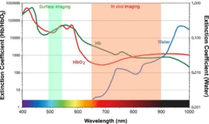
Optical imaging is a molecular imaging modality which describes the behavior of visible, ultraviolet, and infrared light used in medical imaging. It is relatively inexpensive and can provide molecular and functional information, but reveals limited information on a physiological (when organs are deep in the tissue) and anatomical basis. When applying OI, the probe interacts with a biological tissue, and the light then has a resulting photo-physical interactions such as scattering, absorption and emission of light. Each of these interactions can be used in order to obtain information about the various aspects of interest such as biochemical and morphological information about a specific target [Solomon 11]. OI uses light as its medium and often fluorescence is implemented to help view things indirectly that could be seen or ’marked’ otherwise. In order to visualize some of these flu-orophores, an excitation laser must be used. While near infrared wavelengths are used to avoid light absorption from blood mainly, it still must overcome other
as-pects, such as scattering, attenuation, and energy conversion into heat, to remain visible to the high speed CCD cameras. A non-negligible advantage is the lack of radiation when compared to other modalities. Even though there are numerous advantages to OI, there is one which cannot be disregarded: limited tissue pen-etration depth which means that a translation into a clinical setting is difficult to carry out at present. Figure 1.1.2 and 1.1.3 illustrate an example of in vivo imaging in a mouse and the absorption coefficient, respectively. Figure 1.1.2 was acquired with the help of a contrast agent Angiostamp®which is a near infrared fluorescent probe which targets alpha-v-beta-3 integrin specifically and has been used for the labeling of tumors or angiogenesis studies. This image illustrated the Angiostamp®uptake within the implanted tumors, located behind the head of the mouse. However, due to the path of elimination, one can also see the kidneys. Looking at figure 1.1.3, one can understand why the near infrared (NIR) light is of interest as it is within the optical window for in vivo imaging.
Figure 1.1.2: Optical image obtained using fluorescence molecular tomography (FMT) of a mouse injected with Angiostamp®
Figure 1.1.3: Depicted here is a graph illustrating the absorption coefficient of the therapeutic window for in vivo imaging in optical imaging.
1.1.2 Ultrasound
Ultrasound (US) is another technique which has very different advantages and disadvantages and is based on oscillating sound pressure wave with a frequency greater than the upper limit above that of human hearing and is used to measure distances for many applications to include the medical imaging field. It allows for the visualization of various structures to include muscles, tendons, and many inter-nal organs, but to also capture their size, structure. US is a widely used diagnostic tool and has been used for the past half a century as it is relatively inexpensive and portable too. US maps the acoustical properties of the tissue to include the density and compressibility[Cobbold 07]. It is commonly used for echographies in a clinical setting and an be an alternative to MRI and CT scans in some cases. As it is relatively inexpensive, it can be widely used and implemented for the di-agnosing/staging of various diseases [Fenster 01]. However, the image analysis is very user dependent and can be difficult to interpret if not being looked at by an experienced user as the objects can be distorted due to defraction, attenuation, dispersion and other inhomogeneities [Cobbold 07]. Figure 1.1.4 illustrates a cross section of a moving heart and shows that US can be used for the analysis of a functional organ, contrary to CT scans.
Figure 1.1.4: Cross section of a moving heart
1.1.3 Computerized Tomography
When talking about structural imaging, one must not forget to mention CT scan. CT scans, or X-ray computed tomographies, are generated by X-rays which pass through various tissues. These x-rays are then either absorbed or scattered. The 3D images is acquired by a source which rotates around the subject and on the other end, a detector which measures the difference in received transmitted x-rays. CT’s are still the reference modality today when regarding bone imaging. An example of a CT image (figure 1.1.5) shows how clearly one can identify the bone outlines. One advantage of the CT imaging modality is that one can manipulate various parameters of the imaging process to extract different tissue characteristics based on their X-ray absorption. Be this as it may, limitations are still evident when comparing images (A) and (B) in figure 1.1.5. In image (A), the CT image reveals structurally intact bones whereas the MRI (B) using a short T1 inversion recovery image reveals ruptured ligaments where the arrow is located [Fotiadou 11].
Figure 1.1.5: CT (A) & MRI images (B)
1.1.4 Magnetic Resonance Imaging
As can be seen, there are advantages of each modality, and another imaging modal-ity which has gained much attention and has been translated into a clinical setting quite quickly is magnetic resonance imaging (MRI). This is a medical imaging modality which records changes in magnetic fields and depending on the pulse sequence allows for different kinds of images. Figure 1.1.6 illustrates examples of two different sequences, among others, that are possible with MRI (T1 on the left and T2 on the right). MRI can be used for a wide range of applications ranging from neuroimaging to cardiovascular imaging, musculoskeletal, oncological and also functional applications. Beyond that, a distinct advantage of MRI is that there is no use of ionizing radiation. Each sequence allows the user to extract different kinds of information which in turn will assist in the diagnosis.
Figure 1.1.6: MRI with T1 (left) and T2 (right) [Leijser 10]
MRI is used much of the time for brain scans but is also routinely used for whole body image acquisitions. As MRI has exceptional soft tissue contrast prop-erties, the user may be able to determine soft tissue diseases based on an image [Fotiadou 11]. The ability to apply an MRI to various kinds of medical questions shows its wide usage. Beyond this however, there are two more strong argument for this modality; the high resolution and the lack of radiation for the patient.
While it is true that other modalities have a advantages such as not being dependent on a radioactive element, they (OI, US) have depth limitation which become a large obstacle when looking at a living system. MRI, as mentioned previously, is not dependent either on radioactivity, nor is there a depth limitation. All things taken into account, nuclear imaging techniques are able to detect the tracers on a molecular level as depicted in figure 1.1.1.This along with the lack of depth limitation make it of great interest for many applications. The ability to acquire specific information at a molecular level is of great interest in many diseases.
1.1.5 Nuclear Imaging
There are two nuclear imaging modalities to look at. The first being single photon emission computed tomography (SPECT) and the second being positron emission tomography (PET). SPECT and PET are imaging techniques which allow for functional imaging, as oppose to anatomical imaging. In these modalities, there
are advantages which are very desirable, such as being able to obtain functional information at a molecular level while not being limited to the depth at which the functions are taking place. The resolution of a SPECT device is at about is at about 8-10 mm in a clinical setting, but can have competitive resolutions with PET in a preclinical setting. PET resolution is generally anywhere between 1-2 mm in the preclinical field and at 3-4mm in most clinical machines.
The two modalities however are not identical and have, again, different char-acteristics which make a difference in practice. The resolution disadvantage of SPECT imaging makes PET very interesting. Beyond that though, there is the higher sensitivity of PET.
This increased sensitivity is non-negligible as it increased by two to three orders of magnitude[Rahmim 08]. Both PET and SPECT however have different limita-tions when talking about the spatial resolution. SPECT is limited by technology at the moment due to the design of collimators. PET on the other hand is limited by physics. Due to the non-collinearity of the photons. Much of this can however be corrected by reconstruction modeling[Reader 07]. The other aspect that one should take into account, when dealing with nuclear imaging, is the temporal reso-lution. Each modality is able to exploit dynamic information which in many cases is of great importance. The possible alteration in bio-distribution of the radio-tracers/radiopharmaceuticals within a subject, may yield pertinent physiological or pathophysiological information which could not otherwise be observed.
A distinct advantage of SPECT is that the user is able to carry out an acqui-sition of two different parameters if using two different isotopes, each with their own distinct energy. This is of great interest, especially in a clinical setting as it allows both, the hospital/doctor as well as the patient to gain time as well as supplementary information, if required.
While, dual tracer acquisitions are currently not being carried out in PET, it could one day be possible. Using the knowledge of the different half-life dura-tions, it has been shown that this simultaneous acquisition of two different tracers, emitting the same energy, and separation of the two signals is indeed possible [Kadrmas 05] [Rust 06].
1.1.6 Tracers/Contrast agents
Contrast agents, or tracers, are substances which are injected and used to increase the contrast of fluids or structures during an imaging process.
When speaking about the ability to detect something (in regard to PET imag-ing), one must understand the principle behind this. There are multiple ways in which a tracer can detect and or interact with its target. Haberkorn et al [Haberkorn 11] displayed this nicely in figure 1.1.7.
In the image below one can observe that (1) [18F]FDG (fluorodeoxyglucose) is taken up into cells by glucose transporters and subsequently trapped after enzy-matic phosphorylation by hexokinase; (2) radioactive isotopes of iodide are taken up by the sodium iodide symporter on thyroid or breast cancer tissues; (3) the
amino acid [18F]-O-(2-fluoroethyl)-tyrosine (FET) is taken up into cells by the
L-type amino-acid transporter system; (4) radiolabeled antibodies such as Zevalin bind to antigens specifically expressed on the surface of tumor cells.
Figure 1.1.7: Multiple ways for a radiotracer to be taken up and provide contrast [Haberkorn 11] [Damont 08]
Figure 1.1.7illustrated the multiple ways in which various molecules/tracers can be marked, not only in terms of methodology, but also in terms of which isotope is used. There are of course chemical limitations as to which element can be used to mark a given molecule too. This aside though, one should also take into account
that each element has a certain half-life. [11C] for example has a half-life duration
of 20.3 minutes where the half-life of [64Cu] lasts 12.7 hours. One of the most used
elements is [18F] however, which has a half-life of 109.8 minutes. This amount
of time is practical as it allows the user to not have to rush as is the case for
[15O] which is very brief and has a life of a mere 122.24 seconds, but the
half-life of about 2 hours also ensures that the patients/subject will not be exposed to a radioactive substance for a prolonged time period either. Beyond this, it also means that the user would be able to cary out successive experiments as the radioactivity of a given day would be decayed by the following day.
As mentioned previously, [18F]FDG is taken up into cells by glucose transporters
and subsequently trapped after enzymatic phosphorylation by hexokinase.
Glu-cose uptake is a necessary for cellular survival and the use of [18F]FDG is closely
correlated with various types of tissue metabolism. As [18F]FDG is an analog of
glucose, there will be a natural accumulation of the tracer in tissues which contain high-glucose consuming cells. Such cells can be found in the brain, the kidneys,
but also in cancer cells. As the [18F] has replaces a 2’hydroxyl group, once the
[18F]FDG has been included into the cell via phosphorylation, it cannot undergo
once can say with certainty that there is therefore an increased level of glucose con-sumption which is likely to be pathologically related. Figure 1.1.8 exemplifies a
whole-body scan in which one can observe the accumulation of [18F]FDG. Here
one can observe in image (A) that with the help of a PET scan, a spotty or focal accumulation which is more intense than the liver uptake (arrow). (B) reveals a focal accumulation in the area of the lower abdomen (arrow). (C) PET scan demonstrates no definite accumulation of FDG in the stomach. (D) PET scan demonstrates diffuse accumulation (normal physiological accumulation) of FDG in the stomach (arrow).
Figure 1.1.8: Whole-body scan of [F]FDG accumulation [Shoda 07]
PET imaging has the fortune of having a very large selection of tracers to choose
from. Yet [18F]FDG remains the most commonly used. [18F]FDG does have its
advantages however. As previously mentioned, [18F]FDG is a glucose analog and
as such accumulates in cells which have an increased activity. Originally, [18F]FDG
was used for the detection of cancerous cells and has proven time and time again to be very effective in this application. However, as cancerous cells are not the only
ones which have an increased activity, [18F]FDG has thus also been used for other
applications in which there are malfunctioning cells and as such has been used to a great extent in evaluation of various inflammatory diseases [Buscombe 03] [Basu 09] As this tracer has served the medical world well for a very long time in many different applications, one can use better tracers which are better adapted to targets specific to inflammation. The reason for this statement is that increased
glucose metabolism, as previously mentioned, and the subsequent [18F]FDG accu-mulation are not phenomena which are unique to malignant cells [Wu 13]
Benign processes, as can be found in inflammatory pathologies, will also
accu-mulate [18F]FDG and as a result yield false positive results for tumor detection
[Rosenbaum 06]. This is also due to the fact that certain indicators and factors in an inflammatory response require glucose at an elevated rate when compared to the healthy cells in the surrounding tissue (non-inflammatory foci). This in turn
explains why [18F]FDG was of interest initially.
As good as [18F]FDG is and as much as it can tell the user about a given subject,
[18F]FDG is also non-specific and as such can be imprecise by definition. However,
it should not be confused with the non-specificity of the cellular process.
Increased cellular activity is not the only phenomenon occurring in an inflam-matory state however. There various stimuli which are triggered, initiate the in-flammatory cascade which starts with the release of a variety of pro-inin-flammatory mediators, to include cytokines, leukotrienes, chemokines. All these are initiated by resident inflammatory and endothelial cells. Once this has occurred, the vas-cular permeability is increase which in turn allowed for an infiltration on both
neutrophils and macrophages. In an activated environment the [18F]FDG uptake
is increased in many types of cells and is not specific to one population or a single type of molecular event. It has, for example, been shown that fibroblasts increase their metabolic glucose activity when in a stressful environment [CORNELIUS 90].
The fact that [18F]FDG has a nonspecific uptake is inconvenient when investigating
molecular processes related to inflammation.
1.2 Inflammation
The principle of inflammation is not a new discovery. It was just looked at from a more macroscopic stand-point. At the time, one was looking for different signs, other than a cellular level. The tell-tale signs of inflammation have been described over 2000 years ago by Celsius and in his statement (’Rubor et Tumor cum Calore et Dolore’ meaning ’redness and swelling with heat and pain’) described the cardi-nal signs of an inflammatory state. These symptoms are things that most individ-uals have come across at one point or another in their life when they have either been cut or had a sprained ankle [Rocha e Silva 78] [Benaroyo 94]. While these four symptoms are remarkable, the ultimate consequence is the loss of function, which can certainly be a possibility if left undetected and/or untreated.
As much as the these above listed phenomena may inhibit normal functionality in daily life, the inflammatory response is a necessary function and exhibits a healthy immune response. One should note that the symptoms will dissipate relatively quickly in an acute inflammation and thus enable the affected area to
resume a normal functioning state [Mueller 13].
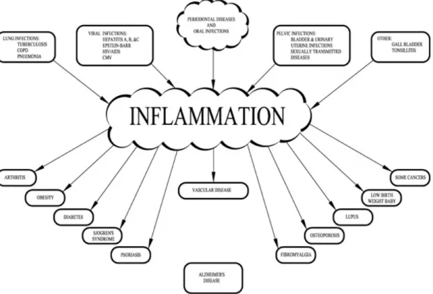
Functionality is compromised if the beneficial aspects have all served their pur-pose, but have not halted their activity, then, what once was of aid to the healthy state, becomes pathological and this uncontrolled continuity of the inflammatory process can then lead to pathogenesis such as, but not limited to, metabolic dis-eases / type 2 diabetes, neurodegenerative disdis-eases, and cardiovascular disdis-eases among others [Mueller 13] [Schwartz 13] [Aguzzi 13]. Figure 1.2.1 illustrates some of the health areas which are concerned and can be affected by inflammation and some which can be a consequence of an inflammation which goes uncontrolled, thus developing a pathological state. Inflammation plays a central role as can be seen in this illustration. The upper 3rd (above the inflammation bubble) lists possible origins for an inflammatory reaction while the lower 3rd (below the inflammation bubble) illustrates possible consequences of a pathological inflammatory state.
Figure 1.2.1: Inflammation plays a central role in pathologies and disorders Figure ?? by Tabas et al. [Tabas 13] illustrate the principle behind the patholog-ical state and how this state becomes a health problem. In an acute inflammatory response (image A) there is a suppression of the pathogen, which in turn brings the stimulus to an arrest. Some reversible tissue damage will also occur which will be
repaired and ultimately bring the damaged tissue to a homeostatic state. In image B, the chronic response, one can observe some of the classical responses, and thus features, of a chronic inflammatory disease. This, non-immune, pathophysiological process triggers a sterile inflammatory response. This is most likely initiated by the production of damage-associated molecular patterns. This response then is amplified by various factors such as chemokines and cytokines. Due to the fact that the initial response is not stopped as would be the case in a physiological situation, it continues and a chronic inflammation takes place.
Figure 1.2.2: Acute inflammatory response (image A) and chronic inflammatory disease
[Tabas 13]
The previous illustration remains vague but allows for a global view of what can go wrong when inflammation is present. It also shows that inflammation is of great importance in various pathologies and that this phenomenon is of utmost importance and as such could be a significant factor in disease development to observe and evaluate.
harmful pathogens, while on the other it is also necessary for the repair of damaged tissue [Foley 13]. The consequences are rather dire as can be seen in some of the better known diseases, such as the inflammatory bowel diseases (be it Crohn’s diseases or Ulcerative Colitis), neurodegenerative diseases such as Alzheimer’s-, Parkinson’s-, and Huntington’s disease, or Sepsis. As much as we feel we ’know’ about the previously mentioned diseases, there is still much work that needs to be done in the future in order to properly understand them.
It is a start to know that there is a problem present and that it is possible to follow the various diseases by various methods. But it is not enough. In order to bring help those suffering, one must be able to do better than just admit that there is a problem. There is a need for precise prognosis, which can be made by a precise diagnosis, which ultimately leads to the global desire for personalized medicine.
Traditionally, the only way to really observe the presence of various aspects or any biological (be it physiological of pathophysiological) process, one was obliged to extract a tissue sample and then analyze it. This meant that in some cases, the patient had to already have been deceased in order to obtain the samples, such as in individuals who ultimately succumb to their neurodegenerative diseases. Thus, the observation was retrospective and too late for the patient. This has another consequence, that there is not much known about the exact nature of the pro-inflammatory stages. Beyond this aspect, there are also a list of regulatory and feedback mechanisms which are also involved in the feedback system, but are not fully understood either. Alas, all that has been researched, has not been able to fully bring to light all the involved phenomena that occur while an inflammatory state is being activated or controlled [Foley 13].
New approaches were needed and this is where the field of imaging comes in; specifically nuclear imaging. This tool has drastically changed the way that re-searchers and medical doctors approach problems. This is in part due to the fact that nuclear imaging enables the user to observe what is happening on a molecular level while in vivo. Figure 1.2.3 illustrates a list of biomarkers, along with their foci, implicated in inflammation [Wu 13].
Figure 1.2.3: PET-radiotracers for inflammatory phenomena with corresponding targets
[Wu 13]
A wide range of studies have been carried out by numerous groups in an effort to better understand inflammation. The first steps were to analyze which aspects were able to be traced with the ultimate goal to one day be able to effectively diagnose and thus prognose an inflammation, no matter what the origin or state. Some of the targets have been inflammatory cytokines, choline metabolism, inflammatory related vessels, and membrane markers.
1.2.1 Inflammatory targets
As inflammation induces a whole cascade of events, one can imagine that there are various participants that could serve as targets which in turn might be able to indicate the level of inflammation currently taking place.
Among the participants are MMP’s (matrix metalloproteinase), inflammatory cytokines, and COX (cyclooxygenase). Each has a different function and is of interest for various reasons.
MMP’s are zinc-depandant endopeptidases and were originally thought to be
exclusively implicated in matrix degradation but have been proven to be be able to modify cytokines (such as TNF-alpha, IL-6) and chemokines (CXCL-8 or CCL2 for example), which are found in inflammatory states and as such is now thought to be or importance for inflammatory conditions [Ryu 12]. This would mean that the imaging of the MMP’s in itself would be of interest, and as such has been
investigated by Zhu et al. [Zhu 11] among others who have also mainly used SPECT as their modality of choice [Hermann 12] [Schäfers 04] [Kuge 10]. The areas in which these radiolabeled tracers have been used was largely in the cancer detection field, but also in vascular inflammation too.
PET has also been used to investigate MMP expression in the field of tumor
imaging using [18F]CGS27023A and [11C]CGS27023A (same compound, marked
with a different radioisotope) while124I-MIP (both are MMP inhibitors) to target
vascular lesions [Wagner 9] [Zheng 02] [Hartung 07]. Initial investigations using
[11C]CGS27023A has led the researchers to continue the development of the tracer
as it has a very high lipophilicity and as such yields characteristics which they deem possible to improve upon. Its counter part (the 18F marked compound) has also shown to be of interest while also having a strong inhibitory effectiveness on MMP-1 in comparison with the parent compound CGS27023A. One should also note the
half-life of 11C (20.33 minutes) and take into consideration the impact that this
time constraint has on clinical applications. 124I-HO-MIP (half-life of 4.8 days) has
also been tested and as might be expected, has the disadvantage of the relatively long half-life. The half-life is something to take into consideration, yet the potential impact does need to be weighed out as to whether or not a patient can benefit from a diagnosis using such a tracer with a long half-life isotope. Yet, MMP’s are maybe not the most advantageous target all inflammatory diseases. There are various MMP’s, some of which promote, and others inhibit the inflammatory process and as such are modulators. This means that the existing MMP radioligands to date are imprecise and as such yield poor specificity.
Inflammatory Cytokines are loosely defined as a group of immunoregulatory
proteins (such as growth factors, interleukins and other lymphocytes) which are small in size, yet important in cell signaling.
There are a few cytokines which have been targeted in the past. Among them are IL-2 (interleukin-2), and the TNF-alpha (tumor necrosis factor alpha). Each has their role within the inflammatory process and as such were targeted for imaging purposes.
The presence of IL-2 is augmented by activated T lymphocytes which are ob-served in multiple types of inflammatory pathologies ranging from tumor inflam-mation to organ specific autoimmune diseases [Kintscher 08]. These activated T
lymphocytes (Th1 CD8+ and CD4+ in large part) not only synthesize the IL-2,
but also express an IL-2 receptor to which the IL-2 bind with a high affinity. This shows the interest of using a marked (radiolabeled) IL-2 as the user is also able to differentiate between activated and non activated T lymphocytes. There have been some SPECT tracers which have been used in the past, but also PET tracers
(99mTc and123I labeled IL-2 for SPECT usage and[18F]SFB to also label IL-2 to
did prove useful and was able to assist the user in detecting areas of inflammation in an in vivo situation. The authors state that the tracer was indeed useful for the detection and quantification of lymphocyte infiltration. However, as imaging is a field in which many areas of science work together, one cannot just look at the final result. There are many difficulties that may be overlooked. Here for example, one must also consider the work that is done when preparing the radiotracer itself too. Even though, the tracer may be of interest, much needs to be done to facilitate the production as the radiolabeled IL-2 tracer is tedious to produce as there is a poor solubility and stability problems [Signore 02].
TNF-alpha is another cytokine that is implicated in the inflammatory
phe-nomenon which is produced by monocytes and macrophages. There are two dis-tinct actions that the presence of TNF-alpha causes. The first being physiological in that it increases the transport of white blood cells to the inflamed area. The second is pathological. There is a delayed inflamed state in which the presence of TNF-alpha leads to organ dysfunction and cellular apoptosis [Mäki-Petäjä 06].
Some of the tracers used in past ex vivo studies has been 64Cu-DOTA-etanercept
among others. 64Cu-DOTA-etanercept for example was able to detect the
TNF-alpha cytokines, but only in the early stages and was not of use for chronic in-flammation as TNF-alpha is not significantly over-expressed outside of an acute inflammation.
The Cyclooxygenases (COX,-1 and -2) family are important enzymes for the conversion of arachidonic acid to prostaglandins along with other lipid mediators. As COX can be induced by inflammatory stimuli, COX-2 has been classically con-sidered as the most appropriate target for anti-inflammatory drugs. The inhibition leads to pain relief [Hawkey 99]. It has been found that the cyclooxygenases are implicated in neuroinflammatory related instances such as cancers, Alzheimer’s Disease, Parkinson’s Disease, and various ischemias [Katori 00] [Minghetti 04].
As this is yet another aspect of the inflammatory process which could possibly be of interest for the imaging in community, multiple tracers have been developed and implemented in a a range of studies. Some of the tracers (each a COX-inhibitor)
produced are: [18F]desbromo-Dup-697, [18F]SC58125 as well as 11C tagged
cele-coxib, and rofecoxib [de Vries 03] [McCarthy 02] [Prabhakaran 05] [de Vries 08]. The studies performed tried to address inflammation in the fields of neuroinflam-mation and tumors[de Vries 08] [Shukuri 11] [Gao 11] [Uddin 11]. The results of the previously mentioned studies revealed that the tracers were not of great value to imaging due to undesired in vivo and ex vivo characteristics such as low binding affinity and unspecific binding.
1.2.2 Inflammatory metabolism
In this area, there are two aspects to take into account. The first being glucose metabolism, which has been addressed in the Tracers/Contrast Agents section, and choline metabolism being the second.
Glucose metabolism, as previously mentioned, has its interests as an imaging target and is currently the most used radiotracer used for nuclear imaging purpose
as it is very versatile. However, as it is ’versatile’, [18F]FDG is non-specific and
often leads to a false positive diagnoses [Rosenbaum 06].
Choline is an amino alcohol found in the synthesis of two types of phospholipids; sphingomylin and phosophatidylcholine. The biosynthesis pathway of these phos-pholipids can be imaged using radiolabeled choline as choline is associated with the phosphorycholine, which is an intermediary step which is involved with the
phospholipid synthesis. The choline, which is radiolabeled to with 11C and 18F,
has been applied to image prostate cancers, but also inflammatory diseases such as atherosclerosis, by targeting the macrophages and monocytes [Schwarzenböck 12]
[Matter 06]. 18F labels choline is more interesting as the half-life is easier to handle
in an clinical environment. The experiments carried out until now have shown that
the 18F labeled choline did not have other marked advantages, but could however
create a point to consider as the kidney have a high choline retention along the nephrons, which must be considered when taking the radiation-dose into account [Witney 12].
In comparison to FDG, tagged choline has an advantage over FDG in that it has much less uptake within the heart and as such would be better adapted for detecting coronary plaques [Roivainen 06]. It is not only in the myocardium that
[18F]choline may have an advantage over the popular [18F]FDG. [18F]choline has
been shown to have a better sensitivity for the detection and characterization of atherosclerotic plaques too [Matter 06].
1.2.3 Vascular biomarkers of inflammation
Another aspect of inflammation related to vessels is the permeability. In a physi-ological situation, the vessels are of the utmost importance in regulating a homeo-static state for the internal environment. This means that the structural integrity can be altered, situation dependent. Upon stimulation, the vessels can become more or less permeable. If an inflammatory state triggering various factors (such as cytokines, chemokines or leukotrienes) to be released by the local inflammatory cells, then the permeability is vastly increased in order to allow for the arrival and infiltration of macrophages or neutrophils, among other immune cells, at the in-flamed site [Tsan 85]. This means that one would not have to distinguish between an infectious inflammation or a sterile one to be able to use a tracer for vessel

![Figure 1.3.2: TSPO implications have a correlation with microglial activation as well [Scarf 09].](https://thumb-eu.123doks.com/thumbv2/123doknet/12860511.368514/49.892.262.635.148.405/figure-tspo-implications-correlation-microglial-activation-scarf.webp)


![Delayed [(18)F]FDG PET imaging of central nervous system lymphoma: is PET better than MRI?](data:image/gif;base64,R0lGODlhAQABAIAAAP///wAAACH5BAEAAAAALAAAAAABAAEAAAICRAEAOw==)