Spontaneous Viral Clearance, Viral Load, and
Genotype Distribution of Hepatitis C Virus (HCV)
in HIV-Infected Patients with Anti-HCV Antibodies
in Europe
Vincent Soriano,1Amanda Mocroft,2Juergen Rockstroh,3Bruno Ledergerber,4Brygida Knysz,5Saulius Chaplinskas,6 Lars Peters,7Anders Karlsson,8Christine Katlama,9Carlos Toro,1Bernd Kupfer,3Martin Vogel,3and Jens Lundgren,7 for the EuroSIDA Study Groupa
1Service of Infectious Diseases, Hospital Carlos III, Madrid, Spain;2Royal Free and University College Medical School, London, United Kingdom; 3Department of Medicine I, University of Bonn, Bonn, Germany;4Division of Infectious Diseases, Department of Medicine, University Hospital, Zurich, Switzerland;5Department of Infectious Diseases, Wroclaw Medical University, Wroclaw, Poland;6Lithuanian AIDS Center, Vilnius, Lithuania;7Copenhagen HIV Program, Copenhagen, Denmark;8Karolinska University Hospital, Stockholm, Sweden;9Pitié-Salpêtrière Hospital, Paris, France
(See the editorial commentary by Bruno and Sacchi, on pages 1262– 4.)
Background. Variables influencing serum hepatitis C virus (HCV) RNA levels and genotype distribution in individuals with human immunodeficiency virus (HIV) infection are not well known, nor are factors determining spontaneous clearance after exposure to HCV in this population.
Methods. All HCV antibody (Ab)–positive patients with HIV infection in the EuroSIDA cohort who had stored samples were tested for serum HCV RNA, and HCV genotyping was done for subjects with viremia. Logistic regres-sion was used to identify variables associated with spontaneous HCV clearance and HCV genotype 1.
Results. Of 1940 HCV Ab–positive patients, 1496 (77%) were serum HCV RNA positive. Injection drug users (IDUs) were less likely to have spontaneously cleared HCV than were homosexual men (20% vs. 39%; adjusted odds ratio [aOR], 0.36 [95% confidence interval {CI}, 0.24 – 0.53]), whereas patients positive for hepatitis B surface antigen (HBsAg) were more likely to have spontaneously cleared HCV than were those negative for HBsAg (43% vs. 21%; aOR, 2.91 [95% CI, 1.94 – 4.38]). Of patients with HCV viremia, 786 (53%) carried HCV genotype 1, and 53 (4%), 440 (29%), and 217 (15%) carried HCV genotype 2, 3, and 4, respectively. A greater HCV RNA level was associated with a greater chance of being infected with HCV genotype 1 (aOR, 1.60 per 1 log higher [95% CI, 1.36 –1.88]).
Conclusions. More than three-quarters of the HIV- and HCV Ab–positive patients in EuroSIDA showed active HCV replication. Viremia was more frequent in IDUs and, conversely, was less common in HBsAg-positive patients. Of the patients with HCV viremia analyzed, 53% were found to carry HCV genotype 1, and this genotype was associated with greater serum HCV RNA levels.
The course of hepatitis C virus (HCV) infection varies widely after initial exposure. Although one-third of in-dividuals may clear the infection spontaneously, the re-maining persons have persistent viremia and develop chronic hepatitis C [1– 6]. In the former group, liver function test results and hepatic tissue remain normal, and HCV antibodies (Abs) are the only evidence of a
past encounter with HCV [7]. In contrast, most patients with chronic hepatitis C show elevations in liver enzyme levels as a consequence of hepatic inflammation, and
Received 14 February 2008; accepted 8 May 2008; electronically published 3 September 2008.
Potential conflicts of interest: none reported.
The Journal of Infectious Diseases 2008; 198:1337– 44
© 2008 by the Infectious Diseases Society of America. All rights reserved. 0022-1899/2008/19809-0013$15.00
DOI: 10.1086/592171
Financial support: EuroSIDA is a network funded by the European Commission. The European Commission BIOMED 1 (grant CT94 –1637), BIOMED 2 (grant CT97– 2713), 5th Framework (grant QLK2–2000-00773), and 6th Framework (grant LSHP-CT-2006 – 018632) programs were the primary sponsors of the study. Unrestricted grants were also provided by Bristol-Myers Squibb, GlaxoSmithKline, Roche, Gilead, Pfizer, Merck, Tibotec, and Boehringer-Ingelheim. The participation of centers from Switzerland was supported by a grant from the Swiss Federal Office for Education and Science.
aStudy group members are listed after the text.
Reprints or correspondence: Dr. Vincent Soriano, Dept. of Infectious Dis-eases, Hospital Carlos III, Calle Sinesio Delgado 10, 28029 Madrid, Spain ([email protected]).
many steadily progress to liver cirrhosis [5, 6, 8 –10]. Although serum HCV RNA levels and HCV genotype do not seem to sig-nificantly influence the natural history of chronic hepatitis C, these virological features largely affect therapeutic outcomes. In-dividuals with low viral load and those infected with HCV geno-type 2 or 3 respond significantly better to pegylated interferon plus ribavirin than do subjects with high HCV RNA levels and infection with HCV genotype 1 or 4 [11, 12].
Because of shared routes of transmission, coinfection with HIV and HCV is relatively common [13–15]. In Western coun-tries, liver disease due to HCV infection has become a leading cause of morbidity and mortality in HIV-infected individuals [16 –19]. In this regard, the virological characterization of HIV-infected patients positive for HCV Abs is important, given that this information may help in making appropriate estimations about the burden of liver disease and the costs of treatment in this population, including the need for liver transplantation in a subset of them. The aim of the present study was to virologically characterize HIV-infected patients positive for HCV Abs in the EuroSIDA cohort, in an attempt to establish the current burden of HCV-related disease and the expected clinical and therapeutic outcomes derived from its main virological features.
METHODS
Patients. EuroSIDA is a prospective study of 14,310 HIV-1– infected patients at 93 centers across Europe, Israel, and Argen-tina; details have been reported elsewhere [20]. Briefly, for each cohort the centers provide data on consecutive patients seen at the outpatient clinics beginning in May 1994 until a predefined number of patients is enrolled from each site. To date, 7 cohorts of patients have been recruited. Data are collected prospectively at clinical sites and are extracted and sent to the coordinating center at 6-month intervals. For cohorts I–III, eligible patients were those who had had a CD4 cell count⬍500 cells/mm3
dur-ing the previous 4 months. The CD4 cell count restriction was removed for cohorts IV–VII. At recruitment, in addition to de-mographic and clinical information, a complete antiretroviral treatment history is obtained, together with the most recent CD4 cell count and plasma HIV RNA measurements. At each follow-up visit, details on all CD4 cell counts and plasma HIV RNA values measured since the last follow-up visit are extracted, as are the dates of starting and stopping each antiretroviral drug received and the use of drugs for prophylaxis against opportu-nistic infections. The dates of diagnosis of all AIDS-defining ill-nesses, non–AIDS-defining malignancies, and other serious in-fections are also recorded. The present analysis includes follow-up to a median date of January 2007.
Information on HCV Ab status has been collected since 1997; patients who died or were lost to follow-up before this date did not routinely have information on HCV Ab status collected. Centers that have determined HCV genotype or that have
mea-sured HCV RNA level can provide that information to the coor-dinating center at any time. The EuroSIDA plasma repository was set up in 1997 and collects plasma samples from all HIV-infected patients at 6-month intervals. Patients with unknown HCV Ab status and a stored plasma sample were identified in 2006, and HCV Ab status was determined. Patients who tested positive for HCV Ab were then tested for serum HCV RNA level and genotype in a reference laboratory. In addition, patients with unknown HCV genotype and stored samples were identi-fied, and serum HCV RNA levels were tested and genotyping was performed.
Serum HCV RNA was investigated and quantified in HCV Ab–positive samples by means of the Versant HCV RNA assay (version 3.0; Bayer Diagnostics), which uses a signal amplifica-tion procedure with a linear dynamic range of 615 to 1⫻ 107
IU/mL. This method uses multiple probes to match several se-quences of the HCV genome, negating the potential for under-estimation of some HCV genotypes due to mismatches caused by genetic polymorphisms [21, 22].
HCV genotyping was performed using the LiPA HCV geno-type assay (version 2.0; Innogenetics), which is a line probe assay that simultaneously detects sequences in the 5' untranslated re-gion (UTR) and the core rere-gion. The examination of the core region along with the 5' UTR allows more-accurate discrimina-tion between genotypes 1 and 6 and improves subtyping across distinct genotypes [23–25].
Statistical analyses. Descriptive values are expressed as ab-solute or median values with interquartile ranges. Categorical variables were compared using the2test, and continuous
vari-ables were compared using nonparametric tests. Logistic regres-sion was used to identify which variables were associated with spontaneous HCV clearance and with HCV genotype 1 infection and which genotype was the most common, after adjustment for demographic (age, sex, HIV exposure group, ethnic origin, re-gion of Europe, date of HCV Ab testing, and date of joining EuroSIDA), clinical (prior AIDS, CD4 cell count, and plasma HIV RNA level), and therapeutic factors (antiretroviral ther-apy). The results are presented using odd ratios (ORs) and ad-justed ORs (aORs). All data were analyzed using SAS software (version 9.1).
RESULTS
Of the 14,310 HIV-infected patients enrolled in EuroSIDA, 3375 (24%) were positive for HCV Ab. Stored specimens were not available for 1435 of these patients, who were excluded from further analyses. Excluded patients were more likely to be of white ethnic origin compared with included patients (95% vs. 89%), more likely to originate from southern Europe/Argentina (39% vs. 34%) or eastern Europe (37% vs. 23%), and less likely to have started antiretroviral therapy (63% vs. 84%). No other significant differences were noted between included and
ex-cluded HCV Ab–positive patients. Samples were not available because the sample had already been used for a different project or because it was not collected at the center, predominantly be-cause the patient had died or was lost to follow-up before the plasma repository was set up.
The main characteristics of the 1940 HCV Ab–positive pa-tients included in the present analyses are shown in table 1. HCV genotype was determined by the central laboratory for 80% of patients. Overall, 1496 patients (77% [95% confidence interval {CI}, 75%–79%]) were positive for serum HCV RNA and/or had a known HCV genotype. For the comparison between the main features of HIV-infected patients with chronic HCV viremia with those who spontaneously cleared HCV, it is remarkable that injection drug users (IDUs) were less likely to spontaneously clear HCV than were men who had sex with men (MSM) (20% vs. 39%), whereas patients who were positive for serum hepatitis B surface antigen (HBsAg) were more likely to clear HCV than were those negative for HBsAg (43% vs. 21%). Finally, patients from southern Europe/Argentina were less likely to have
spon-taneously cleared HCV than were those from the other regions (18% vs. 22%–27%).
Table 2 shows the univariate and multivariate ORs for spon-taneous clearance of HCV. All 26 patients with prior exposure to interferon therapy who subsequently tested negative for HCV RNA were excluded from this analysis. In the multivariate model, females had 39% increased odds of spontaneous HCV clearance compared with males (aOR, 1.39 [95% CI, 1.06 –1.81];
P⫽ .017). Compared with MSM, any other exposure group
had decreased odds of spontaneous HCV clearance. This was particularly true for IDUs, who had 64% reduced odds of HCV clearance compared with MSM (aOR, 0.36 [95% CI, 0.24 – 0.53];
P⬍ .001). Compared with patients from southern
Europe/Ar-gentina, subjects from central or northern Europe had increased odds of spontaneous HCV clearance. This was particularly man-ifest for patients from northern Europe, who had 47% increased odds of HCV clearance compared with those from southern Eu-rope/Argentina (aOR, 1.47 [95% CI, 1.03–2.09]; P⫽ .032). Fi-nally, patients who were positive for HBsAg had increased odds of spontaneous HCV clearance compared with those who were negative (aOR, 2.91 [95% CI, 1.94 – 4.38]; P⬍ .001).
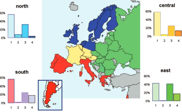
Of patients with detectable serum HCV RNA, 786 (53%) were infected with HCV genotype 1, and 53 (4%), 440 (29%), and 217 (15%) were infected with HCV genotype 2, 3, and 4, respectively. Mixed genotypes were recognized in only 4 (0.2%) patients with viremia. Figure 1 shows the geographical distribution of HCV genotypes across the different regions. Although HCV genotype 1 predominated in all regions, the proportion of patients with HCV genotype 3 infection was significantly higher in eastern Europe than in the rest of Europe. HCV genotype 2 was uncom-mon and was mainly seen in the northern and central European regions. Finally, HCV genotype 4 represented⬃15% of infec-tions but was less frequent in the northern Europe region.
Table 3 shows the main characteristics of patients infected with distinct HCV genotypes. The median viral load in patients for whom the HCV genotype was known was 576,812 IU/mL; it was ⬎500,000 IU/mL in 54%. By genotype, the median serum HCV RNA values were 776,015, 685,258, 393,523, and 389,000 IU/mL for HCV genotypes 1, 2, 3, and 4, respectively (P⬍ .001).
Table 4 shows the univariate and multivariate odds of having HCV genotype 1 infection compared with any other HCV geno-type. In the multivariate model, females had decreased odds of HCV genotype 1 infection compared with males (aOR, 0.76 [95% CI, 0.59 – 0.99]; P⫽ .038), as did patients from eastern Europe compared with those from southern Europe/Argentina (aOR, 0.37 [95% CI, 0.25– 0.56]; P⬍ .001). Older patients had decreased odds of HCV genotype 1 infection (aOR, 0.80 per 10 years older [95% CI, 0.68 – 0.94]; P⫽ .008). Finally, patients with a higher serum HCV RNA level had increased odds of HCV genotype 1 infection compared with patients with a lower serum HCV RNA level (aOR, 1.60 per 1 log higher [95% CI, 1.36 –1.88];
P⬍ .001).
Table 1. Main characteristics of 1940 HIV-infected patients positive for hepatitis C virus (HCV) antibody.
Variable Total Serum HCV RNA P Positive Negative Patients 1940 (100) 1496 (77) 444 (23) . . . Sex .22 Male 1348 (69) 1050 (78) 298 (22) Female 592 (31) 446 (75) 146 (25)
Age, median, years 37.2 37.5 37.0 .47
Risk group ⬍.001 IDUs 1399 (72) 1126 (80) 273 (20) MSM 181 (9) 111 (61) 70 (39) Heterosexuals 233 (12) 164 (70) 69 (30) Other 127 (7) 95 (75) 32 (25) Ethnicity .30 White 1734 (89) 1343 (77) 391 (23) Other 206 (11) 153 (74) 53 (26) Region ⬍.001 Southern Europe/ Argentina 654 (34) 536 (82) 118 (18) Central Europe 466 (24) 339 (73) 127 (27) Northern Europe 378 (19) 274 (72) 104 (28) Eastern Europe 442 (23) 347 (79) 95 (21)
Serum HBsAg status ⬍.001
Positive 137 (7) 78 (57) 59 (43) Negative 1483 (76) 1173 (79) 310 (21) Unknown 320 (16) 245 (77) 75 (23) Antiretroviral therapy .048 Yes 1685 (87) 1287 (76) 398 (23) No 255 (13) 209 (82) 46 (18)
NOTE. Data are no. (%) of patients, unless otherwise indicated. HBsAg, hepatitis B surface antigen; IDUs, injection drug users; MSM, men who have sex with men.
DISCUSSION
This study reports the rate of HCV infection chronicity, genotype distribution, and viral load in a large group of HIV-infected patients across Europe. In excess of three-quarters of HIV-infected patients positive for HCV Ab showed active HCV replication. In HIV-negative individuals, spontaneous HCV clearance occurs overall in approximately one-third of cases [1– 6]. Two factors might explain the higher rate of chronicity we saw in the present HIV-infected population. First, HIV-associated immunodeficiency could impair effective immune control of HCV replication after initial exposure [1, 26, 27], given that it has been shown in other immunocompro-mised conditions. Second, IDUs represented the largest group of HCV Ab–positive patients in EuroSIDA, and repeated exposure to HCV in them could have favored the establishment of persistent HCV infection. Although innate and adaptive immune responses might partially protect against repeated exposure to HCV [28], they might be less effective for exposure to distinct HCV genotypes or in the setting of HIV infection [29]. Given that HCV viremia was more frequent among HCV Ab–positive IDUs enrolled in EuroSIDA than in HCV Ab–positive subjects infected through sexual contact (whether homo- or heterosexual), we hypothesize that repeated ex-posure to HCV rather than HIV-associated immunosuppression was the most likely cause of the higher rate of HCV infection chro-nicity noted on average in EuroSIDA patients. In fact, HCV expo-sure must have preceded HIV infection in most cases, as has been shown in other studies that have examined the incidence of HCV and HIV infections in high-risk groups [30, 31].
Patients with markers of dual hepatitis B virus (HBV) and HCV infections show a worse liver prognosis [32]. In our study, HCV viremia was less frequent in persons with HBsAg than in the rest of the population (54% vs. 75%). This observation is in agreement with the findings of prior studies, in which viral in-terference phenomena have been reported to account for recip-rocal inhibition of HBV and HCV replication [33, 34]. It is note-worthy that control of HBV replication with antiviral agents does not seem to be associated with HCV rebound, while the contrary may occur [35, 36]. The biological basis for this obser-vation relies on the distinct cell cycles for HBV and HCV infec-tion. Although the genetic material of HBV is relatively stable within infected cells as covalently closed circular DNA, the RNA of HCV can be found replicating in the cytosol only, where it is subject to rapid degradation [37, 38]. Therefore, if HBV over-takes HCV replication, there is a high chance of spontaneous HCV eradication. In contrast, treatment of HCV infection with pegylated interferon plus ribavirin may make previously avire-mic HBsAg-positive patients prone to HBV rebound [36].
Of patients with active HCV replication, 53% were infected with HCV genotype 1; this genotype was associated with higher HCV RNA levels and male sex, and it was less prevalent in east-ern Europe. Whether subjects infected with HCV genotype 1 have higher serum HCV RNA levels than do those infected with other genotypes has been debated for a while. Only a few studies have claimed such an association, as in a large US cohort of individuals with hemophilia [39] and, more recently, in the RIB-AVIC trial [40]. Although it has been claimed that
underestima-Table 2. Variables associated with spontaneous hepatitis C virus (HCV) clearance.
Variable
Univariate Multivariate
OR (95% CI) P aOR (95% CI) P
Female sex, vs. male 1.17 (0.93–1.48) .18 1.39 (1.06–1.81) .017 Older age, per 10 years 1.13 (0.99–1.28) .069 1.12 (0.96–1.32) .15 Exposure group
MSM 1.00 (reference) 1.00 (reference)
IDUs 0.39 (0.28–0.55) ⬍.001 0.36 (0.24–0.53) ⬍.001
Heterosexuals 0.66 (0.43–1.00) .050 0.65 (0.40–1.06) .084
Other 0.56 (0.34–0.93) .024 0.42 (0.24–0.73) .002
Serum HBsAg status
Negative 1.00 (reference) 1.00 (reference)
Positive 2.87 (1.99–4.15) ⬍.001 2.91 (1.94–4.38) ⬍.001
Unknown 1.25 (0.93–1.67) .13 1.34 (0.95–1.89) .093
Region
Southern Europe/Argentina 1.00 (reference) 1.00 (reference)
Central Europe 1.68 (1.25–2.26) ⬍.001 1.35 (0.95–1.91) .098 Northern Europe 1.80 (1.32–2.44) ⬍.001 1.47 (1.05–2.09) .032 Eastern Europe 1.26 (0.93–1.72) .14 1.15 (0.75–1.78) .52 NOTE. The model was adjusted for data source, ethnic origin, prior AIDS diagnosis, date of testing, date recruited to EuroSIDA, CD4 cell count at date of HCV testing, and antiretroviral therapy. aOR; adjusted odds ratio; HBsAg, hepatitis B surface antigen; IDUs, injection drug users; MSM, men who have sex with men; OR, odds ratio.
tion of viral load in persons infected with non-1 HCV genotypes is the reason for this observation, at least when some amplifica-tion methods are used, in our study serum HCV RNA level was
measured by a hybridization technique that uses multiple probes and with which all distinct HCV genotypes seem to be reliably quantified [21, 22]. Therefore, we are confident about our
find-Figure 1. Distribution of hepatitis C virus genotypes in the distinct EuroSIDA regions.
Table 3. Main features of 1496 HIV-infected patients with chronic hepatitis C, stratified by hepatitis C virus (HCV) genotype.
Variable
HCV genotype
P
1 2 3 4
Patients with genotype 786 (53) 53 (4) 440 (29) 217 (15) . . .
Male sex 569 (72) 37 (70) 300 (68) 144 (66) .24
Age, median, yearsa 37.1 40.9 37.7 36.2 .001
Risk group, IDUs 569 (72) 28 (53) 356 (81) 173 (80) ⬍.001
White ethnicity 695 (88) 49 (92) 409 (93) 190 (88) .046 Region ⬍.001 Southern Europe/Argentina 306 (57) 12 (2) 131 (24) 87 (16) Central Europe 187 (55) 16 (5) 86 (25) 50 (15) Northern Europe 153 (56) 23 (8) 85 (31) 13 (5) Eastern Europe 140 (40) 2 (0.6) 138 (40) 67 (19)
Serum HBsAg positive 38 (5) 4 (8) 21 (5) 15 (7) .001
Started antiretroviral therapya 693 (88) 45 (85) 353 (80) 196 (90) ⬍.001
Serum HCV RNA levelb
Median, IU/mL 776,015 685,258 393,523 389,000 ⬍.001
Range, IU/mL 728–39,200,000 6772–7,692,310 787–51,999,959 2870–16,399,854 . . .
Level⬎500,000 IU/mL 444 (62) 31 (61) 183 (45) 84 (43) ⬍.001
CD4 cell count, median, cells/mm3 313 349 326 350 .21
Plasma HIV RNA level, median, log copies/mL 2.70 2.30 2.70 2.55 .10
NOTE. Data are no. (%) of patients, unless otherwise indicated. HBsAg, hepatitis B surface antigen; IDUs, injection drug users. aAt date of genotyping.
ing of higher serum HCV RNA levels in patients infected with HCV genotype 1 than with genotype 3 or 4. Given that baseline HCV load predicts treatment outcome [14], our observation further emphasizes the difficulties of successfully treating HCV genotype 1–infected patients.
Serum HCV RNA levels may vary after antiretroviral expo-sure, with a trend toward increasing during the first months after the initiation of therapy and decreasing steadily thereafter [41– 43]. Moreover, differences may be seen between protease inhib-itors (PIs) and nonnucleoside reverse-transcriptase inhibinhib-itors, as was recently observed in the RIBAVIC trial [40], in which 379 HIV/HCV-coinfected patients were examined. Multiple linear regression analysis identified HCV genotype 1 and PI-based reg-imens as independent predictors of higher serum HCV RNA levels. Moreover, it should be noted that antiretroviral therapy in general was independently associated with lower HCV RNA lev-els in the RIBAVIC analysis [40], a finding that we could not reproduce in our study.
Several limitations of our study should be acknowledged. First, HCV Ab status was not known for all patients, and plasma samples were not available for all patients who were HCV Ab positive. Patients with missing data were more likely to have died or to have been lost to follow-up during the earlier years of the study. Second, 2 different data sources were used, and therefore potential biases might have been introduced. It is important to note that the plasma repository was set up in 1997, and it was unlikely that the decision to send a sample for storage was related to HCV Ab status or genotype. Where data were available from both the case report forms and the central laboratory, results from the latest were used and taken as the most accurate results. All models were adjusted for the data source, and overall sensi-tivity analyses including only data from the central laboratory showed results similar to those of the main analyses presented here. Finally, we have no information on when patients were infected with HCV, the duration of drug use in IDUs, or whether
IDUs were using injection drugs at the time blood was drawn. Despite these limitations, we feel that this study provides a com-prehensive overview of the epidemiology of HCV in HIV-infected patients across Europe.
In summary, this extensive virological study of HIV-infected patients positive for HCV Abs enrolled in EuroSIDA has shown that nearly one-quarter of patients spontaneously cleared HCV infection after initial exposure, although this rate was much higher in MSM (39%) than IDUs (20%). Of patients with HCV viremia, 53% were infected with HCV genotype 1, and this ge-notype was associated with higher serum HCV RNA levels. Al-together, these findings highlight the difficult-to-treat profile of chronic HCV infection in HIV-positive individuals.
THE EuroSIDA STUDY GROUP
Study group members. Argentina: M. Losso (national
coordi-nator) and A. Duran, Hospital JM Ramos Mejia, Buenos Aires.
Austria: N. Vetter (national coordinator), Pulmologisches
Zen-trum der Stadt Wien, Vienna. Belarus: I. Karpov (national coor-dinator) and A. Vassilenko, Belarus State Medical University, Minsk. Belgium: N. Clumeck (national coordinator), S. De Wit and B. Poll, Saint-Pierre Hospital, Brussels; .R Colebunders, stitute of Tropical Medicine, Antwerp. Bulgaria: K. Kostov, In-fectious Diseases Hospital, Sofia. Croatia: J. Begovac, University Hospital of Infectious Diseases, Zagreb. Finland: M. Ristola (na-tional coordinator), Helsinki University Central Hospital, Hel-sinki. Czech Republic: L. Machala (national coordinator) and H. Rozsypal, Faculty Hospital Bulovka, Prague; D. Sedlacek, Charles University Hospital, Plzen. Denmark: J. Nielsen (na-tional coordinator), J. Lundgren, T. Benfield, and O. Kirk, Pa-num Institute, Copenhagen; J. Gerstoft, T. Katzenstein, A.-B. E. Hansen, and P. Skinhøj, Rigshospitalet, Copenhagen; C. Peder-sen, Odense University Hospital, Odense; L. Oestergaard, Skejby Hospital, Aarhus. Estonia: K. Zilmer (national coordinator), West-Tallinn Central Hospital, Tallinn; J. Smidt,
Nakkusosa-Table 4. Variables associated with hepatitis C virus (HCV) genotype 1 infection.
Variable
Univariate Multivariate
OR (95% CI) P aOR (95% CI) P
Female sex, vs. male 1.25 (1.00–1.56) .050 0.76 (0.59–0.99) .038 Older age, per 10 years 1.00 (0.88–1.13) .94 0.80 (0.68–0.94) .008 Region
Southern Europe/Argentina 1.00 (reference) 1.00 (reference) Central Europe 1.08 (0.82–1.42) .58 0.83 (0.59–1.16) .26 Northern Europe 1.05 (0.79–1.41) .73 0.90 (0.64–1.26) .54 Eastern Europe 1.97 (1.50–2.59) ⬍.001 0.37 (0.25–0.56) ⬍.001 Higher HCV RNA level, per 1 log higher 1.49 (1.29–1.71) ⬍.001 1.60 (1.36–1.88) ⬍.001 Prior AIDS, yes vs. no 0.82 (0.65–1.03) .086 1.22 (0.93–1.59) .15
NOTE. The model was adjusted for data source, ethnic origin, exposure group, serum hepatitis B surface antigen serostatus, date of HCV genotyping, date recruited to EuroSIDA, and CD4 cell count at the date of HCV testing. aOR, adjusted odds ratio; OR, odds ratio.
kond Siseklinik, Kohtla-Järve. France: C. Katlama (national co-ordinator), Hôpital de la Pitié-Salpétière, Paris; J.-P. Viard, Hôpital Necker-Enfants Malades, Paris; P.-M. Girard, Hospital Saint-Antoine, Paris; J.M. Livrozet, Hôpital Edouard Herriot, Lyon; P. Vanhems, University Claude Bernard, Lyon; C. Pradier, Hôpital de l’Archet, Nice; F. Dabis, Unité INSERM, Bordeaux.
Germany: J. Rockstroh (national coordinator), Universitäts
Klinik Bonn; R. Schmidt, Medizinische Hochschule Hannover; J. van Lunzen and O. Degen, University Medical Center Hamburg-Eppendorf, Infectious Diseases Unit, Hamburg; H. J. Stellbrink, IPM Study Center, Hamburg; S. Staszewski, J. W. Goethe University Hospital, Frankfurt; J. Bogner, Medizinische Poliklinik, Munich; G. Fätkenheuer, Universität Köln, Cologne.
Greece: J. Kosmidis (national coordinator), P. Gargalianos, G.
Xylomenos, and J. Perdios, Athens General Hospital; G. Panos, A. Filandras, and E. Karabatsaki, 1st IKA Hospital; H. Sambat-takou, Ippokration Genereal Hospital, Athens. Hungary: D. Banhegyi (national coordinator), Szent Lásló Hospital, Buda-pest. Ireland: F. Mulcahy (national coordinator), St. James’s Hospital, Dublin. Israel: I. Yust (national coordinator), D. Turner, and M. Burke, Ichilov Hospital, Tel Aviv; S. Pollack and G. Hassoun, Rambam Medical Center, Haifa; S. Maayan, Hadas-sah University Hospital, Jerusalem. Italy: A. Chiesi (national co-ordinator), Istituto Superiore di Sanità, Rome; R. Esposito, I. Mazeu, and C. Mussini, Università Modena, Modena; C. Arici, Ospedale Riuniti, Bergamo; R. Pristera, Ospedale Generale Re-gionale, Bolzano; F. Mazzotta and A. Gabbuti, Ospedale S. Maria Annunziata, Firenze; V. Vullo and M. Lichtner, University di Roma la Sapienza, Rome; A. Chirianni, E. Montesarchio, and M. Gargiulo, Presidio Ospedaliero A. D. Cotugno, Monaldi Hospi-tal, Napoli; G. Antonucci, F. Iacomi, P. Narciso, C. Vlassi, and M. Zaccarelli, Istituto Nazionale Malattie Infettive Lazzaro Spal-lanzani, Rome; A. Lazzarin and R. Finazzi, Ospedale San Raf-faele, Milan; M. Galli and A. Ridolfo, Osp. L. Sacco, Milan; A. d’Arminio Monforte, Istituto Di Clinica Malattie Infettive e Tropicale, Milan. Latvia: B. Rozentale (national coordinator) and P. Aldins, Infectology Centre of Latvia, Riga. Lithuania: S. Chaplinskas (national coordinator), Lithuanian AIDS Centre, Vilnius. Luxembourg: R. Hemmer (national coordinator), T. Staub, Centre Hospitalier, Luxembourg. Netherlands: P. Reiss (national coordinator), Academisch Medisch Centrum bij de Universiteit van Amsterdam, Amsterdam. Norway: J. Bruun (national coordinator), A. Maeland, and V. Ormaasen, Ullevål Hospital, Oslo. Poland: B. Knysz (national coordinator) and J. Gasiorowski, Medical University, Wroclaw; A. Horban, Cen-trum Diagnostyki i Terapii AIDS, Warsaw; D. Prokopowicz and A. Wiercinska-Drapalo, Medical University, Bialystok; A. Boron-Kaczmarska and M. Pynka, Medical Univesity, Szczecin; M. Beniowski and E. Mularska, Osrodek Diagnostyki i Terapii AIDS, Chorzow; H. Trocha, Medical University, Gdansk.
Portu-gal: F. Antunes (national coordinator) and E. Valadas, Hospital
Santa Maria, Lisbon; K. Mansinho, Hospital de Egas Moniz,
Lis-bon; F. Maltez, Hospital Curry Cabral, Lisbon. Romania: D. Dui-culescu (national coordinator), Spitalul de Boli Infectioase si Tropicale; V. Babes, Bucarest. Russia: A. Rakhmanova (national coordinator), Medical Academy Botkin Hospital, St. Petersburg; E. Vinogradova, St. Petersburg AIDS Centre, St. Peterburg; S. Buzunova, Novgorod Centre for AIDS, Novgorod. Serbia: D. Jevtovic (national coordinator), Institute for Infectious and Tropical Diseases, Belgrade. Slovakia: M. Mokrás (national co-ordinator) and D. Staneková, Dérer Hospital, Bratislava. Spain: J. González-Lahoz (national coordinator), V. Soriano, L. Martin-Carbonero, and P. Labarga, Hospital Carlos III, Madrid; B. Clotet, A. Jou, J. Conejero, and C. Tural, Hospital Germans Trias i Pujol, Badalona; J. M. Gatell and J. M. Miró, Hospital Clinic i Provincial, Barcelona; P. Domingo, M. Gutierrez, G. Mateo, and M. A. Sambeat, Hospital Sant Pau, Barcelona.
Swe-den: A. Karlsson (national coordinator), Karolinska University
Hospital, Stockholm; P. O. Persson, Karolinska University Hos-pital, Huddinge; L. Flamholc, Malmö University HosHos-pital, Malmö. Switzerland: B. Ledergerber (national coordinator) and R. Weber, University Hospital, Zürich; P. Francioli and M. Cavassini, Centre Hospitalier Universitaire Vaudois, Lausanne; B. Hirschel and E. Boffi, Hospital Cantonal Universitaire de Ge-neve, Geneve; H. Furrer, Inselspital Bern, Bern; M. Battegay and L. Elzi, University Hospital Basel. Ukraine: E. Kravchenko (na-tional coordinator) and N. Chentsova, Kiev Centre for AIDS, Kiev. United Kingdom: S. Barton (national coordinator), St. Ste-phen’s Clinic, Chelsea and Westminster Hospital, London; A. M. Johnson and D. Mercey, Royal Free and University College London Medical School, London (University College Campus); A. Phillips, M. A. Johnson, and A. Mocroft, Royal Free and Uni-versity College Medical School, London (Royal Free Campus); M. Murphy, Medical College of Saint Bartholomew’s Hospital, London; J. Weber and G. Scullard, Imperial College School of Medicine at St. Mary’s, London; M. Fisher, Royal Sussex County Hospital, Brighton; R. Brettle, Western General Hospital, Edin-burgh.
Virology group. B. Clotet (central coordinator) plus ad hoc virologists from participating sites in the EuroSIDA study.
Steering committee. F. Antunes, B. Clotet, D. Duiculescu, J. Gatell, B. Gazzard, A. Horban, A. Karlsson, C. Katlama, B. Ledergerber (chair), A. D’Arminio Montforte, A. Phillips, A. Rakhmanova, P. Reiss (vice-chair), and J. Rockstroh.
Coordinating center staff. J. Lundgren (project leader), O. Kirk, A. Mocroft, N. Friis- Møller, A. Cozzi-Lepri, W. Bannister, M. Ellefson, A. Borch, D. Podlevkareva, C. Holkmann Olsen, J. Kjær, L. Peters, and J. Reekie.
References
1. Mehta S, Cox A, Hoover D, et al. Protection against persistence of hep-atitis C. Lancet 2002; 359:1478 – 83.
2. Netski D, Mosbruger T, Depla E, et al. Humoral immune response in acute hepatitis C virus infection. Clin Infect Dis 2005; 41:667–75.
3. Thimme R, Oldach D, Chang K, Steiger C, Ray S, Chisari F. Determi-nants of viral clearance and persistence during acute hepatitis C virus infection. J Exp Med 2001; 194:1395– 406.
4. Hofer H, Watkins-Riedel T, Janata O, et al. Spontaneous viral clearance in patients with acute hepatitis C can be predicted by repeated measure-ments of serum viral load. Hepatology 2003; 37:60 – 4.
5. Thomas D, Astemborski J, Rai R, et al. The natural history of hepatitis C virus infection: host, viral, and environmental factors. JAMA 2000; 284: 450 – 6.
6. Alter H, Seeff L. Recovery, persistence, and sequelae in hepatitis C virus infection: a perspective on long-term outcome. Semin Liver Dis 2000; 20:17–35.
7. Takaki A, Wiese M, Maertens G, et al. Cellular immune responses persist and humoral responses decrease two decades after recovery from a single-source outbreak of hepatitis C. Nat Med 2000; 6:578 – 82. 8. Vandelli C, Renzo F, Romano L, et al. Lack of evidence of sexual
trans-mission of hepatitis C among monogamous couples: results of a 10-year prospective follow-up study. Am J Gastroenterol 2004; 99:855–9. 9. Villano S, Vlahov D, Nelson K, Cohn S, Thomas D. Persistence of
vire-mia and the importance of long-term follow-up after acute hepatitis C infection. Hepatology 1999; 29:908 –14.
10. Seeff L, Hollinger F, Alter H, et al. Long-term mortality and morbidity of transfusion-associated non-A, non-B, and type C hepatitis: a National Heart, Lung, and Blood Institute collaborative study. Hepatology 2001; 33:455– 63.
11. Pawlotsky JM. HCV genetic variability: pathogenic and clinical implica-tions. Clin Liver Dis 2003; 7:45– 66.
12. Hadziyannis H, Koskinas J. Differences in epidemiology, liver disease and treatment response among HCV genotypes. Hepatol Res 2004; 29: 129 –35.
13. Alberti A, Clumeck N, Collins S, et al. Short statement of the First Eu-ropean Consensus Conference on the treatment of chronic hepatitis B and C in HIV co-infected patients. J Hepatol 2005; 42:615–24. 14. Soriano V, Puoti M, Sulkowski M, et al. Care of patients coinfected with
HIV and hepatitis C virus: 2007 recommendations from the HCV-HIV International Panel. AIDS 2007; 21:1073– 89.
15. Shepard C, Finelli L, Alter M. Global epidemiology of hepatitis C virus infection. Lancet Infect Dis 2005; 5:558 – 67.
16. Hammer G, Kellogg T, McFarland W, et al. Low incidence and preva-lence of hepatitis C virus infection among sexually active non-intravenous drug-using adults, San Francisco, 1997–2000. Sex Transm Dis 2003; 30:919 –24.
17. Browne R, Asboe D, Gilleece Y, et al. Increased numbers of acute hepa-titis C infections in HIV positive homosexual men: is sexual transmis-sion feeding the increase? Sex Transm Infect 2004; 80:326 –7. 18. Ghosn J, Pierre-Francois S, Thibault V, et al. Acute hepatitis C in
HIV-infected men who have sex with men. HIV Med 2004; 5:303– 6. 19. Rodger A, Roberts S, Lanigan A, Bowden S, Brown T, Crofts N.
Assess-ment of long-term outcomes of community-acquired hepatitis C infec-tion in a cohort with sera stored from 1971 to 1975. Hepatology 2000; 32:582–7.
20. Mocroft A, Ledergerber B, Katlama C, et al. Decline in the AIDS and death rates in the EuroSIDA study: an observational study. Lancet 2003; 362:22–9.
21. Colson P, Motte A, Tamalet C. Broad differences between the COBAS Ampliprep Total Nucleic Acid Isolation-COBAS TaqMan 48 HCV and COBAS HCV Monitor v2.0 assays for quantification of serum HCV RNA of non-1 genotypes. J Clin Microbiol 2006; 44:1602–3.
22. Chevaliez S, Bouvier-Alias M, Brillet R, Pawlotsky JM. Overestimation and underestimation of HCV RNA levels in a widely used real-time PCR-based method. Hepatology 2007; 46:22–31.
23. Nadarajah R, Khan G, Miller S, Brooks G. Evaluation of a new genera-tion line probe assay that detects 5' untranslated and core regions to
genotype and subtype hepatitis C virus. Am J Clin Pathol 2007; 128: 300 – 4.
24. Ross R, Viazov S, Roggendorf M. Genotyping of HCV isolates by a new line probe assay using sequence information form both the 5' untrans-lated and the core regions. J Virol Methods 2007; 143:153– 60. 25. Bouchardeau F, Cantaloube J, Chevalier S, et al. Improvement of HCV
genotype determination with the new version of the Inno-LiPA HCV assay. J Clin Microbiol 2007; 45:1140 –5.
26. Grebely J, Raffa J, Lai C, Krajden M, Conway B, Tyndall M. Factors associated with spontaneous clearance of hepatitis C virus among illicit drug users. Can J Gastroenterol 2007; 21:447–51.
27. Klenerman P, Kim A. HCV-HIV coinfection: simple messages from a complex disease. PLoS Med 2007; 4:e240.
28. Vogel M, Rockstroh J. Repeated HCV clearance in an HIV-infected pa-tient. Infection 2007; 35:120 –1.
29. Vispo E, Martinez-Alarcon J, Poveda E, Toro C, Soriano V. Initial spon-taneous clearance and re-infection with a distinct hepatitis C virus ge-notype leading to chronic hepatitis C in an HIV⫹ intravenous drug user. AIDS 2008; 22:318.
30. Garfein R, Vlahov D, Galai N, Doherty M, Nelson K. Viral infections in short-term injection drug users: the prevalence of hepatitis C, hepatitis B, human immunodeficiency and human T-lymphotropic viruses. Am J Pub Health 1996; 86:655– 61.
31. Des Jarlais D, Diaz T, Perlis T, et al. Variability in the incidence of HIV, hepatitis B virus, and hepatitis C virus infection among young injecting drug users in New York City. Am J Epidemiol 2003; 157:467–71. 32. Bonacini M, Bonacini M, Louie S, Bzowej N, Wohl A. Survival in
pa-tients with HIV infection and viral hepatitis B or C: a cohort study. AIDS
2004; 18:2039 – 45.
33. Sagnelli E, Coppola N, Scolastico C, et al. Virologic and clinical expres-sions of reciprocal inhibitory effects of hepatitis B, C and delta virus in patients with chronic hepatitis. Hepatology 2000; 32:1106 –10. 34. Liu Z, Hou J. Hepatitis B virus and hepatitis C virus dual infection. Int
J Med Sci 2006; 3:57– 62.
35. Soriano V, Barreiro P, Martin-Carbonero L, et al. Treatment of chronic hepatitis B or C in HIV-infected patients with dual viral hepatitis. J In-fect Dis 2007; 195:1181–3.
36. Chakvetadze C, Bani-Sadr F, Le Pendeven C, Lamontagne F, Vincensini J, Pialoux G. Reactivation of hepatitis B virus replication during peginterferon-ribavirin therapy in an HIV/hepatitis C virus-co-infected patient with isolated anti-hepatitis B core antibodies. AIDS 2007; 21: 393– 4.
37. Pawlotsky JM, Chevaliez S, McHutchison J. The hepatitis C virus life cycle as a target for new antiviral therapies. Gastroenterology 2007; 132: 1979 –98.
38. Soriano V, Perelson AS, Zoulim F. Why are there different dynamics in the selection of drug resistance in HIV and hepatitis B and C viruses? J Antimicrob Chemother 2008; 62:1– 4.
39. Yoo T, Donfield S, Lail A, Lynn H, Daar E. Effect of hepatitis C virus (HCV) genotype on HCV and HIV-1 disease. J Infect Dis 2004; 191:4 – 10.
40. Bani-Sadr F, Goderel I, Morand P, et al. High hepatitis C virus viral load in HIV/hepatitis C virus-co-infected patients: a different influence of protease inhibitor and non-protease inhibitor-based HAART? AIDS
2007; 21:1645– 8.
41. Rutschmann O, Negro F, Hirschel B, Hadengue A, Anwar D, Perrin L. Impact of antiretroviral treatment with HIV protease inhibitors on hep-atitis C viremia in patients coinfected with HIV. J Infect Dis 1998; 177: 783–5.
42. Ragni M, Bontempo F. Increase in hepatitis C virus load in hemophiliacs during treatment with HAART. J Infect Dis 1999; 180:2027–9. 43. Perez-Olmeda M, Garcia-Samaniego J, Soriano V. Hepatitis C viremia
in HIV/HCV coinfected patients having immune restoration with HAART. AIDS 2000; 14:212.