Do colonization by dark septate endophytes and elevated
temperature affect pathogenicity of oomycetes?
Christoph Tellenbach1& Thomas N. Sieber2
1Aquatic Ecology, Eawag, Du¨bendorf, Switzerland; and2Institute of Integrative Biology, Forest Pathology and Dendrology, ETH Zu¨rich, Zu¨rich,
Switzerland
Correspondence: Christoph Tellenbach, Aquatic Ecology, Eawag, U¨berlandstr. 133,
CH-8600, Dübendorf, Switzerland. Tel.:
+41 58 765 5096; fax: +41 58 765 5315; e-mail: [email protected]
Received 31 August 2011; revised 7 May 2012; accepted 8 May 2012.
Final version published online 27 June 2012. DOI: 10.1111/j.1574-6941.2012.01415.x Editor: Angela Sessitsch
Keywords
mutualism–antagonism continuum; plant– microorganism interactions; qPCR; disease suppression; DSE; Phialocephala fortinii.
Abstract
Phialocephala subalpina is one of the most frequent dark septate root endo-phytes in tree roots but its function in forest ecosystems is largely unknown. A full-factorial infection experiment was performed, using six P. subalpina iso-lates, two pathogenic oomycetes (Phytophthora plurivora [syn. Phytophthora citricola s.l.] and Elongisporangium undulatum [syn. Pythium undulatum]) and two temperature regimes (17.9 and 21.6°C) to examine the ability of P. sub-alpina to protect Norway spruce seedlings against root pathogens. Seedling sur-vival, disease intensity and seedling growth were affected by P. subalpina genotype, temperature and pathogen species. Some P. subalpina isolates effec-tively reduced mortality and disease intensity caused by the two pathogens. Ele-vated temperature adversely affected seedling growth but did not aggravate the effect of the pathogens. Elongisporangium undulatum but not P. plurivora sig-nificantly reduced plant growth. Colonization density of P. subalpina measured by quantitative PCR was not affected by temperature or the presence of the pathogens. In conclusion, P. subalpina confers an indirect benefit to its host and might therefore be tolerated in natural ecosystems, despite negative effects on plant health and plant growth.
Introduction
Microorganisms are interacting in complex ways with plants and each other in natural communities. Aerial plant surfaces are colonized by epiphytic microorgan-isms, the rhizosphere hosts a plethora of soil microor-ganisms, and most functional plant tissues are colonized by endosymbionts. Microbial endosymbionts are ubiqui-tous, and depending on their effects on the host, they are considered as mutualistic, pathogenic or neutral. However, the nature of host–endosymbiont interactions is usually not fixed and depends on a multitude of fac-tors, such as environmental conditions, time of observa-tion of the interacobserva-tion or host and microorganism genotype. For instance, mycorrhizal fungi reduce plant growth during the early establishment of the symbiosis compared to mycorrhiza-free plants, but when the mycorrhiza is functional, these plants gain relatively more biomass and overgrow nonmycorrhized plants
(Johnson et al., 1997). In contrast, the nature of host– endosymbiont interactions is less clear for nonmycorrhi-zal endosymbionts.
Fungal endophytes are endosymbionts that colonize most functional plant tissues but do not cause disease symptoms, either for a prolonged period of time or never (Saikkonen et al., 1998; Brundrett, 2004). Some endo-phytes can have beneficial effects on their plant hosts, such as increased tolerance to drought, heat or high levels of metal concentrations (Clay, 2001; Rodriguez et al., 2009), the production of compounds that are toxic for herbivores or make the infected tissues unpalatable for them or the deterrence of harmful pathogens (Carroll, 1988; Clay, 2001; Arnold et al., 2003; Selosse et al., 2004; Sieber, 2007; Miller et al., 2008). Whilst these effects have been relatively well studied for common leaf and needle endophytes, the ecological significance of naturally occur-ring root endophytes in protecting plants against patho-gens is not well understood.
MICR
Yet, the presence of fungal root endophytes in plants, in particular of dark septate endophytes (DSE), is very common. DSE are abundant root colonizers of a wide range of mycorrhizal and nonmycorrhizal plant species (Stoyke et al., 1992; Sieber, 2002; Addy et al., 2005) and form a polyphyletic group of ascomycete fungi with mel-anized, septate hyphae (Stoyke et al., 1992; Ahlich & Sieber, 1996; Jumpponen & Trappe, 1998; Newsham, 2011). In conifers and ericaceous shrubs, the most preva-lent DSE fungi belong to the Phialocephala fortinii s.l.–Acephala applanata cryptic species complex (PAC) (Wang & Wilcox, 1985; Ahlich & Sieber, 1996; Gru¨nig et al., 2008). All known PAC species are widely distrib-uted across the northern hemisphere without showing any biogeographic pattern (Queloz et al., 2011). PAC communities are composed of up to ten species, but spe-cies composition neither correlates with host spespe-cies (Ah-lich & Sieber, 1996; Addy et al., 2005; Gru¨nig et al., 2008; Walker et al., 2011) nor with climate and is assumed to be mainly driven by stochastic effects (Queloz et al., 2011). Despite the wide distribution and frequent occur-rence, PAC behaves in a range from nearly neutral to highly virulent on Norway spruce seedlings, showing only small differences among species (Gru¨nig et al., 2008; Tellenbach et al., 2011). One possible explanation for this apparent contradiction might be that PAC provides indi-rect benefits to their host by protecting it against harmful root pathogens. Such protection could be vital for plant seedlings, as these are particularly susceptible to patho-gens (Newhook & Podger, 1972). In nurseries, damping-off of conifer seedlings occurs frequently and is often due to oomycete root pathogens of the genera Pythium and Phytophthora (Hendrix & Campbell, 1973; Hamm & Han-sen, 1982; Lilja et al., 1992). Moreover, oomycetes are globally involved in dieback of forest trees (Newhook & Podger, 1972; Brasier, 1996; Jung et al., 1996, 2005; Nechwatal & Osswald, 2001; Brasier et al., 2004; Chavar-riaga et al., 2007). Two widely distributed species are Elongisporangium undulatum (syn. Pythium undulatum, Uzuhashi et al., 2010) and Phytophthora plurivora (syn. Phytophthora citricola, Jung & Burgess, 2009). Elongispo-rangium undulatum is often found in nurseries causing remarkable loss to pine and spruce seedlings (Lilja et al., 1992), and it was shown to cause disease and mortality in conifer seedlings in infection trials (Lilja, 1994; Shafizadeh & Kavanagh, 2005). Phytophthora plurivora is primarily known as a pathogen of broadleaved tree species (Jung et al., 1996, 2005; Nechwatal & Osswald, 2001; Jung & Burgess, 2009), but can also cause significant root loss and mortality in Norway spruce seedlings (Nechwatal & Osswald, 2001). Oomycetes seem to react strongly to environmental conditions (Newhook & Podger, 1972), and might gain even more importance under climate
change, as warming combined with high amounts of rain-fall is predicted to create favourable conditions for their growth and development (Brasier, 1996; Desprez-Loustau et al., 2007). The effects of climate change on different pathosystems are variable, but diseases in general are expected to become more damaging (Ayres & Lombarder-o, 2000; Harvell et al., 2002; Garrett et al., 2006; Walther, 2010). The effect of climate change on neutral or benefi-cial interactions is little understood, but is also predicted to vary considerably (Compant et al., 2010; Van der Putten et al., 2010).
Norway spruce forests are considered to be highly vul-nerable to climate change (Ohlemu¨ller et al., 2006), and the question arises whether disturbance of the endophyte –pathogen–host equilibrium might enhance this effect. Thus, the goal of this study was to examine in a tripartite host–endophyte–pathogen system whether Phialocephala subalpina can protect Norway spruce seedlings against P. plurivora and E. undulatum and whether this effect is altered by elevated temperature.
Materials and methods
Host plant and fungal isolates
The experiment was performed with Norway spruce (Picea abies) seedlings from a central alpine provenance (Fully, Switzerland) and six genetically distinct PAC iso-lates representing different multilocus microsatellite hapl-otypes (6_16_1, 6_2_7v, 6_37_6v, 6_53_6v, 6_70_4, 6_8_7v) (Queloz et al., 2010). These isolates belong to the globally most widely distributed PAC species, P. sub-alpina (Queloz et al., 2011), and originate from a single population (Bo¨dmeren, Switzerland). Moreover, they have previously been shown to vary in strength of interaction with Norway spruce from neutral to highly virulent (Tellenbach et al., 2011). Phytophthora plurivora (syn. P. citricola) isolate Bu 137/7a (Nechwatal & Osswald, 2001; Jung & Burgess, 2009) and E. undulatum (syn. P. undulatum) (CBS 101728) (Uzuhashi et al., 2010) were included as soilborne root pathogens.
Experimental procedures
Phialocephala subalpina isolates were grown on terramy-cine-malt agar [TMA; 15 g L 1 malt extract (Hefe Schweiz AG, Stettfurt, Switzerland), 20 g L 1 agar, 50 mg L 1 terramycine] in Petri dishes at 20°C in the dark. After 1 week, one colonized agar plug (diame-ter= 4 mm) from the margin of the growing colony was transferred to 50 mL 2% malt broth (20 g L 1 malt extract) in 100-mL Erlenmeyer flasks and incubated at 20°C on a rotary shaker at 100 r.p.m. After 23 days, the
mycelium was harvested and washed with sterile nano-pure water. Mycelia were blotted dry on a sieve and weighed. Then, they were homogenized with a blender for 30 s, and the thallus-to-water ratio was adjusted to 55 g L 1 (fresh weight) with sterile nanopure water. Fifty millilitre Falcon tubes containing a sterile 1 : 1 : 1 (v/v/v) silica sand/vermiculite/sphagnum peat mixture and a 1-mL pipette tip as spacer for the addition of P. plurivora or E. undulatum later on were inoculated with 1 mL of the homogenized P. subalpina mycelium and rinsed with 3 mL sterile nanopure water to distribute the inoculum more evenly in the substrate, whereas 4 mL sterile nano-pure water was added to uninoculated control tubes. Then, tubes were incubated for 14 days at 20 °C in the dark. Thirteen-day-old sterile spruce seedlings were planted in the tubes. Seedlings had been produced from surface-sterilized seeds to exclude seedborne fungi and bacteria. Surface sterilization occurred by immersion in 30% H2O2 for 30 min, followed by rinsing with sterile
nanopure water. Germination occurred within 12 days on water agar at 18°C in the dark. After planting the seed-lings into the Falcon tubes, plants were transferred to a phytotron [16-h day (120–140 lE m 2
s 1)/8-h night, temperature (24°C/15 °C) and relative humidity (rH 45%/rH 85%)]. Tubes were randomly distributed in non-heated or non-heated water baths to expose them to two dif-ferent temperature regimes. Thus, temperature in the tubes fluctuated between 21.0 and 14.7°C (day/night; mean 17.9°C) for the low temperature treatment, corre-sponding to average June temperatures recorded at the climate-measuring station in Sion (http://www.meteo schweiz.admin.ch/), which lies about 20 km apart from the seed origin in Fully, and between 26.2 and 16.5°C (day/night; mean 21.6°C) for the elevated temperature regime. After planting, all seedlings were fertilized with 5 mL of a 0.2% dilution (v/v) of complete fertilizer (Wuxal, Maag, Switzerland). Thereafter, plants exposed to low temperature were watered every other day with 3–4 mL deionized water. Plants exposed to elevated tem-perature were watered equally but once a week they were given an additional 1 mL deionized water to compensate for higher evaporation. In the first month, plants of both treatments were given 4 mL 0.2% complete fertilizer weekly and thereafter once every 3 weeks.
The inoculum of P. plurivora and E. undulatum was prepared as follows. Two colonized plugs were punched out with a cork borer (diameter = 4 mm) from the mar-gin of 3-day-old cultures growing on 10% carrot juice agar (CA; 100 mL L 1 carrot juice, 20 g L 1 agar, pH = 7.0) plates and used to inoculate sterile vermiculite –millet–carrot juice [VMC, vermiculite/millet/carrot juice= 50 : 4 : 35 (v/v/v)] inoculation medium in 50-mL Falcon tubes. The cultures were incubated at room
temperature. After 2 months, the VMC medium was rinsed with nanopure water to remove excess nutrients and used for inoculation. Inoculation of the seedlings with P. plurivora and E. undulatum occurred 49 days after planting. The pipette tip was removed and replaced by 1 mL inoculated VMC medium, and controls received sterile VMC medium. Then, tubes were flooded with de-ionized water for 72 h to induce sporangia formation of P. plurivora and E. undulatum. Thereafter, watering and fertilizing occurred as described above. We applied a full-factorial experimental design, consisting of two different temperature regimes with six different P. subalpina iso-lates and an uninoculated control treatment and with two pathogens and a sterile inoculate. Each treatment was replicated 10 times, resulting in 420 experimental units.
Data collection
Twenty-two days after pathogen inoculation, virulence, that is, the degree of damage caused to the seedlings, was assessed as disease intensity and reduction of seedling performance. Disease intensity, which includes the two components disease incidence, that is, the number of affected seedlings (e.g. mortality rate), and disease sever-ity, that is, the percentage of necrotic or chlorotic surface area of the needles, was scored: (0) seedlings without any apparent disease symptoms, (1) seedlings with < 50% of the needle surface necrotic, (2) seedlings with more than 50% of the needle surface necrotic or chlorotic, (3) seed-lings dead. Dead seedseed-lings were removed and processed as described below. Scoring of the seedlings and removal of dead plants was repeated weekly. The experiment was terminated 70 days after pathogen inoculation, disease intensity was scored again, and plants were harvested.
The shoot was cut off, and roots were washed under running tap water and scanned on a standard flatbed scanner to measure root lengths, using the root analysis software WinRhizo (Pro 2009c, Regent Instrument Inc., Canada). Then, three 5-mm-long root segments were excised from the root system of all dead seedlings that had been removed during the experiment and from two randomly chosen seedlings at harvest: one from the periphery of the root system, one from the middle of the root system and one close to the hypocotyl. Two seg-ments were surface-disinfected (1 min in 30% H2O2, 10 s
in 98% EtOH) and incubated on TMA to re-isolate P. subalpina and the third one without surface disinfec-tion was laid on PARP medium (Jeffers & Martin, 1986) to re-isolate P. plurivora and E. undulatum to verify Koch’s postulates. Similarly, from the same two seedlings and an additional seedling per treatment, four 5-mm-long root segments from the same root regions (an extra seg-ment was excised from the middle of the root system)
were pooled, freeze-dried, weighed and analysed, using a nested quantitative PCR (qPCR) to estimate the coloniza-tion density of P. subalpina (Tellenbach et al., 2010). Afterwards, the seedlings were dried in an oven at 50°C for two consecutive days, and the following plant growth –related parameters were measured: total biomass, needle mass, shoot mass, root mass, the root-to-shoot ratio (R/S ratio, i.e. root mass divided by shoot mass) and specific root length (SRL, i.e. root length divided by root mass).
DNA extraction and qPCR conditions
Conditions for DNA extraction and qPCR were the same as in the study by Tellenbach et al. (2010): samples were fro-zen at 80°C and freeze-dried for 2 days. A few grains of DNA-free silica sand (dried for 4 h at 180°C) were added to the lyophilized root samples to facilitate the disruption of plant cell walls. Then, samples were frozen again at 80°C and homogenized using a bead mill. DNA was extracted using a CTAB/DNeasy plant mini kit (Qiagen, Basel, Switzerland) extraction, following the protocol of Gru¨nig (2003), with adjustments for small sample sizes: only 250lL lysis buffer and 3 lL RNAse A were added to the ground tissue, and there was only one washing step with 500 lL chloroform–isoamyl alcohol (1 : 24).
A nested qPCR was performed using the external prim-ers pPF-076_F1 and pPF-076_R1 (Gru¨nig et al., 2007) in step 1 and the qPCR primers and probe pPF-076_qPCR_F, pPF-076_R, pPF-076_P (Tellenbach et al., 2010) in step 2. The PCR in step 1 was performed in a total volume of 25lL containing 5 lL 1 : 25 diluted sample DNA, 2.5lL 109 PCR buffer (GE, Switzerland), 200 lM dNTPs, 0.5 units of Taq polymerase (GE, Switzerland) and 500 nM each external primer. PCR amplification conditions were as follows: one cycle at 94°C for 2 min; 15 cycles at 94°C for 30 s, 60 °C for 45 s and 72 °C for 30 s; and a final cycle at 72 °C for 6 min. In step 2, reac-tions were performed in a total volume of 25 lL contain-ing 5lL of the PCR product of step 1, 12.5 lL of 29 reaction buffer (qPCR MasterMix Plus Low ROX; Euro-gentec, Belgium), 600 nM each primer and 150 nM probe. PCR amplification conditions consisted of a dena-turation step at 95°C for 10 min, 40 cycles at 95 °C for 15 s and 60 °C for 1 min, fluorescence was monitored during the 60°C phase, as a standard serially diluted genomic DNA with known concentrations was added to each sample plate, and each sample was run in triplicate.
Statistical analyses
All statistical analyses were performed using the R statisti-cal package (R Development Core Team, 2009). Seedling survival in relationship to temperature, P. subalpina
isolate and pathogens was analysed in a survival analysis, with a Cox proportional hazard model using the coxph function in the Survival library. An individual plant was censored in the analysis when it survived until the end of the experiment. Similarly, ordinal categories of disease intensity were analysed with a proportional odds model using the lrm function in the Design library. Plant growth –related parameters were analysed using an ANOVA. Prior
to the analysis, all plant growth–related variables were transformed in order to reduce skewness and achieve homogeneity in variance. R/S ratio, SRL and DNA con-tent were transformed with the logarithm, and the remaining variables with the square-root. For theANOVAs,
the best model was selected according to Akaike Informa-tion Criterion (AIC) comparisons. In the survival analysis and the proportional odds model, the best model was selected using likelihood ratio tests that are asymptotically chi-squared distributed. Significant differences in the ANO-VAs were further investigated using Tukey’s HSD post hoc
test.
Results
Koch’s postulates were fulfilled for P. subalpina and the two pathogens. On average, P. subalpina was successfully re-isolated from 65% of all seedlings at low and 70% at elevated temperature, P. plurivora from 63% and 39% and E. undulatum from 90% and 77%.
Seedling survival and disease intensity
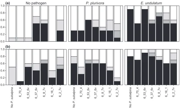
Survival and disease intensity varied considerably among the two temperature treatments, P. subalpina isolates, and the presence or absence of the two pathogens (Fig. 1). Mortality of plants inoculated with P. subalpina ranged from 0% to 50% at low and 10% to 60% at elevated tem-perature, when no pathogen was inoculated. In the absence of P. subalpina, mortality of seedlings inoculated with P. plurivora was 30% at low and 60% at high tem-perature, whereas mortality of seedlings inoculated with E. undulatum was 90% and 80% at the respective temper-ature (Fig. 1). Significance of the parameters was the same in the survival analysis and the proportional odds model (Table 1): elevated temperature, inoculation with P. subalpina and/or the two pathogens significantly increased mortality and disease intensity of Norway spruce seedlings. Moreover, the significant interaction between P. subalpina and pathogens (I9 P) indicates that some P. subalpina isolates suppressed P. plurivora and/or E. undulatum disease more effectively than others. For example, inoculation of seedlings with the isolate 6_2_7v reduced mortality caused by P. plurivora, whereas inoculation with isolates 6_70_4, 6_8_7v, 6_16_1 and
6_2_7v reduced mortality caused by E. undulatum (Fig. 1). Elongisporangium undulatum was highly virulent on P. subalpina-free controls and seedlings inoculated with isolate 6_53_6v, resulting in survival of fewer than three seedlings at both temperatures (Figs 1 and 2). Ele-vated temperature led to a general increase in disease intensity, but this increase was the same for all P. subalp-ina isolates as indicated by the absence of a significant interaction between temperature and P. subalpina. Like-wise, temperature did not alter the expression of disease symptoms caused by the two pathogens.
Plant growth–related parameters
Noninoculated seedlings (control) had the highest bio-mass (i.e. total biobio-mass, needle, shoot, and root bio-mass)
at both temperatures. Biomass varied strongly between the two temperature regimes and among seedlings inoc-ulated with different P. subalpina isolates and the two pathogens (Supporting Information, Table S1). Total biomass ranged from 4.96 to 368.9 mg, needle mass from 2.45 to 154.98 mg, shoot mass from 4.71 to 186.60 mg and root mass from 0.25 to 191.54 mg per plant. ANOVAs were performed for each pathogen
sepa-rately. One analysis was performed with all seedlings inoculated with P. plurivora and any P. subalpina isolate (including the P. subalpina-free control inoculates) (analysis 1, Table 2a) and another one with a reduced data set containing the data of seedlings inoculated with E. undulatum comprising only those P. subalpina isolates (i.e. 6_16_1, 6_2_7v, 6_70_4, 6_8_7v) that led to sur-vival of at least three seedlings at either temperature (analysis 2, Table 2b).
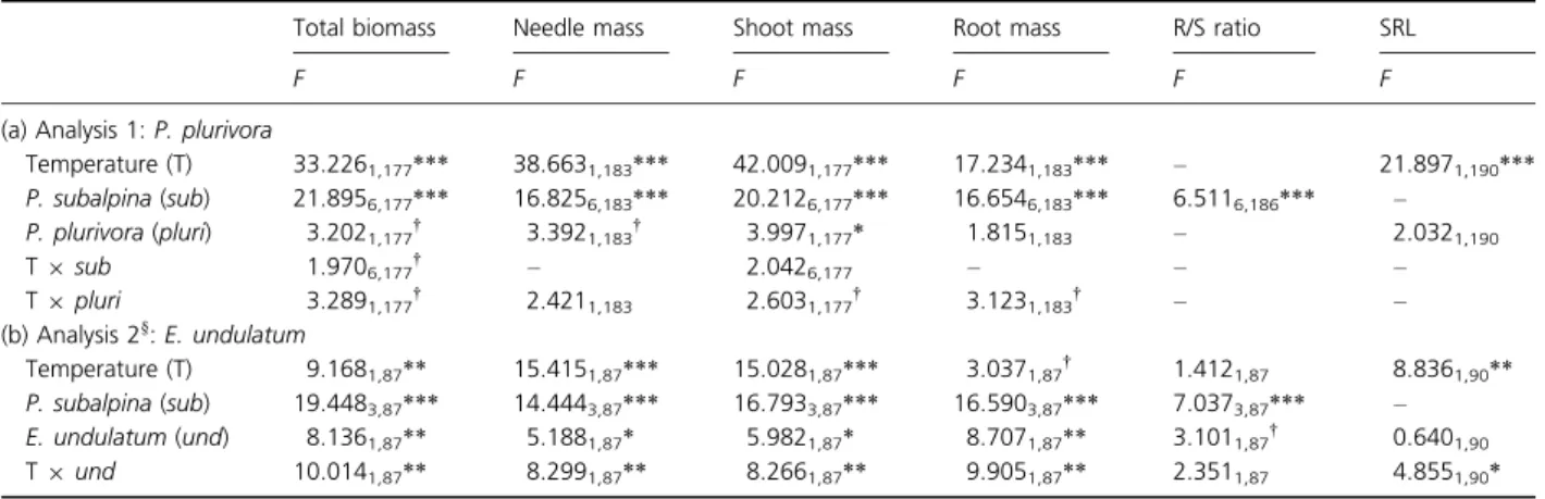
Analysis 1 showed that P. plurivora had no effect on biomass except for a slightly significant reduction in shoot weight, whereas elevated temperature and P. sub-alpina isolate significantly decreased all biomass parame-ters (Table 2a). Tukey’s HSD test revealed that seedlings inoculated with P. subalpina isolate 6_2_7v had signifi-cantly lower biomass than the controls or seedlings inoc-ulated with any other isolate. Moreover, biomass of seedlings inoculated with isolate 6_16_1 differed signifi-cantly from the controls at lower temperature. Significant
No P. subalpina 6_70_4 6_53_6v 6_37_6v 6_8_7v 6_16_1 6_2_7v 0.0 0.2 0.4 0.6 0.8 1.0(b) 6_70_4 6_53_6v 6_37_6v 6_8_7v 6_16_1 6_2_7v 6_70_4 6_53_6v 6_37_6v 6_8_7v 6_16_1 6_2_7v 0.0 0.2 0.4 0.6 0.8
1.0(a) No pathogen P. plurivora E. undulatum
No
P. subalpina
No
P. subalpina
Fig. 1. Proportional distribution of the disease intensity of seedlings at (a) low and (b) elevated temperature inoculated with and without pathogens. Phialocephala subalpina isolate names and a P. subalpina-free control are indicated below each pathogen treatment (no pathogen, Phytophthora plurivora and Elongisporangium undulatum). Black indicates dead, dark grey heavily diseased, grey mildly diseased and white
healthy seedlings. In total, there were n= 10 seedlings per treatment.
Table 1. Effects of isolate, temperature and presence of a pathogen on survival and disease intensity of the seedlings
Parameter df
Survival analysis Odds model
Waldv2 Waldv2 Isolate 6 22.526** 46.004*** Temperature 1 8.114** 9.458** Pathogen 2 93.114*** 19.782*** Isolate9 Pathogen 12 40.695*** 28.771** df, degrees of freedom. **P < 0.01; ***P < 0.001.
differences among other isolates or the control were less consistent and depended on the biomass parameter.
In the reduced data set of analysis 2, inoculation with different P. subalpina isolates distinctly affected seedling biomass parameters. Likewise, all seedling biomass param-eters were significantly decreased by inoculation with E. undulatum. In contrast, temperature significantly
decreased total biomass, needle and shoot mass, but not root mass (Table 2b). However, the interaction between temperature and E. undulatum was significant for all four biomass parameters, indicating that temperature differ-ently affected E. undulatum-free and infected seedlings at either temperature. Post hoc tests with Tukey’s HSD revealed for each biomass parameter that this significant
10 10 10 5 6 10 7 10 9 5 6 5 8 6 7 7 4 6 7 7 9 5 7 4 4 4 6 9 5 5 3 6 5 2 3 2 3 4 4 No P. subalpina 6_70_4 6_53_6v 6_37_6v 6_8_7v 6_16_1 6_2_7v 6_70_4 6_53_6v 6_37_6v 6_8_7v 6_16_1 6_2_7v 6_70_4 6_53_6v 6_37_6v 6_8_7v 6_16_1 6_2_7v 6_70_4 6_53_6v 6_37_6v 6_8_7v 6_16_1 6_2_7v 6_70_4 6_53_6v 6_37_6v 6_8_7v 6_16_1 6_2_7v 6_70_4 6_53_6v 6_37_6v 6_8_7v 6_16_1 6_2_7v 0 20 40 60 80 100 120 X X X
No pathogen P. plurivora E. undulatum
Root mass (mg) Lower temperature Elevated temperature No P. subalpina No P. subalpina No P. subalpina No P. subalpina No P. subalpina
Fig. 2. Bar charts depicting the effect of temperature and inoculation with the root pathogens Phytophthora plurivora or Elongisporangium undulatum in combination with Phialocephala subalpina on mean root mass (± standard error) of Norway spruce seedlings. Root mass is grouped
by pathogen treatment (no pathogen, P. plurivora and E. undulatum) and temperature regime. In total, there were n= 10 seedlings per
treatment. Numbers above the bars indicate the number of surviving seedlings, and ‘X’ indicates treatments with less than two surviving seedlings.
Table 2. Effects of isolate, temperature and presence of a pathogen on plant growth–related parameters‡
Total biomass Needle mass Shoot mass Root mass R/S ratio SRL
F F F F F F
(a) Analysis 1: P. plurivora
Temperature (T) 33.2261,177*** 38.6631,183*** 42.0091,177*** 17.2341,183*** – 21.8971,190*** P. subalpina (sub) 21.8956,177*** 16.8256,183*** 20.2126,177*** 16.6546,183*** 6.5116,186*** – P. plurivora (pluri) 3.2021,177† 3.3921,183† 3.9971,177* 1.8151,183 – 2.0321,190 T9 sub 1.9706,177† – 2.0426,177 – – – T9 pluri 3.2891,177† 2.4211,183 2.6031,177† 3.1231,183† – – (b) Analysis 2§: E. undulatum Temperature (T) 9.1681,87** 15.4151,87*** 15.0281,87*** 3.0371,87† 1.4121,87 8.8361,90** P. subalpina (sub) 19.4483,87*** 14.4443,87*** 16.7933,87*** 16.5903,87*** 7.0373,87*** – E. undulatum (und) 8.1361,87** 5.1881,87* 5.9821,87* 8.7071,87** 3.1011,87† 0.6401,90 T9 und 10.0141,87** 8.2991,87** 8.2661,87** 9.9051,87** 2.3511,87 4.8551,90*
Type III F–statistics from a linear model, followed by the effect and residual degrees of freedom (lowercase numbers).
†P< 0.1; *P < 0.05; **P < 0.01; ***P < 0.001 – parameters excluded by model reduction according to the AIC criterion.
‡Interactions that were excluded by the AIC criterion for all plant growth–related parameters are not shown.
§Excluding Phialocephala subalpina-free seedlings and seedlings inoculated with P. subalpina isolates 6_37_6v and 6_53_6v (< 3 surviving
interaction was due to a significant biomass reduction of E. undulatum-infected seedlings at low temperature, whereas there were no differences at elevated temperature between E. undulatum-free and infected seedlings. As in analysis 1, P. subalpina isolate 6_2_7v infected seedlings had significantly less biomass than the other seedlings in analysis 2. Moreover, there was a distinct protective effect of isolates 6_70_4, 6_8_7v, 6_16_1 and 6_2_7v against E. undulatum (Fig. 2). For any of these isolates, at least 30% of E. undulatum-infected seedlings survived, whereas almost all P. subalpina-free seedlings were killed by E. undulatum (Figs 1 and 2). Moreover, root mass of surviving E. undulatum-infected seedlings colonized by P. subalpina isolates 6_70_4 or 6_8_7v was almost as high or higher (at elevated temperature) as root mass of E. un-dulatum-free seedlings inoculated with the same P. subalp-ina isolate (Fig. 2).
The root-to-shoot (R/S) ratio ranged from 0.05 to 2.62 and differed significantly only among seedlings inoculated with different P. subalpina isolates in both ANOVAs
(Table 2). This significant effect was due to a R/S ratio reduction by isolate 6_2_7v, indicating that seedlings infected with this particular isolate had proportionally less root than shoot mass compared to plants inoculated with the other isolates and the controls.
SRL ranged from 13.04 to 101.22 mm mg 1 and was significantly increased by elevated temperature for both pathogens (Table 2). However, there was a significant interaction between temperature and E. undulatum, which was due to differences in SRL at low and elevated temperature. These differences were significant for seed-lings inoculated with E. undulatum, but not for E. undul-atum-free seedlings.
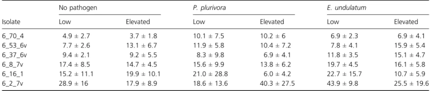
Fungal colonization density
The amount of DNA of P. subalpina in the root samples was determined using a nested qPCR (Tellenbach et al., 2010) and used as a measure for colonization density of P. subalpina. Colonization density varied among different P. subalpina isolates, but was within the same order of
magnitude (Table 3). It was neither significantly affected by elevated temperature nor by the presence of a patho-gen. After model reduction according to the AIC crite-rion, both P. subalpina isolate and pathogen were retained as factors, but only P. subalpina isolate was sig-nificant (F5,100= 8.99, P < 0.001). Moreover, the absence
of a significant pathogen by isolate interaction indicated that pathogen presence did not significantly alter endo-phyte colonization density. As revealed by Tukey’s HSD test, isolate 6_2_7v had a significantly higher colonization density than all other isolates except isolate 6_8_7v (Table 3). Moreover, isolates 6_70_4 and 6_8_7v differed significantly from each other.
Discussion
Protection of Norway spruce byP. subalpina against oomycete root pathogens
The ability of six genetically distinct P. subalpina isolates to control two oomycete root pathogens of Norway spruce was tested in vitro. The degree of protection depended on P. subalpina isolate and pathogen, and the protective effect was not an artefact due to the failure of the pathogen to establish in the plants, because presence and viability of both pathogens were demonstrated by high re-isolation rates. To estimate the efficacy of P. sub-alpina to control the pathogens, disease was quantified by estimating incidence and severity and by measuring dif-ferent plant growth parameters. All these components must be assessed to fully appreciate the ability of an endophyte to control disease. For instance, the number of surviving plants alone is a poor predictor for the fitness of a plant population as demonstrated for isolate 6_2_7v. This isolate conferred the best protection against the two pathogens in terms of mortality. However, biomass of the surviving seedlings was very poor. Under field conditions, these seedlings would certainly be outcompeted. In con-trast, the nonaggressive isolate 6_70_4 also provided very good protection against both pathogens combined with good plant growth. Thus, there is no linear relationship
Table 3. Mean Phialocephala subalpina colonization density± standard deviation (ng DNA per mg root dry weight) of each pathogen treatment
at low and elevated temperature
Isolate
No pathogen P. plurivora E. undulatum
Low Elevated Low Elevated Low Elevated
6_70_4 4.9± 2.7 3.7± 1.8 10.1± 7.5 10.2± 6 6.9± 2.3 6.9± 4.1 6_53_6v 7.7± 2.6 13.1± 6.7 11.9± 5.8 10.4± 7.2 7.8± 4.1 15.9± 5.4 6_37_6v 9.4± 2.1 9.2± 5.5 8.3± 9.8 6.9± 4.1 11.8± 3.5 15.1± 4.7 6_8_7v 17.4± 8.5 14.7± 4.5 15.6± 9.9 13.8± 6.2 19.7± 4.5 16.1± 5.8 6_16_1 15.2± 11.1 19.9± 10.1 21.0± 28.8 6.0± 4.2 22.7± 15.7 10.7± 5.9 6_2_7v 28.9± 16 17.9± 8.9 18.6± 13.6 40.3± 27.5 43.9± 9.8 25.5± 19.6
between interaction strength of P. subalpina and protec-tion against root pathogens. This is also supported by the absence of a relationship between protective effects and P. subalpina colonization density, which was previously shown to be positively correlated with seedling mortality (Tellenbach et al., 2011).
The six P. subalpina isolates used in this study were selected based on their virulence exhibited on Norway spruce seedlings in a previous experiment (Tellenbach et al., 2011). The virulence of the selected isolates ranged from neutral to highly virulent, as this corresponds to a more natural situation where isolates differing in strength of interaction with their host co-occur within the same plot (Queloz et al., 2011; Tellenbach et al., 2011). More-over, plant response to isolates differing in virulence is dis-tinct (Niks & Marcel, 2009), which in turn might affect the interaction with pathogens. The effect of the P. subalpina isolates was similar in the current study compared to the study of Tellenbach et al. (2011) with some isolates being slightly more and others slightly less aggressive. However, comparability of these two studies might be limited because experimental conditions were not exactly the same (i.e. substrate composition, temperature regime, duration). In general, the protection against pathogens by endo-phytes and nonpathogenic isolates of fungal pathogens occurs either directly by antagonism or indirectly by the induction of host resistance. Antagonism occurs when the fungus attacks and penetrates the hyphae of a pathogenic fungus (i.e. by mycoparasitism), when it sequesters antibi-otics and/or toxins that limit pathogen growth or when it competes for nutrients or space in the host tissue. These different mechanisms of protection against pathogens have been well documented in agricultural systems (Whipps, 2001; Harman et al., 2004; Tripathi et al., 2008; Alabouvette et al., 2009). In our experiment, we cannot determine the exact mechanisms of antagonism, but the data suggest that competition between P. subalpina and the two root pathogens for infection sites is not the sole protection mechanism as there was no relationship between P. subalpina colonization density and protective capability. Moreover, mechanical- and chemical-induced resistance is well known in conifers and could be another protection mechanism conferred to Norway spruce seed-lings by P. subalpina (Bonello & Blodgett, 2003). But as there were no significant differences in SRL, indicating the formation of thicker cell walls (e.g. by thickening of the exodermis; Eissenstat & Achor, 1999), mechanical-induced resistance seems to be less likely. In contrast, the production of toxins with antifungal activities against P. plurivora and E. undulatum as shown for Pinus strobus needle endophytes in disc diffusion assays against Saccha-romyces cerevisiae and Microbotryum violaceum (Sumarah et al., 2011) is likely and needs further consideration.
In contrast to mycorrhizal fungi, PAC can colonize roots of all age classes and can thus also be detected in the bark of coarse roots close to the stem base of mature trees (Gru¨nig et al., 2008). PAC frequently colonizes root tips (Menkis et al., 2004; Gru¨nig et al., 2008; Wagg et al., 2008) where they have to compete with ectomycorrhizal fungi. Interestingly, the primary roots of fungus-free coni-fer seedlings planted into forest soil as bait are usually much faster colonized by PAC than by ectomycorrhizal fungi (Ahlich et al., 1998). However, colonization of established ectomycorrhizae (ECM) by PAC is signifi-cantly less frequent than that of nonmycorrhizal root tips (Gru¨nig et al., 2008). Mycorrhizal fungi are known to protect plant seedlings against pathogens (Newsham et al., 1995; Azco´n-Aguilar & Barea, 1997; Whipps, 2004), and Richard et al. (1971) showed that ECM formed by Suillus granulatus prevented DSE (probably PAC) from colonizing and adversely affecting Picea mariana seed-lings. Similarly, PAC colonization of ECM of P. abies and Pseudotsuga menziesii was less frequent and less dense than that of nonmycorrhizal roots (V. Reininger, unpub-lished). ECM certainly form a physical barrier against col-onization by other fungi including PAC. However, as PAC seems to be the faster colonizers, they might play an important protective role against pathogens early in the life of a spruce seedling, but complete exploitation of the root might be controlled by delayed colonization by ECM.
The effect of elevated temperature
Control of either pathogen by any P. subalpina isolate was not influenced by elevated temperature. Moreover, the relative among-isolate differences in the reduction in plant growth remained the same at both temperatures, although plant biomass was reduced at higher tempera-ture. Studies that examined the influence of temperature on tripartite interactions are scarce, and many of them dealt in fact with bipartite systems (Ko¨hl et al., 1999; Singh et al., 2009; Thomson et al., 2010). Nonetheless, some studies were carried out using true tripartite sys-tems demonstrating dependency of plant protection from the interaction between temperature, other environmental parameters, pathogen isolate and antagonist species as shown for the protection of Douglas fir seedlings against Fusarium oxysporum or bean plants against Botrytis cine-rea (Strobel & Sinclair, 1991; Hannusch & Boland, 1996).
Elevated temperature did not significantly affect P. sub-alpina colonization and its influence on Norway spruce. However, the effect of elevated temperature on endo-phytes and mycorrhiza seems to be ruled by a complex set of additional factors as contradictory effects were found in different studies (e.g. Staddon et al., 2004; Ju
et al., 2006; Compant et al., 2010; Antunes et al., 2011; Brosi et al., 2011), and whether temperature affects endo-phyte as well as mycorrhiza colonization and behaviour further depends on host species (Rillig et al., 2002; Com-pant et al., 2010; Olsrud et al., 2010).
Neither P. plurivora nor E. undulatum disease intensity increased on Norway spruce at elevated temperature, although both pathogens show increased vegetative growth in culture at higher temperature. The growth optimum of P. plurivora lies at approximately 25°C (Jung & Burgess, 2009), whereas the growth optimum of E. undulatum lies at 30°C (Robertson, 1980). Sporan-gium production by E. undulatum, however, was shown to occur only at temperatures between 18 and 20°C (Goldie-Smith, 1952), whilst nothing is known of the optimum temperature for sporangium production in P. plurivora. In oomycetes, the critical step for pathogene-sis and rapid colonization of uninfected host tissues is the proliferation of asexual zoospores, which are considered the major infectious units (Erwin & Ribeiro, 1996). Therefore, reduced sporangium production might explain the better performance of surviving seedlings infected with E. undulatum at elevated temperature. Nonetheless, the mortality caused by E. undulatum at elevated temper-ature was still high. In contrast, elevated tempertemper-ature did not aggravate P. plurivora disease symptoms of the seed-lings, and it is therefore likely that P. plurivora does not pose a major threat to Norway spruce seedlings in tem-perate forests, when temperature increases.
In our experiment, Norway spruce seedling biomass was reduced by approximately 30% at elevated tempera-ture, indicating that these seedlings were more stressed under this condition. Similarly, mean biomass of black spruce (P. mariana) seedlings grown at 30°C/24 °C was reduced by about 60% compared to biomass of seedlings grown at 22 °C/16 °C (Way & Sage, 2008). Moreover, soil heating experiments showed bell-shaped growth curves for conifer trees with a maximum at around 20°C soil temperature (Lopushinsky & Max, 1990). Therefore, seedlings outside this temperature range might be slightly stressed and develop some mechanisms to evade heat stress, which in turn might also induce disease resistance (Sandermann, 2004; Wang et al., 2006).
Conclusions
In this study, we have demonstrated that P. subalpina, the most abundant and widespread PAC species, can reduce disease intensity caused by the two oomycete root rot pathogens E. undulatum and P. plurivora in Norway spruce seedlings. However, suppression of pathogens is strongly P. subalpina strain dependent, and only two of the six strains constantly reduced mortality (6_2_7v,
which, however, had a strong negative impact on host biomass) or both mortality and adverse effects on plant growth (6_70_4). Therefore, protection provided by PAC against more harmful pathogens might prompt plants to tolerate PAC despite negative effects of some strains on plant growth and health, and could explain why this symbiosis is globally widely distributed (Ahlich & Sieber, 1996; Queloz et al., 2011). Moreover, PAC might also play a stabilizing role in protecting Norway spruce against oomycete root pathogens under climate change. However, because this study was performed in vitro, the findings must be verified in field trials as well, and it is further not clear whether this protective effect also occurs in adult trees, which are densely colonized by PAC (Ahlich & Sieber, 1996; Sieber & Gru¨nig, 2006; Gru¨nig et al., 2008). Our study also gives valuable insights into the interaction between plants and endo-phytes, because data on the effects of elevated temperature are still scarce and inconclusive (Compant et al., 2010; Van der Putten et al., 2010). However, there may be other confounding factors like host and endophyte genotype or species, or other biotic and abiotic factors that need fur-ther consideration, before any conclusions about the wide distribution of PAC and their role in natural ecosystems can be drawn. Consequently, the protection of plants against pathogens by fungal endophytes under changing environmental conditions is complex. In both cases, basic knowledge of the components leading to this complexity can only be gathered with experiments performed under strictly controlled conditions, and it will be challenging, setting up field experiments to study the PAC–host inter-action in a more natural environment.
Acknowledgements
We would like to thank Veronika Netzer, Christine Rupf-lin and Angelo Duo` for technical assistance and Pascal Zaffarano, Lioba Paul, Ottmar Holdenrieder and Steve Woodward for their helpful comments. We also thank Thomas Jung for providing the P. plurivora isolate, Aria Minder and Tania Torossi and the Genetic Diversity Cen-ter (GDC), ETH, Zu¨rich, Switzerland, where qPCR was performed. This work was supported by SNF grant 3100A0-113977 from the Swiss National Science Founda-tion, Bern, Switzerland. This work was also partly sup-ported by the GEDIHAP project of the Competence Center Environment and Sustainability (CCES) of the ETH Domain, Zu¨rich, Switzerland.
References
Addy HD, Piercey MM & Currah RS (2005) Microfungal endophytes in roots. Can J Bot83: 1–13.
Ahlich K & Sieber TN (1996) The profusion of dark septate endophytic fungi in non-ectomycorrhizal fine roots of forest trees and shrubs. New Phytol132: 259–270.
Ahlich K, Rigling D, Holdenrieder O & Sieber TN (1998) Dark septate hyphomycetes in Swiss conifer forest soils surveyed using Norway-spruce seedlings as bait. Soil Biol Biochem30: 1069–1075.
Alabouvette C, Olivain C, Migheli Q & Steinberg C (2009) Microbiological control of soil-borne phytopathogenic fungi with special emphasis on wilt-inducing Fusarium oxysporum. New Phytol184: 529–544.
Antunes PM, Koch AM, Morton JB, Rillig MC & Klironomos JN (2011) Evidence for functional divergence in arbuscular mycorrhizal fungi from contrasting climatic origins. New Phytol189: 507–514.
Arnold AE, Mejia LC, Kyllo D, Rojas EI, Maynard Z, Robbins N & Herre EA (2003) Fungal endophytes limit pathogen damage in a tropical tree. PNAS100: 15649–15654. Ayres MP & Lombardero MJ (2000) Assessing the
consequences of global change for forest disturbance from herbivores and pathogens. Sci Total Environ262: 263–286. Azco´n-Aguilar C & Barea JM (1997) Arbuscular mycorrhizas and biological control of soil-borne plant pathogens– an overview of the mechanisms involved. Mycorrhiza6: 457–464.
Bonello P & Blodgett JT (2003) Pinus nigra-Sphaeropsis sapinea as a model pathosystem to investigate local and systemic effects of fungal infection of pines. Physiol Mol Plant Pathol 63: 249–261.
Brasier CM (1996) Phytophthora cinnamomi and oak decline in southern Europe. Environmental constraints including climate change. Ann For Sci53: 347–358.
Brasier CM, Kirk SA, Delcan J, Cooke DEL, Jung T & Man in’tVeld WA (2004) Phytophthora alni sp nov and its variants: designation of emerging heteroploid hybrid pathogens spreading on Alnus trees. Mycol Res108: 1172– 1184.
Brosi GB, McCulley RL, Bush LP, Nelson JA, Classen AT & Norby RJ (2011) Effects of multiple climate change factors on the tall fescue–fungal endophyte symbiosis: infection frequency and tissue chemistry. New Phytol189: 797–805. Brundrett M (2004) Diversity and classification of mycorrhizal
associations. Biol Rev79: 473–495.
Carroll G (1988) Fungal endophytes in stems and leaves: from latent pathogen to mutualistic symbiont. Ecology69: 2–9. Chavarriaga D, Bodles WJA, Leifert C, Belbahri L &
Woodward S (2007) Phytophthora cinnamomi and other fine root pathogens in north temperate pine forests. FEMS Microbiol Lett276: 67–74.
Clay K (2001) Symbiosis and the regulation of communities. Am Zool41: 810–824.
Compant S, van der Heijden, MGA & Sessitsch A (2010) Climate change effects on beneficial plant-microorganism interactions. FEMS Microbiol Ecol73: 197–214.
Desprez-Loustau M-L, Robin C, Reynaud G, De´que´ M, Badeau V, Piou D, Husson C & Marçais B (2007) Simulating the
effects of a climate-change scenario on the geographical range and activity of forest-pathogenic fungi. Can J Plant Pathol29: 101–120.
Eissenstat DM & Achor DS (1999) Anatomical characteristics of roots of citrus rootstocks that vary in specific root length. New Phytol141: 309–321.
Erwin DC & Ribeiro OK (1996) Phytophthora Diseases Worldwide. APS Press, St. Paul., USA.
Garrett KA, Dendy SP, Frank EE, Rouse MN & Travers SE (2006) Climate change effects on plant disease: genomes to ecosystems. Annu Rev Phytopathol44: 489–509.
Goldie-Smith EK (1952) The sporangial phase of Pythium undulatum Petersen. J Elisha Mitchell Sci Soc Chapel Hill N C68: 273–292.
Gru¨nig CR (2003) Population Biology of the Tree-Root Endophyte Phialocephala fortinii. Swiss Federal Institute of Technology, Zurich.
Gru¨nig CR, Brunner PC, Duo` A & Sieber TN (2007) Suitability of methods for species recognition in the Phialocephala fortinii-Acephala applanata species complex using DNA analysis. Fungal Genet Biol44: 773–788. Gru¨nig CR, Queloz V, Sieber TN & Holdenrieder O (2008)
Dark septate endophytes (DSE) of the Phialocephala fortinii s.l.– Acephala applanata species complex in tree roots: classification, population biology, and ecology. Botany86: 1355–1369.
Hamm PB & Hansen EM (1982) Pathogenicity of
Phytophthora species to Pacific Northwest conifers. Eur J For Pathol12: 167–174.
Hannusch DJ & Boland GJ (1996) Interactions of air temperature, relative humidity and biological control agents on grey mold of bean. Eur J Plant Pathol102: 133– 142.
Harman GE, Howell CR, Viterbo A, Chet I & Lorito M (2004) Trichoderma species– opportunistic, avirulent plant symbionts. Nat Rev Microbiol2: 43–56.
Harvell CD, Mitchell CE, Ward JR, Altizer S, Dobson AP, Ostfeld RS & Samuel MD (2002) Ecology– climate warming and disease risks for terrestrial and marine biota. Science296: 2158–2162.
Hendrix FF & Campbell WA (1973) Pythiums as plant pathogens. Annu Rev Phytopathol11: 77–98.
Jeffers SN & Martin SB (1986) Comparison of two media selective for Phytophthora and Pythium species. Plant Dis70: 1038–1043.
Johnson NC, Graham JH & Smith FA (1997) Functioning of mycorrhizal associations along the mutualism-parasitism continuum. New Phytol135: 575–585.
Ju HJ, Hill NS, Abbott T & Ingram KT (2006) Temperature influences on endophyte growth in tall fescue. Crop Sci46: 404–412.
Jumpponen A & Trappe JM (1998) Dark septate endophytes: a review of facultative biotrophic root-colonizing fungi. New Phytol140: 295–310.
Jung T & Burgess TI (2009) Re-evaluation of Phytophthora citricola isolates from multiple woody hosts in Europe and
North America reveals a new species, Phytophthora plurivora sp. nov. Persoonia22: 95–110.
Jung T, Blaschke H & Neumann P (1996) Isolation, identification and pathogenicity of Phytophthora species from declining oak stands. Eur J For Pathol26: 253–272. Jung T, Hudler GW, Jensen-Tracy SL, Griffiths HM,
Fleischmann F & Osswald W (2005) Involvement of Phytophthora species in the decline of European beech in Europe and the USA. Mycologist19: 159–166.
Ko¨hl J, Lombaers-van der Plas CH, Molhoek WML, Kessel GJT & Goossen-Van Der Geijn HM (1999) Competitive ability of the antagonists Ulocladium atrum and Gliocladium roseum at temperatures favourable for Botrytis spp. development. Biocontrol44: 329–346.
Lilja A (1994) The occurrence and pathogenicity of uni- and binucleate Rhizoctonia and Pythiaceae fungi among conifer seedlings in Finnish forest nurseries. Eur J For Pathol24: 181–192.
Lilja A, Lilja S, Poteri M & Ziren L (1992) Conifer seedling root fungi and root dieback in Finnish nurseries. Scand J For Res7: 547–556.
Lopushinsky W & Max TA (1990) Effect of soil temperature on root and shoot growth and on budburst timing in conifer seedling transplants. New Forest4: 107–124. Menkis A, Allmer J, Vasiliauskas R, Lygis V, Stenlid J & Finlay
R (2004) Ecology and molecular characterization of dark septate fungi from roots, living stems, coarse and fine woody debris. Mycol Res108: 965–973.
Miller J, Sumarah M & Adams G (2008) Effect of a rugulosin-producing endophyte in Picea glauca on Choristoneura fumiferana. J Chem Ecol34: 362–368.
Nechwatal J & Osswald W (2001) Comparative studies on the fine root status of healthy and declining spruce and beech trees in the Bavarian Alps and occurrence of Phytophthora and Pythium species. Forest Pathol31: 257–273.
Newhook FJ & Podger FD (1972) The role of Phytophthora cinnamomi in Australian and New Zealand forests. Annu Rev Phytopathol10: 299–326.
Newsham KK (2011) A meta-analysis of plant responses to dark septate root endophytes. New Phytol190: 783–793. Newsham KK, Fitter AH & Watkinson AR (1995)
Multi-functionality and biodiversity in arbuscular mycorrhizas. Trends Ecol Evol10: 407–411.
Niks RE & Marcel TC (2009) Nonhost and basal resistance: how to explain specificity? New Phytol182: 817–828. Ohlemu¨ller R, Gritti ES, Sykes MT & Thomas CD (2006)
Quantifying components of risk for European woody species under climate change. Glob Change Biol12: 1788– 1799.
Olsrud M, Carlsson BA˚, Svensson BM, Michelsen A & Melillo JM (2010) Responses of fungal root colonization, plant cover and leaf nutrients to long-term exposure to elevated atmospheric CO2and warming in a subarctic birch forest
understory. Glob Change Biol16: 1820–1829. Queloz V, Duo` A, Sieber TN & Gru¨nig CR (2010)
Microsatellite size homoplasies and null alleles do not affect
species diagnosis and population genetic analysis in a fungal species complex. Mol Ecol Resour10: 348–367.
Queloz V, Sieber TN, Holdenrieder O, McDonald BA & Gru¨nig CR (2011) No biogeographical pattern for a root-associated fungal species complex. Glob Ecol Biogeogr20: 160–169.
R Development Core Team (2009) R: A Language and Environment for Statistical Computing. R Foundation for Statistical Computing, Vienna, Austria.
Richard C, Fortin JA & Fortin A (1971) Protective effect of an ectomycorrhizal fungus against the root pathogen Mycelium radicis atrovirens. Can J For Res1: 246–251.
Rillig MC, Wright SF, Shaw MR & Field CB (2002) Artificial climate warming positively affects arbuscular mycorrhizae but decreases soil aggregate water stability in an annual grassland. Oikos97: 52–58.
Robertson GI (1980) The genus Pythium in New Zealand. NZ J Bot18: 73–102.
Rodriguez RJ, White JF Jr, Arnold AE & Redman RS (2009) Fungal endophytes: diversity and functional roles. New Phytol182: 314–330.
Saikkonen K, Faeth SH, Helander M & Sullivan TJ (1998) Fungal endophytes: a continuum of interactions with host plants. Annu Rev Ecol Syst29: 319–343.
Sandermann H (2004) Molecular ecotoxicology: from man-made pollutants to multiple environmental stresses. Molecular Ecotoxicology of Plants, Vol. 170. Springer, Berlin, pp. 1–16.
Selosse M-A, Baudoin E & Vandenkoornhuyse P (2004) Symbiotic microorganisms, a key for ecological success and protection of plants. CR Biol327: 639–648.
Shafizadeh S & Kavanagh JA (2005) Pathogenicity of Phytophthora species and Pythium undulatum isolated from Abies procera Christmas trees in Ireland. Forest Pathol35: 444–450.
Sieber TN (2002) Fungal root endophytes. Plant Roots: The Hidden Half (Waisel Y, Eshel A & Kafkafi U, eds), pp. 887–917. Marcel Dekker, New York and Basel.
Sieber TN (2007) Endophytic fungi in forest trees: are they mutualists? Fungal Biol Rev21: 75–89.
Sieber TN & Gru¨nig CR (2006) Biodiversity of fungal root-endophyte communities and populations in particular of the dark septate endophyte Phialocephala fortinii. Microbial Root Endophytes (Schulz B, Boyle C & Sieber TN, eds), pp. 107–132. Springer, Berlin.
Singh DP, Backhouse D & Kristiansen P (2009) Interactions of temperature and water potential in displacement of Fusarium pseudograminearum from cereal residues by fungal antagonists. Biol Control48: 188–195.
Staddon PL, Gregersen R & Jakobsen I (2004) The response of two Glomus mycorrhizal fungi and a fine endophyte to elevated atmospheric CO2, soil warming and drought. Glob
Change Biol10: 1909–1921.
Stoyke G, Egger KN & Currah RS (1992) Characterization of sterile endophytic fungi from the mycorrhizae of subalpine plants. Can J Bot70: 2009–2016.
Strobel NE & Sinclair WA (1991) Influence of temperature and pathogen aggressiveness on biological control of Fusarium root-rot by Laccaria bicolor in Douglas-fir. Phytopathology81: 415–420.
Sumarah MW, Kesting JR, Sørensen D & Miller JD (2011) Antifungal metabolites from fungal endophytes of Pinus strobus. Phytochemistry72: 1833–1837.
Tellenbach C, Gru¨nig CR & Sieber TN (2010) Suitability of quantitative real-time PCR to estimate biomass of fungal root endophytes. Appl Environ Microbiol 76: 5764– 5772.
Tellenbach C, Gru¨nig CR & Sieber TN (2011) Negative effects on survival and performance of Norway spruce seedlings colonized by dark septate root endophytes are primarily isolate-dependent. Environ Microbiol13: 2508–2517. Thomson LJ, Macfadyen S & Hoffmann AA (2010) Predicting
the effects of climate change on natural enemies of agricultural pests. Biol Control52: 296–306.
Tripathi S, Kamal S, Sheramati I, Oelmuller R & Varma A (2008) Mycorrhizal fungi and other root endophytes as biocontrol agents against root pathogens. Mycorrhiza (Varma A, ed), pp. 281–306. Springer, Berlin, Heidelberg. Uzuhashi S, Tojo M & Kakishima M (2010) Phylogeny of the
genus Pythium and description of new genera. Mycoscience 51: 337–365.
Van der Putten WH, Macel M & Visser ME (2010) Predicting species distribution and abundance responses to climate change: why it is essential to include biotic interactions across trophic levels. Philos Trans R Soc Lond B Biol Sci365: 2025–2034.
Wagg C, Pautler M, Massicotte HB & Peterson RL (2008) The co-occurrence of ectomycorrhizal, arbuscular mycorrhizal, and dark septate fungi in seedlings of four members of the Pinaceae. Mycorrhiza18: 103–110.
Walker JF, Aldrich-Wolfe L, Riffel A, Barbare H, Simpson NB, Trowbridge J & Jumpponen A (2011) Diverse Helotiales
associated with the roots of three species of Arctic Ericaceae provide no evidence for host specificity. New Phytol191: 515–527.
Walther G-R (2010) Community and ecosystem responses to recent climate change. Philos Trans R Soc Lond B Biol Sci 365: 2019–2024.
Wang CJK & Wilcox HE (1985) New species of ectendomycorrhizal and pseudomycorrhizal fungi -Phialophora finlandia, Chloridium paucisporum, and Phialocephala fortinii. Mycologia77: 951–958.
Wang D, Eyles A, Mandich D & Bonello P (2006) Systemic aspects of host-pathogen interactions in Austrian pine (Pinus nigra): a proteomics approach. Physiol Mol Plant Pathol68: 149–157.
Way DA & Sage RF (2008) Elevated growth temperatures reduce the carbon gain of black spruce [Picea mariana (Mill.) B.S.P.]. Glob Change Biol14: 624–636.
Whipps JM (2001) Microbial interactions and biocontrol in the rhizosphere. J Exp Bot52: 487–511.
Whipps JM (2004) Prospects and limitations for mycorrhizas in biocontrol of root pathogens. Can J Bot82: 1198–1227.
Supporting Information
Additional Supporting Information may be found in the online version of this article:
Table S1. Mean values and standard deviations of differ-ent plant growth related parameters.
Please note: Wiley-Blackwell is not responsible for the content or functionality of any supporting materials sup-plied by the authors. Any queries (other than missing material) should be directed to the corresponding author for the article.