Protein Engineering vol.6 no.6 pp.585-592, 1993
The nature of the ion binding interactions in EF-hand peptide
analogs: free energy simulation of Asp to Asn mutations
Blaise Prod'hom1 and Martin Karplus
Department of Chemistry, Harvard University, Cambridge, MA 02138, USA
'Present address: Institut de Physiologie, Faculty de Medecine, Univershd de Lausanne, CH-1005 Lausanne, Switzerland
The binding of the La3+ ion to a tridecapeptide, which is a
model for the EF-hand in calcium-binding proteins, is studied hi solution by free energy simulations. The calculations analyze the effect on the La3+ ion binding of the mutation
of Asp to Asn for side chains that interact directly with the ion. The results are compared with the measurements of Marsden.B.J., Hodges,R.S. and Sykes.B.D. (1989) Biochemistry, 28,8839, on the same system. They found that the Asp to Asn mutation has only a small effect on the binding; the observed differences in the free energies on changing one Asp to an Asn are between - 0 . 3 and 1.8 kcal/ mol. This result is analyzed by alchemical simulations for the tridecapeptide in the bound Qoop) structure and free (extended) form. The free energy changes due to the muta-tion of an Asp to an Asn are large and positive for both the bound and free forms. However, since the values of the free energy changes are calculated to be similar hi the two forms, the difference in the binding free energy of Asp and Asn peptides is found to be small, in agreement with experiment. By use of thermodynamic integration, the various contribu-tions to the free energy changes are estimated. In the com-plexed form, the Asp to Asn mutation is favored by the reduction in the repulsive interaction with other charged residues of the peptide; it is disfavored by the reduction of the stabilization of the ion and the surrounding water has a small effect. When the peptide adopts an extended con-formation in the absence of the ion, the mutation Asp to Asn is strongly disfavored by the interactions with the water and is favored by the interactions within the peptide. The results demonstrate the essential role of contributions to the binding of EF-hands from interactions other than those between the ion and the charged amino acid side chains. The results obtained from the simulations suggest, in accord with crystal structures of La3+ bound to various ligands, that the
calcium-binding loop complexed with La3+ in solution has a
significantly different structure from that observed hi proteins.
Key words: calcium binding motif/ion binding/EF
hands/thermo-dynamic integration
Introduction
Free energy simulations in macromolecular systems have been shown to be useful for understanding the mechanisms of drug selectivity, protein stability and pathological mutations (Brooks
etal., 1988; Karplus and Petsko, 1990; Straatma and
McCammon, 1992). Such simulations provide a bridge between biochemical and diermodynamic measurements at the macro-scopic level and descriptions of phenomena at the molecular level.
A problem of biological interest that can be analyzed by free energy simulations is ion binding to proteins. Since calcium plays an essential role in the control of cellular mechanisms from metabolism and cell signalling to secretion and contraction (Evered and Whelon, 1986; Smith and Augustin, 1988; Rasmussen, 1989; Papahadjopoulos et al., 1990; Wier, 1990), it is of importance to have a quantitative understanding of the binding mechanism. Among several families of calcium-binding proteins, the troponin C superfamily, which includes calmodulin and parvalbumin, is by far the most thoroughly studied (Forse"n, 1989; Strynadka and James, 1989). The proteins of this family have binding sites with characteristic sequences and structures (Kretsinger and Nockolds, 1973; Herzberg and James, 1985; Staryshur etal., 1988; Godzik and Sander, 1989). Generally, the binding sites consist of a loop of 12 consecutive amino acids flanked by two a-helices. These supersecondary structures, often called EF-hands (Kretsinger and Nockolds, 1973; Godzik and Sander, 1989), are grouped in pairs in many calcium-binding proteins. As crystal structures have improved [e.g. compare the recent structures of carp parvalbumin (Swain and Amma, 1989; Swain et al., 1989; Kumat et al., 1990) with the pioneering study of Kretsinger and Nockolds (1973)], the ion coordination has been shown to be more similar throughout the superfamily (Strynadka and James, 1989).
It has been observed that the affinity of mese proteins for calcium depends in part on the number of acidic (negatively charged) residues of the loop participating in the coordination of the ion (Reid and Hodges, 1985; Marsden etal., 1990). However, the binding is not simply related to the number of carboxylate groups involved. So far no mutant studies have been made to quantitate this observation. Marsden et al. (1988) have conducted NMR studies of the interaction between rare earth ions and several tridecapeptides with sequences based on that of die calcium-binding site IJJ of rabbit skeletal muscle troponin C. They showed diat such peptides in solution do not form well-defined complexes with Ca2+, although at high calcium concentration
there is evidence from circular dkhroism that the peptide structure is affected. When trivalent lanthanide ions are used, the peptides adopt a conformation similar to the loop of an EF-hand (Marsden
etal., 1989). Using nuclear Overhauser enhancement (NOE)
measurements they were able to estimate 33 hydrogen-hydrogen distances of the peptide in solution in the presence of the lanthanide ion. With gadolinium-induced *H relaxation measure-ments, they determined 31 gadolinium-hydrogen distances. The root mean square difference (r.m.s.d.) between these distances and mose measured in the crystal structure of turkey troponin C was 1.1 A. Minimization and molecular dynamics simulations
in vacuo with the NOE- and Gd-induced relaxation constraints
yielded mean structures that had r.m.s.d.s from the crystal structure of 1.1 A for the main chain and 1.8 A for all heavy atoms. Small differences in the conformations of die peptides have been observed with different landianides (Gariepy et al., 1983). There was no evidence for a stable loop structure in the absence of ions, suggesting that the charge repulsions of me side chains leads to an extended random coil.
Since the solution structures of these peptides appear to be similar to that observed in calcium-binding proteins, Marsden
et al. (1988) synthesized several tridecapeptide homologs of the
loop segment of site HI of rabbit skeletal troponin C with some aspartate residues of the binding site substituted by asparagine. By studying a series of related peptides, they were able to estimate the contribution of each of these ligands to the lanthanum ion affinity. Although no detailed structural studies of the modified peptides were made, it appears likely that the structures of the liganded loops are close to that of the peptide sequence whose structure was studied by NMR. Based on this assumption, Marsden et al. (1988) estimated the relative contribution of an Asn versus an Asp at the various positions and obtained values in the range —0.3 to 1.8 kcal/mol.
To understand the origin of the small difference in ion binding resulting from Asp to Asn substitutions, we have performed alchemical free energy simulations (Gao et al., 1989; Tidor and Karplus, 1991; Straatma and McCammon, 1992) on a peptide, whose sequence corresponds to peptide I in the terminology of Marsden et al. (1988),
1 2 3 4 5 6 7 8 9 10 11 12 13 (I)
Asp - Arg - Asp - Ala - Asp - Gly - Tyr - lie - Asp - Ala - Glu - Glu - Leu Free energy simulations were performed using the exponential formula and thermodynamic integration (Tidor and Karplus, 1991) in which residues 3 or 5 [peptide N and HI in the Marsden
et al. (1988) nomenclature], were transformed from Asp to Asn;
both of these residues participate in the ion coordination. The strong Ca2+-binding site (HI) in rabbit troponin C, in fact, has
an Asn instead of an Asp at position 3. In turkey and chicken troponin C, for which crystal structures are available (Strynadka and James, 1989), Tyr7 is replaced by Phe and AlalO by lie. The residues participating in the coordination shell of the calcium in these structures correspond to positions 1, 3, 5, 7 (all monodentate) and 12 (bidentate); the ligands of 1, 3, 5 and 12 to Ca2+ are side chain oxygens, while for Tyr7 the backbone
carbonyl is ligated to the Ca2+ (Herzberg and James, 1985;
Satyshur et al., 1988). In addition, one water is coordinated with the calcium. The coordination number is 7 and the ligand atoms have an approximately pentagonal bipyramidal arrangement. The most recent refinements of the carp parvalbumin coordinates (Swain and Amma, 1989; Swain et al., 1989; Kumat et al., 1990) show a similar seven-coordinate arrangement of the CD loop around the Ca2+ ion, which differs somewhat from the earlier
results (Kretsinger and Nockolds, 1973).
Although the overall structure of the loop appears to be similar in the crystal structure of the protein and in the peptide in solution, there is no evidence that the detailed geometry and the ion coordination are the same. The NMR results give little information on the side chain geometry and, in particular, the distances between the ion and possible ligands have not been measured. It is likely that when the Ca ion is replaced by La3+ die coordination will change; the ionic radius of Ca2+ is
0.99 A versus 1.15 A for La3+ (Pauling, 1948) and the charge
increases from + 2 to + 3 . Significant differences were observed between the NMR spectra of Lu3+- and La3+-bound peptides
(Gariepy et al., 1983). In addition, there is the possibility that alterations in the loop structure are induced by the absence of the protein. Thus, the peptide loop studies of bound La3+ must
be considered as approximate models of the Ca2+-binding site
in proteins, although they are of interest in themselves. The simulations show that the free energy of mutation of a
charged amino acid (Asp) into a polar amino acid (Asn) leads to nearly the same free energy change in two environments as different as a relatively protected cation-binding site and an extended chain where the amino acid is fully exposed to aqueous solution. To analyze this result, the individual contributions to the change in free energy of binding were estimated by the thermodynamic integration method (Gao et al., 1989; Tidor and Karplus, 1991).
Methodology
Alchemical free energy simulations (Gao et al., 1989; Tidor and Karplus, 1991; Straatma and McCammon, 1992) of the mutation of Asn into Asp at positions 3 and 5 of peptide I were performed. This direction was chosen because it was simpler to orient the oxygen of Asn to coordinate wim the La3+, although in an exact
treatment the direction of the mutation should make no difference. We use the terms 'alchemical' and 'computer alchemy' for this type of simulation (Gao et al., 1989; Tidor and Karplus, 1991; Straatma and McCammon, 1992) because we transmute one amino acid into another along a reversible path that makes it possible to calculate the free energy difference. Since no such path exists for the real system, the calculated free energy is not directly accessible by experiment. However, when alchemical simulations are done for the peptide in two different conforma-tions (a loop structure corresponding to the bound system and an extended structure corresponding to the system without an ion), die difference between the two alchemical free energy changes can be measured. This is made clear by the thermo-dynamic cycle:
AGi(Asp—Asn) Loop (La3+; Asp)
Ext (Asp) + La
•-Loop (La3+; Asn)
- e ) (U)
••Ext (Asn) + La,3 + AGe(Asp—Asn)
where AG^Asp—Asn) and AGe(Asp—Asn) correspond to the
alchemical free energy change associated with the transformation Asp—Asn in the loop conformation (with die ion) and the extended conformation (widiout the ion) respectively and A G A ^ I — e ) and A G ^ l — e) correspond to the chemical free energy change associated with the transition from the loop conformation (with the ion) to the extended conformation (without the ion) for die peptides containing Asn and Asp respectively. We use the notation LI for the La3+ binding loop with the
peptide I sequence (see Introduction) and L3, L5 for the loop with Asp3—Asn and Asp5 — Asn respectively. The correspond-ing notation El, E3 and E5 is used for the extended chain without LJr+. Thus, the top horizontal line in scheme II corresponds to
die mutation LI to L3 or L5 and the bottom horizontal line to die mutation El to E3 or E5.
The chemical and alchemical free energy values can be compared by use of die double free energy difference, AAG,
AAG = A G ^ l - e ) - A G ^ l - e )
AGe(Asp— Asn) - AG^Asp—Asn) (1)
Free energy simulation of Asp to Asn mutations
Since the atomic coordinates of rabbit skeletal muscle troponin C were unavailable when we undertook this study and the published NMR data are not sufficient to generate accurate coordinates, the crystal coordinates of the CD site (residues 51 - 6 3 ) of carp muscle parvalbumin (Kretsinger and Nockolds, 1973) were used to generate the starting structure of the bound system; the coordinates were obtained from the Brookhaven Protein Data Bank (the structure used has the file name 3PTP; Bernstein et al., 1977). The r.m.s.d. between the main chains of the corresponding loops of troponion C and carp muscle j3 parvalbumin is stated to be 0.428 A (Herzberg and James, 1985; Satyshur et al., 1988), suggesting that the choice of main chain coordinates is appropriate. To correspond to the chemical structure of the synthetic peptides, an acetyl group was introduced at the N-terminus, an amide group was introduced at the C-terminus and the side chains of the residues were modified to match the sequence of peptide I that was used for the simu-lation. The changes required are Glu2 — Arg, Lys4 — Ala, Ser5 - Asp, Phe7 - Tyr, Glu9 - A s n , GlulO - A l a and Asp 11 — Glu. In the original crystal structure (Kretsinger and Nockolds, 1973), which we used to build the loop model, the Ca2+ ion is coordinated approximately octrahedrally by six
oxygen ligands (OD1 of Aspl, OD1 of Asp3, OG of Ser5, O of Phe7, OE1 of Glu9 and OE1 of Glul2 following the numbering system in scheme I). As already mentioned, this ligation scheme differs from the more recent structural data (Swain and Amma, 1989; Kumat et al., 1990), in which the ion is seven-coordinated (OD1 of Aspl, OD1 of Asp3, OG of Ser5, O of Phe7, OE1 of Glu9 and OE1 and OE2 of Glu 12), which is essentially the same as in troponin C(m). Although the interactions in the model-built tridecapeptide differ significantly from those in the loop of troponin C, the structural rearrangements that occur in the simulation and the similarity of the resulting structures to those found in crystal complexes of La3+ (Hoard et al., 1965; Lind
et al., 1965) suggest that the procedure used is meaningful. In
going from parvalbumin to troponin C all of the side chains of the altered residues except Asp5 and Glu9 point towards the outside of the loop and do not interact significantly with the ion or the rest of the peptide. The modified residues were built so that common atoms coincide and the remaining atoms were added in the most probable conformation and then minimized with the rest of the peptide fixed. Thus, for Asp5 in place of Ser, the C/3 position was overlapped. Polar hydrogen atoms were constructed with the HBUILD facility (Briinger and Karplus, 1988). The loop was embedded in a sphere of water with a radius of 14 A centered on the ion. The system includes 351 molecules of water introduced by three overlays with a box of TIP3P water and was confined by use of the stochastic boundary method (Brooks and Karplus, 1989); an 11 A molecular dynamics region and 3 A Langevin dynamics boundary region were used. While the water molecules close to the surface of the sphere were acted on by a boundary potential (Brooks and Karplus, 1989), the peptide loop was far enough from the border of the sphere (i.e. all of it inside the 11 A radius) to be simulated solely by molecular dynamics. For long-range truncation, the shift option was used for electrostatics and switch for van der Waals with a cut-off of 9.0 A for both electrostatic and van der Waals interactions. The shift option has been shown to yield satisfactory results in comparison with simulations without truncation (Loncharich and Brooks, 1989; Stote and Karplus, 1991). The SHAKE algorithm (van Gunsteren and Berendsen, 1977) was used to constrain the bonds between hydrogen atoms and heavy atoms. The system was minimized and equilibrated at 327 K. The water was first
equilibrated for 1 ps while the peptide atoms were kept fixed. Then 5 ps of simulations were done while imposing the NOE-and Gd-induced relaxation constraints of Marsden et al. (1988) on the hydrogen-hydrogen and ion—hydrogen distances. Finally, 7 ps of equilibration were done without constraints. The free energy simulations were performed with no constraints.
As already mentioned, Marsden et al. (1989) have shown that peptide I does not form a loop in the absence of a lanthanide ion. We made the assumption that the side chains of the acidic residues would be fully exposed to solvent and that the value of the free energy of mutation in an extended peptide would be representative of the mean free energy change calculated from an ensemble of structures. An extended peptide was built by setting the <f> and $ dihedral angles of the main chain to 180° and minimizing the structure in vacuo; the other parameters had the default values in the CHARMM topology file (Brooks et al., 1983). The peptide was placed in a 13 A radius sphere of water; the region between 11 and 13 A was used as a buffer region. Because of its extended structure, only five residues were within the sphere; the rest of the peptide was deleted. The conditions and protocols of the simulations of the peptide in the loop conformation, were also applied to the extended conformation simulations; a total of 310 water molecules was included.
The simulations were carried out with the BLOCK facility (Tidor and Karplus, 1991) in the program CHARMM (Brooks
et al., 1983). The force constants, atomic partial charges and
Lennard- Jones parameters of the peptide atoms were taken from the parameters of the CHARMM 19 polar hydrogen set. A modified 1TP3P model (TIP3P/C) (A.MacKerrell and M.Karplus, personal communication), which differs slightly from the original Jorgensen TIP3P model (Jorgensen, 1981), was used to represent the water molecules. A simple point charge model was used for La3+ since it has no f electrons outside the core.
More generally it has been suggested on structural and thermo-dynamic grounds that the f electrons play a relatively small role in the rare earth chelates (MacKay et al., 1962; Hoffmann et al., 1977; Cossy et al., 1989). The R^ll and e Lennard-Jones parameters of the lanthanum ion were taken to be 1.71 A and - 0 . 1 2 kcal/mol respectively. These parameters yield a lanthanum-TIP3P/C interaction of -75.5 kcal/mol at 2.35 A. For comparison, a recent ab initio effective core potential calcula-tion of the lanthanum—water interaccalcula-tion gives a minimum of -83.5 kcal/mol at 2.42 A (Meier et al., 1990). Although electronic polarization effects are expected due to the presence of La3+, the calculations are simplified by choosing parameters
that yield the approximately correct interaction energies without explicit polarization. This is analogous to the introduction of implicit polarization by the use of large charges in TIP3P water, for example (Jorgensen, 1981).
For the free energy simulations, a hybrid potential energy function of the form K(X) = (1 - X) KA + XKB was introduced
where X is a parameter that goes from zero [K(0) = VA, the
energy function for the initial system] to one [K(l) = VB, the
final system]. To determine the alchemical free energy changes, AG(A—B), both the exponential formula and thermodynamic integration (Mezei and Beveridge, 1986; Gao et al., 1989; Tidor and Karplus, 1991) were used; the exponential formula is
AG = Gs - GA = - (2)
AG = GB - GA = - \l < A K >X dX = £ <AK> XAA, (3)
In equations (2) and (3) the expression < >x represents a
simulation with the energy function V(\); AV = KB — VA. The
mutations were done in five steps, corresponding to X = 0.1, 0.3, 0.5, 0.7 and 0.9. For each step, the system was equilibrated for 10 ps and then sampled for 10 ps.
To obtain a correctly oriented Asn side chain with its oxygen coordinating with the ion, the simulations were started with a model-built structure for Asn and mutating it to Asp. In the starting structure for the free energy simulation, the duplicated side chain had the common atoms of Asn and Asp in the same positions. Thus, the only difference between VA and VB is that
the former has an Asn side chain and the latter an Asp side chain. Although the Asn and Asp side chains interact with the rest of die system during the simulation, they do not interact widi each other.
Results
We first describe the structural results and then consider the binding thermodynamics and its analysis.
Structural aspects
Figure la and b shows the loop and extended structures with the Asn and Asp copies of the mutated side chain. The main chain r.m.s.d.s between the initial model-built structure (LI) and the L3 and L5 structures were 1.68 A and 2.40 A respectively after equilibration in a sphere of water. Asp9 has moved out of the ion coordination sphere and is exposed to solvent. In all three peptide loops (LI, L3 and L5), the La3+ ion coordinates 10
oxygens. In L3, both Aspl and Asp5 interact in a bidentate mode with the La3+ ion; Glul2 is coordinated in a monodentate mode,
although the second oxygen is also relatively close (see Table I). Asn3 has its oxygen coordinated to the ion. In addition, the ion is coordinated with the main chain carbonyl oxygen of Tyr7 and three water molecules. The mean distance between La3+
and the three waters is 2.64 A (2.55, 2.49 and 2.88 A), while the Tyr7 carbonyl oxygen is 2.35 A from the ion. In L5, there is a corresponding coordination involving Aspl, Asp3, Asn5 and Glul2, though the roles of OE1 and OE2 are interchanged. Also, the backbone carbonyl oxygen of Tyr7 is not in direct contact widi die ion. It is replaced by a water oxygen, bringing die number of water molecules in the coordinates sphere to four. The mean ion-water distance is 2.60 A (2.67, 2.55, 2.55 and 2.64 A).
Since the simulations done were Asn3—Asp for L3 and Asn5—Asp for L5, both L3 and L5 have the peptide sequence LI as meir final state. Thus, the structures of diese two LI loops can be compared. They are similar but not identical (the main chain r.m.s.d. is 1.89 A) and the La3+ ion is not coordinated
in exactly the same way. In both cases, die ion is coordinated by 10 oxygen atoms, but die oxygens involved are not all the same. LI (L5) coming from the mutation Asn5 —Asp has three carboxylate groups liganding the ion in a bidentate fashion [Aspl, Asp3 and Glul2; Glul2 changes from unidentate to bidentate while going from L5—LI (L5)], die mutated Asp5 stays in a unidentate mode and diree water molecules supply the three additional oxygen ligands [mean ion-water distance, 2.53 A (2.56, 2.52 and 2.51 A)]. LI (L3) resulting from the mutation Asn3 —Asp has two carboxylate groups in a bidentate mode (Aspl and Glul2), while Asp3, die mutated residue, and Asp5 interact widi die ion in a unidentate mode; die remainder of die coordination sphere is made up by the Tyr7 main chain oxygen (ion—oxygen distance 2.63 A) and three water molecules widi mean ion-oxygen distance of 2.57 A (2.55, 2.49 and 2.88 A). Thus, die LI loops essentially retain die structure diat mey had in me L3 and L5 form, except diat in L5 die additional oxygen of Glul2 replaces one of the water molecules. It is likely diat die two different LI structures reflect two possible forms of nearly equal free energy. Because die essential coordination is very similar, the effect on the free energy simulation results is expected to be small.
Fig. 1. (a) Cross-eyed stereo plot of the loop structure of peptide I with the ion (black dot) and peptide Asn5 superimposed on Asp5. (b) Cross-eyed stereo
plot of the extended structure with Asn3 superimposed on Asp3; only the five residues included in the simulation sphere are shown. 588
Free energy simulation of Asp to Asn mutations
The peptide in the bound conformations forms a bowl inside which the La3+ ion is located and the water molecules form a
cap. The structure is thus significantly different from the Ca2+-binding loops found in the proteins. However, it is
reminiscent of La3+ complexes with EDTA for which crystal
structures are available. In the crystal structure of the complexes La3+EDTA(H2O)3(26) and La3+EDTAH(H2O)4 (Lind et al.,
1965), La3+ is coordinated by one oxygen of each of the four
carboxyl groups and the two nitrogens, which form a bowl and three or four H2O molecules which form a cap. The observed
L a3 +- x distances are 2.51 (2.54) A for x equal to O~, 2.76
(2.87) A for x equal to N and 2.58 (2.59) A for x equal to O of H2O; the two values given in each case correspond to those
observed in the two different compounds. Thus, the overall structure and distances for the oxygen ligands are very similar to those found here. In lanthanide crystals formed with guanidinhim acetate or nicotinate where the La3+ is surrounded by oxygen
atoms the coordination number can vary from 7 to 10 (Sinha, 1976). Some considerations of the structure and energetics of the lanthanides as a function of ion size are given in MacKay
et al. (1962) and Hoffmann et al. (1977). It has been observed
that the same general type of cage structure is found for the EDTA chelates of the different lanthanides. However, the details of the structure (e.g. number of coordinated H2O molecules) are
sensitive to the ion radius. There is a decrease in coordina-tion from 10 to eight as the ion size decreases from La3+ (ionic
radius 1.15 A) to Lu3+ (ionic radius 0.85 A). These radii are
to be compared with the value of 0.99 A for Ca2+. X-ray
diffraction measurements of lanthanum chloride solutions give an ion to water oxygen distance of 2.48—2.58 A and a co-ordination number of eight to nine (Smith and Wertz, 1975; Habenschuss and Spedding, 1980).
The mean numbers of water molecules, whose oxygen is at a distance smaller than 3.3 A from OD, OE, ND of Asp, Asn or Glu were examined in the bound and extended structures. The mean number of water molecules around an Asp in the extended peptide is 6.4, slightly less than the value (7.1) found for a non-ligating Asp in the loop with bound La3+. The ligating Asp
residues in the loop structure have a mean of 3.2 water molecules around them, of which approximately half (mean 1.4) are also in the coordination sphere of the ion. In the extended conforma-tion the mean number of water molecules around an Asp is 6.0; in the loop structures, all of the Asn side chains are coordinated with the La3+ ion. There are no experimental data or other
simulation results for comparison.
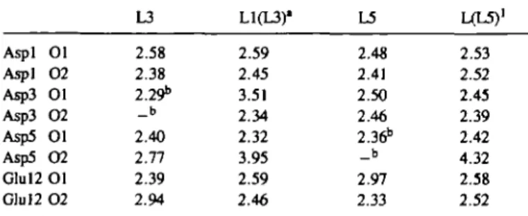
Table I. Distances between La34 the loop conformations (A)
and the oxygens of the acid residues in
L3 L1(L3)" L5 LCL5)1 Aspl 01 Aspl 0 2 Asp3 01 Asp3 O2 Asp5 01 Asp5 0 2 Glul2 Ol Glu 12 O2 2.58 2.38 2.29b _ b 2.40 2.77 2.39 2.94 2.59 2.45 3.51 2.34 2.32 3.95 2.59 2.46 2.48 2.41 2.50 2.46 2.36b _ b 2.97 2.33 2.53 2.52 2.45 2.39 2.42 4.32 2.58 2.52 "L1(L3) corresponds to the structure LI obtained from a simulation that started with L3; L1(L5) is defined in the same way.
bThe residue corresponds to an Asn in L3 or L5 and so has only a carbonyl oxygen that can coordinate the ion.
Thermodynamics
To relate the alchemical simulation results to the measurements by Marsden et al. (1988) of the difference in the La3+-binding
free energy between LI and L3 or L5, we make use of the thermodynamic cycle shown in Scheme II. The top line represents the mutations of the peptide in the loop conformation in the presence of the ion, while the bottom line represents the mutation in the extended conformation of the peptide in the absence of the ion. Since the solvation free energy of the free ion is constant, its contribution cancels in AAG, which appears on the right-hand side of equation (1). The vertical lines reflect the difference in free energy of binding for peptide I and the mutants determined from the experimental binding affinity data; this difference gives the AAG on the left-hand side of equation (1). The measured values of the affinity constant and the corresponding free energies are shown in Table n . As can be seen L3 has the highest affinity, LI is intermediate and L5 has the lowest affinity, though all of the differences are small.
The free energy differences calculated for the alchemical change (Asp to Asn) in the loop or extended conformation by thermodynamic integration (TI) and the exponential formula (EF) are presented in Table HI. The differences between the free energy values calculated by the two methods vary between 0.7 and 2.9 kcal/mol. This sets a lower limit to the uncertainty of the results for this system with the present protocol. Although these values cannot be compared directly with the experiment, they are of considerable interest. The alchemical changes are sufficiently large that the inherent error (of the order of 10%) is tolerable. The mutation of the negatively charged Asp into the polar Asn is always very unfavourable, as indicated by the positive sign of the free energy differences. The values are rather similar in the loops and in the extended structures, ranging from 41.9 kcal/mol in El to E5 with the EF calculation to 48.1 kcal/mol in LI (L5) to L5 with the TI calculation. The values are much smaller than the difference in the La3+
inter-action energies for Asp and Asn. The calculated interinter-action between La3+ and a single acid group in the binding site varies
between - 2 4 6 kcal/mol for Asp3 in LI (L3) to - 3 2 7 kcal/mol for Aspl in L5; the much weaker La3+ - A s n interaction varies
between - 6 3 and - 7 0 kcal/mol. These values are obtained by averaging the interaction energy over 10 ps of the simulation.
Table D. Experimental Marsden et al. (1988)* nomenclature I N
m
*AG(1 — e) refers to the affinities'
Type of loop Af(mol LI L3 L5 1.4 X 2.5 x 1.8 x vertical arrows in Scheme D".
Table ID. Calculated alchemical free
AG(Asp3-Asn) AG(Asp5-Asn) Loop (with TI 47.6 48.1 energy changes ( La3*) EF 46.4 45.3 ~I } 105 105 10* >cal/nx A G ( l - e ) (kcaVmol)* 7.11 7.46 5.88 Extended TI 44.3 42.6 EF 47.2 41.9 'Although the simulations were done in the direction Asn to Asp, the free energy changes are listed as Asp to Asn.
Table IV shows the calculated and experimental AAG values. The difference between the calculated AAG values obtained with the two methods (TI and EF) is larger than the experimental binding free energy differences. Thus, predicting the relative affinity of the different peptides for the lanthanide ion is not possible. The only important result is that the differences in affinities are small and of the same magnitude as the experimental results. Nevertheless, we note that comparison of the AAG values of L3/L1 and L5/L1 shows that the calculated order agrees with that found experimentally.
Decomposition of the free energy change resulting from the Asp to Asn mutation into contributions from the components of the system has been performed with the thermodynamic integration method, as described previously (Gao et al., 1989; Tidor and Karplus, 1991). The results are shown in Table V, which gives the contributions of the water, the peptide and the ion; the self-term is dominated by the change in the number of degrees of freedom and makes only a small contribution. Although there are quantitative differences between the results for the two mutants, the overall features are very similar and, as already mentioned, the differences may not be significant. Thus, we concentrate on aspects of the results that are common to the two sets of calculations.
Of particular interest is the possibility of interpreting the fact that the free energy of mutation of a charged residue (Asp) into a polar one (Asn) is similar in the aqueous solvent environment and in the cation-binding site (see Table V). In the bound form the largest free energy term is the loss of the interaction between La3+ and the Asp side chain, which is replaced by the much
smaller interaction between La3+ and the neutral, although
polar, Asn side chain. A significant fraction of this destabilization (approximately 100 out of 150 kcal/mol) is regained by the reduction in repulsive interactions between the closely spaced negatively charged side chains in the loop conformation in going from Asp to Asn (see also below). The solvent contribution is small in the loop configuration with the bound ion. There is some indication, in accord with the structural results (see above) that there is a compensation between the solvent and the peptide contributions for the two mutants in the bound form, i.e. the
solvent plus peptide values are very similar (—95.1 and - 9 3 . 7 kcal/mol for L 1 - L 3 and LI —L5 respectively), while the individual terms differ by more than 14 kcal/mol.
For the extended configuration in the absence of the La3+ ion,
the qualitative features of the interactions are very different. There is of course no La3+ interaction term, the reduction in the
repulsive interactions is much smaller (in the order of 30 rather than 100 kcal/mol) and the solvent contribution is dominant. This is not unexpected because the residues are further from each other and completely immersed in solvent. The calculated values (87.8 kcal/mol for L3 and 68.3 kcal/mol for L5) are of the order of the free energy of solvation of acetate (84 kcal/mol) (Yu and Karplus, personal communication) and similar to the value obtained in a free energy simulation form of acetate to acetamide (-71.6 kcal/mol) in aqueous solution (Bash et al., 1987).
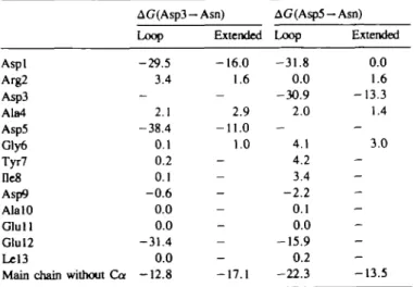
Further decomposition of the peptide free energy change into contributions from the individual amino acids is shown in Table VI. The side chain atoms and the Ca atom of each residue are included. The overall changes in backbone interactions (excluding Ca) are listed separately. Although not considered in the previous discussion, the latter make a significant contribution. The Asp to Asn mutation is favored due to the decrease in repulsion between the side chain and the main chain carbonyl groups. For Asp3 — Asn, the effect is to destabilize the bound form relative to the extended by 4.3 kcal/mol, while there is a reverse effect of 8.8 kcal/mol for Asp5 — Asn. The side chains which strongly favor the mutation Asp—Asn are the amino acids with a carboxylic group that is close to the mutated residue. For analysis, we define the distance between two acidic side chains as the mean distance between the oxygens. In the folded state, the charged residues tend to be closer together than in the extended form. Thus, they have a large influence on the free energy of mutation; the values range from -16.2 kcal/mol for Glul2 at 5.4 A from the mutated residue in L5 to -39.1 kcal/mol for Asp5 at 4.02 A from the mutated residue of L3. Carboxylate residues which are far from the mutated residue (and from the ion) make only a small contribution, i.e. Asp9 and Glul 1, whose oxygens are 10.0 and 12.7 A way from Asn3 and 8.4 and 13.8 A away from Asn5, have almost no effect. Correspondingly, the interaction of the Asp residues in the extended form of the peptide is smaller,
Table IV. Calculated and experimental AAG values (kcal/mol)1
System Calculation Experiment
TI EF Mean Asp3 — Asn Asp5 —Asn - 3 . 3 - 5 . 5 +0.8 - 3 . 4 - 1 . 3 - 4 . 5 +0.35 - 1 . 2 3 *AAG is defined in equation (1); see also footnote to Table HI.
Table V. Contributions to the mutation free energies (kcal/mol)"
AG(Asp3-Asn) AG(Asp5-Asn)
Loop Extended Loop Extended
Solvent Peptide Self Total 150.1 9.6 -106.7 - 5 . 4 47.6 87.8 - 3 8 . 5 - 5 . 0 44.3 145.9 - 4 . 5 -89.2 - 4 . 0 48.1 68.3 - 2 0 . 8 - 4 . 9 42.6 •Values are calculated by the thermodynamic integration method (see text); see also footnote • of Table m .
Table VI. Peptide side chain contributions to the mutant free energies
(kcal/mol)
AG(Asp3-Asn) AG(Asp5-Asn)
Loop Extended Loop Extended
Aspl -29.5 - 1 6 . 0 - 3 1 . 8 0.0 Arg2 3.4 1.6 0.0 1.6 Asp3 - - -30.9 - 1 3 . 3 Ala4 2.1 2.9 2.0 1.4 Asp5 -38.4 - 1 1 . 0 Gly6 0.1 1.0 4.1 3.0 Tyr7 0.2 - 4.2 De8 0.1 - 3.4 Asp9 - 0 . 6 - - 2 . 2 AlalO 0.0 - 0.1 Glull 0.0 - 0.0 Glul2 -31.4 - - 1 5 . 9 Lel3 0.0 - 0.2
Main chain without Ca - 1 2 . 8 - 1 7 . 1 - 2 2 . 3 - 1 3 . 5 •Values are calculated by the thermodynamic integration method (see text); see also footnote * of Table m .
Free energy simulation of Asp to Asn mutations
-11.4 to -16.1 kcal/mol. There is an approximate inverse correlation between the distance separating a given carboxylate side chain from the mutated Asp and die contribution of the residue to the free energy of mutation. If an effective dielectric constant Z)efr is used to describe the dependence of AG on the
distance r (in A) between two charges qx and q2 (in electronic
units) (Russell etal., 1987; Bashford and Karplus, 1990),
AG = 332 qxq2
rD,'eff
the calculated free energies yield an effective dielectric constant between 2 and 4.
In both conformations, loop and extended, the contributions of the other side chains are very small (see Table VI). The largest contribution of the non-coordinating amino acids comes from Tyr7 in L5 with a value of 4.2 kcal/mol. The contribution of the van der Waals term to the alchemical free energy changes per side chain is also very small. The values range from 0.3 kcal/mol for L1(L5)-L5 to 1.0 kcal/mol for L1(L3)-L3. Thus, the van der Waals contribution is less than 3% of the total, which is dominated by the electrostatic term. However, for the AAG values, the van der Waals contribution, which is - 0 . 3 kcal/mol for Asp3 —Asn and 0.4 kcal/mol for Asp5 — Asn, is 10-25% of the total. As found in other studies (Gao et al.,
1989; Tidor and Karplus, 1991), the cancellation of the larger electrostatic term in AAG is much greater than that for the van der Waals contribution.
The analysis of Marsden etal. (1988) and of Reid and Hodges (1985) on the effect of a mutation of Asp to Asn in different positions on the stability of the complex can be examined in light of the above results. The observation that the mutant Asp3—Asn leads to stronger binding, in contrast to all other cases where there is a reduction in binding when an Asp is replaced by an Asn, is explained by Marsden et al. (1988) on the basis of the conclusion of Reid and Hodges (1985) that Asn3, which corresponds to the wild type, acts as a spacer to reduce the electrostatic repulsion between the charged groups, i.e. it is argued that the complex is stabilized by having Asn instead of Asp, even though the interaction with the ion is weaker. Although the simulations are not precise enough to draw conclusions concerning the difference in the overall binding free energy (the difference is only 0.3 kcal/mol), an analysis of the model of Reid and Hodges (1985) is possible. From Table V, we see that AG (Asp3—Asn) in the bound loop has indeed a larger stabilizing effect due to the reduction of charged side chain repulsions (—106.7 kcal/mol) than the Asp5 —Asn mutation (-89.2 kcal/mol). The difference of - 1 7 . 5 kcal/mol is much larger than the observed change in binding free energy. The calculations show essentially the same difference (—17.7 kcal/mol) in the extended form, so that the overall con-tribution of this term is small. Thus, the implicit assumption in the model of Reid and Hodges (1985) that only the bound configuration has to be considered is apparently not valid. From the decomposition in Table VI, it is clear that the major difference between the effect of the two mutations in the bound loop is that the repulsive interaction between Asp3 and Glul2, which are neighbors in the complex (at Y and — Z), is much larger than that of Asp5 and Glul2, which are more distant neighbors (at Z and - Z ) . In the extended form, neither Asp3 nor Asp5 interact with Glul2, which is far away. The differences arise in the extended form because Asp3 interacts repulsively with both Aspl and Asp5 while Asp5 has a repulsive interaction only with Asp3,
again due to the distances involved. Thus, the phenomenon being considered involves a complex cancellation of several factors, suggesting that any simple rule may be valid for only a small number of cases.
Conclusion
Molecular dynamics free energy simulations of the mutation of aspartate residues into asparagine have been performed and used to analyze the binding of an La3+ ion to a tridecapeptide that
is a possible model for a calcium-binding loop in proteins. The results are compared with NMR structural and binding studies of the same system. Although the NMR work was undertaken to model the EF-hands of calcium proteins such as troponin C, it is not clear that the solution structure of the calcium-binding site corresponds to that in a protein. Nevertheless, the results are of interest because they provide an understanding of the effect on ion binding of a charged to uncharged mutation. The differences in the alchemical free energy of the mutation Asp—Asn in two different conformations of the peptide are calculated; one corresponds to a loop structure for the peptide complexed with a lanthanum ion and the other to an extended structure that is a model for the peptide in the absence of the lanthanum ion. In qualitative agreement with the experiment, it was found that the overall change in the free energy of binding due to an Asp to Asn mutation is small.
Decomposition of the calculated free energy values by the thermodynamic integration method shows that the small effect of the mutation arises from the cancellation of large free energy differences that exist in the bound loop and in die free extended peptide. In each case, the electrostatic contribution, which dominates, leads to a free energy change of approximately 40 kcal/mol. Although the overall magnitude of the free energy change is nearly the same, die contributions are very different in the two environments. With the bound ion, the largest effect of the Asp to Asn mutation is a decrease in the favorable interaction with the ion, which is partly cancelled by a reduction in the repulsion between charged side chains; the water contribution is small in comparison to the contribution of die ion and the peptide. In the unfolded peptide, the main contribution to the free energy change comes from the water surrounding the mutated aspartate and the peptide contribution is small, though not negligible. Clearly, the 'bound loop conformation' in die absence of die ion is very unstable, relative to an extended solvated form, although no direct simulation was done to determine this free energy difference. If a loop structure is preserved in the protein in die absence of die ion, strong destabilizing repulsions are expected, though it is possible diat die pA, values of the interacting carboxylic acids are sufficiendy perturbed mat some of diem would be protonated at normal pH values.
The present results, in correspondence with earlier calculations on odier mutants (Gao et al., 1989; Tidor and Karplus, 1991), suggest diat die cancellation of large effects is a general phenomenon when electrostatic interactions are involved. This complicates simple interpretations of die small differences in die observed free energy changes (die ion binding constants in diis case). It is to be hoped that an understanding of the apparent simplicity observed in experimental studies will emerge from die insights provided by free energy simulations.
Acknowledgements
Most of the calculations were done at the Pittsburgh Supercomputer Center. We thank K.Kuczera for help with the calculations and J.Gao and B.Tidor for useful
discussions. The work was supported in part by a grant from the National Institutes of Health. B.P. was supported by the Swiss National Science Foundation.
References
Bash.P.A., Singh.U.C, Langridge.R. and Kollman.P.A. (1987) Science, 236,
564.
Bashford.D. and Karplus.M. (1990) Biochemistry, 29, 10219.
Bemstein.F.C, Koetzle.T.F., Williams.G.J.B., Meyer.E.F., Jr, Brice.M.D., Rodgers,J.R., Kennard.O., Shimanouchi.T. and Tasumi.M. (1977) J. Mol.
Bid., 112, 535.
Brooks.B.R., Bruccoleri.R.E., Oiafson.B.D., States.D.J., Swaminathan.S. and Karplus.M. (1983) /. Comput. Chem., 4, 187.
Brooks.C.L. and Karplus.M. (1989) J. Mol Biol., 208, 159.
Brooks,C.L.,in, Karplus.M. and PettiU.B.M. (1988) Proteins: A Theoretical
Perspective of Dynamics, Structure, and Thermodynamics. Adv. Chem. Phys.
LXXI, John Wiley & Sons, New York.
Brflnger.A.T. and Karplus.M. (1988) Proteins, 4, 148.
Cossy.C, Barnes A C EnderbyJ.E. and Merbach.A.E. (1989)/ Chem. Phys., 90, 3254.
Evered.D. and WhelonJ. (1986) Calcium and the Cell. Ciba Foundation Symposium 122, John Wiley & Sons, New York.
Forsen,S. (1989) In Jardetsky.O. (ed.), Protein Structure and Engineering. Plenum Press, New York, pp. 291-308.
Gao.J., Kuzcera.K., Tklor.B. and Martin.K. (1989) Science, 244, 1069. Gariepy,J., Sykes.B.D. and Hodges.R.S. (1983) Biochemistry, 22, 1765. Godzik,A. and Sander.C. (1989) Protein Engng, 2, 589.
Habenschuss.A. and Spedding.F.H. (1980) /. Chem. Phys., 73, 442. Herzberg.O. and James.M.N.G. (1985) Nature, 313, 653-659. HoanU.L., Lee,B. and Lind.M.D. (1965) /. Am. Chem. Soc., 87, 1612. Hoffinarm.R., Buer.B.F., Muettertjes.E.C. and Rossi.A.R. (1977) Inorg. Chan.,
16, 511.
Jorgensen.W.L. (1981) J. Am. Chem. Soc., 103, 335. Karplus.M. and Petsko.G.A. (1990) Nature, 347, 631.
Kretsinger.R.H. and Nockolds.C.E. (1973) / Biol. Chem., 248, 3313. Kumat.V.D., Lee.L. and Edwards.B.F.P. (1990) Biochemistry, 29, 1404. Lind.M.D., Lee.B. and HoardJ.L. (1965) / Am. Chem. Soc, 87, 1611. Loncharich.R.J. and Brooks.B.R. (1989) Proteins, 6, 32.
MacKayJ.L., PowellJ.E. and Spedding.F.H. (1962) /. Am. Chem. Soc., 84, 2047.
Marsden.B.J., Hodges.R.S. and Sykes.B.D. (1988) Biochemistry, 27, 4198. Marsden.B.J., Hodges.R.S. and Sykes.B.D. (1989) Biochemistry, 28, 8839. Marsden.B.J., Shaw.G.S. and Sykes.B.D. (1990) Biochem. Cell BioL, 68, 587. Meier.W., Bopp.P., Probst,M.M., Spohr.E. and UnJ.L. (1990)/ Phys. Chem.,
94, 4672.
Mezei.M. and Beveridge.D.L. (1986) Ann. N.Y. Acad. Sci., 482, 1. Papahadjopoulos.D., Nir.S. and Nejat,D. (1990) /. Bioenerget. Biomembrane,
22, 157.
Pauling,L. (1948) The Nature of the Chemical Bond. Cornell University Press, Ithaca, NY.
Rasmussen.H. (1989) Sci. Am., 261, 66.
Reid.R.E. and Hodges.R.S. (1985) /. Theoret. Biol, 84, 5298.
Russell.A.J., Thomas.P.G. and Fersht,A.R. (1987) /. Mol. Biol, 193, 803. Satyshur.K.A., Rao.S.T., Pyzalska.D., Drendel.W., Greaser,M. and
Sundaralingamjrf. (1988) /. Biol Chem., 263, 1628. Sinha.S.P. (1976) Struct. Bond., 25, 69.
Smith.L.S. and Wem.D.L. (1975) /. Am. Chem. Soc., 97, 2365. Smith,S.J. and Augustin.G.J. (1988) Trends Neurosd., 11, 458. Stote.R- and Karplus.M. (1991) /. Own. Phys., 88, 2419.
Straatma.T.P. and McCammon^.A. (1992) Annu. Rev. Phys. Chem., 43, 407. Strynadka.N.C.J. and James.M.N.G. (1989) Annu. Rev. Biochem., 58, 951. Swain.A.L. and Amma.E.L. (1989) Inorg. Chun. Acta, 163, 5.
Swain.A., Amma.S. and Kretsinger.R.H. (1989) /. Biol. Chem., 264, 16620. Tidor.B. and Karplus.M. (1991) Biochemistry, 30, 3217.
van Gunsteren.W.F. and Berendsen.H.J.C. (1977) Mol Phys., 34, 1311. Wier.G. (1990) Annu. Rev. Physiol, 52, 467.
Received on March 3, 1993; revised on April 16, 1992; accepted on April 20, 1993