HAL Id: hal-02990084
https://hal-amu.archives-ouvertes.fr/hal-02990084
Submitted on 9 Dec 2020HAL is a multi-disciplinary open access archive for the deposit and dissemination of sci-entific research documents, whether they are pub-lished or not. The documents may come from
L’archive ouverte pluridisciplinaire HAL, est destinée au dépôt et à la diffusion de documents scientifiques de niveau recherche, publiés ou non, émanant des établissements d’enseignement et de
Lipolytic enzymes inhibitors: A new way for
antibacterial drugs discovery
Jean-François Cavalier, Christopher Spilling, Thierry Durand, Luc Camoin,
Stéphane Canaan
To cite this version:
Jean-François Cavalier, Christopher Spilling, Thierry Durand, Luc Camoin, Stéphane Canaan. Lipoly-tic enzymes inhibitors: A new way for antibacterial drugs discovery. European Journal of Medicinal Chemistry, Elsevier, 2020, pp.112908. �10.1016/j.ejmech.2020.112908�. �hal-02990084�
Lipolytic Enzymes Inhibitors: a new way for Antibacterial Drugs
1
Discovery
2
Jean-François Cavalier,*
,aChristopher D. Spilling,
bThierry Durand,
cLuc
3
Camoin,
dand Stéphane Canaan*
,a 45
a Aix-Marseille Univ., CNRS, LISM, Institut de Microbiologie de la Méditerranée FR3479,
6
Marseille, France 7
b Department of Chemistry and Biochemistry, University of Missouri−St. Louis, One
8
University Boulevard, St. Louis, Missouri 63121, United States 9
c IBMM, Univ Montpellier, CNRS, ENSCM, Montpellier, France
10
d Aix-Marseille Univ., INSERM, CNRS, Institut Paoli-Calmettes, CRCM, Marseille
11
Protéomique, Marseille, France 12
13
* Corresponding authors: Jean-François Cavalier ([email protected]), and Stéphane 14 Canaan ([email protected]) 15 16 17 18 19
Abstract
20Tuberculosis (TB) caused by Mycobacterium tuberculosis (M. tb) still remains the deadliest 21
infectious disease worldwide with 1.5 million deaths in 2018, of which about 15% are attributed 22
to resistant strains. Another significant example is Mycobacterium abscessus (M. abscessus), a 23
nontuberculous mycobacteria (NTM) responsible for cutaneous and pulmonary infections, 24
representing up to 95% of NTM infections in cystic fibrosis (CF) patients. M. abscessus is a 25
new clinically relevant pathogen and is considered one of the most drug-resistant mycobacteria 26
for which standardized chemotherapeutic regimens are still lacking. Together the emergence of 27
M. tb and M. abscessus multi-drug resistant strains with ineffective and expensive therapeutics, 28
have paved the way to the development of new classes of anti-mycobacterial agents offering 29
additional therapeutic options. In this context, specific inhibitors of mycobacterial lipolytic 30
enzymes represent novel and promising antibacterial molecules to address this challenging 31
issue. The results highlighted here include a complete overview of the antibacterial activities, 32
either in broth medium or inside infected macrophages, of two families of promising and potent 33
anti-mycobacterial multi-target agents, i.e. oxadiazolone-core compounds (OX) and 34
Cyclophostin & Cyclipostins analogs (CyC) ; the identification and biochemical validation of 35
their effective targets (e.g., the antigen 85 complex and TesA playing key roles in mycolic acid 36
metabolism) together with their respective crystal structures. To our knowledge, these are the 37
first families of compounds able to target and impair replicating as well as intracellular bacteria. 38
We are still impelled in deciphering their mode of action and finding new potential therapeutic 39
targets against mycobacterial-related diseases. 40
Keywords
: mycobacteria; tuberculosis; cystic fibrosis; antibiotics; oxadiazolone-core 42derivatives; Cyclipostins and Cyclophostin analogs 43
Abbreviations:
45ABP: activity-based probe 46
ABPP: Activity-based protein profiling 47
AChE: acetylcholinesterase 48
CC50: compound concentration leading to 50% host cell toxicity 49
CF: cystic fibrosis 50
CyC: Cyclophostin & Cyclipostins analogs 51 FM: foamy macrophages 52 GPL: glycopeptidolipids 53 HSL: hormone-sensitive lipase 54
IC50: compound concentration leading to 50% enzyme activity inhibition 55
ILI: intracytoplasmic lipid inclusions 56
ILI+3: foamy macrophages displaying many ILI occupying most of the mycobacterial cytoplasm 57
LB: lipid bodies 58
Lip-HSL: enzymes belonging to the hormone-sensitive lipase family members proteins 59
mBMDM: murine bone-marrow-derived macrophages 60
MIC: minimum inhibitory concentration 61
MIC50: compound concentration leading to 50% mycobacterial growth inhibition 62
MIC50Raw: compound concentration leading to 50% bacterial growth inhibition inside Raw264.7 63
macrophages as compared to untreated infected cells 64
M. abscessus: Mycobacterium abscessus 65
M. tb: Mycobacterium tuberculosis 66
NTM: nontuberculous mycobacterium 67
OX: Oxadiazolone-core derivatives 68
PDIM: phthiocerol dimycocerosate 69
PGL: phenolic glycolipids 70
RFU: relative fluorescence unit 71
SI: Stereoselectivity Index 72 TAG: triacylglycerols 73 TB: tuberculosis 74 TDM: trehalose dimycolate 75
VLDL: Very Low-Density Lipoproteins 76
xI: inhibitor molar excess related to 1 mol of enzyme 77
xI50: inhibitor molar excess leading to 50% lipase inhibition 78
1. Introduction
79A hallmark of Mycobacterium tuberculosis (M. tb), the etiologic agent of tuberculosis (TB), is 80
its ability to metabolize host lipids. M. tb hydrolyzes triacylglycerols (TAG) contained in host 81
lipid bodies (LB) [1-3], and uses the released fatty acyl chains to resynthesize TAG which are 82
stored in its own cytoplasm in the form of intracytoplasmic lipid inclusions (ILI) to serve as 83
carbon source and energy. At this stage, lipid-loaded mycobacteria stop their replication and 84
enter in a persistent and non-dividing state [1, 4, 5]. During the reactivation phase, these ILIs 85
are hydrolyzed by M. tb and used to fuel the regrowth leading to its exit from the non-replicating 86
state [4, 6]. 87
These findings imply that assimilation of fatty acids from LB degradation, as well as TAG 88
biosynthesis and hydrolysis are key aspects of mycobacterial metabolism. ILI formation has 89
been described in many mycobacterial species like M. tb [1, 2], M. bovis BCG [6, 7], M. leprae 90
[8], M. abscessus [9], and M. smegmatis [4]. However, their origin at the time of infection is 91
poorly understood. It could result from the presence of intracellular and secreted/membrane-92
anchored mycobacterial lipolytic enzymes capable of degrading LBs, from the absorption of 93
fatty acids available at the caseum center and originating from host cells degradation, or from 94
de novo synthesis [1, 10-12]. 95
Given the importance of lipid metabolism, the complete analysis of the M. tb genome 96
revealed that this bacterium possesses 250 enzymes involved in lipid metabolism representing 97
6% of the full genome [13]. This characteristic strongly suggests that lipids and mycobacterial 98
lipolytic enzymes play an essential role in the life cycle and the virulence of the tubercle bacilli. 99
Over the past decade, it has been well established by our group and others, that such 100
enzymes, possessing a catalytic serine or cysteine residue in their catalytic site (i.e. (Ser/Cys)-101
based enzymes), are involved in the host-pathogen cross-talk [14] and play essential roles in 102
the physiopathology of the disease [15]. These lipolytic enzymes are indeed involved in 103
bacterial growth [4, 16, 17], virulence (reactivation and propagation) [2, 7, 18, 19], dormancy 104
[1, 6], cell wall biosynthesis [15, 16], and in lipid storage and degradation [1, 4, 5, 10]. 105
More specifically, the physiological processes related to lipid accumulation/consumption are 106
crucial to the M. tb infectious life-cycle for the propagation of the infection, the establishment 107
of the dormancy state and the reactivation of the disease [2, 4]. Moreover, we have recently 108
demonstrated that the presence of ILIs substantially enhanced bacterial burden and granulomas 109
size in zebrafish embryos infected with M. abscessus lipid-rich vs. lipid-poor strains, suggesting 110
that ILIs contribute actively to mycobacterial virulence and pathogenesis [11]. 111
Therefore, finding ways to inhibit or control the activity of such mycobacterial lipolytic 112
enzymes may open the way to new chemotherapeutic developments against pathogenic 113
mycobacterial-related infections, especially against M. tb and M. abscessus, the two most drug-114
resistant and clinically relevant mycobacterial species. 115
116
2. Mycobacterial Lipolytic Enzyme Inhibitors are Promising
Anti-117tuberculous Candidates
1182.1. Orlistat, β-lactones and related compounds. Among the potent lipolytic enzyme
119
inhibitors, β-lactones bearing the strained 2-oxetanone 4-membered ring represent an important 120
class of compounds that display potent inhibitory activity against (Ser/Cys)-based enzymes. 121
The most representative member of this family of inhibitors, is the FDA-approved drug Orlistat 122
(also known as Tetrahydrolipstatin, THL, Scheme 1). Orlistat is an active site-directed inhibitor 123
that forms a stoichiometric but reversible long-lived acyl-enzyme complex with lipolytic 124
enzymes as a result of nucleophilic attack of the catalytic serine (or cysteine) residue on the -125
lactone ring [20-22]. Since 1997, Orlistat was known to inhibit microbial lipases [23]. 126
Functioning as a versatile (Ser/Cys)-hydrolase inhibitor, Orlistat was indeed found to inhibit 127
phospholipase/thioesterase Cut6 (Rv3802c) [24-26]; enzymes belonging to the hormone-129
sensitive lipase (HSL) family member proteins (i.e., Lip-HSL) [27, 28]; as well as the 130
mycolyltransferase Antigen 85C [29, 30]. When tested as a possible anti-mycobacterial agent, 131
Orlistat impaired M. tb growth with a minimum inhibitory concentration (MIC) of around 15-132
30 µM [26, 28, 31, 32], and displayed a strong synergistic effect with vancomycin resulting in 133
a MIC drop of around 16-fold [31]. Lipids analysis confirmed that Orlistat destabilized the outer 134
membrane of the cell envelope by reducing the amount of phthiocerol dimycocerosate (PDIM) 135
content in the mycobacterial cell wall, therefore facilitating the action of vancomycin [31]. 136
Similar to Orlistat, the human lysosomal acid lipase inhibitor Lalistat was found to not only 137
inhibit M. tb growth with moderated MIC values of 25-50 µM, but to also act in synergy with 138
vancomycin. Activity-based protein profiling (ABPP) approach using an alkyne-modified 139
Lalistat probe allowed identification of a variety of hydrolases as molecular targets, including
140
8 Lip-HSL enzymes [33]. 141
Various structural modifications based on the Orlistat pharmacophore have been further 142
investigated in order to improve the specificity and antibacterial potency of the new synthesized 143
analogs (Scheme 1) [26, 32, 34]. Of interest, compound Cpd-12, bearing an L-thiazolidyl ester 144
side chain, and analogs Cpd-17 to Cpd-20 bearing L- and D-prolyl ester side chains displayed 145
a 10-fold lower MIC against M. tb growth and also improved inhibitory concentrations, i.e., 146
IC50 values of 0.2-0.8 µM toward Cut6, compared with Orlistat (IC50 = 3.8 µM) [26]. More 147
recently, the -lactone EZ120 was identified as hit compound inhibiting M. tb growth with 148
bactericidal activity of 1.6 µM, and low cytotoxicity against mouse macrophages [34]. 149
Chemical proteomics with the alkyne-modified EZ120P activity-based probe (ABP) identified 150
the antigen 85 enzymes [35] and the thioesterase domain of Pks13 [36], which are essential 151
enzymes involved in mycolic acid biosynthesis, as major targets of EZ120 [34]. 152
153
Scheme 1. Chemical structures of Orlistat & Orlistat probe [27], and related -lactones Cpd-154
12, Cpd-17-20 [26], EZ120 & EZ120P probe [34]; as well as the human lysosomal acid lipase 155
inhibitor Lalistat and its corresponding alkyne-modified probe [33]. 156
157
All these above-mentioned results, strongly support the therapeutic potential of lipolytic 158
enzyme inhibitors targeting (Ser/Cys)-based hydrolases involved in the global mycobacterial 159
lipid metabolism. Given such findings, this review will now focus on and discuss the 160
development of two new families of promising anti-mycobacterial molecules exhibiting potent 161
anti-lipolytic enzyme activity: Oxadiazolone (OX) derivatives (Figure 1) and the Cyclophostin 162
& Cyclipostins (CyC) analogs (Figure 3). 163
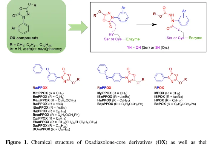
2.2. Oxadiazolone-core derivatives (OX). 3,5-substituted 1,3,4-oxadiazol-2(3H)-one
165
derivatives were first described in 1954 as active anti-TB molecules [37, 38]. Second generation 166
compounds were further found to exhibit interesting anti-mycobacterial activity with MIC 167
ranging from 8 to 50 µM [39, 40]. Few years later, we reported that a new substitutive OX 168
derivative, the MmPPOX (Figure 1), was also able to inhibit M. tb growth on solid medium 169
with moderated MIC values of 50-90 µM [28]. 170
171
Figure 1. Chemical structure of Oxadiazolone-core derivatives (OX) as well as their
172
mechanism of action on (Ser/Cys)-based enzymes in inset. 173
174
We further showed that MmPPOX efficiently inhibited pure recombinant M. tb enzymes 175
belonging to the hormone-sensitive lipase (HSL) family member proteins (i.e., Lip-HSL) [28], 176
including LipY (Rv3097c) the major M. tb Lip-HSL lipase involved in TAG acquisition from 177
the host and in ILI breakdown [2, 10, 17, 41]. The mechanism of action of MmPPOX involving 178
the formation of a covalent bond with the catalytic serine residue of the enzymes (Figure 1 179
inset) thus resulting in a total abolition of their activities was also confirmed [28]. Overall, the 180
fact that MmPPOX alters mycobacterial growth and abolishes the activity of Lip-HSL proteins 181
suggest that HSL family member proteins fulfill essential metabolic and/or physiologic 182
functions in M. tb life cycle. 183
Interestingly, MmPPOX has also proven its ability to prevent ILI catabolism using the 184
experimental ex vivo model of foamy macrophages developed in the lab [5, 10, 42]. Following 185
M. bovis BCG infection, murine bone-marrow-derived macrophages (mBMDM) were fed with 186
Very Low-Density Lipoproteins (VLDL), allowing the differentiation of these infected BMDM 187
into foamy macrophages (FM) [5, 10, 42]. In this specific environment, the bacteria are able to 188
i) accumulate lipids from host LBs to form ILIs; ii) enter in persistence phase depicted by a
189
typical absence of septation; and iii) hydrolyze stored ILIs after VLDL removal from the culture 190
medium thus mimicking reactivation and propagation of the disease. When infected cells were 191
exposed to VLDL in the presence of 50 µM MmPPOX, macrophages still retained the ability 192
to become foamy, but M. bovis BCG was unable to form ILIs (Figure 2A). Conversely, when 193
VLDL-treated M. bovis BCG-infected cells were further incubated for 24h in fresh medium, 194
nearly 90% of the ILI+3 profiles were retained in the presence vs. absence of MmPPOX (Figure 195
2B) [10]. These findings support MmPPOX as effective inhibitor of mycobacterial lipolytic 196
enzymes, including LipY (Rv3097c) [28]. involved in LB and ILI degradation. Such results 197
were further confirmed using an in vitro model of mycobacteria growing under carbon excess 198
and nitrogen-deprived conditions allowing ILI biosynthesis and hydrolysis. Incubation of the 199
resulting lipid-rich mycobacteria (i.e., M. smegmatis and M. abscessus) with MmPPOX also 200
resulted in nearly complete blockage of TAG lipolysis [11]. 201
202
Figure 2. (A) Foamy cell after 24h-exposure to VLDL showing large amounts of LB and M.
203
bovis BCG-containing ILI. (B-D) Effect of MmPPOX during exposure to VLDL on host LB 204
formation and ILI accumulation. At day 6 post-infection with M. bovis BCG, mBMDM were 205
exposed for 24h to VLDL in the absence or presence of MmPPOX. When exposed to VLDL 206
only (C) many mycobacterial profiles are ILI+3; whereas exposure to VLDL+MmPPOX (D) 207
results in small ILI with no ILI+3 profiles. (E-G) Effect of MmPPOX during TAG consumption 208
within ILIs. At day 6 post-infection with M. bovis BCG, mBMDM were exposed to VLDL and 209
re-incubated in VLDL-free culture medium alone or with MmPPOX for 24h. After VLDL 210
treatment followed by a 24-h chase in medium devoid of inhibitor, (F) cells contain few ILI+3 211
profiles. Conversely, in the presence of MmPPOX (G) cells still contain ILI+3 profiles. (B, E) 212
Both bar graphs represent the quantitative evaluation of the percentage of each category of ILI 213
profiles ±SD (*, p<0.05; **,##, p<0.01; ***, p<0.001). Bars in panels A, 1 µm; C-D-E-F, 0.5 214
µm. Adapted from [10, 42]. 215
216
Based on these data, a new series of 18 lipophilic OX derivatives were designed and 217
synthesized (Figure 1) [43]. A specific nomenclature was set up for these OXs noted Rm(or 218
p)PPOX; where m(or p)PP (= R1) represents the meta (or para)-PhenoxyPhenyl group, when 219
present; OX the oxadiazol-2(3H)-one core; and R the alkyl chain (Figure 1) [43]. Data reported 220
in Table 1 point out to the potent inhibitory activity of this new series of OXs towards the 221
TAG-lipase LipY. Their inhibitory power, defined here as the inhibitor molar excess leading to 222
50% lipase inhibition (xI50), ranges from 0.59 to 0.93 (Table 1) implying that the best OXs react 223
in close stoichiometry with this lipase, being thus more potent than MmPPOX (xI50 = 2.4), but 224
also than Orlistat (xI50 = 7.1) used as reference inhibitor [28, 44]. 225
226
2.3. The Cyclipostins & Cyclophostin analogs (CyC). In parallel, we investigated the
227
inhibitory properties of new monocyclic analogs of Cyclophostin and Cyclipostins (CyC) 228
(Figure 3), derived from phosphorus-containing natural products isolated from fermentation 229
broths of Streptomyces sp [45, 46], on pure mycobacterial lipases and various mycobacterial 230
strains. 231
Members of the Cyclipostins family (Figure 3 – X=O, R3=C15-C18) possess a core structure 232
similar to that of Cyclophostin (Figure 3 – X=O, R3=CH3) but are phosphate esters of long 233
chain lipophilic alcohols. The Cyclipostins were described to inhibit the growth of various 234
mycobacteria (including M. smegmatis, M. phlei, Nocardia abscessus, and Corynebacterium 235
diphtheriae) with MIC values similar to that of rifampicin and penicillin G [47]. These natural 236
products were initially described as potent inhibitors of either human acetylcholinesterase 237
(AChE) (i.e., Cyclophostin) [45, 48] or human and rat HSL (i.e., Cyclipostins) [46, 49]. 238
240
Figure 3. Chemical structure of Cyclipostins & Cyclophostin analogs (CyC), as well as their
241
mechanism of action on (Ser/Cys)-based enzymes in inset. For a review on the CyC synthesis, 242
please refer to [50]. 243
244
From these findings, we first reported the total synthesis of natural Cyclophostin CyC1, its 245
phosphonate analogues CyC2 [48, 49], and related monocyclic enol-phosphonates 246
CyC3-10,19-22[51-53] and phosphates CyC23-30 [53]; as well as Cyclipostin P CyC18, its trans-247
() diastereoisomer CyC18α [49], and the corresponding monocyclic enol-phosphonates 248
CyC11-13 [52, 54], difluorophosphonates CyC14-15 and phosphates CyC16-17[55, 56] (Figure 3). 249
Of particular importance, diastereomeric cis- and trans-monocyclic enolphosphonates CyC6-10 250
were screened against six representative Ser-based enzymes belonging to distinct lipolytic 251
enzyme families [52]. None of these enolphosphonates inhibited the mammalian AChE [55], 252
HSL [56], or gastric and pancreatic lipases [52]. The microbial enzymes; i.e., Fusarium solani 253
Cutinase [57] and lipolytic enzymes from M. tb (i.e., Rv0183 [16, 58] and LipY); were, 254
however, all fully inactivated by formation of a covalent and irreversible bond between the 255
enol-phosphorous atom and the catalytic serine residue [51, 52]. Moreover, modulation of the 256
lipophilicity by varying the nature of the alkyl group, either at the C-5 carbon atom (i.e., R2 257
group – Figure 3) or at the phosphorous center (i.e., R3 group – Figure 3), strongly impacted 258
the inhibitory efficiency of these CyCs [52]. This property has been exploited to significantly 259
attenuate or increase the affinity of one inhibitor towards a specific enzyme [52, 59]. 260
In order to shed more light on the influence of the chirality on enzyme inhibition, CyC7 261
bearing a C10-side alkyl chain was chosen for its significant inhibitory potency towards the 262
Cutinase, Rv0183 and LipY, but also for the high diastereoselectivity (51.9%-78.3%) exerted 263
by these enzymes in favor of the cis-(β)-isomer [52]. The four stereoisomers of CyC7 were 264
prepared by asymmetric synthesis, and the absolute configuration at both the phosphorus and 265
C-5 carbon stereocenters were assigned unambiguously [59]. Pure compounds at phosphorus 266
were obtained with a diastereoisomeric excess of around 95%, together with enantiomeric 267
excess of >85% related to the cyclized C-5 carbon center [59]. 268
Cutinase displayed a high diastereoselectivity for the (Sp) configuration with a 269
Stereoselectivity Index (SI) derived from xI50 values of 94.9% (Table 2) when using CyC7-(Sc) 270
inhibitors, whereas no obvious stereopreference at the phosphorus center was observed with the 271
CyC7-(Rc) inhibitors. On the contrary, Rv0183 strongly discriminated the (Sp) configuration (SI 272
= 72-81.4%) independently of the absolute (Rc) or (Sc) configuration on the asymmetric C-5 273
carbon atom; and thus, exhibited the classical enantiopreference of lipolytic enzymes [60]. The 274
discriminated only the unusual diastereoisomeric configuration (Rc,Rp), which led to the most 276
potent CyC7β-(Rc,Rp) inhibitor (SI > 80.7% - Table 2). Modulation of the lipophilicity at the C-277
5 carbon atom combined with this unusual high enantiopreference displayed by LipY for the 278
(Rp) and (Rc) associated absolute configurations, should open new prospects in the design of 279
specific inhibitors of this mycobacterial lipase [59]. Overall, these results raised significant 280
achievements in the understanding of the stereoselective relationships between pure non-281
racemic compounds and their inhibitory activity towards several microbial lipases of interest. 282
To summarize, these CyC derivatives have not only proved to be powerful bacterial 283
(Ser/Cys)-based enzymes inhibitors [52, 59], but above all, they had lost their inhibitory 284
activity towards the mammalian enzymes initially targeted by the natural parent molecules
285
[52, 55, 56]. 286
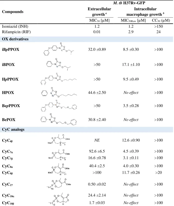
287
2.4. The OX and CyC derivatives are novel promising multi-target inhibitors of M. tb
288
growth. The set of 19 OX derivatives and 26 CyC analogues were further evaluated for their
289
anti-mycobacterial activity on a M. tb H37Rv-GFP strain using a high-content screening assay 290
based on the fluorescence measurement of GFP-expressing bacteria [54, 61]. In vitro growth of 291
M. tb H37Rv-GFP was monitored by directly measuring GFP fluorescence after 5 days at 37 °C 292
in presence of increasing concentrations of candidate inhibitors. Intracellular growth of M. tb 293
H37Rv-GFP was also assessed following a 5-day exposure of infected Raw264.7 murine 294
macrophage cell lines to the different compounds. In the latter case, the percentage of infected 295
cells and the number of living host cells allowed determining the values of both MIC50 296
(compound concentration leading to 50% growth inhibition) and CC50 (compound 297
concentration leading to 50% host cell toxicity) [62, 63]. 298
Among all molecules tested, 14 potential candidates (6 OXs and 8 CyCs – Table 3) 299
exhibited very promising anti-tubercular activities with moderate (15-50 µM) to good (3-4 µM) 300
and to excellent (500 nM for CyC17) MIC50 values. Of great importance, beside their inhibitory 301
activity against bacterial growth, both set of compounds exhibited very low toxicity towards 302
host macrophages (CC50 > 100 µM). Data show that both OXs and CyCs can be divided into 303
two different classes based on their antibacterial activity (Figure 4 and Table 3). 304
305
306
Figure 4. (A-C) Activity of HPOX, CyC7 & CyC17 against GFP-labelled M. tb H37Rv 307
replicating in culture medium expressed as normalized relative fluorescence units (RFU%). (B-308
D) Activity of iBPOX and CyC7 against M. tb H37Rv-GFP replicating inside Raw264.7 309
macrophages. Adapted from [54, 61]. 310
311
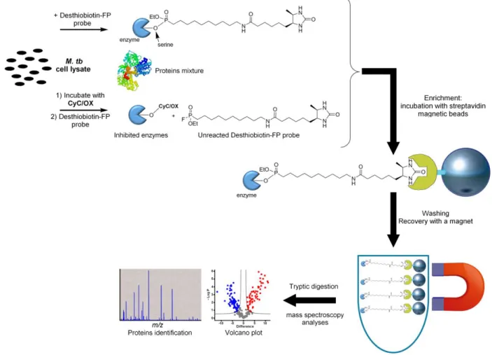
First, 9 out of 14 compounds exhibited higher activity against intracellular bacteria than 312
against extracellular ones: i.e., iBpPPOX, iBPOX, HpPPOX, BepPPOX, CyC6β, CyC7α,β and 313
CyC8α,β. Such feature supposes that the intracellular mode of action of these inhibitors may 314
differ from that of those acting on extracellularly-replicating bacilli. It can therefore be 315
hypothesized that the vulnerability of the corresponding bacterial target(s) of these 4 OXs and 316
extracellularly-growing bacteria. Alternatively, a specific stringent response of the macrophage 318
caused by the compounds and leading to bacterial death can, however, not be excluded. The 319
second type of compounds are active against extracellular bacteria and display only poor or 320
even no activity against intracellular M. tb: HPOX, BePOX, CyC17, CyC18α,β; a property 321
already reported for 1,2,4-Oxadiazole EthR inhibitors [63]. The observed differences in the 322
behavior of studied compounds, particularly the high ratio between the intracellular vs. 323
extracellular antibacterial effects, may translate in the prevention of entry of actively replicating 324
bacilli into the persistence phase and/or prevent reactivation of dormant bacilli within FM. 325
326
Figure 5. General scheme for the identification of the target enzymes of our inhibitors using
327
ABPP approach. 328
329
Based on these aforementioned results, and given the strong affinity of our OXs and CyCs 330
for M. tb lipolytic enzymes, these inhibitors might target and impair the activity of various 331
(Ser/Cys)-based enzymes involved in several processes of M. tb life cycle, thus resulting in 332
bacterial death without any (or only very low) cytotoxicity towards host cells. Accordingly, 333
target(s) identification experiments were next conducted by applying ABPP approach [27, 33, 334
34, 64-67] (Figure 5). Here, HPOX and CyC17, which selectively inhibit M. tb growth in 335
culture broth medium only, were selected for such experiments [54, 61]. M. tb total cell lysate 336
was incubated with each inhibitor and then subjected to competitive probe labelling/enrichment 337
assays using an activity-based probe (ABP), i.e., the ActivX Desthiobiotin-FP widely exploited 338
to screen for reversible and irreversible inhibitors of drug targets [54, 61, 64, 68]. This resulted 339
in the identification of a panel of 18 and 23 distinct proteins for the HPOX- and 340
CyC17-pretreated lysate, respectively [54, 61]. Remarkably, these 41 identified proteins were 341
all (Ser/Cys)-based enzymes, most of them participating in M. tb lipid metabolism and in cell
342
wall biosynthesis. Among them, several are annotated as essential enzymes for the in vitro 343
growth of M. tb and/or its survival of inside macrophages [69-71]. These included the antigen 344
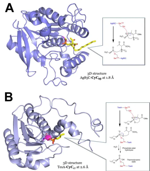
85 complex, Ag85A (Rv3804c), Ag85B (Rv1886c) and Ag85C (Rv0129c) [35]; the 345
thioesterase TesA (Rv2928) [72]; the carboxylesterase CaeA (Rv2224c) [73]; the β-ketoacyl 346
synthase KasA (Rv2245) [74]; a bifunctional thioesterase-phospholipase enzyme LipG 347
(Rv0646c) [75], and the hydrolase HsaD (Rv3569c) [76, 77]. 348
Through biochemical and structural studies, the antigen 85 complex (i.e., Ag85A/B/C) was 349
validated as an effective target of CyC17, but also of CyC7β and CyC8β [78]. These three CyCs 350
are indeed able to block the synthesis of trehalose dimycolate (TDM) as well as the 351
mycolylation of arabinogalactan in M. tb which results in the disruption of the cell envelope 352
integrity [78]. Among the targets of both the CyCs and OXs, TesA, a putative thioesterase 353
involved in the synthesis of phthiocerol dimycocerosates (PDIM) and phenolic glycolipids 354
(PGL), has been identified. These two lipids (PDIM and PGL), non-covalently linked to the 355
this context, TesA was validated as an effective pharmacological target of CyC17 (xI50 = 12.4) 357
[79] and HPOX (xI50 = 0.59) [61]. 358
359
360
Figure 6. Crystal structures of (A) Ag85C in complex with CyC8β (PDB ID: 5OCJ; 1.8Å 361
resolution) [78] and (B) TesA in complex with CyC17 (PDB ID: 6FVJ; 2.6Å resolution) [79]. 362
The mechanism of action of the phosphonate (CyC8β) and the phosphate (CyC17) analog based 363
on mass spectrometry analyses are illustrated in inset. Each inhibitor is in stick representation, 364
and the catalytic Serine residue is colored in magenta. Structures were redrawn as ribbon 365
representations from their respective modified PDB IDs, with PyMOL Molecular Graphics 366
System (Schrödinger, LLC). 367
From a molecular point of view, the obtained crystal structures of Ag85C inhibited by CyC8β 369
(Figure 6A) [78] and TesA in complex with CyC17 (Figure 6B) [79], together with biochemical 370
and mass spectrometry experiments, have clearly stated that the inhibition of these enzymes 371
results from the phosphorylation of their respective catalytic Serine residue (Figure 6). 372
Interestingly, following CyC17 phosphorylation of the TesA-Ser104 or Ag85C-Ser124, further 373
rearrangement of the inner structure of the covalently bound inhibitor occurs resulting in the 374
loss of the -ketoester moiety (Figure 6B). Such chemical modification can therefore be 375
considered as a signature of the CyC17 reactivity with Ser- and Cys-based enzymes [78, 79]. 376
Finally, the bifunctional thioesterase-phospholipase enzyme LipG (Rv0646c), involved in the 377
modification and remodeling of the mycobacterial envelope and described as essential for the 378
survival and intracellular persistence of M. tb [75], has been biochemically characterized [80]. 379
Inhibition kinetics indeed demonstrated that LipG was able to react with CyC7α (xI50 = 5.0) and 380
in near stoichiometry with CyC17 (xI50 = 0.98), but not with CyC7β and CyC8α,β therefore 381
exhibiting a certain selectivity of action. 382
383
3. The CyC And Ox Analogs are New Compounds for the Treatment of
384Mycobacterial-Related Infections
385In view of the results obtained on M. tb, the specificity of our CyC compounds against various 386
bacteria was further investigated. Susceptibility testing conducted on 7 bacterial strains, P. 387
aeruginosa, E coli, B. subtilis, M. abscessus, M. marinum, and M. bovis BCG against the 26 388
available CyC1-18 surprisingly showed that these compounds block specifically the growth of 389
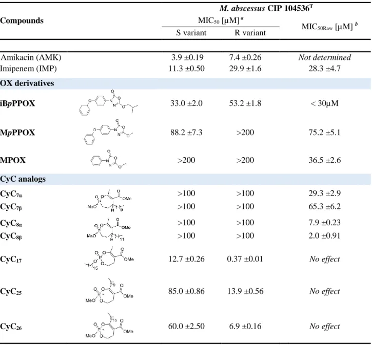
the mycobacterial species without affecting the growth of Gram-positive and Gram-negative 390
bacteria [81]. More particularly, CyC17 exhibited MIC50 values of 12.7 µM and 0.37 µM 391
towards the smooth and rough morphotype of M. abscessus, respectively; the latter value being 392
20- to 37-fold lower than that of reference antibiotics, amikacin (7.4 µM), imipenem (12 µM) 393
or cefoxitin (27 µM) [81]. 394
M. abscessus, a rapidly growing mycobacterium, is an opportunistic pathogen responsible 395
for chronic lung infection in patients with respiratory diseases such as cystic fibrosis (CF) [82-396
84]. M. abscessus exists in two variants, a smooth (S) and a rough (R) one, the latter being 397
considered the most virulent form in humans [85, 86]. Nicknamed the "antibiotics nightmare", 398
this nontuberculous mycobacterium (NTM) is one of the most drug-resistant mycobacteria for 399
which most standardized chemotherapeutic regimens are poorly effective, often leading to high 400
treatment failures and death [86-88]. 401
When tested on 37 bacterial strains isolated from CF patients, including 26 clinical isolates 402
belonging to the M. chelonae-abscessus complex, the best candidate (CyC17) showed high 403
selectivity for mycobacteria only, and MICs (<2-40 µg/mL) comparable with those of clinically 404
used antibiotics [81]. To refine the structure-activity relationships of this family of compounds, 405
12 new CyC analogs (CyC19-30 – Figure 3) were synthesized by varying the R4 chain on the 406
enolphosphorus ring [53]. Such structural modification was also guided by the isolation of the 407
antimalarial Salinipostin A, a natural product produced by a Salinispora sp. bacterium, which 408
is a Cyclipostin-like compound with variations in the alkyl enol substituent [89, 90]. Among 409
these 12 new CyCs, CyC25 and CyC26 exhibited very promising MIC50 values against M. 410
abscessus R (Table 4) [53]. Remarkably, these latter three best inhibitors of M. abscessus 411
growth were all phosphate esters bearing a long lipophilic C10/C16 alkyl chain either at the R3 412
or R4 position (Figure 3). We also demonstrated that the absence of surface exposed 413
glycopeptidolipids (GPL) in the R variant was responsible for the higher susceptibility (up to 414
34×) to the CyCs as compared to the S strain. More recently, by combining fast quantitative 415
imaging (QI) atomic force microscopy (AFM) with hydrophobic tips, Viljoen et al. 416
demonstrated that GPL modulated the nanoscale surface hydrophobicity of M. abscessus [91]. 417
While S morphotype displayed unusual variations of nanoscale hydrophobic properties, the R 418
variant showed homogeneous surface hydrophobicity conferred by surface exposed mycolic 419
acids when GPL were lacking [91]. Given together, the antibacterial activity of the CyCs 420
against the R and S variants, respectively, can thus be directly correlated with the GPL-421
dependent variation in the nanoscale distribution of M. abscessus hydrophobicity. 422
This was, however, not the case when using the OX derivatives, for which M. abscessus R 423
was nearly 1.6-times less sensitive to these compounds than the S morphotype, iBpPPOX being 424
the best growth inhibitor of both strains (Table 4) [92]. Interestingly, M. abscessus S 425
susceptibility to this latter inhibitor was similar to that of M. tb (Table 3) [61]. Such increased 426
tolerance of virulent M. abscessus R variant towards the OX compounds is in line with its high 427
resistance to classical antibiotics [86] compared to M. tb. 428
M. abscessus, like M. tb, is also able to survive and replicate inside macrophages [93, 94]. 429
Altogether, high intrinsic resistance and intracellular forms of M. abscessus are thus mostly 430
contributing to treatment failures since only few antimicrobials can penetrate the cell membrane 431
of phagocytes [95]. In this context, the 38 CyCs and 19 OXs were tested for their capability to 432
inhibit the intramacrophagic growth of M. abscessus inside Raw264.7 macrophages. Only 433
CyC7α,β and CyC8α,β were found active against intracellularly replicating M. abscessus (Table 434
4). With MIC50Raw values of 7.9 and 2.0 μM, respectively, CyC8α and CyC8β were 3.6- and 14-435
fold more potent against intracellular M. abscessus than imipenem (28.3 μM), used as reference. 436
It is noteworthy that such MIC50Raw are of the same order of magnitude than those previously 437
obtained with the same CyC8α,β against intracellular M. tb (4-12 μM – Table 3). Regarding the 438
OXs, only MPOX and MpPPOX (inactive against extracellular bacterial growth) and
439
iBpPPOX were able to block intracellularly-growing M. abscessus. As previously observed for
440
M. tb [61], iBpPPOX impairs both extracellular (MIC50 = 33.0 μM) and intracellular (~59% 441
bacterial clearance at 30 μM) replicating bacteria with similar potency/efficiency to that of 442
imipenem (Table 4). 443
By using similar strategy applied for M. tb (Figure 5), 39 and 21 potential target enzymes 444
of CyC17 and iBpPPOX, respectively, have been identified from a M. abscessus R cell culture. 445
Interestingly, 11 out of the 60 identified proteins possess orthologs annotated as essential 446
enzymes in the M. tb genome. Moreover, consistent with our previous works [54, 61], most of 447
them are (Ser/Cys)-based enzymes mainly involved in M. abscessus lipid metabolism or cell-448
wall synthesis. These include several Lip-HSL enzymes, some Cutinase-like family proteins, 449
and the members of the antigen 85 (Ag85) complex which play a central role in the 450
mycobacterial cell wall biosynthesis and in the survival of mycobacteria [96, 97]. 451
The fact that Ag85A/B/C proteins have been identified and validated as effective targets of 452
both the CyC and OX compounds in M. tb [61, 78] and M. abscessus [53, 92], but also of 453
Orlistat and related analogs [25, 30, 34], suggest that these mycolyltransferases represent a 454
common primary target of such inhibitors, regardless of the mycobacterial strain tested. Indeed, 455
as Ag85A/B/C proteins are secreted enzymes, they may be easily accessible to the CyCs and 456
OXs. Due to their importance in mycolic acid metabolism, the Ag85 enzymes have often been
457
proposed as attractive targets for future chemotherapeutic developments against mycobacteria 458
[35, 78]. Overall, our studies provide compelling evidence that both the CyC analogs and the 459
OX compounds inhibit the activity of the Ag85 complex in vitro and in mycobacteria [78, 92], 460
therefore opening the way to a new strategy to fight against pathogenic mycobacteria. 461
462
4. Conclusion and future perspectives
463Collectively all our results strengthen the fact that the OX and the CyC inhibitors are not only 464
non-toxic for mammalian cells, but above all represent a novel class of selective and efficient 465
multi-target inhibitors [98] leading to the arrest of M. tb and M. abscessus, two major infectious 466
pathogens. By impairing simultaneously, the activity of various (Ser/Cys)-base enzymes 467
participating in important physiological processes related to the whole bacterial lipid 468
metabolism, including the Ag85 complex proteins, both the CyCs and the OXs will inhibit 469
extracellular and/or intracellular M. tb and M. abscessus growth, therefore leading to bacterial 470
death. Consequently, the probability that a strain would develop resistance to such inhibitors 471
would be very low, because resistant mutants would require the simultaneous acquisition of 472
several mutations in the same bacterial genome; thus, making it difficult or impossible for the 473
bacteria to adapt themselves and survive. 474
These data also demonstrated that our inhibitors are able to penetrate both the bacteria and 475
the host macrophage (without any cytotoxic effect). Such a dual activity of the CyCs and OXs 476
is a key point as it may affect the different stages of the infection process: i.e., entry of bacilli 477
into the persistence phase, and/or interfere by blocking the lipid accumulation from foamy 478
macrophages or the lipid consumption during the reactivation of dormant bacilli. 479
Given these promising properties, these two families of inhibitors will be chemically 480
modified to allow, in living cells and via click chemistry, the direct capture of specific proteins 481
that lead to M. tb or M. abscessus growth arrest during the mycobacterial phases of active 482
replication, latency and/or reactivation of the bacilli in infected foamy as well as non-foamy 483
macrophages [11, 42]. Accordingly, the in vivo identification of the proteins inactivated by our 484
potent antibacterial activity-based probes will reveal new potential targets for treating 485
mycobacterial-related diseases, and contribute to background information for the development 486
of new therapeutic strategies for elimination of either actively replicating or latent bacilli from 487
infected individuals. Deciphering the physiological role of mycobacterial lipolytic enzymes in 488
lipid metabolism, notably focusing on the accumulation and consumption of lipids at key stages 489
of the bacterial development will generate results that are directly relevant for deciphering the 490
492
Declaration of competing interest
493The authors report no declarations of interest. 494
495
Acknowledgments
496This work was supported by the CNRS, Aix Marseille University, the Association Grégory 497
Lemarchal and Vaincre la Mucoviscidose (project n°RF20160501651), and the Agence 498
Nationale de la Recherche (grant N°ANR-19-CE44-0011). Proteomics analyses were done 499
using the mass spectrometry facility of Marseille Proteomics supported by IBISA, the 500
Cancéropôle PACA, the Provence-Alpes-Côte d'Azur Région, the Institut Paoli-Calmettes, and 501
Fonds Européen de Développement Régional (FEDER). The CyC synthesis was supported by 502
the National Institute of General Medical Studies (grant N°R01-GM076192), and more recently 503
by UMSL. Equipment used in this synthesis was supported by grants from the National Science 504
Foundation (grant 9974801 and CHE-0959360 for NMR spectrometers, grant N°CHE-505
0420497 for X-ray diffractometer, and grant N°CHE-9708640 for mass spectrometer). The 506
authors wish to thanks Dr. Chantal De Chastellier for providing the electron microscopy photos. 507
508 509
References
510[1] J. Daniel, H. Maamar, C. Deb, T.D. Sirakova, P.E. Kolattukudy, Mycobacterium 511
tuberculosis uses host triacylglycerol to accumulate lipid droplets and acquires a dormancy-like 512
phenotype in lipid-loaded macrophages, PLoS Pathog, 7 (2011) e1002093. 513
[2] C. Deb, J. Daniel, T. Sirakova, B. Abomoelak, V. Dubey, P. Kolattukudy, A Novel Lipase 514
Belonging to the Hormone-sensitive Lipase Family Induced under Starvation to Utilize Stored 515
Triacylglycerol in Mycobacterium tuberculosis, J. Biol. Chem., 281 (2006) 3866-3875. 516
[3] D.G. Russell, P.J. Cardona, M.J. Kim, S. Allain, F. Altare, Foamy macrophages and the 517
progression of the human tuberculosis granuloma, Nat Immunol, 10 (2009) 943-948. 518
[4] R. Dhouib, A. Ducret, P. Hubert, F. Carriere, S. Dukan, S. Canaan, Watching intracellular 519
lipolysis in mycobacteria using time lapse fluorescence microscopy, Biochim Biophys Acta, 520
1811 (2011) 234-241. 521
[5] P. Santucci, F. Bouzid, N. Smichi, I. Poncin, L. Kremer, C. De Chastellier, M. Drancourt, 522
S. Canaan, Experimental Models of Foamy Macrophages and Approaches for Dissecting the 523
Mechanisms of Lipid Accumulation and Consumption during Dormancy and Reactivation of 524
Tuberculosis, Front Cell Infect Microbiol., 6 (2016) 122. 525
[6] K.L. Low, P.S. Rao, G. Shui, A.K. Bendt, K. Pethe, T. Dick, M.R. Wenk, Triacylglycerol 526
utilization is required for regrowth of in vitro hypoxic nonreplicating Mycobacterium bovis 527
bacillus Calmette-Guerin, J Bacteriol, 191 (2009) 5037-5043. 528
[7] K.L. Low, G. Shui, K. Natter, W.K. Yeo, S.D. Kohlwein, T. Dick, S.P. Rao, M.R. Wenk, 529
Lipid droplet-associated proteins are involved in the biosynthesis and hydrolysis of 530
triacylglycerol in Mycobacterium bovis bacillus Calmette-Guerin, J Biol Chem, 285 (2010) 531
21662-21670. 532
[8] K.A. Mattos, F.A. Lara, V.G. Oliveira, L.S. Rodrigues, H. D'Avila, R.C. Melo, P.P. Manso, 533
E.N. Sarno, P.T. Bozza, M.C. Pessolani, Modulation of lipid droplets by Mycobacterium leprae 534
in Schwann cells: a putative mechanism for host lipid acquisition and bacterial survival in 535
phagosomes, Cell Microbiol, 13 (2011) 259-273. 536
[9] A. Viljoen, M. Blaise, C. de Chastellier, L. Kremer, MAB_3551c encodes the primary 537
triacylglycerol synthase involved in lipid accumulation in Mycobacterium abscessus, Mol 538
Microbiol, 102 (2016) 611-627. 539
Lipid Consumption in Mycobacterium-Infected Foamy Macrophages, Infect Immun, 86 (2018) 542
e00394-00318. 543
[11] P. Santucci, M.D. Johansen, V. Point, I. Poncin, A. Viljoen, J.-F. Cavalier, L. Kremer, S. 544
Canaan, Nitrogen deprivation induces triacylglycerol accumulation, drug tolerance and 545
hypervirulence in mycobacteria, Sci Rep, 9 (2019) 8667. 546
[12] J.G. Rodriguez, A.C. Hernandez, C. Helguera-Repetto, D. Aguilar Ayala, R. Guadarrama-547
Medina, J.M. Anzola, J.R. Bustos, M.M. Zambrano, Y.M.J. Gonzalez, M.J. Garcia, P. Del 548
Portillo, Global adaptation to a lipid environment triggers the dormancy-related phenotype of 549
Mycobacterium tuberculosis, MBio, 5 (2014) e01125-01114. 550
[13] J.C. Camus, M.J. Pryor, C. Medigue, S.T. Cole, Re-annotation of the genome sequence of 551
Mycobacterium tuberculosis H37Rv, Microbiology, 148 (2002) 2967-2973. 552
[14] B. Brust, M. Lecoufle, E. Tuaillon, L. Dedieu, S. Canaan, V. Valverde, L. Kremer, 553
Mycobacterium tuberculosis lipolytic enzymes as potential biomarkers for the diagnosis of 554
active tuberculosis, PLoS ONE, 6 (2011) e25078. 555
[15] G. Johnson, The alpha/beta Hydrolase Fold Proteins of Mycobacterium tuberculosis, With 556
Reference to their Contribution to Virulence, Curr Protein Pept Sci, 18 (2017) 190-210. 557
[16] K. Côtes, R. Dhouib, I. Douchet, H. Chahinian, A. De Caro, F. Carriere, S. Canaan, 558
Characterization of an exported monoglyceride lipase from Mycobacterium tuberculosis 559
possibly involved in the metabolism of host cell membrane lipids, Biochem J., 408 (2007) 417-560
427. 561
[17] K.C. Mishra, C. de Chastellier, Y. Narayana, P. Bifani, A.K. Brown, G.S. Besra, V.M. 562
Katoch, B. Joshi, K.N. Balaji, L. Kremer, Functional role of the PE domain and immunogenicity 563
of the Mycobacterium tuberculosis triacylglycerol hydrolase LipY, Infect Immun, 76 (2008) 564
127-140. 565
[18] G. Xu, H. Jia, Y. Li, X. Liu, M. Li, Y. Wang, Hemolytic phospholipase Rv0183 of 566
Mycobacterium tuberculosis induces inflammatory response and apoptosis in alveolar 567
macrophage RAW264.7 cells, Can J Microbiol, 56 (2010) 916-924. 568
[19] M. Schue, D. Maurin, R. Dhouib, J.C. Bakala N'Goma, V. Delorme, G. Lambeau, F. 569
Carriere, S. Canaan, Two cutinase-like proteins secreted by Mycobacterium tuberculosis show 570
very different lipolytic activities reflecting their physiological function, FASEB J, 24 (2010) 571
1893-1903. 572
[20] P. Hadvary, W. Sidler, W. Meister, W. Vetter, H. Wolfer, The lipase inhibitor 573
tetrahydrolipstatin binds covalently to the putative active site serine of pancreatic lipase, J Biol 574
Chem, 266 (1991) 2021-2027. 575
[21] H. Stalder, Oesterhelt, Borgström, Tetrahydrolipstatin: degradation products produced by 576
human carboxyl-ester lipase, Helvetica Chimica Acta, 75 (1992) 11. 577
[22] A. Benarouche, V. Point, F. Carriere, J.F. Cavalier, Using the reversible inhibition of 578
gastric lipase by Orlistat for investigating simultaneously lipase adsorption and substrate 579
hydrolysis at the lipid-water interface, Biochimie, 101 (2014) 221-231. 580
[23] L. Haalck, F. Spener, On the inhibition of microbial lipases by tetrahydrolipstatin, Methods 581
Enzymol, 286 (1997) 252-263. 582
[24] S.K. Parker, R.M. Barkley, J.G. Rino, M.L. Vasil, Mycobacterium tuberculosis Rv3802c 583
encodes a phospholipase/thioesterase and is inhibited by the antimycobacterial agent 584
tetrahydrolipstatin, PLoS One, 4 (2009) e4281. 585
[25] P.K. Crellin, J.P. Vivian, J. Scoble, F.M. Chow, N.P. West, R. Brammananth, N.I. 586
Proellocks, A. Shahine, J. Le Nours, M.C.J. Wilce, W.J. Britton, R.L. Coppel, J. Rossjohn, T. 587
Beddoe, Tetrahydrolipstatin Inhibition, Functional Analyses, and Three-dimensional Structure 588
of a Lipase Essential for Mycobacterial Viability, J. Biol. Chem., 285 (2010) 30050-30060. 589
[26] N.P. West, K.M. Cergol, M. Xue, E.J. Randall, W.J. Britton, R.J. Payne, Inhibitors of an 590
essential mycobacterial cell wall lipase (Rv3802c) as tuberculosis drug leads, Chem Commun 591
(Camb), 47 (2011) 5166-5168. 592
[27] M.S. Ravindran, S.P. Rao, X. Cheng, A. Shukla, A. Cazenave-Gassiot, S.Q. Yao, M.R. 593
Wenk, Targeting lipid esterases in mycobacteria grown under different physiological conditions 594
using activity-based profiling with tetrahydrolipstatin (THL), Mol Cell Proteomics, 13 (2014) 595
435-448. 596
[28] V. Delorme, S.V. Diomande, L. Dedieu, J.-F. Cavalier, F. Carriere, L. Kremer, J. Leclaire, 597
F. Fotiadu, S. Canaan, MmPPOX inhibits Mycobacterium tuberculosis lipolytic enzymes 598
belonging to the hormone-sensitive lipase family and alters mycobacterial growth, PLoS ONE, 599
7 (2012) e46493. 600
[29] C.M. Goins, T.D. Sudasinghe, X. Liu, Y. Wang, G.A. O'Doherty, D.R. Ronning, 601
Characterization of Tetrahydrolipstatin and Stereoderivatives on the Inhibition of Essential 602
[30] C.M. Goins, S. Dajnowicz, M.D. Smith, J.M. Parks, D.R. Ronning, Mycolyltransferase 604
from Mycobacterium tuberculosis in covalent complex with tetrahydrolipstatin provides 605
insights into antigen 85 catalysis, J Biol Chem, 293 (2018) 3651-3662. 606
[31] C. Rens, F. Laval, M. Daffe, O. Denis, R. Frita, A. Baulard, R. Wattiez, P. Lefevre, V. 607
Fontaine, Effects of lipid-lowering drugs on vancomycin susceptibility of mycobacteria, 608
Antimicrob Agents Chemother, 60 (2016) 6193-6199. 609
[32] P. Santucci, C. Dedaki, A. Athanasoulis, L. Gallorini, A. Munoz, S. Canaan, J.-F. Cavalier, 610
V. Magrioti, Synthesis of long chain β-lactones and their antibacterial activities against 611
pathogenic mycobacteria, ChemMedChem, 14 (2019) 349-358. 612
[33] J. Lehmann, J. Vomacka, K. Esser, M. Nodwell, K. Kolbe, P. Ramer, U. Protzer, N. 613
Reiling, S.A. Sieber, Human lysosomal acid lipase inhibitor lalistat impairs Mycobacterium 614
tuberculosis growth by targeting bacterial hydrolases, MedChemComm 7(2016) 1797-1801. 615
[34] J. Lehmann, T.Y. Cheng, A. Aggarwal, A.S. Park, E. Zeiler, R.M. Raju, T. Akopian, O. 616
Kandror, J.C. Sacchettini, D.B. Moody, E.J. Rubin, S.A. Sieber, An Antibacterial beta-Lactone 617
Kills Mycobacterium tuberculosis by Disrupting Mycolic Acid Biosynthesis, Angew Chem Int 618
Ed Engl, 57 (2018) 348-353. 619
[35] J.T. Belisle, V.D. Vissa, T. Sievert, K. Takayama, P.J. Brennan, G.S. Besra, Role of the 620
major antigen of Mycobacterium tuberculosis in cell wall biogenesis, Science, 276 (1997) 621
1420-1422. 622
[36] A. Aggarwal, M.K. Parai, N. Shetty, D. Wallis, L. Woolhiser, C. Hastings, N.K. Dutta, S. 623
Galaviz, R.C. Dhakal, R. Shrestha, S. Wakabayashi, C. Walpole, D. Matthews, D. Floyd, P. 624
Scullion, J. Riley, O. Epemolu, S. Norval, T. Snavely, G.T. Robertson, E.J. Rubin, T.R. Ioerger, 625
F.A. Sirgel, R. van der Merwe, P.D. van Helden, P. Keller, E.C. Böttger, P.C. Karakousis, A.J. 626
Lenaerts, J.C. Sacchettini, Development of a Novel Lead that Targets M. tuberculosis 627
Polyketide Synthase 13, Cell, 170 (2017) 249-259.e225. 628
[37] A.E. Wilder Smith, The Action of Phosgene on Acid Hydrazides to Give 1, 3, 4-629
Oxdiazolones of Interest in the Treatment of Tuberculosis, Science, 119 (1954) 514. 630
[38] A.E. Wilder Smith, H. Brodhage, Biological spectrum of some new tuberculostatic 1,3,4-631
oxadiazolones with special reference to cross-resistance and rates of emergence of resistance, 632
Nature, 192 (1961) 1195. 633
[39] M.G. Mamolo, D. Zampieri, L. Vio, M. Fermeglia, M. Ferrone, S. Pricl, G. Scialino, E. 634
Banfi, Antimycobacterial activity of new 3-substituted 5-(pyridin-4-yl)-3H-1,3,4-oxadiazol-2-635
one and 2-thione derivatives. Preliminary molecular modeling investigations, Bioorg Med 636
Chem, 13 (2005) 3797-3809. 637
[40] D. Zampieri, M.G. Mamolo, E. Laurini, M. Fermeglia, P. Posocco, S. Pricl, E. Banfi, G. 638
Scialino, L. Vio, Antimycobacterial activity of new 3,5-disubstituted 1,3,4-oxadiazol-2(3H)-639
one derivatives. Molecular modeling investigations, Bioorg Med Chem, 17 (2009) 4693-4707. 640
[41] P. Santucci, N. Smichi, S. Diomande, I. Poncin, V. Point, H. Gaussier, J.-F. Cavalier, L. 641
Kremer, S. Canaan, Dissecting the membrane lipid binding properties and lipase activity of 642
Mycobacterium tuberculosis LipY domains, The FEBS journal, 286 (2019) 3164-3181. 643
[42] I. Caire-Brandli, A. Papadopoulos, W. Malaga, D. Marais, S. Canaan, L. Thilo, C. de 644
Chastellier, Reversible lipid accumulation and associated division arrest of Mycobacterium 645
avium in lipoprotein-induced foamy macrophages may resemble key events during latency and 646
reactivation of tuberculosis, Infect Immun, 82 (2014) 476-490. 647
[43] V. Point, A. Benarouche, J. Zarrillo, A. Guy, R. Magnez, L. Fonseca, B. Raux, J. Leclaire, 648
G. Buono, F. Fotiadu, T. Durand, F. Carriere, C. Vaysse, L. Couedelo, J.-F. Cavalier, Slowing 649
down fat digestion and absorption by an oxadiazolone inhibitor targeting selectively gastric 650
lipolysis, Eur J Med Chem, 123 (2016) 834-848. 651
[44] S. Ulker, C. Placidi, V. Point, B. Gadenne, C. Serveau-Avesque, S. Canaan, F. Carriere, 652
J.-F. Cavalier, New lipase assay using Pomegranate oil coating in microtiter plates, Biochimie, 653
120 (2016) 110-118. 654
[45] T. Kurokawa, K. Suzuki, T. Hayaoka, T. Nakagawa, T. Izawa, M. Kobayashi, N. Harada, 655
Cyclophostin, acetylcholinesterase inhibitor from Streptomyces lavendulae, J Antibiot 656
(Tokyo), 46 (1993) 1315-1318. 657
[46] L. Vertesy, B. Beck, M. Bronstrup, K. Ehrlich, M. Kurz, G. Muller, D. Schummer, G. 658
Seibert, Cyclipostins, novel hormone-sensitive lipase inhibitors from Streptomyces sp. DSM 659
13381. II. Isolation, structure elucidation and biological properties, J Antibiot (Tokyo), 55 660
(2002) 480-494. 661
[47] G. Seibert, L. Toti, J. Wink, Treating mycobacterial infections with cyclipostins., in: 662
Patent, 2008. 663
[48] S. Bandyopadhyay, S. Dutta, C.D. Spilling, C.M. Dupureur, N.P. Rath, Synthesis and 664
biological evaluation of a phosphonate analog of the natural acetyl cholinesterase inhibitor 665
[49] R.K. Malla, S. Bandyopadhyay, C.D. Spilling, S. Dutta, C.M. Dupureur, The first total 667
synthesis of (+/-)-cyclophostin and (+/-)-cyclipostin P: inhibitors of the serine hydrolases acetyl 668
cholinesterase and hormone sensitive lipase, Org Lett, 13 (2011) 3094-3097. 669
[50] C.D. Spilling, The Chemistry and Biology of Cyclophostin, the Cyclipostins and Related 670
Compounds, Molecules, 24 (2019) 2579. 671
[51] S. Dutta, R.K. Malla, S. Bandyopadhyay, C.D. Spilling, C.M. Dupureur, Synthesis and 672
kinetic analysis of some phosphonate analogs of cyclophostin as inhibitors of human 673
acetylcholinesterase, Bioorg Med Chem, 18 (2010) 2265-2274. 674
[52] V. Point, R.K. Malla, S. Diomande, B.P. Martin, V. Delorme, F. Carriere, S. Canaan, N.P. 675
Rath, C.D. Spilling, J.-F. Cavalier, Synthesis and kinetic evaluation of cyclophostin and 676
cyclipostins phosphonate analogs as selective and potent inhibitors of microbial lipases, J Med 677
Chem, 55 (2012) 10204-10219. 678
[53] A. Madani, J.N. Ridenour, B.P. Martin, R.R. Paudel, A. Abdul Basir, V. Le Moigne, J.L. 679
Herrmann, S. Audebert, L. Camoin, L. Kremer, C.D. Spilling, S. Canaan, J.-F. Cavalier, 680
Cyclipostins and Cyclophostin Analogues as Multitarget Inhibitors That Impair Growth of 681
Mycobacterium abscessus, ACS Infect Dis, 5 (2019) 1597-1608. 682
[54] P.C. Nguyen, V. Delorme, A. Benarouche, B.P. Martin, R. Paudel, G.R. Gnawali, A. 683
Madani, R. Puppo, V. Landry, L. Kremer, P. Brodin, C.D. Spilling, J.-F. Cavalier, S. Canaan, 684
Cyclipostins and Cyclophostin analogs as promising compounds in the fight against 685
tuberculosis, Sci Rep, 7 (2017) 11751. 686
[55] B.P. Martin, E. Vasilieva, C.M. Dupureur, C.D. Spilling, Synthesis and comparison of the 687
biological activity of monocyclic phosphonate, difluorophosphonate and phosphate analogs of 688
the natural AChE inhibitor cyclophostin, Bioorg Med Chem, 23 (2015) 7529-7534. 689
[56] E. Vasilieva, S. Dutta, R.K. Malla, B.P. Martin, C.D. Spilling, C.M. Dupureur, Rat 690
hormone sensitive lipase inhibition by cyclipostins and their analogs, Bioorg Med Chem, 23 691
(2015) 944-952. 692
[57] S. Longhi, M. Mannesse, H.M. Verheij, G.H. De Haas, M. Egmond, E. Knoops-Mouthuy, 693
C. Cambillau, Crystal structure of cutinase covalently inhibited by a triglyceride analogue, 694
Protein Sci, 6 (1997) 275-286. 695
[58] P. Aschauer, R. Zimmermann, R. Breinbauer, T. Pavkov-Keller, M. Oberer, The crystal 696
structure of monoacylglycerol lipase from M. tuberculosis reveals the basis for specific 697
inhibition, Sci Rep, 8 (2018) 8948. 698
[59] V. Point, R.K. Malla, F. Carriere, S. Canaan, C.D. Spilling, J.-F. Cavalier, Enantioselective 699
inhibition of microbial lipolytic enzymes by nonracemic monocyclic enolphosphonate 700
analogues of cyclophostin, J Med Chem, 56 (2013) 4393-4401. 701
[60] J.-F. Cavalier, G. Buono, R. Verger, Covalent inhibition of digestive lipases by chiral 702
phosphonates, Acc. Chem. Res., 33 (2000) 579-589. 703
[61] P.C. Nguyen, V. Delorme, A. Benarouche, A. Guy, V. Landry, S. Audebert, M. Pophillat, 704
L. Camoin, C. Crauste, J.M. Galano, T. Durand, P. Brodin, S. Canaan, J.-F. Cavalier, 705
Oxadiazolone derivatives, new promising multi-target inhibitors against M. tuberculosis, 706
Bioorg Chem, 81 (2018) 414-424. 707
[62] T. Christophe, M. Jackson, H.K. Jeon, D. Fenistein, M. Contreras-Dominguez, J. Kim, A. 708
Genovesio, J.P. Carralot, F. Ewann, E.H. Kim, S.Y. Lee, S. Kang, M.J. Seo, E.J. Park, H. 709
Skovierova, H. Pham, G. Riccardi, J.Y. Nam, L. Marsollier, M. Kempf, M.L. Joly-Guillou, T. 710
Oh, W.K. Shin, Z. No, U. Nehrbass, R. Brosch, S.T. Cole, P. Brodin, High content screening 711
identifies decaprenyl-phosphoribose 2' epimerase as a target for intracellular antimycobacterial 712
inhibitors, PLoS Pathog, 5 (2009) e1000645. 713
[63] M. Flipo, M. Desroses, N. Lecat-Guillet, B. Dirie, X. Carette, F. Leroux, C. Piveteau, F. 714
Demirkaya, Z. Lens, P. Rucktooa, V. Villeret, T. Christophe, H.K. Jeon, C. Locht, P. Brodin, 715
B. Deprez, A.R. Baulard, N. Willand, Ethionamide boosters: synthesis, biological activity, and 716
structure-activity relationships of a series of 1,2,4-oxadiazole EthR inhibitors, J Med Chem., 717
54 (2011) 2994-3010. 718
[64] C. Ortega, L.N. Anderson, A. Frando, N.C. Sadler, R.W. Brown, R.D. Smith, A.T. Wright, 719
C. Grundner, Systematic Survey of Serine Hydrolase Activity in Mycobacterium tuberculosis 720
Defines Changes Associated with Persistence, Cell Chem Biol., 23 (2016) 290-298. 721
[65] K.R. Tallman, S.R. Levine, K.E. Beatty, Small Molecule Probes Reveal Esterases with 722
Persistent Activity in Dormant and Reactivating Mycobacterium tuberculosis, ACS Infect. Dis., 723
2 (2016) 936-944. 724
[66] F. Faucher, J.M. Bennett, M. Bogyo, S. Lovell, Strategies for Tuning the Selectivity of 725
Chemical Probes that Target Serine Hydrolases, Cell Chem Biol, 27 (2020) 937-952. 726
[67] L.J. Keller, B.M. Babin, M. Lakemeyer, M. Bogyo, Activity-based protein profiling in 727
bacteria: Applications for identification of therapeutic targets and characterization of microbial 728
communities, Curr Opin Chem Biol, 54 (2019) 45-53. 729
[68] D.A. Bachovchin, S.J. Brown, H. Rosen, B.F. Cravatt, Identification of selective inhibitors 730
of uncharacterized enzymes by high-throughput screening with fluorescent activity-based 731
probes, Nat Biotechnol, 27 (2009) 387-394. 732
[69] C.M. Sassetti, D.H. Boyd, E.J. Rubin, Genes required for mycobacterial growth defined 733
by high density mutagenesis, Mol Microbiol, 48 (2003) 77-84. 734
[70] C.M. Sassetti, E.J. Rubin, Genetic requirements for mycobacterial survival during 735
infection, Proc Natl Acad Sci U S A, 100 (2003) 12989-12994. 736
[71] J.E. Griffin, J.D. Gawronski, M.A. Dejesus, T.R. Ioerger, B.J. Akerley, C.M. Sassetti, 737
High-resolution phenotypic profiling defines genes essential for mycobacterial growth and 738
cholesterol catabolism, PLoS Pathog, 7 (2011) e1002251. 739
[72] L. Alibaud, Y. Rombouts, X. Trivelli, A. Burguiere, S.L. Cirillo, J.D. Cirillo, J.F. 740
Dubremetz, Y. Guerardel, G. Lutfalla, L. Kremer, A Mycobacterium marinum TesA mutant 741
defective for major cell wall-associated lipids is highly attenuated in Dictyostelium discoideum 742
and zebrafish embryos, Molecular Microbiology, 80 (2011) 919-934. 743
[73] S. Lun, W.R. Bishai, Characterization of a Novel Cell Wall-anchored Protein with 744
Carboxylesterase Activity Required for Virulence in Mycobacterium tuberculosis, J. Biol. 745
Chem., 282 (2007) 18348-18356. 746
[74] R.A. Slayden, C.E. Barry, 3rd, The role of KasA and KasB in the biosynthesis of 747
meromycolic acids and isoniazid resistance in Mycobacterium tuberculosis, Tuberculosis 748
(Edinb), 82 (2002) 149-160. 749
[75] J. Rengarajan, B.R. Bloom, E.J. Rubin, Genome-wide requirements for Mycobacterium 750
tuberculosis adaptation and survival in macrophages, Proc Natl Acad Sci U S A, 102 (2005) 751
8327-8332. 752
[76] N.A. Lack, K.C. Yam, E.D. Lowe, G.P. Horsman, R.L. Owen, E. Sim, L.D. Eltis, 753
Characterization of a carbon-carbon hydrolase from Mycobacterium tuberculosis involved in 754
cholesterol metabolism, J Biol Chem., 285 (2010) 434-443. 755
[77] A. Ryan, E. Polycarpou, N.A. Lack, D. Evangelopoulos, C. Sieg, A. Halman, S. Bhakta, 756
O. Eleftheriadou, T.D. McHugh, S. Keany, E.D. Lowe, R. Ballet, A. Abuhammad, W.R. Jacobs, 757
A. Ciulli, E. Sim, Investigation of the mycobacterial enzyme HsaD as a potential novel target 758
for anti-tubercular agents using a fragment-based drug design approach, British J Pharmacol, 759
174 (2017) 2209-2224. 760
[78] A. Viljoen, M. Richard, P.C. Nguyen, P. Fourquet, L. Camoin, R.R. Paudal, G.R. Gnawali, 761
C.D. Spilling, J.-F. Cavalier, S. Canaan, M. Blaise, L. Kremer, Cyclipostins and cyclophostin 762
analogs inhibit the antigen 85C from Mycobacterium tuberculosis both in vitro and in vivo, J 763
Biol Chem, 293 (2018) 2755-2769. 764
[79] P.C. Nguyen, V.S. Nguyen, B.P. Martin, P. Fourquet, L. Camoin, C.D. Spilling, J.-F. 765
Cavalier, C. Cambillau, S. Canaan, Biochemical and Structural Characterization of TesA, a 766
Major Thioesterase Required for Outer-Envelope Lipid Biosynthesis in Mycobacterium 767
tuberculosis, J Mol Biol, 430 (2018) 5120-5136. 768
[80] P. Santucci, V. Point, I. Poncin, A. Guy, C. Crauste, C. Serveau-Avesque, J.M. Galano, 769
C.D. Spilling, J.-F. Cavalier, S. Canaan, LipG a bifunctional phospholipase/thioesterase 770
involved in mycobacterial envelope remodeling, Biosci Rep, 38 (2018) BSR20181953. 771
[81] P.C. Nguyen, A. Madani, P. Santucci, B.P. Martin, R.R. Paudel, S. Delattre, J.L. Herrmann, 772
C.D. Spilling, L. Kremer, S. Canaan, J.-F. Cavalier, Cyclophostin and Cyclipostins analogues, 773
new promising molecules to treat mycobacterial-related diseases, Int J Antimicrob Agents, 51 774
(2018) 651-654. 775
[82] M. Osmani, D. Sotello, S. Alvarez, J.A. Odell, M. Thomas, Mycobacterium abscessus 776
infections in lung transplant recipients: 15-year experience from a single institution, Transpl 777
Infect Dis, 20 (2018) e12835. 778
[83] M.-L. Wu, D.B. Aziz, V. Dartois, T. Dick, NTM drug discovery: status, gaps and the way 779
forward, Drug Discov Today, 23 (2018) 1502-1519. 780
[84] M.D. Johansen, J.L. Herrmann, L. Kremer, Non-tuberculous mycobacteria and the rise of 781
Mycobacterium abscessus, Nat Rev Microbiol, 18 (2020) 392-407. 782
[85] E. Catherinot, J. Clarissou, G. Etienne, F. Ripoll, J.F. Emile, M. Daffe, C. Perronne, C. 783
Soudais, J.L. Gaillard, M. Rottman, Hypervirulence of a rough variant of the Mycobacterium 784
abscessus type strain, Infect Immun, 75 (2007) 1055-1058. 785
[86] R. Nessar, E. Cambau, J.M. Reyrat, A. Murray, B. Gicquel, Mycobacterium abscessus: a 786
new antibiotic nightmare, J Antimicrob Chemother, 67 (2012) 810-818. 787
[87] P.H. Candido, S. Nunes Lde, E.A. Marques, T.W. Folescu, F.S. Coelho, V.C. de Moura, 788
M.G. da Silva, K.M. Gomes, M.C. Lourenco, F.S. Aguiar, F. Chitolina, D.T. Armstrong, S.C. 789
Leao, F.P. Neves, F.C. Mello, R.S. Duarte, Multidrug-resistant nontuberculous mycobacteria 790