Crystallization Studies of Pictet-Spenglerases
ByWilliam C. Hillmann
B.S., Biochemistry (2006)
Boston College
Submitted to the Department of Chemistry in June 2008 in Partial Fulfillment of
the Requirements for the Degree of
Master of Science
in Biological Chemistry
At the
Massachusetts Institute of Technology
June 2008
© 2008 Massachusetts Institute of Technology
All rights reserved
'V Signature of Author Department of Chemistry May 2008 Certified by_ Sarah E. O'Connor Assistant Professor Thesis supervisor Accepted by_. Robert W. Field Chairman, Departmental Committee on Graduate Students
MASSACHUSETTS NSTITUTE OF TEOHNOLOGY
JUN 1 0 2008
Crystallization Studies of Pictet-Spenglerases By
William C. Hillmann
Submitted to the Department of Chemistry on May 23, 2008 in Partial Fulfillment of the Requirements for the Degree of Master of Science in Biological Chemistry
ABSTRACT
Natural products are a rich source of medicinally important molecules. Monoterpene indole alkaloids from plants are an especially important source of therapeutic molecules. Due to the complexity of these molecules, biosynthesis of derivatives is an attractive way of obtaining molecules with potentially new or improved functionality. The rational design of mutants with altered/expanded substrate scope is an important step in engineering organisms to produce such compounds.
In monoterpene indole alkaloid biosynthesis, the enzyme strictosidine synthase catalyzes the first committed reaction. This reaction is a Pictet-Spengler coupling between tryptamine and
secologanin and produces the biosynthetic intermediate strictosidine, common to all
monoterpene indole alkaloids. To better understand the structural features that impart binding selectivity, crystallization studies of this enzyme were performed. The native enzyme and several interesting mutants were studied; co-crystallization experiments with inhibitors and substrates were also performed. Diffraction quality crystals of the native enzyme were obtained following optimization by grid screening, additive screens, and macroseeding. Data on the optimized crystals was collected at the Argonne National Labs synchrotron radiation source. In addition to monoterpene indole alkaloids, the benzylisoquinoline alkaloids are another class of
medicinally important plant derived natural products. In a reaction analogous to that catalyzed by strictosidine synthase, the first committed step of benzylisoquinoline biosynthesis is a
Pictet-Spengler reaction between 4-hydroxyphenylactetaldehyde and dopamine, catalyzed by the enzyme norcoclaurine synthase. Two different forms of this enzyme have been identified, neither of which shows any homology to strictosidine synthase. Structural information for these enzymes could provide general structural features required for enzymatic Pictet-Spengler
reactions. Before crystallization, the enzymes were expressed and tested for activity. Once active preparations of protein were available, crystallization studies were performed and crystals were obtained.
Thesis supervisor: Sarah E O'Connor
Acknowledgements
First, thank you to my research advisors Professor Sarah O'Connor and Professor Cathy Drennan for all their help and support during my time. The effect of their help and advice, regarding the directions of my project and my life, has been invaluable.
Thanks also to the Drennan and O'Connor lab members for always taking the time to help out or answer questions. They helped make my experiences both enjoyable and interesting.
Thanks to Caroline for her unwavering support over the years, for always being willing to listen, and for keeping me sane and grounded when I most needed it.
Table of Contents A bstract ... . 2 Acknowledgements... 3 Table of Contents... 4 List of Abbreviations ... 6 1. Introduction 1. I1 Medicinal Natural Products ... ... 7
1.1.1 Biosynthetic pathways to modified natural products 1.2 Monoterpene Indole Alkaloids ... 9
1.2.1 Biosynthesis of monoterpene indole alkaloids 1.3 Strictosidine Synthase ... .. 13
1.3.1 Reaction mechanism of strictosidine synthase 1.3.2 Crystal structure of R. serpentina strictosidine synthase 1.3.3 Obtaining modified monoterpene indole alkaloids 1.4 Benzylisoquinoline Alkaloids ... . 21
1.4.1 Biosynthesis of benzylisoquinoline alkaloids 1.5 Norcoclaurine Synthase... 23
1.5.1 Multiple forms and their relation to strictosidine synthase 1.5.2 Reaction mechanism of T. flavum norcoclaurine synthase 1.5.3 NMR based homology model of T. flavum norcoclaurine synthase 1.6 Sum m ary and goals ... .. 27
1.7 R eferences... ... 30
2. Strictosidine synthase crystallization studies 2.1 C. roseus strictosidine synthase ... 32
2.1.1 Kinetic characterization of wild-type STS 2.2 Crystallization experiments with C. roseus STS mutants... 35
2.2.1 Sparse matrix screening with D 177A and F226L 2.2.2 Optimizing crystallization of F226L 2.3 Crystallization experiments with wild-type C. roseus STS... . 39
2.3.1 Sparse matrix screening for initial conditions 2.3.2 Optimizing crystallization of wild-type STS 2.4 Data processing with C. roseus STS... 49
2.4.1 Indexing with Denzo 2.4.2 Integration and scaling with Denzo/Scalepack 2.5 R. serpentina strictosidine synthase ... 57
2.5.1 Preparation of R. serpentina STS for crystallization 2.5.2 R. serpentina STS activity assays 2.6 Summary and future work... 60
2.7 Materials and Methods ... 61
3. Norcoclaurine synthase crystallization studies
3.1 Assay design and substrate synthesis ... ... ... 75
3.1.1 Synthesis of 4-hydroxyphenylacetaldehyde 3.1.2 Monitoring reaction progress 3.2 T. flavum norcoclaurine synthase ... 80
3.2.1 Expression of T. flavum NCS 3.2.2 Dynamic light scattering 3.3 Crystallization experiments with A 19 T. flavum NCS ... 84
3.3.1 Sparse matrix screening for initial conditions 3.3.2 Optimization of initial hits 3.3.3 Co-crystallization experiments 3.4 C. japonica norcoclaurine synthase ... ... 89
3.4.1 Expression and thrombin cleavage of C. japonica NCS 3.4.2 Enzyme activity assays 3.5 Summary and future work ... ... ... ... 94
3.6 Materials and Methods ... ... 96
3.7 References ... ... 108
List of Abbreviations BIA CjNCS CrSTS HCOONa 4-HPAA HPE MBS MIA MPD NCE NCS PDB PEG RsSTS STS TfNCS Benzylisoquinoline alkaloid
C. japonica norcoclaurine synthase C. roseus strictosidine synthase
Sodium formate
4-hydroxyphenylacetaldehyde 2-(4-hydroxyphenyl)ethanol Mutational biosynthesis Monoterpene indole alkaloid (+/-)-2-methyl-2,4-pentandiol New chemical entitity Norcoclaurine synthase
Precursor directed biosynthesis Polyethylene glycol
R. serpentina strictosidine synthase
Strictosidine synthase
1. Introduction
1.1 Medicinal Natural Products
Natural products provide a rich source of bioactive molecules used for treating a wide range of different human diseases.' Even in light of new methodologies for screening large, diversity oriented small molecule libraries, natural products still provide a large number of lead compounds used for developing new drugs. In a testament to the continuing utility of natural products, most combinatorial libraries rely on the combination of structural scaffolds and modifiers derived from natural products.2 In an analysis of 1010 new chemical entities (NCEs) from 1981-2006, Newman and Cragg found that 43 were natural products (4.2%), 232 were derived from natural products by semi-synthesis (22.9%), 108 were synthetically made natural
Figure 1.1 - Medicinally relevant natural products.
o Artemisinin Terpene Anti-malaria Polyketide l Anti-cancer Antibiotic C02H Dn~nirillin
r-Modified Peptide Morphine Vinblastine
Antibiotic Alkaloid Alkaloid
Analgesic Anti-cancer
product mimics (10.6%), 47 were synthetic with the pharmacophore derived from a natural product (4.6%), and 107 were synthetic natural product mimics with the pharmacophore from a
natural product (10.5%).' In total, more than half of the NCEs were derived from or inspired by natural products.
Medicinal natural products come from a wide array of natural product classes and exhibit a staggering array of structural diversity. Some of the classes that have yielded natural product drugs include aromatic polyketides, polyethers, coumarins, flavonoids, terpenoids, alkaloids, nonribosomal peptides, and aminoglycosides.3 A sampling of relevant medicinal natural products can be seen in Figure 1.1.
1.1.1 Biosynthetic Pathways to Modified Natural Products
Making modified natural products is an important way of obtaining new drugs. However, synthesis of these modified compounds is in many cases a technically challenging task due to the structural complexity of the molecules involved. Total synthesis of modified compounds is not an optimal solution, since these syntheses are often complicated and give very low yields of product. In view of these difficulties, biosynthetic pathways to modified natural products seem to be an attractive way to investigate new functionality with potential to scale up the production to industrial scales. In a recent review, several different biosynthetic approaches to modified natural products were detailed, including precursor directed biosynthesis and mutational biosynthesis.4
In precursor directed biosynthesis (PDB), modified starting materials are fed to wild-type organisms. Any modified natural products are then isolated and characterized. While no genetic manipulations are necessary, this strategy is limited by the ability of the producer organism biosynthetic pathway to accept the modified starting material and incorporate the modified products in downstream steps in the production pathway. Another biosynthetic approach to modified natural products is mutational biosynthesis (MBS), or mutasynthesis, to use the
terminology of Rinehart.5 This strategy involves the creation of mutant enzymes which are then supplemented to the producing organism. These mutants are selected so that they incorporate non-native substrates into the biosynthetic pathway. As with PDB, any modified natural products produced can be isolated from cultures, characterized, and assayed for new or improved activity.
1.2 Monoterpene Indole Alkaloids
Alkaloids compose one of the largest and most structurally diverse classes of secondary metabolite natural products. These compounds are defined by the presence of a basic nitrogen atom (the term "alkaloid" comes from "alkali"); the different alkaloid classes can be differentiated based on the source of the nitrogen atom or the structural scaffold present in the final product.6 The source is usually an amino acid, including both proteinogenic amino acids
N H HO
MeO2C
Ajmalicine
Strychnine (hypertension) Vinblastine
(convulsant) C. roseus (Anti-cancer)
Strychnos nux-vomica R. serpentina C. roseus
M
HO OH N H
N HO
MeO H N H
C HH H
SMe H MeO2 "OH
N Me Me
Quinine Ajmaline Yohimbine
(anti-malarial) (cardiac arrhythmia) (adrenergic receptor antagonist)
Cinchona R. serpentina R. serpentina
Figure 1.2 - A selection of biologically active monoterpene indole alkaloids
(i.e. lysine, tryptophan, and tyrosine) and non-proteinogenic amino acids (i.e. ornithine).3 Alkaloids are produced by an array of different organisms, including plants, animals, bacteria,
and fungi. Due to the prevalence of the use of plants in traditional therapies for a large number of illnesses, plant alkaloids include some of the most historically important pharmaceutical
compounds.7
The monoterpene indole alkaloids (MIAs) are one major class of alkaloids, so named due to the presence of an indole (derived from tryptophan) and a monoterpene (secologanin, itself a complex secondary metabolite).8 There are approximately 2000 members of this class of natural products, and they display remarkable structural diversity
Figure 1.3 - Vinblastine and and a myriad of different biological activities.9'"o Figure vinflunine.
1.2 shows a selection of MIAs and their associated medicinal uses.
Most notable in this selection are the Vinca
alkaloids vincristine and vinblastine, anti-cancer Vinblastine compounds from Catharanthus roseus. The Vinca
alkaloids are used to treat leukemia, lymphomas, and several solid tumors." They have been shown to arrest
Vinflunine
the cell cycle by destabilizing microtubules, and have
been implicated in apoptosis mediated by an NF-KB mediated signaling pathway." Also notable is that a recent semi-synthetic derivative, vinflunine (shown in Figure 1.3), has been shown to have improved activity and act through a novel mechanism.12 In addition to demonstrating the
importance of MIAs in medical treatment, the new findings regarding vinflunine demonstrate the ability of modification to impart new and interesting activities.
1.2.1 Biosynthesis of Monoterpene Indole Alkaloids
In many ways, studying the biosynthesis of secondary metabolites in plants is a daunting prospect. Unlike bacteria and fungi, the biosynthetic genes encoding plant natural products are not clustered. Instead of identifying a single gene and being able to find the rest of the enzymes
in a particular pathway by sequencing genomic DNA on either side of the initial hit, each enzyme in a plant biosynthetic pathway must be identified, cloned, and isolated individually.13
Scheme 1.1 - Initial steps in MIA biosynthesis.
COOH Tryptophan Decarboxylase NH2 - NH2 H H Strictosidine Tryptamine Synthase Non-mevalonate terpene biosynthesis ".a MIAs Strictosidine Secologanin
Within the group of MIAs, there are several different subclasses of alkaloids that have common structural features. The abundance of these different subtypes varies between producer organisms that include Rauwolfia serpentina, Catharanthus roseus, Ophiorrhiza pumila, and the
Cinchona species.8 From these diverse subclasses, it is clear that in different organisms the biosynthesis of MIAs can follow various different branching pathways. However, the MIAs are united by their common precursors (tryptamine and secologanin) and a common committed enzymatic step. This step is the coupling of tryptamine and secologanin through a stereospecfic Pictet-Spengler reaction catalyzed by the enzyme strictosidine synthase (STS).14 Tryptamine is derived from the amino acid tryptophan by an enzymatic decarboxylation catalyzed by tryptophan decarboxylase and secologanin is itself a terpenoid natural product thought to be derived from the non-mevalonate pathway.'1516 The f3-carboline produced by STS is known as
strictosidine, and is the common intermediate in MIA biosynthesis. This first reaction step is common of all MIAs, regardless of their eventual final structures. These initial steps are illustrated in Scheme 1.1.
After this initial coupling step, another common (but not ubiquitous) feature of MIA biosynthesis is the deglycosylation of strictosidine by the enzyme strictosidine glucosidase (SGD).'7 The glucose moiety serves multiple functions in secologanin and strictosidine. These functions include recognition and proper positioning of secologanin in the STS active site. The glucose also acts as a protecting group in the strictosidine product. Removal of the glucose reveals a reactive intermediate that can undergo spontaneous rearrangement to give 4,21-dehydrogeissoschizine.'8 This intermediate can be converted into a range of alkaloid structures from the different subclasses. An interesting exception to this general pathway can be seen in the biosynthesis of the quinoline alkaloid camptothecin.19 In addition to a rearrangement of the indole ring system
Scheme 1.2 - Deglycosylation reveals a reactive intermediate.
Strictosidine Glucosidase 'H N MH H Me02C OHOH MIAs ` 4,21 -dehydrogeissoschizine (enol)
into a quinoline system, following STS the first step of camptothecin biosynthesis is formation of strictosamide by attack of the 13-carboline nitrogen on the methyl ester of secologanin. It is
currently not known whether the biosynthetic pathway for the other quinoline MIA, quinine, features a similar first step.8 The degree to which the enzymes involved in the downstream biosynthetic steps are characterized varies based on the particular pathway, although feeding studies have been useful in identifying plausible intermediates.
1.3 Strictosidine Synthase
Strictosidine synthase is a Pictet-Spenglerase that catalyzes the stereoselective coupling of tryptamine and secologanin to give the 13-carboline product strictosidine. It is a 344 amino acid protein and contains no metal or cofactors. As described above, STS represents the first committed step in MIA biosynthesis. Due to its central role in the synthesis of a number of medicinally important alkaloids, early efforts were made to identify and clone STS. These efforts are described in an excellent review by T.M. Kutchan.20 STS was identified by traditional reverse genetic methods, meaning that the isolation of the enzyme preceded the isolation of the gene. STS was initially purified and characterized using cell-free extracts from C. roseus and R.
serpentina cell suspension cultures.2 1,22 Once cDNA libraries for producing organism became available, the gene for STS was isolated from both C. roseus and R. serpentina.23'24 STS has
since been cultured and expressed in a variety of heterologous systems, including E. coli and S.
cerevisiae.25 Notably, STS from C. roseus and R. serpentina features a predicted signal peptide
used for intracellular localization. This must be removed before active protein may be obtained from heterologous systems.
1.3.1 Reaction mechanism of strictosidine synthase
The Pictet-Spengler reaction is a general reaction between aldehydes and electron rich aromatic amines that involves two basic steps.26',27 The first is the condensation of the amine and the aldehyde to form an iminium species. The electrophilic carbon of the iminium is then subject
to attack by the aromatic ring in a classical electrophilic aromatic substitution reaction. The final intermediate species is deprotonated to restore aromaticity and complete the reaction sequence. The enzymatic Pictet-Spengler reaction has been kinetically characterized from several different species (including R. serpentina and C. roseus), and the mechanism of C. roseus STS has been studied in detail.28
In the Pictet-Spengler reaction between tryptamine and secologanin, a chiral center is formed in the product and as such there are two possible stereoisomeric products - strictosidine (S configuration) and vincoside (R configuration). In model chemical reaction systems, both strictosidine and vincoside are produced, however in the enzyme catalyzed system strictosidine is produced asymmetrically. The initially proposed mechanism for STS involved several acid/base catalyzed steps. An X-ray crystal structure of STS from R. serpentina suggested that Glu309 (R. serpentina sequence numbering) was the most likely residue for this catalytic proton shuttle.29 Indeed, mutagenesis of this residue to an alanine resulted in the greatest loss of activity seen for a number of different active site mutants which were tested.29
Kinetic analysis of the mechanism of STS was performed by using kinetic isotope effects determined with deuterium labeled tryptamine substrates.28 The data from these reactions showed that the rate-determining step of the reaction was the final deprotonation to reform aromaticity. While this may seem counter-intuitive at first, it is important to note that following the electrophilic aromatic substitution, both the forward reaction (to 3-carboline product) and the reverse reaction (to the iminium precursor) would accomplish the energetically favorable reformation of aromaticity. As such, the rate of the reverse reaction is substantial, slowing the rate of the forward reaction enough to make it the rate-limiting step. This is a rare occurrence, but is not unprecedented.30'3' In addition to the kinetic isotope effects, detailed analysis of the
pH dependence of the reaction catalyzed by STS shows multiple different acid and base catalyzed steps, thought to be the different proton transfers mediated by Glu309.28 The current mechanistic proposal for the mechanism of STS is shown in Scheme 1.3.
Scheme 1.3 - Mechanism of the enzymatic Pictet-Spengler reaction catalyzed by STS.
E309 E309 O) HO 0 0
iH
o NH3 NH2 NN N N R) H H H E309 0 N H - E309 H- E309 0HO0 NH2 NH -N R N R H H1.3.2 Crystal structure of R. serpentina strictosidine synthase
The crystallization of strictosidine synthase from R. serpentina was first reported in 2004, crystallization of the enzyme in complex with tryptamine was reported in 2005, and the structure of R. serpentina STS (rsSTS) was reported in 2006.29, 32-33 Three structures were reported - the native protein (PDB 2FP8) and the protein in complex with each of its substrates individually (PDB 2FPB, PDB 2FPC for tryptamine and secologanin, respectively). The structure of the native protein is at 2.3 A resolution, while the complexes with tryptamine and secologanin are at 2.8 and 3.0 A resolution, respectively. Phases were obtained using Se-Met derived protein and MAD phasing. Due to the small number of methionine residues present in rsSTS (only two in the native protein), suitable phases could only be obtained after several Leu and Ile residues were mutates to Met. The best results were obtained from a 4-Met derivative (L 116M, 11 90M).
The crystal structure of rsSTS revealed a six-bladed four-stranded 3-propeller fold, shown in Figure 1.4.29 The six blades of the propeller are roughly arranged in a six-fold axis around the center of the protein. The blades are composed of four twisted antiparallel P-sheets.
The structure is primarily P-sheet, but there are a few small a-helices, one of which is joined by a disulfide bond between two cysteine residues. There are two molecules in the asymmetric unit and the interfacial area between the two molecules is 1937 A2. This contact surface is fairly substantial and may be suggestive of a dimer, however there is evidence from size exclusion chromatography that STS is stable and acts as a monomer in solution.22
Figure 1.4 - X-ray crystal structure of STS. Each 1-sheet is shown in a different color (1-6); a-helices are shown in orange (a-c). The active site is in the central cavity (PDB 2FP8).
6
The active site of rsSTS is found in the center of the rough barrel formed by the six 1-propellers, and is a pocket lined mostly with hydrophobic residues. Tryptamine binds deeper in the pocket than secologanin with the amino group positioned proximally to Glu309, one of the
few hydrophilic residues and the essential catalytic residue involved in acid/base chemistry in the active site. In addition to being surrounded by hydrophobic residues, the aromatic indole ring of tryptamine is surrounded by the aromatic rings of Phe226 and Tyrl 51. These aromatic residues may help to stabilize the positively charged intermediates formed during the course of the reaction. Secologanin binds in an extended conformation, with the aldehyde pointing in to the pocket located near the catalytic Glu309. The terpenoid ring is in the in the middle and the glucose is sticking out of the pocket. The glucose is solvent accessible and makes several important contacts with His307.
1.5 - Active site contacts made by substrates. Tryptamine binds deeper in the pocket than secologanin. a) Tryptamine binding (PDB 2FPB) b) Secologanin binding (PDB 2FPC)
This initial structure suggested important roles for several active site residues in binding and catalysis. These roles were probed by making mutants at the residues in questions and performing steady-state kinetic analysis of the resulting enzymes.29 Three mutants were made -Y151F, H307A, and E309A. As expected, the Y151F mutant showed decreased affinity for tryptamine (-3 fold less than wild type), unchanged affinity for secologanin, and only slightly reduced catalysis. The H307A mutant showed unchanged affinity for tryptamine, but
dramatically reduced affinity for secologanin (-130 fold less than wild type) and catalysis. The drop in catalysis may be due to conformation changes in the active site caused by making such a substantial mutation so close to the catalytic residue. Finally, the E309A mutant showed unchanged affinity for tryptamine, slightly decreased affinity for secologanin, and almost not catalytic activity. This makes sense in light of its validated mechanistic role in the acid/base
catalysis of STS.28
One of the ways of further probing the active site of enzymes is through crystallization and analysis of structures with substrate analogs or inhibitors bound. Such an inhibitor was synthesized by reducing the iminium intermediate formed in the non-enzymatic Pictet-Spengler reaction between tryptamine and
IJ'MA U27 =,Ilna
secologanin using sodium cyanoborohydride.28 The crystal structure of the inhibitor bound complex shows that the glucose ring and the terpenoid ring overlay
fcn;rll ~x1ll r;th the i cronlornnin
28 Figure 1.6 - Inhibitor bound structure. The overlay bound structure (PDB VAQ). shows the tryptamine structure in blue, the However, the indole ring of the secologanin structure in cyan, and the inhibitor structure in lavender. Secologanin and tryptamine inhibitor is twisted almost 900 with are in green, and the inhibitor is in orange.
respect to the native tryptamine structure, and neither of the nucleophilic indole carbons is in the proper position for cyclization.28 An overlay of the inhibitor structure with the tryptamine and secologanin bound structures is shown in Figure 1.6. It appears that the binding pocket of STS can accept substrate in a variety of different binding modes, not all of which are catalytically
competent. This is an intriguing result, and suggests that further structural characterization with different inhibitors/substrates bound could provide information pertaining to the scope of different binding conformations allowed in strictosidine synthase.
1.3.3 Obtaining modified monoterpene indole alkaloids
As previously discussed, there are several different ways to obtain biosynthetically derived modified natural products. Two of these approaches, precursor directed biosynthesis (PBD) and mutational biosynthesis (MBS) have yielded promising results for obtaining modified monoterpene indole alkaloids from C. roseus. Initial efforts towards PDB focused on determining the substrate scope of STS.34 As the first committed step in MIA biosynthesis, it was possible that an "unnatural" substrate accepted by this enzyme could be accepted by downstream biosynthetic enzymes to produce complex modified MIAs. Results from this work showed that for tryptamine, indole analogs (benzofurans and benzothiophenes) could be accepted, as could a variety of substitutions on the benzene ring of the indole. For secologanin, several different esters were accepted, but no other changes were tolerated. Kinetic characterization showed that the unnatural substrates had a wide range of different affinities, from equivalent to native substrate to orders of magnitude worse, Figure 1.7 - Tryptamine
analogs used in PDB. as well as a wide range of activities and catalytic efficiencies. Accepted:
4 NH2 5 = F
5 , 6 = F These promising results were followed by feeding studies in which
7 N 5 =OH
7 H the modified tryptamine substrates were fed to
C. roseus cell
cultures. These feeding studies showed that numerous unnatural substrates could be incorporated into downstream steps, including those shown in Figure 1.7 albeit with varying degrees of incorporation based on the particular tryptamine derivative used.35
In addition to work towards PDB of modified alkaloids, progress has been made towards the biosynthesis of modified alkaloids by MBS. Several mutants have been made of C. roseus crSTS that feature altered or expanded substrate specificity. One such mutant is crSTS D177A
(C. roseus numbering used; subtract six to obtain the R. serpentina numbering), which has been
.... ..1 L. . . . - . . .. -. . .1
foun tO ac;cept a peitiynyl
secologanin derivative with greater catalytic efficiency than the natural substrate.36 This mutant was identified from a rationally generated library of active site mutants.
Several different ester derivatives Figure 1.8 - Residue D177 was mutated, allowing STS to accept secologanin ester derivatives. The ester were tested, but the best results were
points out towards D177, while the aldehyde projects achieved for the pentyny ester. This back into the active site towards E309.
achieved for the pentynyl ester. This
is especially useful as a tag for further derivatization through copper-catalyzed cycloadditions between azides and alkynes, or "click" chemistry.37 This can be used to add new functionality to alkaloid products, or to add affinity purification tags to small molecules (i.e. biotin).
Other studies have shown that the substrate specificity of crSTS can be further altered. Several mutants with expanded substrate scopes were identified from a saturation-mutagenesis library of active site residues.38 The residues to be mutagenized were selected based on their position in the crystal structure of rsSTS. Following expression and purification of the mutants, a high-throughput assay identified variants that accepted unnatural substrates. These include F232L and V214M (C. roseus numbering), which accept 2'-(R)-tryptophanol and 5-methyltryptamine respectively. The wild-type enzyme does not accept either of these
compounds. Kinetic analysis of the mutants showed that they showed much lower affinity for both tryptamine and the modified substrates than the wild type enzyme showed for tryptamine. Also, the mutants were not as catalytically active as wild type (with either tryptamine or the unnatural Figure 1.9 - a) STS active site with mutated residues; b) Unnatural
substrates accepted by STS mutants substrate).38 Despite
LIII•3•; [IIIIILitLIV IIY ,
these mutants orovide the potential
2'-(R)-Tryptophanol
NH2 for incorporating
H even more
5-methyltryptamine
B modifications into
the MIA biosynthetic pathway. Transgenic plant cell cultures including these mutants should be able to produce modified alkaloids.
1.4 Benzylisoquinoline alkaloids
The benzylisoquinoline alkaloids comprise another diverse and medically important class of alkaloid natural products, a selection of which are shown in Figure 1.10. Benzylisoquinoline alkaloids (BIAs) are primarily produced by five different plant families, including
Papaveraceae, Fumariaceae, Ranunculaceae, Berberidaceae, and Menispermaceae.39 One of
the most important and most well studied of these is the opium poppy (Papaver somnifernum). In addition to morphine, a molecule for which the medical and historical importance cannot be understated, this plant produces the analgesic codeine, the muscle relaxant papaverine, the antibiotic sanguinarine, and the antineoplastic noscapine.39 The number of biologically active molecules isolated from this single species is staggering. Another benzylisoquinoline alkaloid of
note is berberine, a potent antimicrobial compound.4 0 Outside of the five main plant families,
there are several other species that have been found to produce BIAs in appreciable amounts. Most interesting among these is Thalictrum flavum,
Figure 1.10 - Medicinally important common meadow rue, which has been found to benzylisoquinoline alkaloids. produce berberine and a potential anti-HIV drug HO,,
(+
0
-
N,magnofluorine.41 As with MIAs, the 0O', Ne
benzylisoquinoline alkaloids are classified on the HO" OMe
Morphine Berberine
basis of the structural feature that the final products Analgesic Antibiotic exhibit, which can be very different. A clear contrast
of these different structural classes is drawn between
MeO
MeO
MeO
MeO
morphine and berberine, seen in Figure 1.10. Noscapine Papaverine Antineoplastic Muscle relaxant
1.4.1 Biosynthesis of benzylisoquinoline alkaloids
Elucidating the biosynthesis of BIAs shares the same challenges as studying any other plant biosynthetic pathway. However, due to the historical importance of morphine and the other medicinally relevant BIA alkaloids, much is known about these pathways. The first committed step in BIA biosynthesis is a Pictet-Spengler reaction between dopamine (the aromatic amine) and 4-hydroxyphenylacetaldehyde (4-HPAA, the aldehyde).42 Both of these precursors are derived from the amino acid tyrosine by a series of decarboxylation and oxidation reactions.43 This reaction is stereoselective, and is catalyzed by the enzyme norcoclaurine synthase (NCS) to give the product (S)-norcoclaurine. Following this reaction, a series of methylations and oxidations occur to give a variety of different intermediate molecules.3 The biosynthetic pathway of papaverine branches off early in the sequence from the intermediate (S)-coclaurine. After several more steps, the berberine-type BIAs and morphine-type BIAs diverge from the
intermediate reticuline. Interestingly, berberine synthesis proceeds from the (S)-reticuline enantiomer, while morphine biosynthesis proceeds from the (R)-reticuline form.3 In the morphine biosynthetic pathway, two coupling reactions between the two aromatic rings produce the intermediate thebaine, which is further converted into codeine and morphine.3
Scheme 1.3 - Early steps of BIA biosynthesis.
NH2
NH NH
HO HO HO
L-Tyrosine HO Norcoclaurine HO H BIAs
Synthase
HO HO
HO (S)-Norcoclaurine (S)-Coclaurine
There are several parallels that can be drawn between monoterpene indole alkaloid and benzylisoquinoline alkaloid biosynthesis. Both biosynthetic pathways begin with a
stereoselective, enzyme catalyzed Pictet-Spengler reaction in the first committed step. In one step, a highly functionalized molecule is synthesized for transformation into a wide range of different product structures. Also, both pathways are highly divergent, producing a large number of compounds with many different structural types. However, while the (S)-norcoclaurine molecule produced by NCS is quite reactive, featuring two electron rich aromatic rings, there is no reactive intermediate similar to that produced upon the deglycosylation of strictosidine. The relationship between these two pathways illustrates both the parallels and the differences among plant alkaloid natural product biosynthetic pathways.
1.5 Norcoclaurine synthase
1.5.1 Multiple forms and their relation to strictosidine synthase
The structure of the common precursor to benzylisoquinoline biosynthesis was not identified until recently, hampering the identification of the enzyme catalyzing this transformation. Norcoclaurine synthase was first isolated from cultures of Eschscholzia
tenuifolia, but its ability to accept incorrect substrates (3,4-dihydroxyphenylacetaldehyde in
addition to 4-HPAA) led to the incorrect assignment of the common precursor.44 Once the correct precursor molecule of (S)-norcoclaurine was identified in the early 1990s, this mistake was apparent.42 However, little work was done on NCS throughout this decade. More detailed studies of this enzyme, including isolation from several plant species, activity assays, and kinetic characterization, were published in 2001 and 2002.3 9,4 5 The enzyme was initially purified from
Thalictrumflavum, and as such will be referred to as tfNCS. tfNCS is a -25 kDa protein that
feature a 19 amino acid N-terminal signal peptide with no associated metals or cofactors. The gene has been cloned and the protein has been expressed heterologously in E. coli (truncated to remove the signal sequence).46 No sequence homology has been detected between tfNCS and strictosidine synthase (from any species), however tfNCS does have 50-60% homology with members of class 10 pathogenesis related proteins, which are a class of pollen and food allergens.47 The relationship between tfNCS and STS appears to represent an example of convergent evolution.
In addition to enzymes characterized from T. flavum, enzymes involved in the initial steps of BIA biosynthesis have been characterized from another plant species, Coptisjaponica.48 In this work, screening of expressed sequence tag (EST) clones in a cell culture line selected for enhanced BIA production revealed a new form of NCS, bearing no homology to tfNCS or STS. This new enzyme, termed cjNCS, was isolated from cell cultures, purified to homogeneity through chromatographic methods, and sequenced. Following sequencing, comparison of the cjNCS sequence to other Pictet-Spenglerase sequences showed no homology. However, this enzyme did show homology to 2-oxoglutarate dependant non-heme iron dioxygenases. Interestingly, assays showed that the enzyme did not require oxygen or 2-oxoglutarate to
function, but assays performed in the presence of the chelator o-phenanthroline suggested that Fe2
+ is required for activity.4 8 These results were rationalized in view of a sequence alignment of cjNCS with other plant 2-oxoglutarate-dependent non-heme iron enzymes, which showed that the iron binding residues of cjNCS were conserved, but the 2-oxoglutarate binding region, conserved in all other enzymes of this class, was not conserved in cjNCS.
Along with cjNCS, another enzyme was identified from C. japonica on the basis of
sequence homology with tfNCS and class 10 pathogenesis related proteins.48 This enzyme, called cjPRIOA, was also demonstrated to have Pictet-Spenglerase activity, although it was not up-regulated in the BIA producing cell cultures. cjPRIOA was shown to have an expanded substrate scope compared to cjNCS, accepting 4-hydroxyphenylpyruvate in addition to the native substrate 4-HPAA.4 8 Also, the product produced by cjPRIOA had the same mass as that produced by cjNCS, but a different elution time by LC/MS. Finally, cjPRIOA was found to have an N-terminal signal sequence like tfNCS, while cjNCS showed no such signal sequence. There are still several unresolved issues raised by this work, but further studies of a Pictet-Spenglerase like cjNCS with a potentially novel mechanism may lead to a broader understanding of the requirements for enzyme-catalyzed stereoselective Pictet-Spengler reactions.
1.5.2 Reaction mechanism of T. flavum norcoclaurine synthase
Recently, a detailed mechanistic study has illuminated the mechanism of the Pictet-Spengler reaction catalyzed by tfNCS.49 This work used alternative substrates and kinetic isotope effects to differentiate between two possible reaction mechanisms. Also, analysis of substituent effects helped to support the mechanistic conclusions. While this proposed mechanism is similar to that proposed for STS, the hydroxyl groups present on the aromatic ring of dopamine allow for the reaction intermediate to be charge neutral. Also similar to STS, the
deprotonation of the reaction intermediate appears to be the rate-limiting step. These seem to be common features for enzyme catalyzed Pictet-Spengler reactions. Also of note, this study determined that dopamine exhibited positive cooperative binding, with a Hill coefficient of 1.8. This supports the previous evidence that tfNCS functions as a dimer in solution.45
1.5.3 NMR based homology model of T. flavum norcoclaurine synthase
Recently, a report
HO. ,
HO NHformation O NH
HO HO&
cescnrilng the structural characterization of tfNCS was published.5 ° A truncated version of the protein missing the N-terminal signal sequence was used; in total, 29 amino
Scheme 1.4 - Proposed reaction mechanism of NCS. acids were removed trom the
N-terminus. Using NMR
spectroscopy, a semi-experimental homology model was constructed based on homology with the major birch pollen allergen Betvl. Betvl was selected based on the significant homology between this protein and tfNCS, and the availability of an X-ray crystal strucutre.51 CD spectroscopy verified that Betvl and tfNCS had similar secondary structure and thermal stability properties, supporting the choice of this protein as a starting point for homology modeling.
Following the expression and purification of isotopically labeled protein, NMR measurements yielded signals for 86% of the tfNCS sequence. In addition to native measurements, the protein was also studied in the presence of substrates to attempt to localize the binding pocket. To investigate 4-HPAA binding, several different analogs were employed
including methyl(4-hydroxyphenyl)acetate and 2-(4-hydroxyphenyl)ethanol. Upon inclusion of these analogs, widespread changes in chemical shifts were observed, suggesting that 4-HPAA binding induces a significant structural rearrangement. For dopamine binding, a much smaller set of residues exhibited chemical shift changes. These included residues 69-72 and 150-155. Within these regions, several hydrophobic residues may be involved in interactions with the aromatic ring of dopamine, including F71 and M155. Near the putative dopamine binding site, there is one acidic residue, E75, that may play the same role in tfNCS as E309 plays in STS, namely that of a general acid/base catalyst. This seems plausible in light of the mechanistic
studies showing similar mechanisms for these two enzyme catalyzed reactions.
While many new insights can be drawn from this structural characterization, there are still numerous unanswered questions. For instance, what are the specific interactions involved in substrate binding? What are the structural rearrangements caused by 4-HPAA binding? It has been shown that the two substrates show ordered binding, with 4-HPAA binding first.45 How do the structural changes affect dopamine binding and catalysis? Is E75 really poised to be a catalytic residue, or is it just in the vicinity of the binding pocket without playing a role in the reaction? Further structural characterization would aid in answering these questions, in addition to suggesting residues for mutation for enzyme engineering.
1.6 Summary and goals
Natural products are a rich source of bioactive molecules used for treating a wide range of human diseases. Alkaloids compose one of the largest and most structurally diverse classes of natural products. Two sub-classes of plant-derived alkaloids, the terpene indole alkaloids and the benzylisoquinoline alkaloids, have provided several well-known therapeutics that are widely used today. The structural diversity and complexity of these compounds makes chemical
synthesis difficult and expensive. As such, the synthesis of analogs that may have modified or improved biological activity and the eventual large-scale production of these potentially improved compounds present a significant challenge.
An attractive solution is the production of modified alkaloids by utilizing mutational biosynthesis, or mutasynthesis. This methodology entails altering the enzymes in a biosynthetic pathway, feeding a reengineered producing organism a modified substrate, and isolating the "unnatural" product. Mutational biosynthesis requires a detailed understanding of the enzymes that function in alkaloid biosynthetic pathways, and an understanding of how mutations to these enzymes affect their function and substrate specificity. Structural characterization of enzymes involved in alkaloid biosynthesis by x-ray crystallography is critical for this task, as is structural characterization of mutants with expanded or altered substrate specificities.
The first committed step in terpene indole alkaloid biosynthesis is catalyzed by strictosidine synthase, an enzyme that catalyzes a Pictet-Spengler reaction coupling tryptamine and secologanin to form strictosidine. Several STS mutants have been identified with expanded substrate specificity, accepting tryptamine analogs that are not turned over by the wild type enzyme. One STS mutant has been found with altered substrate specificity, accepting a secologanin analog preferentially over the natural secologanin substrate. Understanding how these mutations confer different binding activities at atomic resolution will allow for catalysis of substrate analogs by maximizing productive binding modes through rational mutagenesis.
Analogous to terpene indole alkaloid biosynthesis, the first step in benzylisoquinoline alkaloid biosynthesis is the Pictet-Spengler coupling of dopamine and 4-HPAA to form norcoclaurine catalyzed by norcoclaurine synthase. Interestingly, there is no sequence homology between STS and NCS, even though they catalyze the same reaction. Even more interesting is
the fact that the two enzymes (cjNCS and tfNCS) known to carry out norcoclaurine synthesis share no sequence homology. One of these enzymes (cjNCS) shows similarity to 2-oxoglutarate-dependant oxygenases and dependence on ferrous iron, but not 2-oxoglutarate or oxygen. Structural characterization of cjNCS, and further structural characterization of tfNCS, should provide insight into the mechanism, as well as suggest residues for mutation to change the substrate specificity of this enzyme and allow mutational biosynthesis of benzylisoquinoline alkaloids.
1.7 References
1. Newman DJ, Cragg GM. (2007) J. Nat. Prod. 70, 461-477. 2. Reayi A, Arya P. (2005) Curr. Opin. Chem. Biol. 9, 240-247.
3. Dewick, PM. (2001) Medicinal Natural Products: A Biosynthetic approach. John Wiley and Sons, Ltd., Hoboken, NJ.
4. Kirschning A, Taft F, Knobloch T. (2007) Org. Biomol. Chem. 5, 3245-3259. 5. Rinehart KL. (1977) Pure Appl. Chem. 49, 1361-1384.
6. Cordell, GA. (1998) The Alkaloids: Chemistry and Biology. Academic Press, San Diego. 7. Tyler, VE. (1994) Herbs of Choice. Haworth Press, Inc., New York.
8. O'Connor SE, Maresh JM. (2006) Nat. Prod. Rep. 4, 532-547. 9. Leonard, J. (1999) Nat. Prod. Rep. 16, 319-338.
10. Saxton, EJ. (1997) Nat. Prod. Rep. 14, 559-590.
11. Huang Y, Fang Y, Wu J, Dziadyk JM, Zhu X, Sui M, Fan W. (2004) Mol. Cancer Ther. 3, 271-277.
12. Makarov, AA, Tsvetkov, PO, Villard C, Esquieu D, Pourroy B, Fahy J, Braguer D, Peyrot V, Lafitte D. (2007) Biochemistry. 46, 14899-14906.
13. Hashimoto T, Yamada Y. (2003) Curr. Opin. Biotechnol. 14, 163-168. 14. Treimer JF, Zenk MH. (1979) FEBS Lett. 97, 159-162.
15. Leete E. (1961) Tetrahedron. 14, 35-41.
16. Contin A, van der Heijden R, Lefeber AWM, Verpoorte R. (1998) FEBS Lett. 434, 413-416. 17. Gerasimenko I, Sheludko Y, Ma X, Stoeckigt J. (2002) Eur. J. Biochem. 269, 2204-2213. 18. Heinstein P, Holfe G, Stoeckigt J. (1979) Planta Med. 37, 349-357.
19. Lorence A, Nessler CL. (2004) Phytochemistry. 65, 2735-2749. 20. Kutchan TM. (1993) Phytochemistry. 32, 493-506.
21. Pfitzner U, Zenk MH. (1989) Planta Med. 55, 525-530. 22. Hampp N, Zenk MH (1988) Phytochemistry. 27, 3811-3815.
23. Kutchan TM, Hampp N, Lottspeich F, Beyreuther K, Zenk MH. (1998) FEBS Lett. 237, 40-44.
24. McKnight TD, Roessner CA, Devagupta R, Scott AI, Nessler CL. (1990) Nucl. Acids Res. 18, 4939.
25. Kutchan TM, Dittrich H, Bracher D, Zenk MH. (1991) Tetrahedron. 47, 5945-5954. 26. Cox ED, Cook JM. (1995) Chem. Rev. 95, 1797-1842.
27. Pictet A, Spengler T. (1911) Ber. Dtsch. Chem. Ges. 44, 2030-2036.
28. Marash JJ, Giddings L, Friedrich A, Loris EA, Panjikar S, Trout BL, Stoeckigt J, Peters B, O'Connor SE. (2008) J. Am. Chem. Soc. 130, 710-723.
29. Ma X, Panjikar S, Koepke J, Loris EA, Stoeckigt J. (2006) The Plant Cell. 18, 907-920. 30. Jackson AH, Lynch PP. (1987) J. Chem. Soc., Perkin Trans. II. 1483-1488.
31. Zollinger, H. (1955) Helv. Chim. Acta. 38, 1617-1631.
32. Ma X, Koepke J, Fritzsch G, Diem R, Kutchan TM, Michel H, Stoeckigt J. (2004) Biochim.
Biophys. Acta. 1702, 121-124.
33. Koepke J, Ma X, Fritzsch G, Michel H, Stoeckigt J. (2005) Acta Cryst. D61, 690-693. 34. McCoy EM, Galan MC, O'Connor SE. (2006) Bioorg. Med. Chem. Lett. 16, 2475-2478.
35. McCoy EM, O'Connor SE. (2006) J. Am. Chem. Soc. 128, 14276-14277.
36. Chen S, Galan MC, Coltharp C, O'Connor SE. (2006) Chemistry and Biology. 13, 1137-1141.
37. Kolb HC, Sharpless KB. (2003) Drug Discov. Today. 8, 1128-1138.
38. Bernhardt P, McCoy EM, O'Connor SE. (2007) Chemistry and Biology. 14, 888-897. 39. Samanani N, Facchini PJ. (2001) Planta. 213, 898-906.
40. Birdsall TC, Kelly GS. (1997) Alt. Med. Rev. 2, 94-103.
41. Rashid MA, Gustafson KR, Kashman Y, Cardellina JH, McMahon JB, Boyd MR. (1995)
Nat. Prod. Lett. 6,153-156.
42. Stadler R, Kutchan TM, Zenk M. (1989) Phytochemistry. 28, 1083-1086. 43. Rueffer M, Zenk MH. (1987). Z. Naturforschteil. C. 42, 319-332.
44. Rueffer M, El-Shagi H, Nagakura N, Zenk MH. (1981) FEBS Lett. 129, 5-9. 45. Samanani N, Facchini PJ. (2002) J. Biol. Chem. 277, 33878-33883.
46. Samanani N, Liscombe DK, Facchini PJ. (2004) Plant J. 40, 302-313.
47. Ebner C, Hoffmann-Sommergruber K, Breiteneder H. (2001) Allergy. 56 suppl. 67, 43-44. 48. Minami H, Dubouzet E, Iwasa K, Sato F. (2007) J. Biol. Chem. 282, 6274-6282.
49. Luk LYP, Bunn S, Liscombe DK, Facchini PJ, Tanner ME. (2007) Biochemistry. 46, 10153-10161.
50. Berkner H, Schweimer K, Matcko I, Rosch P. (2008) Biochem. J. Immediate publication, doi: 10. 1042/BJ20080306.
51. Gajhede M, Osmark P, Poulsen FM, Ipsen H, Larsen JN, Joost van Neerven RJ, Schou C, Lowenstein H, Spangfort MD. (1996) Nat. Struct. Biol. 3, 1040-1045.
2. Strictosidine Synthase Crystallization Studies 2.1 C. roseus strictosidine synthase
Strictosidine synthase (STS) is the first committed enzymatic step towards making the wide variety of terpene indole alkaloid natural products.' As the first committed step, it serves as a gateway to the monoterpene indole alkaloids; modified starting materials for use in precursor directed biosynthesis or mutational biosynthesis must pass through STS before they can proceed to more complex alkaloid structures. Efforts towards precursor directed biosynthesis have shown that STS has some degree of promiscuity, however the scope of substrate analogs accepted will ultimately limit the modified alkaloids that can be produced.2'3 Mutational biosynthesis provides a way around this limitation, providing the potential for an even broader
spectrum of modified natural products.4
To identify mutants of STS with potentially expanded substrate specificity, a saturation mutagenesis library of relevant residues (as identified in the crystal structure of R. serpentina
STS) was constructed.5 Following expression and assay, several mutants were found which could accept substrate analogs unaccepted by native STS. Most notable among these analogs are V214M, F232L, and D177A.5' 6 Structural characterization of these analogs will provide insight into why these particular mutations accept these analogs, as well as provide rationale for further mutagenesis studies. Additionally, crystallography of the native enzyme with substrate analog inhibitors will allow us to probe the active site further and elucidate productive and unproductive binding conformations.
2.1.1 Kinetic characterization of wild-type STS
Previous experience in the O'Connor lab has shown that the activity of crSTS is highly dependent on the specific construct. For example, the native protein has an N-terminal signal
sequence that functions in cellular localization in the producing organisms. To obtain active protein from E. coli, the protein must be expressed as a truncated form without this leader
peptide sequence. Also, several different constructs featuring a maltose binding protein (MBP) affinity purification tag at the N-terminus showed strongly reduced activity. For the purposes of crystallography, a much smaller affinity tag was desired. Because of the reduction of activity associated with substitution at the N-terminus, a C-terminal His-tag was selected. Since this was the first C-terminal His-tagged construct to be expressed and purified, its activity was verified with a brief kinetic characterization. Assays were only performed once so an analysis of error is not possible, however the results are consistent with previous kinetic characterizations of crSTS.7,8
crSTS was cloned, expressed, and purified as detailed in the Materials and Methods section. To determine the steady-state kinetic parameters for crSTS, assays were run in which the concentration of tryptamine was varied under pseudo-first order conditions ([secologanin] >>
Km,sec). Previous kinetic characterization of crSTS showed that the Km,trp to be between 2-10 tM. In light of this, the following tryptamine concentrations were used: 1, 2.5, 5, 10, 50 ýtM. Data were analyzed using the following formula:
([(Astrict/ANAA)*ANAA(Avg.)]/Estrict)/Time = (v0o pM/min)/[crSTS] = kcat (min- )
Astrict = integrated area under the curve for strictosidine ANAA = integrated area under the curve for NAA
ANAA(Avg.) = average area under the curve for all NAA measurements Estrict = extinction coefficient for strictosidine at 280 nm = 12000 jtM-lcm -vo = initial rate
Time = time of quench in minutes
Plotting vo vs. [tryptamine] produced a plot characteristic of Michaelis-Menton kinetics. Non-linear regression was performed using the Origin software package (version 7.0) to give kinetic parameters.
Kinetic characterization was performed twice using two different preparations of crSTS. The first was done using partially purified (Ni-NTA) crSTS, the second was done using electrophoretically homogenous crSTS following size exclusion chromatography. The non-linear regression plots for these two trials can be seen in Figure 2.1. The different kinetic parameters can be seen in Table 2.1.
Figure 2.1 - Kinetic characterization of crSTS. The left plot shows Ni-NTA purified kinetics, the right plot shows pure kinetics.
Table 2.1 - Kinetic parameters for different preparations of crSTS
Ni-NTA purified SEC purified
kcat (min-') 27.8 142.5
Kin, tryptamine M) 15.5 7.7
kcat/Km (min-g M-') 1.8 18.5
As expected, the kinetic parameters were shown to improve upon further purification of crSTS. Data analysis for the fully purified crSTS was complicated by the fact that in some HPLC traces there was an unidentified peak that partially obscured the NAA standard. This peak did not show time dependence, nor did it seem to be related to the concentration of any of the reaction components. In some traces, the peak was distinct enough to be integrated separately, however in others it overlapped with the NAA peak to such a degree that independent integration was impossible. It is hypothesized that this peak could be due to polymerization of un-reacted
20 -E 0.O -•.O- M-BWI y=Pl*.kP2-x 0.52 -a =2tF = y PJ'x = 2 Ira 2M19 ±bO5EBS KM 15E49M1 ta7875 0 10 20 30 40 50 [Tryptamine] (miacomdar) 120 - 120- 100- 40-2O 0-Eqarlo y =V\tTx'SKKm+ S ~waprtn y Nb-mq-• CA• c =2207E8 R"2 = 60M8 kca 1425ML tarlm Km TMW ta3MM3mB 80- B :• TC >60- 7=V tUB• :f1S6 0 W8* 0 10 20 30 40 50 [Tryptamine]
"'
I
,,,secologanin, but no characterization was done to investigate this. Because the kinetic parameters for fully purified crSTS were similar to those of the native enzyme and the main aim of this project is structural and not kinetic, these results were deemed satisfactory.
2.2 Crystallization experiments with C. roseus STS mutants
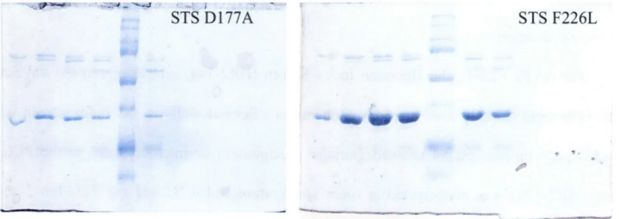
The crSTS mutants V214M, F226L, and D177A were obtained by site-directed mutagenesis performed on the gene in the pET-28a expression vector. Purification of the mutants proceeded as with the native protein. Following MonoQ chromatography, relevant fractions were collected and analyzed by SDS-PAGE. Even though the protein still contained some contamination, as shown in Figure 2.2, it was decided to attempt crystallization with the current preparation rather than perform further chromatography and potentially reduce the ability of STS to crystallize. Based on the success of subsequent crystallization screens, these mutants should be further purified before crystallization is attempted again. Also, future purifications should not include glycerol unless long-term freezer storage is anticipated.
STS D177A STS F226L
Figure 2.2 - SDS PAGE of crSTS mutants after Mono Q chromatography
Fractions from MonoQ chromatography were pooled and concentrated using an Amicon Ultra Centrifugal Filter device. crSTS F226L was concentrated to 15 mg/mL (from - 10 g cell pellet). Repeating the purification using -30 g cells (wet weight) afforded enough protein to set
up screens at 7.5 mg/mL. Using crSTS F226L and D177A, initial crystallization screens were begun.
2.2.1 Sparse matrix screening with D177A and F226L
All crystallization screens were set up in 24 well plates using siliconized glass cover slides and sealed with vacuum grease. For each well, 500 gIL of the pre-mixed condition was used as mother liquor. To construct the hanging drop, 1.5 giL of protein was mixed with 1.5 gtL of mother liquor on the glass cover slide and then inverted and sealed on top of the well. Equilibration was allowed to occur, and wells were observed after 2, 4, and 6 days for scoring. Wells were observed at intermittent time periods after the six days, but were not officially scored.
The Hampton Index Screen (HR2-144) was used for crSTS D177A. Numerous drops showed a light precipitate, and many others stayed clear. No hits appeared promising for optimization. Due to the faintness of even the amorphous precipitates, it is thought that the concentration was too low. Further work would include attempting these screens again at a higher concentration.
For crSTS F226L, the Hampton Index Screen (HR2-144) and Hampton Crystal Screen (HR2-110) were initially used at room temperature. Several wells in the index screen which included polyethylene glycol showed granular precipitation or microcrystals, so the PEG/Ion screen (HR2-126) was investigated at room temperature and 4 'C and the PEG/Ion 2 screen (HR2 - 098) was investigated at room temperature. Numerous wells in both screens showed heavy amorphous precipitation, although some drops remained clear for the length of the observation period. There were few granular precipitates, other that those in the PEG containing
conditions. However it seemed that the protein was well behaved and might eventually be optimized to produce crystals.
A potential hit was Figure 2.3 - Pictures of crystals in PEG/Ion condition 1
before and after "Izit" staining. No staining is observed, seen in PEG/Ion condition 1 after 5 days at both room temperature and 4 "C. Pictures of these crystals are shown in Figure 2.3. This condition contains 0.2 M NaF, 20% w/v PEG 3350; because of the NaF it is especially prone to forming salt crystals. However, several crystals were picked and analyzed following cryo-protection in 0.2 M NaF, 20% w/v PEG 3350, 20% ethylene glycol. One image was collected over 15 minutes with a 10 oscillation and a detector distance of 300 cm starting at 900. This was repeated with a 20 minute exposure starting at 1800. The maximum resolution was
to 3.3
A,
and very few spots were visible. A representative image may be seen in Figure 2.4. Based on the x-ray diffraction, it was concluded that these crystals were salt.Despite the x-ray results, the crystals were investigated further. Addition of 1 gL of mother liquor resulted in the formation of new crystals, strongly
Figure 2.4 - X-ray diftion fraction of supporting the case for salt. The crystals showed no PEG/Ion Condition 1 crystals.
staining with Izit dye and no protein was visible on SDS-PAGE, indicating that these were salt crystals.
2.2.2 Optimizing crystallization of F226L
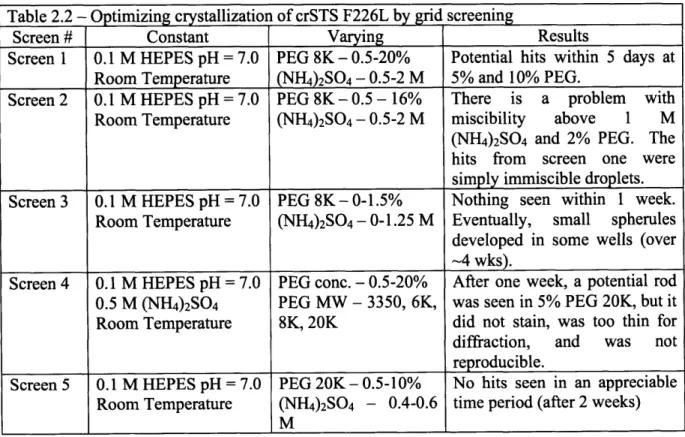
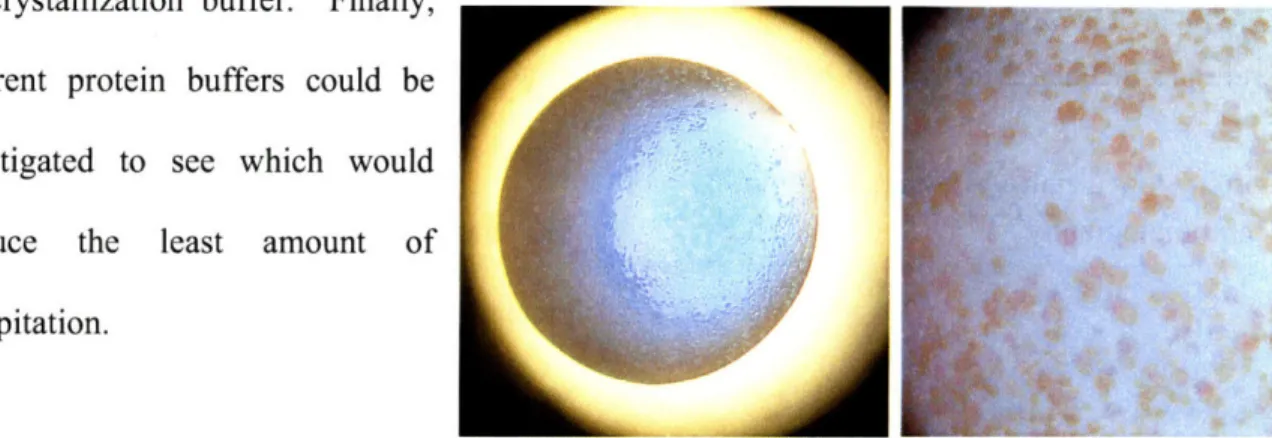
At this point, first index screens set up were re-examined. Numerous small spherules were observed in condition 35. This condition included 1.0 M ammonium sulfate, 0.1 M HEPES pH = 7.0, 0.5% w/v PEG 8000. These spherules were too small to screen for diffraction, but they were well stained by Izit dye (Figure 2.5). To optimize the crystallization for speed (1-2 weeks) and quality (larger, better morphology), several new screens were performed varying different components of the mixture. The components were pre-mixed in 1 mL Eppendorf tubes by vortexing. As before, 500 gL was used as mother liquor and the drop size was 1.5 IL.
Table 2.2 - Optimizing crystallization of crSTS F226L by grid screening
Screen # Constant Varying Results
Screen 1 0.1 M HEPES pH = 7.0 PEG 8K-0.5-20% Potential hits within 5 days at
Room Temperature (NH4)2SO4 - 0.5-2 M 5% and 10% PEG.
Screen 2 0.1 M HEPES pH = 7.0 PEG 8K - 0.5 - 16% There is a problem with Room Temperature (NH4)2SO4 - 0.5-2 M miscibility above 1 M (NH4)2SO4 and 2% PEG. The hits from screen one were simply immiscible droplets. Screen 3 0.1 M HEPES pH = 7.0 PEG 8K- 0-1.5% Nothing seen within I week.
Room Temperature (NH4)2SO4 -0-1.25 M Eventually, small spherules
developed in some wells (over -4 wks).
Screen 4 0.1 M HEPES pH = 7.0 PEG conc. - 0.5-20% After one week, a potential rod 0.5 M (NH4)2SO4 PEG MW - 3350, 6K, was seen in 5% PEG 20K, but it
Room Temperature 8K, 20K did not stain, was too thin for diffraction, and was not reproducible.
Screen 5 0.1 M HEPES pH = 7.0 PEG 20K- 0.5-10% No hits seen in an appreciable Room Temperature (NH4)2SO4 - 0.4-0.6 time period (after 2 weeks)
M
Future work towards crystallizing crSTS F226L would include repeating the initial screens at lower protein concentrations (i.e. 10 mg/mL). Also, glycerol should not be included in
L1IC YtlLULU1l y; UUIL•I. rlIaldly,
different protein buffers could be investigated to see which would produce the least amount of precipitation.
Figure 2.5 - Izit staining of crSTS F226L Index 35
2.3 Crystallization experiments with wild-type C. roseus STS 2.3.1 Sparse matrix screening for initial conditions
It is essential to verify the activity of a protein before attempting crystallization screens. This ensures that any structural information obtained will be of a relevant form of the protein. A detailed kinetic characterization was not necessary; a qualitative assay was all that was required. To assay crSTS, a previously described colorimetric assay was performed.5,9' 10 The reaction mixture for this assay includes crSTS, both tryptamine and secologanin substrates, and strictosidine glucosidase (SG), the next step in the biosynthetic pathway of the terpene indole alkaloids. Deglycosylation of the strictosidine produced by STS yields a reactive intermediate that undergoes a series of rearrangements to eventually yield a yellow and insoluble precipitate. The yellow chromophore forms only in the presence of active STS when the reaction is incubated at 30 oC. Compared to a no enzyme control, crSTS showed a strong yellow color and substantial precipitation after a 10 minute incubation at 30 oC.
After verifying the activity, electrophoretically homogenous crSTS was used for initial sparse matrix screens. The Hampton Index Screen (HR2-144) was used with the following protein solutions- 10 mg/mL crSTS, 10 mg/mL crSTS + 1 mM tryptamine, 5 mg/mL crSTS, 5
I
i