113
Protective Effect of a 21-Aminosteroid during Experimental Pneumococcal
Meningitis
Stefan Lorenzi, Uwe Koedel, Karl Frei, Andrea Bernatowicz, Adriano Fontana, and Hans-Walter Pfister
Department of Neurology, Klinikum Grof3hadern, Ludwig-Maximilians-University of Munich, Germany; Section of Clinical Immunology,
University Hospital, Zurich, Switzerland
This study investigated whether the 21-aminosteroid U74389F, an inhibitor of lipid peroxidation, attenuates pathophysiologic changes in experimental pneumococcal meningitis. Infected rats in-jected intravenously with vehicle and U74389F developed increases in regional cerebral blood flow (rCBF), intracranial pressure (ICP), brain water content, and white blood cells (WBC) in cerebrospi-nal fluid (CSF) within 8 h after intracistercerebrospi-nal challenge. Pretreatment with or administration of U74389F 4 h after infection significantly reduced the increase in ICP but had no effect on rCBF increase. Moreover, U74389F pretreatment significantly reduced brain water content and CSF WBC count. In vitro, U74389F inhibited iron-dependent lipid peroxidation of astrocyte cultures and the production of tumor necrosis factor-a, interleukin-6, and nitric oxide by stimulated macrophages. These data suggest that U74389F modulates early pathophysiologic alterations in experimental pneumococcal meningitis.
Animal models of bacterial meningitis have increased our knowledge of the complex pathophysiologic mechanisms of the disease [1- 5]. In a rat model of meningitis, we showed that intracisternal (ic) inoculation of live pneumococci or pneu-mococcal cell wall components induces an early increase in regional cerebral blood flow (rCBF), intracranial pressure (lCP), and brain water content [6]. Pretreatment with free su-peroxide dismutase (SOD), polyethylene glycol (PEG)-conju-gated SOD, deferoxamine, and catalase greatly attenuates these pathophysiologic changes; the strongest effects are shown with SOD and PEG-SOD [6-8]. Findings by other investigators support a role for reactive oxygen species in the pathophysiol-ogy of bacterial meningitis [9, 10]. Cellular injury caused by reactive oxygen species may involve direct damage to proteins and DNA as well as lipid peroxidation [11]. Here we tested the effect of the novel 21-aminosteroid U74389F [12, 13], an inhibitor of lipid peroxidation, for its capacity to alter rCBF, ICP, and brain edema formation and to reduce meningeal in-flammation in experimental pneumococcal meningitis.
Materials and Methods
Animal preparation. We used a well-characterized rat menin-gitis model that has been described in detail [6]. Adult male Wistar rats (250- 330 g) were intraperitoneally anesthetized with 100 mg!
Received 8 February 1994; revised 13 December 1994.
Presented in part: 33rd Interscience Conference on Antimicrobial Agents and Chemotherapy, New Orleans, 17-20 October 1993 (abstract 793).
Financial support: Deutsche Forschungsgemeinschaft (Pf 246/3-2). Reprints of correspondence: Dr. Hans-Walter Pfister, Dept. of Neurology, Klinikum Grosshadem, Marchioninistrasse 15, 81377 Munich, Germany. The Journal of Infectious Diseases 1995; 172:113-8
© 1995 byThe University of Chicago. All rights reserved. 0022-1899/95/7201-0015$01.00
kg thiobutabarbiturate (lnactin; Byk Gulden, Konstanz, Germany), tracheotomized, and artificially ventilated (small animal ventilator, model 683; Harvard, South Natick, MA). End-expiratory CO2was
continuously monitored by infrared CO2 analyzer (model 2200; Heyer, Bad Ems, Germany). Mean arterial blood pressure (MABP) was measured by pressure transducer (Statham P23; Viggo-Spec-tramed, Oxnard, CA) connected to the femoral artery cannula. Arterial blood gases and hematocrit were determined before ic inoculation and every 2 h thereafter (gas check model 1304; Instru-mentation Laboratory, Kirchheim, Germany). Body temperature was maintained at 38°C by a rectal thermometer-controlled heat-ing pad. Rats were placed in a stereotaxic frame, and a burr hole was made in the occipital bone for placement of the cisterna magna catheter. A 5-mm-diameter craniotomy was made in the right pari-etal bone for the placement of a laser-Doppler probe. rCBF was measured continuously by laser-Doppler flowmetry (model BPM 403a; Vasamedics, St. Paul, MN). Changes in rCBF were ex-pressed as percent changes from baseline. The dura was left intact in all preparations.
Cerebrospinal fluid (CSF; 100 ILL) was withdrawn through the cisterna magna catheter, and meningitis was induced by ic injection of 100 p,L of pneumococci (=106 cfu). We used Streptococcus pneumoniae type 3 (no. 17260), an isolate from an endotracheal aspiration of a patient with septic infection that was maintained at -20°C in trypticase soy broth (Oxoid, Wesel, Germany) supple-mented with 10% glycerol and 1% IsoVitale (Becton Dickinson Microbiology Systems, Heidelberg, Germany). Before use, the bacteria were subcultured on blood-agar plates, checked for purity, inoculated into brain-heart infusion broth (Oxoid), supplemented with 3% horse serum and 1% bovine albumin (Serva, Heidelberg, Germany), and incubated overnight at 35°C. The broth was centri-fuged for 20 min at 2500 g, and the sediment was washed once with 0.85% saline and resuspended in saline. The final suspension was turbidimetrically adjusted to an optical density of 0.5 at 546 nm (photometer with 13-mm filter; Eppendorf, Hamburg, Ger-many), thus achieving a concentration of = 107cfulmL.
The following parameters were continuously monitored for 8 h after ic injection by a personal computer system after
analog-digital conversion for signal processing: ICP (measured by Statham P23 pressure transducer connected to the cisterna magna catheter), rCBF, and MABP. Blood and CSF white blood cell (WBC) counts were determined at baseline and 4 and 8 h after ic injection. At the end of the experiment, the reactivity of the cerebral circulation to CO2 was tested. Hypercapnia was produced with 10% CO2,
21 % O2 ,and the balance ofN2 .Before and 10 min after hypercap-nia, arterial blood samples were drawn for blood gas and pH analysis. To determine brain water content, brains were weighed in glass dishes then dried for 16 h at 130°C to stable weight. Brain water content was calculated by the formula [(wet weight - dry weight)/wet weight] X 100 [6].
We studied 6 groups of rats: There were 6 infected rats in groups 1-3, 6 uninfected animals in group 4, and 3 and 4 infected rats in groups 5 and 6, respectively. Animals in group I were injected intravenously (iv) with vehicle of U74389F: 21-[ 4-(2,6-di-I-pyr-rolidinyl-4-pyrimidinyl)-I-piperazinyl]-pregna-I,4,9 (II )-triene-3,20-dione, monomethansulfonate (provided by Upjohn, Kalama-zoo, MI). The vehicle was citric acid and citrate (U74389Fyeh ) . Group 2 animals were treated iv with U74389F 15 min before (3 mg/kg) and 2 h after (1.5 mg/kg) ic pneumococcal challenge. Rats in group 3 were treated iv with U74389F 4 h after (3 mg/kg) and 6 h after (1.5 mg/kg) ic infection. Group 4 rats were injected iv with U74389F 15 min before (3 mg/kg) and 2 h after (1.5 mg/kg) ic injection of PBS. Rats in group 5 were treated iv with U74389F 15 min before (30 mg/kg) and 2 h after (15 mg/kg) ic infection (5-h measurement period). Group 6 rats were pretreated iv with U74389F 15 min before (3 mg/kg) and 2 h after (1.5 mg/kg) ic infection, the NO synthase inhibitor N-nitro-L-arginine (L-NA; 10 mg/kg) was administered iv 4 h after ic infection (5-h measurement period).
In vitro experiments. We investigated whether U74389F is a
potent inhibitor of lipid peroxidation when central nervous system (CNS) cells are used. For this purpose, primary rat astrocytes were stimulated with heat-killed pneumococci; lipid peroxidation was assessed by the formation ofthiobarbituric acid-reactive products. The data were compared with those of methylprednisolone, a known potent inhibitor of lipid peroxidation.
For initiation of lipid peroxidation, primary rat astrocytes were incubated with 200 f-LM Fe3+ and 50 f-LM Fe2
+ in 0.9% saline for 0.5 h at 37°C [14]. Fe3
+ and Fe2
+ solutions (Aldrich Chemie, Steinheim, Germany) were prepared fresh in argon-purged H20
and used immediately. Astrocytes and the supernatant were re-moved, homogenized, and sonicated for 30 s. After centrifugation (800g, 5 min, 4°C), 150 f-LL of the cell suspension was incubated with I mL of 0.5% thiobarbituric acid (Aldrich Chernie, Steinheim, Germany) in 12.5% trichloracetic acid (Aldrich Chemie) for 10 min at 90°C. After samples were centrifuged (800g, 5 min, room temperature), the formation of thiobarbituric acid-reactive oxida-tion products was determined at A53 2 (Ultrospec III; Pharmacia LKB, Freiburg, Germany). Quantification was based upon a molar extinction coefficient of 1.56 X 10⁵ [14]. The following groups were investigated: addition of U74389Fyc h (n = 16); U74389F, 100 f-LM (n = 6) and 1mM(n= 6), 10mM(n= 4); methylpredni-solone vehicle (n = 6); methylprednisolone, 1mM(n = 6) and 10 mM (n = 4); controls (n = 6; addition of the diluent of Fe2+ /
Fe3+ plus U74389Fv c h ) .
Primary astrocyte cultures were prepared from the cerebral cor-tex of I-day-old neonatal Wistar rats and grown on 6-well plates
(Falcon; Becton Dickinson, Plymouth, UK) at 37°C in a 5% CO2 incubator with 95% oxygen. During the first 7 days, the astrocytes were maintained in Dulbeccos modified Eagle medium (DMEM) supplemented with 20% heat-inactivated fetal calf serum (FCS). On day 8, the medium was replaced and the astrocytes were main-tained in DMEM plus 5% FCS. Astrocytes were cultivated in 6-well plates in culture medium until confluence. When confluence was documented (days 12-14), the astrocytes were shaken at 400 rpm for 3 h to remove microglia and oligodendrocytes. Astrocytes were then maintained I day in G5 medium [15] to eliminate mi-croglia and oligodendrocytes. Astrocyte cultures were character-ized on the basis of morphologic criteria and by the expression of glial fibrillary acidic protein (GF AP) as detected by immunostain-ing. These primary astrocyte cultures consisted of >95% GFAP-positive cells. We used only astrocytes without any passage. Before stimulation, astrocytes were washed with PBS without Ca2+ and Mg2
+ and maintained in DMEM with 5% FCS without phenol red and antibiotics.
Peritoneal macrophage cultures. We also questioned whether
the activity of U74389F was restricted to inhibition oflipid peroxi-dation. Thus, we tested to see if it interfered with the production of known mediators of bacterial meningitis, such as cytokines and NO. Peritoneal macrophages were stimulated with heat-killed pneumococci, lipopolysaccharide (LPS), and cytokines. Peritoneal exudate cells were obtained from 8-week-old female Wi star rats that were injected intraperitoneally with 6 mL of Brewer's thiogly-collate medium 3 days before isolation. The cells were cultured in DMEM (Biochrom, Berlin) supplemented with 1% FCS (Bio-chrom), 10 f-Lg/mL gentamicin, and I mM N-acetyl-L-alanyl-L-glutamine. Cells were stimulated with heat-killed (60°C, 4 h) unen-capsulated pneumococci (HKP; isogenic mutant of S.pneumoniae
type 3, no. 17260) in three different concentrations (105, 106 , and 107
cfu/ml.), LPS (Escherichia coli 0127:B8; I f-Lg/mL) or murine recombinant interferon-v (rIFN-y; 100 U/mL) plus murine recom-binant tumor necrosis factor-a(rTNF-a; 10 ng/mL). Murine
rTNF-a and rIFN-y were both purchased from Boehringer (Mannheim, Germany). E. coli LPS was obtained from Difco (Detroit). Cell cultures were untreated or treated with U74389F (I, 10, or 100
f-LM)or U74389Fv e h 'Interleukin (IL)-6 andTNF-a were measured
using 7TDI cells [16] and L-M cells [17], respectively. NO produc-tion in the cell culture supernatant was assessed by measuring nitrite, a stable metabolic product of NO, by the Griess reaction [18].
Statistical methods. Data on rCBF, ICP, and CSF WBC count
obtained 4 and 8 h after ic pneumococcal challenge in rats injected iv with U74389Fv e h or U74389F (groups 1 and 2) were compared by unpaired Student's t test; Pvalues were corrected for repeated measurements using the Bonferroni-Holm procedure. Data on brain water content and CO2 reactivity at 8 h after ic challenge from groups I and 2 were compared by the unpaired Student's t test. Data on rCBF, ICP, and CSF WBC count at 4 h after ic challenge in infected rats injected iv with U74389F in two different dosages (groups 2 and 5) were compared by unpaired Student's t test. Data on rCBF, ICP, and CSF WBC count at 4 and 5 h after ic challenge in infected rats injected iv with U74389F or with U74389F plus L-NA (groups 2 and 6) were compared by unpaired Student'sttest;Pvalues were corrected for repeated measurements using the Bonferroni-Holm procedure. One-way analysis of vari-ance and Student-Newman-Keuls multiple comparisons were used
JID 1995; 172 (July) 21-Aminosteroid in Pneumococcal Meningitis 115
Table 1. Pathophysiologic parameters in different experimental groups of rats.
Regional cerebral blood Intracranial pressure CSF white blood cell count
flow (%) (mm Hg) (cells/pL) Brain water CO2
content (%) reactivity*
Group 4h 8h 4h 8h 4h 8 h at 8 h at 8 h
Infected
U74389Fvehinjected 175.3 :±: 7.0 211.6 :±: 5.2 13.3 :±: 1.7 15.4:±: 1.2 2611 :±: 551 6710 :±: 729 79.09 :±: 0.03 1.38 :±: 0.56
U74389F pretreated 151.8 :±: 7.0 204.4 :±: 3.1 9.0 :±: 1.4 11.0:±:LOt 1095 :±: 163t 4297 ::±:289t 78.80 :±: 0.06t
0.86 :±: 0.26 Uninfected, U74389F injected 107.9 :±: 5.6 115.6 :±: 6.5 2.1 :±: 0.3 3.8:±: 0.9 6 :±: 2 23 :±: 10 77.91 :±: 0.10 1.34 :±: 0.45
NOTE. Each group, n = 6.
* Change in regional cerebral blood flow (%)/change inPco,(mm Hg).
tP < .05, vs. infected, U74389Fvch-injectedrats.
to compare data on thiobarbituric acid reactive products. Differ-ences were considered significant when P
<
.05. Data are ex-pressed as mean ± SE.Figure 1. Time course of regional cerebral blood flow (rCBF) and intracranial pressure (lCP) in infected rats injected with U74389F or its vehicle (U74389Fve h ) before infection. rCBF and ICP increased in both groups. Statistical analysis was done 4 and 8 h after intracisternal injection(arrows) by unpaired Student'sttest.Pvalues were corrected for repeated measurements by Bonferroni-Holm procedure. Pretreat-ment with U74389F significantly reduced increase in ICP 8 h after infection (see table I). Data are mean ± SE.
220
0' • pneumococci + U74389Fveh
~ 200
o
pneumococci + U74389F ~ 0 180 ...J LL 0::: 160 W ...Ja..
a..
140 0 Cl 0::: 120 Wen
100-c
...Jfr
80 0; 20:c
• pneumococci + U74389F veh E 18o
pneumococci + U74389FoS
w 16 0::: ::J 14en
en
12 W 0:::a..
10 ...J-c
8Z
-c
6 0:::o
-c
4fr
0:::fr
.-
2 Z --,----1 0 2 3 4 5 6 7 8TIME [h]
Physiologic variables. MABP,Paz, Pco-,pH, hematocrit, and body temperature were normal throughout the experiment in all groups (data not shown). CerebrovascularCOz reactivity did not differ significantly between groups (table 1).
rCBF, ICP, brain water content, and CSF WBC count. There was an increase in rCBF in U74389Fveh-injectedinfected
rats (group 1) from a baseline of 100% to 211.6% ± 5.2% within 8 h after pneumococcal challenge (table 1; figure 1). In infected rats pretreated with U74389F (group 2), the mean values of rCBF at 4 and 8 h after ic injection did not differ significantly from that of infected rats pretreated with U74389Fv eh (table 1).
rcp
markedly increased in infected U74389Fveh-injectedrats(group 1) within 8 h after infection from a baseline of 3.6 ±
0.4 mm Hg to 15.4 ± 1.2 mm Hg (figure 1, table 1). Pretreat-ment with U74389F (group 2) significantly attenuated the in-crease in
rcp
at 8 h (table 1). There was no change in rCBF andrcp
in uninfected rats pretreated with U74389F and injected ic with PBS (group 4; table 1). Pretreatment with U74389F (group 2) significantly reduced brain water content and CSF WBC counts (P<
.05; table 1).The inhibitory effect ofU74389F (group 2) on ICl' and CSF pleocytosis in infected rats was not enhanced when infected rats were pretreated with the higher dosage ofU74389F (group 5). For example, at 4 h after infection,
rcp
was 10.7 ± 4.2 mm Hg in group 5 versus 11.0 :::!:::: 1.0 mm Hg in group 2, andCSF WBC count was 1021 :::!:::: 305 versus 1095 :::!:::: 163 cells/ pL. There was also no effect on rCBF (171.1 % :::!:::: 26.0% vs.
151.8% :::!:::: 7.0% at 4 h after infection).
Compared with effects observed in U74389Fveh-injectedrats
(group 1), the administration ofU74389F 4 h after ic infection (group 3) significantly attenuated the increase in
rcp
(7.6 :::!::::1.5 vs. 15.4 :::!:::: 1.2 mm Hg, groups 3 and 1, respectively) but
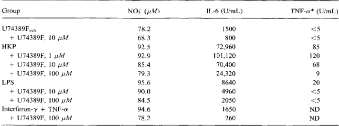
Table 2. Effect of different doses of U74389F on the production of nitric oxide, interleukin-6 (IL-6), and tumor necrosis factor-a (TNF-a) by rat peritoneal macrophages.
Group NOi (pM) IL-6 (U/mL) TNF-a*(U/mL)
U74389Fve b 78.2 1500 <5 +U74389F, 1011M 68.3 800 <5 HKP 92.5 72,960 85 +U74389F, 111M 92.9 101,120 120 +U74389F, 1011M 85.4 70,400 68 +U74389F, 10011M 79.3 24,320 9 LPS 95.6 8640 20 +U74389F, 10f.1M 90.0 4960 <5 + U74389F, 100f.1M 84.5 2050 <5 Interferon-y+TNF-a 94.6 1650 ND +U74389F, 100f.1M 78.2 260 ND
NOTE. NOi = nitrite; veh = vehicle of U74389F; HKP = heat-killed pneumococci (107
cfu/mL); LPS
Escherichia coli lipopolysaccharide; ND = not done.
*Detection limit ofTNF-aassay was 5 U/mL.
content. The anti edematous effect of U74389F when given prior to infection corresponds with findings in other pathophys-iologic CNS models that documented a protective effect of 21-amino steroids on brain edema formation [19]. The increase in ICP in the early phase of experimental pneumococcal meningi-tis is thought to be mainly due to an increase in brain water content and an increase in cerebral blood volume [20]. Our finding that the increase in
rcp
was reduced but not completelyI
I*
I I,
I*
\ I,
I I # I #f
I,
I § If
I I I I I I I I §+ I+
+
#+ I I•
Ihad no effect on CSF pleocytosis (7240 ± 700 vs. 6710 ± 729 cells/pL), rCBF (189.3% ± 13.3% vs. 211.6% ± 5.2%), or brain water content (79.04% ± 0.03% vs. 79.09% ± 0.03%). Administration ofL-NA 4 h after infection in U74389F-pre-treated rats (group 6) produced an increase in MABP from 98 ±
4 to 124 ± 5 rom Hg. The rCBF increase at 4 h after infection (149.1% ± 14.4% for group 6 vs. 151.8% ± 7.0% for group 2) was completely reversed by L-NA (97.4% ± 12.8% for group 6 vs. 167.7% ± 3.4% for group 2 5 h after infection).
Inhibition of release ofNO, 1L-6, and TNF-a by peritoneal macrophages stimulated with HKP, E. coli LPS, and cytokines. HKP (in a dose-dependent fashion, data shown only for 107
cfu/mL), E. coli LPS, and murine rIFN-y plus murine rTNF-a stimulated production of NO, IL-6, andTNF-ain the macro-phage cultures (table 2). U74389F inhibited dose dependently NO, IL-6, and TNF-a production by macrophages stimulated with HKP,E. coli LPS, or murine rIFN-y plus murine rTNF-a. HKP (107
cfu/mL)-induced production of NO, IL-6, and TNF-a by macrophages was inhibited by U74389F (100 J-lM)
by 92%, 89%, and 67%, respectively.
Inhibition of lipid peroxidation by U74389F in primary astrocytes. Iron-dependent lipid peroxidation in primary rat astrocyte cultures was inhibited by U74389F and methylpred-nisolone (figure 2). 0.8 ~ 3 0.7
en
w 0.6o
Z~
0.5en
m
:::::> 0.4en
w>
0.3 i= o~
0.2 a::~
0.1 t-control vehU 0.1mM 1mM 10mM U74389F vehM 1mM 10rnM Methylprednisolone DiscussionThe major finding of this study was that pretreatment with the novel 21-aminosteroid U74389F significantly attenuated increases in brain water content and ICP and CSF leukocytosis during the early phase of experimental pneumococcal meningi-tis in the rat. U74389F had no effect on the increase in rCBF. Administration ofU74389F to animals with established menin-gitis significantly attenuated the increase in ICP but had no effect on CSF pleocytosis or increases in rCBF and brain water
Figure 2. Inhibition of iron-dependent lipid peroxidation in pri-mary rat astrocytes. Lipid peroxidation was assessed by formation of thiobarbituric acid (TBA)-reactive substances (mean ± 99% confi-dence interval). Addition of Fe2+IFe3+(+vehicle of U74389F [veh.j] or methylprednisolone [vehMD to astrocytes induced significant in-crease of TBA-reactive substances vs. action of diluent of Fe2
+/Fe3+ (control;
*
P< .05). Increase was significantly attenuated by methyl-prednisolone, known inhibitor of lipid peroxidation (§t ,
P< .05 vs. veh., and 1 mM, respectively). U74389F also blocked increase of TBA-reactive substances (# +,P < .05 vs. veh., and vs. 0.1 and 10mM,respectively. All P values determined by analysis of variance and Student-Newman-Keuls multiple comparisons.
JID 1995; 172 (July) 21-Aminosteroid in Pneumococcal Meningitis 117
inhibited might be explained by the lack of influence of U74389F on an increase in rCBF, which, in tum, may be caused at least in part by an increase in cerebral blood volume.
The 21-aminosteroids exhibit protective effects in a variety of pathologic conditions, including cerebral ischemia [21], sub-arachnoid hemorrhage [22], traumatic brain injury [23], vaso-genic edema [19], and endotoxemia [24]. Their protective ef-fects have been primarily attributed to their capacity to inhibit membrane lipid peroxidation [12, 25] . We investigated whether U74389F is capable of inhibiting iron-dependent lipid peroxi-dation in a CNS cell culture system. Our in vitro experiments used primary rat astrocytes and showed that the ability of U74389F to suppress lipid peroxidation is comparable to that of methylprednisolone, a potent inhibitor of lipid peroxidation. By inhibiting lipid peroxidation, the 21-aminosteroids may in-directly exert further inhibitory actions. For example, inhibition of 5-lipoxygenase may prevent the formation of leukotrienes [19], inhibition ofNADPH oxidase may prevent the generation of superoxide radical and hydrogen peroxide [26], and inhibi-tion of the destrucinhibi-tion of cell membranes may prevent the release of arachidonic acid [27]. Recently, others have sug-gested that some 21-aminosteroids (e.g., U743 89F, U78518F) may have an oxygen radical scavenging effect [28, 29].
We tested whether U74389F, apart from inhibiting lipid per-oxidation, also interferes with other mediators (e.g., cytokines and NO) known to be involved in the pathophysiology ofbacte-rial meningitis [4,30, 31]. Peritoneal macrophages, which we used in our in vitro experiments, are an established cell culture system for the induction of cytokines and NO. There are simi-larities between rat peritoneal macrophages and brain macro-phages and microglia: Both cell types produce cytokines [32, 33] and NO [34, 35] upon stimulation with LPS and Staphylo-coccus aureus. Our data provide evidence that the activity of U74389F is not restricted to inhibition of lipid peroxidation. U74389F inhibited the production of NO, IL-6, andTNF-a by rat peritoneal macrophages stimulated with heat-killed pneu-mococci, E. coli LPS, and cytokines. A previous study showed that another 21-aminosteroid compound (U74500A) interferes with the production of cytokines [36]. Our in vitro studies showed that U74389F inhibited NO production by rat perito-neal macrophages; however, in vivo, U74389F (in two different dosages) surprisingly had no effect on blood flow changes, which are known to be NO-mediated [30, 31]. Thus, in our meningitis model, U74389F did not affect NO production, al-though the addition of the NO synthase inhibitor L-NA reversed the blood flow increase.
We found that U74389F reduced meningeal inflammation. This observation is consistent with a recent study in which the 21-aminosteroid tirilazad mesylate attenuated the accumulation of neutrophils in ischemic gerbil brain [37]. In contrast, others have reported that U74389F does not inhibit the hyperoxia-induced accumulation of neutrophils in bronchoalveolar lavage fluid [38]. One explanation for the effect observed in our study may be that 21-aminosteroids inhibit the generation of
superox-ide radical and thereby reduce the expression of adhesive glyco-proteins [39, 40] and the adherence of neutrophils. Another explanation might be that damage of biomembranes by lipid peroxidation could lead to the release of chemoattractant sub-stances, such as arachidonic acid metabolites or platelet-activat-ing factor, which could be prevented by inhibitors of lipid peroxidation.
Reactive oxygen species are known to play a role in early pathophysiologic changes during experimental bacterial men-ingitis [6-10]. Along with the data from our previous experi-ments, the results of the current study suggest that lipid per-oxidation induced by oxygen free radicals is involved in the pathophysiologic mechanisms during the early phase of pneu-mococcal meningitis. However, other activities of the 21-amino steroid U74389F, such as interference with cytokine pro-duction, might contribute to the modulation of the pathophysio-logic changes in early experimental pneumococcal meningitis.
Acknowledgments
We thank G. Ruckdeschel (Max-von-Pettenkofer Institute, Uni-versity of Munich) for pneumococci, Dagmar Forth and Judy Ben-son for help with manuscript preparation, Alex Heng for excellent assistance with the experiments, and Peter Dirschedl (Institute for Medical Informatics, Biometrie and Epidemiology, University of Munich) for statistical help.
References
1. Scheld WM, Dacey RG Jr, Winn HR, Welsh JE, Jane JA, Sande MA. Cerebrospinal fluid outflow resistance in rabbits with experimental men-ingitis. Alterations with penicillin and methylprednisolone. J Clin Invest 1980; 66:243-53.
2. Tauber MG, Borschberg U, Sande MA. Influence of granulocytes on brain edema, intracranial pressure, and cerebrospinal fluid concentrations of lactate and protein in experimental meningitis. J Infect Dis 1988; 157:456-64.
3. Tuomanen EI, Saukkonen K, Sande S, Cioffe C, Wright SD. Reduction of inflammation, tissue damage, and mortality in bacterial meningitis in rabbits treated with monoclonal antibodies against adhesion-promoting receptors of leucocytes. J Exp Med 1989; 170:959-69.
4. Mustafa M, Ramilo0,Olsen KD, et al. Tumor necrosis factor in mediating experimentalHaemophilus influenzae type b meningitis. J Clin Invest
1989;84:1253-9.
5. Smith AL. Pathogenesis ofHaemophilus influenzae type b meningitis.
In: Keusch G, Wad strom T, eds. Experimental bacterial and parasitic infections. New York: Elsevier, 1983:295-301.
6. Pfister HW, Koedel U, Haberl RL, et al. Microvascular changes during the early phase of pneumococcal meningitis in the rat. J Cereb Blood Flow Metab 1990; 10:914-22.
7. Pfister HW, Koedel U, Dimagl U, Haberl RL, Ruckdeschel G, Einhaupl KM. Effect of catalase on regional cerebral blood flow and brain edema during the early phase of experimental pneumococcal meningitis. J Infect Dis 1992; 166:1442-5.
8. Pfister HW, Koedel U, Lorenzl S, Tomasz A. Antioxidants attenuate micro-vascular changes in the early phase of experimental pneumococcal men-ingitis in rats. Stroke 1992;23:1798-804.
9. McKnight AA, Keyes WG, Hudak ML, Jones MD. Oxygen free radicals and the cerebral arteriolar response to group B streptococci. Pediatr Res 1992; 31 :640-4.
10. Berkowitz ID, Traystman RJ. Oxygen radical scavengers prevent impair-ment of microvascular autoregulation inH. influenzaetype B meningitis in rats [abstract]. Faseb J 1993; 7:A530.
11. Halliwell B. Reactive oxygen species and the central nervous system. J Neurochem 1992; 59: 1609-23.
12. Haghighi SS, Hall ED, Geng XZ, Oro JJ, Johnson GC. Therapeutic value of 21-aminosteroid U74389F in acute spinal cord injury. Neurol Res 1993; 15:321-6.
13. Bernstein M, Ginsberg H, Glen J. Protection of iodine-125 brachytherapy brain injury in the rat with the 21-aminosteroid U-74389F. Neurosurgery 1992; 31 :923 - 8.
14. Braughler JM, Duncan LA, Chase RL. The involvement of iron in lipid peroxidation: importance of ferric to ferrous ratios in initiation. J Bioi Chern 1986;261:10282-9.
15. Michler-Stuke A, Wolff J, Bottenstein J. Factors influencing astrocyte growth and development in defined media. Int J Dev Neurosci 1984;2:575-84.
16. Frei K, Leist TP, Meager A, et a1. Production of B cell stimulatory factor-2 and interferon gamma in the central nervous system during viral meningitis and encephalitis. Evaluation in a murine model infection and in patients. J Exp Med 1988; 168:449-53.
17. Frei K, Siepl C, Groscurth P, Bodmer S, Schwerdel C, Fontana A. Antigen presentation and tumor cytotoxicity by interferon-gamma-vtreated mi-croglial cells. Eur J Immuno11987; 17:1271-8.
18. Stuehr DJ, Marletta MA. Mammalian nitrate biosynthesis: mouse macro-phages produce nitrite and nitrate in response toEscherichia coli
lipo-polysaccharide. Proc Nat! Acad Sci USA 1985; 82:7738-42. 19. Hall ED, Travis MA. Inhibition of arachidonic acid-induced va so genic
brain edema by the non-glucocorticoid 21-aminosteroid U74006F. Brain Res 1988;451:350-2.
20. Tureenill,Dworkin RJ, Kennedy SL, Sachdeva M, Sande MA. Loss of cerebrovascular autoregulation in experimental meningitis in rabbits. J Clin Invest 1990; 85:577-81.
21. Young W, Wojak JC, DeCrescitoV. 21-aminosteroid reduces ion shifts and edema in the rat middle cerebral artery occlusion model of regional ischemia. Stroke 1988; 19:1013-9.
22. Zuccarello M, Anderson DK. Protective effect of a 21-aminosteroid on the blood-brain barrier following subarachnoid hemorrhage in rats. Stroke 1989; 20:367 - 71.
23. Hall ED, Yonkers PA, McCall JM, Braughler JM. Effects of the 21-aminosteroid U74006F on experimental head injury in mice. J Neuro-surg 1988; 68:456-61.
24. Siegfried MR, Ma X, Lefer AM. Splanchnic vascular endothelial dysfunc-tion in rat endotoxemia: role of superoxide radicals. Eur J Pharmacal 1992;212:171-6.
25. Horton JW, Walker PB. Oxygen radicals, lipid peroxidation, and perme-ability changes after intestinal ischemia and reperfusion. J Appl Physiol 1993; 74:1515-20.
26. Thomas PD, Mao GD, Rabinovitch A, Poznansky MJ. Inhibition of super-oxide-generating NADPH oxidase of human neutrophils by lazaroids (2l-aminosteroids and 2-methylaminochromans). Biochem Pharmacol 1993;45:241-51.
27. Braughler JM, Chase RL, Neff GL, et a1. New 21-aminosteroid antioxidant lacking glucocorticoid activity stimulates adrenocorticotropin secretion and blocks arachidonic acid release from mouse pituitary tumor (AtT-20) cells. J Pharmacol Exp Ther 1988;244:423-7.
28. Barnard ML, Gurdian S, Turrens JF. Activated polymorphonuclear leuko-cytes increase low-level chemiluminescence of isolated perfused rat lungs. J Appl Physiol 1993; 75:933-9.
29. Tanigaki T, Suzuki Y, Heimer D, Sussman HH, Ross WG, Raffin TA. Attenuation of acute lung injury and oxygen radical production by the 21-aminosteroid, U-78518F. J Appl Physiol 1993;74:2155-60. 30. Pfister HW, Koedel U, Bematowicz A, Frei K, Fontana A. Role of reactive
nitrogen intermediates in experimental pneumococcal meningitis [ab-stract 796]. In: Program and ab[ab-stracts of the 33rd Interscience Confer-ence on Antimicrobial Agents and Chemotherapy (New Orleans). Wash-ington, DC: American Society for Microbiology, 1993.
31. Haberl RL' Anneser F, Kadel U, Pfister HW. Is nitric oxide involved as a mediator of cerebrovascular changes in the early phase of experimental pneumococcal meningitis? Neurol Res 1994; 16:108-12.
32. Giulian D, Bakerrr, Shih LN, Lachman LB. 1nterleukin 1 of the central nervous system is produced by ameboid microglia. J Exp Med 1986; 164:594-604.
33. Chung IY, Norris JG, Benveniste EN. Differential tumor necrosis factor-a expression by astrocytes from experimental allergic encephalomyelitis-susceptible and -resistant rat strains. J Exp Med 1991; 173:801-1 1. 34. Merrill JE, IgnarroLJ,Sherman MP, Melinek J, Lane TE. Microglial cell
cytotoxicity of oligodendrocytes is mediated through nitric oxide. J ImmunoI1993; 151:2132-41.
35. Bosca L, Lazo PA. Induction of nitric oxide release by MRC OX-44 (anti-CD53) through a protein kinase C-dependent pathway in rat macro-phages. J Exp Med 1994; 179: 1119-26.
36. Fisher M, Plante GM, Doyle EM. Inhibition of inflammatory cell-mediated myelin oxidation and interleukin I-beta generation by a 21-aminoste-roid, U74500A. JNeurol Sci 1993;119:189-94.
37. Williams LR, Oostveen JA. Tirilazad mesylate attenuates the accumulation of neutrophils in ischemic brain. Ann NY Acad Sci 1993; 679:330-4. 38. Richards 1M, Griffin RL, Fidler SF, Jacobsen EJ. Effect of the 21-aminoste-roid, U-74389F, on hyperoxic lung injury in rats. Agents Actions 1993; 39:CI36-8.
39. Suzuki M, Grisham MB, Granger DN. Leukocyte-endothelial cell adhesive interactions: role of xanthine oxidase-derived oxidants. J Leukoc Bioi 1991; 50:488-94.
40. Patel KD, Zimmerman GA, Prescott SM, McEver RP, McIntyre TM. Oxygen radicals induce human endothelial cells to express GMP-140 and bind neutrophils. J Cell Bioi 1991; 112:749-59.