Carrfnogenesis vol.14 no.10 pp.2119-2122, 1993
p53 mutations in phenacetin-associated human urothelial
carcinomas
Iver Petersen, Hiroko Ohgaki, Barbara I.Ludeke and
Paul Kleihues
1Institute of Neuropatbology, Department of Pathology, University of Zurich, CH-8091 Zurich, Switzerland
'To whom correspondence should be addressed
Chronic abuse of the analgesic drug phenacetin is associated
with an increased risk of development of transitional cell
carcinomas of the urinary tract. It is unclear whether
phenacetin acts through chronic tissue damage (phenacetin
nephropathy) or via a genotoxic metabolite causing
promutagenk DNA lesions. In the present study, we
investigated 15 urothelial carcinomas from 13 patients with
evidence of phenacetin abuse. Tumors were screened for p53
mutations in exons 5 - 8 by single-strand conformation
polymorphism (SSCP) analysis, followed by direct sequencing
of PCR-amplified DNA. p53 Mutations were detected in 8/14
primary tumors (57%). All except one were missense
mutations located in exon 5 (three mutations), exon 6 (one),
exon 7 (two) and exon 8 (one). The type of mutation varied,
with a preference for CpG sites. A frameshift mutation
resulting from the insertion of a single cytosine at codons
151/152 was detected in a bladder tumor and its lung
metastasis. Urothelial carcinomas located in the renal pelvis
and hi the ureter of the same patient exhibited two different
mutations, strongly suggesting that they developed
independently. Another patient had tumors in the renal pelvis
and bladder, both of which contained the same p53 mutation,
indicating intracavitary metastatic spread. This demonstrates
that screening of p53 mutations allows the clonal origin of
tumors in patients with multiple primary and metastatic
lesions to be determined. None of the tumors investigated
contained mutations in codons 12,13 or 61 of H-ras or K-ras
protooncogenes.
Introduction
Habitual use of phenacetin or phenacetin-containing analgesics
is known to cause renal papillary necrosis, chronic interstitial
nephritis and capillary sclerosis of the lower urinary tract (1).
In addition, some patients with long-term addiction develop
urothelial carcinomas ( 2 - 5 ) . Moreover, patients with a history
of phenacetin abuse have a significantly higher risk of developing
multiple tumors of the urinary tract than non-abusers (4). There
is increasing evidence that the type and genomic distribution of
mutations in oncogenes and tumor suppressor genes may be
typical or even specific for the carcinogen which initiated the
multi-step process of malignant transformation. Examples of this
type of genetic tumor epidemiology include aflatoxin Bj-induced
hepatocellular carcinomas (6), tobacco versus uranium-associated
lung cancer (7) and squamous cell carcinomas of the skin
•Abbreviations: SSCP, single-strand conformation polymorphism; PCR, polymerase chain reaction.following UV exposure (8). The objective of the present study
was, therefore, to identify possible specific p53 mutations in
urothelial carcinomas resulting from phenacetin intake.
Materials and methods
Tumors from 13 patients were investigated (Table I). Surgical biopsies were obtained from seven patients (cases 1 —4,7, 8 and 10). Samples from five nAtitvmal patients (cases 5, 6, 9, 11 —13) were collected at post mortem examination. Two separate urinary tract tumors were analyzed in each of two patients (cases 10 and 13), and in one pattern (case 11) a bladder tumor and a lung metastasis were investigated. Twelve out of 13 patients had a clinically documented history of long-term phenacetin abuse and kidney lesions typical for phenacetin nephropathy, i.e. renal capillary sclerosis, renal papillary necrosis and chronic interstitial nephritis (Table I). In one patient (case 9), there was no proven history of phenacetin abuse but the presence of capillary sclerosis and papillary necrosis was considered sufficient evidence of phenacetin nephropathy (1,4). Tumors were graded according to WHO guidelines (9) into well-differenriated (grade I), moderately differentiated (grade IT) and poorly differentiated carcinomas (grade 10).
DNA was extracted from formalin-fixed, paraffin-embedded tissue according to the protocol described by Wright and Manos (10). Briefly, tumor tissue was scraped off serial histoiogical sections after comparison with a representative section stained with H&E. Samples were deparaffinized with xylene and washed with absolute ethanol. Dried samples were treated with 200 /ig/ml of proteinase K (Boehringer Mannheim GmbH, Mannheim, Germany) in 100 /il digestion buffer (50 mM Tris, pH 8.5; 1 mM EDTA; 0.5% Tween 20) for 3 h at 55°C. After inactrvatkm of proteinase K by heating at 95°C for 10 min, samples were kept at -20°C until PCR amplification. DNA from frozen tumor tissue was extracted by phenol and chloroform (11).
Single-strand conformation polymorphism (SSCP*) analysis was performed using a modification of the method of Orita et al (12). Polymerase chain reaction (PCR) was performed with 2 /d of DNA solution extracted from formalin-fixed tissue or 200 ng of genomic DNA from frozen tissue, 10 pmol of each primer, 125
fiM of dNTPs, 1 ^Ci of [a-32PJdCTP (Amersham, sp. act. 3000 Ci/mmol), 10 mM Tris (pH 8.3), 50 mM KC1, 1.25 mM MgCl2, and 0.5 U Taq polymerase (Perkin-Eliner Cetus) in a final volume of 20 /J. After adding 10 pi of mineral oil (Sigma), 35 cycles of denaturation (95°C) for 50 s, annealing (58°Q for 50 s and extension (72°Q for 70 s were carried out using an automated DNA Thermal Cycler (Perkin-Elmer Cetus). The primer sequences for thep55 gene were reported previously (13-15). Aliquots (5 /J) of the amplification mixture were mixed with 5 (J of sequencing stop solution (United States Biochemical, Cleveland, OH), heated at 95°C for 5 min and immediately loaded onto a 6% polyacrylamide non-denaturing gel containing 1096 glycerol. Gels were run at 8 W for 1 3 - 1 5 h at room temperature. Gels were dried and autoradiography was performed with an intensifying screen for 12-72 h.
For the samples scored positive with SSCP analysis, PCR was performed with 10 yX of DNA extracted from formalin-fixed tissue or 1 /ig of genomic DNA extracted from frozen tissue, 20 pmol each primer, 125 JJM dNTPs, 10 mM Tris (pH 8.3), 50 mM KC1, 1.25 mM MgCl2 and 2.5 U Taq polymerase (Perkin-Elmer Cetus) in a final volume of 100 /d. Exons 1 and 2 of H-, K-ras genes were also amplified and directly sequenced as described above. Primer sequences for raj genes were described previously (16). After amplification, 70 /d of the PCR reaction were electrophoresed on a 4% agarose gel (3% low melting and 1% regular agarose). The specific bands were excised, electroeluted in 0.5 x TBE buffer, purified by Ehmp-D columns (Schkicher and Schuefl GmbH, Dassd, Germany) and precipitated with ethanol. Dried DNA was resuspended in 12 /d of distilled water.
Direct sequencing was performed using amplification primers forp53 exons. The following internal sequencing primers were used for rat genes: 5'-TCCAC-AAAATGGTTCTGGAT for H-rai codon 12/13; 5'-AGACGTGCCTGTTGG-ACATC for H-rai codon 61; 5'-CGTCCACAAAATGATTCTGA for K-ras codon 12/13; 5'-GTAATTGATGGAGAAACCTG for K-raj codon 61. A modified Sanger dideoxynucleotide sequencing method was used that allows efficient extension labeling of the sequencing primer. Briefly, template DNA (4 /d) and primer (10 pmol) in 10 /J buffer (20 mM Tris-HCl, pH 7.5, 10 mM Mgd2, 25 mM NaCl; United States Biochemical) containing 10% (v/v) DMSO,
I.Petersen et al.
Table I. p53 mutations in human phenacetin-induced urothelial carcinomas
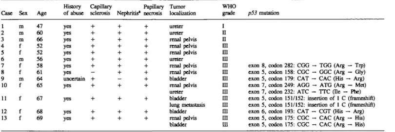
History Capillary Papillary Tumor WHO
Case Sex Age of abuse sclerosis Nephritis* necrosis localization grade p53 mutation 1 2 3 4
5
6 7a
9 10 i i 12 13 m m m f f m f f m f f f f 47 60 66 52 52 56 58 61 64 65 67 68 69 yes yes yes yes yes yes yes yes uncertain yes yes yes yes ureter ureter renal pelvis renal pelvis renal pelvis ureter renal pelvis renal pdvis bladder renal pelvis ureter bladder lung metastasis bladder renal pelvis bladder In
n
m
m
m
m
m
m
m
m
m
m
ra
in
m
exon 8, codon exon 5, codon exon 5, codon exon 7, codon exon 7, codon exon 5, codon exon 5, codon exon 6, codon exon 5, codon exon 5, codon 282: CGG - TGG (Arg - Trp) 158: CGC - GGC (Arg - Gly) 179: CAT - CAC (His - Arg) 249: AGG - ATG (Arg - Met) 232: ATC - TTC (Be - Phe) 151/152: insertion of 1 C (frameshift) 151/152: insertion of 1 C (frameshift) 193: CAT - CGT (His - Arg) 175: CGC - CAC (Arg - His) 175: CGC - CAC (Arg - His) •Chronic interstitial nephritis.were denatured at 95°C for 5 min and immediately frozen in dry ice/ethanol. Six microliters containing an appropriate a-^P-labeled dideoxynucleotide (Amersham, sp. act. 3000 Ci/mmol) and 4 U of Sequenase Version 2.0 (United States Biochemical) in 17 mM dithiothreitol were added. The radioactive nudeocide was chosen to be the one immediately incorporated adjacent to the primer. After preincubation for 2 min at room temperature, 3.6 /»1 aliquots were mixed with 2.5 id termination mix (80 pM dNTPs, 8 ^M of one ddNTP) and incubated for 10 min at 37°C. Samples were loaded onto 6% denaturing polyacrylamide gels containing 7 M urea. Autoradiography was carried out as above.
Results
SSCP analysis and direct sequencing revealed p53 point mutations
in 10/16 tumors (Table I). All tumors with a mutation of the/>53
gene were poorly differentiated (WHO grade HI). All except one
consisted of missense mutations resulting in single amino acid
changes. Three of these were GC — AT transitions at CpG
dinucleotide sites. In addition, a frameshift mutation resulting
from the insertion of a single cytosine residue in a run of five
cytosines was detected at codons 150/151 of exon 5. One bladder
tumor (case 9) and one renal pelvis tumor (case 10) appeared
to have undergone loss of heterozygosity as evidenced by the
absence of the wild-type base in the sequencing autoradiographs.
In the other tumors, both the mutated and the wild-type base were
detected, suggesting that these tumors had retained one normal
allele. Two typical sequencing autoradiographs are shown in
Figure 1. Two patients had multiple tumors of the urinary tract.
In one case (case 10) one tumor was located in the renal pelvis
and a second tumor in the ureter. The two tumors contained
different mutations in exon 7, located in codons 249 and 232
respectively. In case 13, two tumors localized in the renal pelvis
and the bladder both carried the same mutation, a GC — AT
transition at codon 175 in exon 5. In one patient (case 11) we
found the same mutation in both a primary urothelial carcinoma
of the bladder and a lung metastasis.
Mutations at codons 12, 13 or 61 of the H-ras and K-ras
protooncogenes were not detected in any of the tumors.
Discussion
Chronic abuse of phenacetin causes tumor formation in the human
urinary tract ( 3 - 5 ) . Often, multiple tumors are observed, with
preferential location in the bladder (>50%), the renal pelvis
(30-40%) and in the ureter (5-15%). In contrast, >90% of
A c G T A C G T
codon 158 codon 282
Fig. 1. DNA sequence analysis of p53 mutations in phenacetin-induced urothelial carcinomas. Left, tumor of the renal pelvis (Table I, case 8) with a CG — GC transversion at codon 158 in exon 5. Right, tumor of the renal pelvis (Table I, case 7) with a CG — TA transition at a CpG pair at codon 282 in exon 8. Mutated with wild-type sequences are visible in both tumors.
urothelial carcinomas not associated with phenacetin abuse are
located in the bladder (4). It has been calculated that after intake
of a total dose of — 5 kg of phenacetin, the mean latency period
for tumor formation in the renal pelvis and ureter is 25 years
(5). Phenacetin also induces benign and malignant tumors in the
urinary tract of rats (17,18) and mice (19), but the mechanism
of malignant transformation has remained enigmatic.
Tumorigenicity studies in rodents suggested that phenacetin may
act as a complete genotoxic carcinogen when fed chronically at
doses of 0.6-2.5% in the diet (17,19). It is weakly mutagenic
in bacteria but only after metabolic activation by hamster liver
microsomes (20—22). Following phenacetin administration to
rats, a marginal though statistically significant increase in DNA
fragmentation was observed in the kidney (21). In vitro DNA
binding of the phenacetin metabolite N-hydroxyphenacetin has
been reported (23), but to date no DNA adducts of phenacetin
or its metabolites have been identified. On the other hand, there
is considerable evidence that phenacetin may act as a potent
co-carcinogen. It enhances the induction of urothelial carcinomas
in rats exposed to A'-(4-<5-nitro-2-furyl)-2-thiazolyl)formamide
(FANFT) (24) or A'-butyl-AK4-hydroxybutyl)-nitrosamine (25).
2120
p53 mutations in urothelial cardnomas
It is assumed that this effect is due to the phenacetin-induced
stimulation of urothelial cell proliferation (26,27).
The present study revealed that phenacetin-associated urothelial
carcinomas contain a high frequency (57%) of p53 mutations,
which thus may significantly contribute to the multi-step process
of malignant transformation. However, there were neither specific
mutations nor mutational hotspots, in contrast to various other
tumors induced by chemical carcinogens and UV irradiation
(6-8,28). Three of eight mutations were GC — AT transitions
at CpG pairs, a type of mutation thought to result from
spontaneous deamination of 5-methylcytosine (29). In addition,
two were AT — GC transitions which may arise through
deamination of adenine to hypoxanthine: Chronic inflammation
is associated with activation of macrophages which in turn
produce high levels of nitric oxide, a known deaminating agent
(30). Chronic inflammation of the urothel as a cause of malignant
transformation is illustrated by schistosomiasis, a chronic parasitic
disease known to be associated with an increased risk for bladder
cancer in humans (31). The results of this study do not clarify
the mechnism by which phenacetin induces urothelial carcinomas
in man. The absence of specific mutations and of mutational
hotspots may be suggestive of a mechanism involving chronic
tissue damage and stimulation of urothelial proliferation;
however, our results do not exclude tumor induction via specific
promutagenic DNA lesions generated by phenacetin metabolites.
Our data are compatible with previous molecular genetic studies
of human bladder tumors showing that p53 mutations occur at
an incidence of 50—60% and prevail in invasive transitional cell
carcinomas rather than in superficial bladder cancers (32,33).
In a recent study on bladder carcinomas in patients with
long-term exposure to cigarette smoke, GC — CG transversions were
most frequent. In contrast to our data on phenacetin-associated
carcinomas, one third of neoplasms with p53 mutations had
double mutations, usually tandem mutations on the same allele
which were not observed in similar tumors from non-smokers
(34).
Mutations at codons 12, 13 or 61 of the H-ras and K-ras
protooncogenes were not detected in any of the tumors. This is
consistent with previous reports showing a low incidence or even
absence of H-ras and K-ras mutations in urothelial carcinomas
not associated with exposure to phenacetin (35—42).
p53 mutations have been reported to persist and are usually
identical in primary tumors and their metastases (43). This may
serve to distinguish between the occurrence of multiple primary
tumors and metastatic spread, particularly when the tumors are
localized in the same tissue. In the present series, we identified
an identical frameshift mutation in a bladder tumor and its lung
metastasis (case 11). In another patient (case 13), two urothelial
tumors in the renal pelvis and the bladder were diagnosed as
multiple primary tumors but contained the same p53 point
mutation. This suggests that the renal tumor was the primary
neoplasm which had spread via the urine to the bladder. This
contrasts with a third case (case 10) in which urothelial
carcinomas were located in the renal pelvis and ureter. They
carried different p53 point mutations, located in codons 249 and
232 respectively, indicating that the two tumors arose
independently. Multifocal neoplasms that differed in p53
mutations have also been reported for human esophageal cancer
(44). These examples demonstrate that screening for p53 point
mutations may prove to be very useful in determining the clonal
origin of tumors in patients with multiple primary and metastatic
lesions, in addition to identifying or excluding putative causative
agents or their carcinogenic metabolites.
References
l.Mihatsch.M.J., TorhorstJ., Steinmann.E., Hofer.H., Stkkelberger.M., Bianchi.L., Bemeis^K. and Zollinger.H.U. (1979) The morphologic diagnosis of analgesic (phenacetin) abuse. PathoL Res. Pract., 164, 6 8 - 7 9 . 2.Hullengren,N., Lagergren.C. and Ljungquist^A. (1965) Carcinoma of thermal
pelvis in renal papillary necrosis. Acta Chir. Scand., 130, 314-320. 3. Johansson,S., Angervall.L., Bengtsson.U. and Wahlquist.L. (1974)
Uroepithelial tumors of the renal pelvis associated with abuse of phenacetin containing analgesics. Cancer, 33, 743-753.
4. Mihatsch.MJ. and Knusli, C. (1982) Phenacetin abuse and malignant tumors.
Klin. Wochemchr., 60, 1339-1349.
5. SteffensJ. and Nagel.R. (1988) Tumours of the renal pelvis and ureter. Br.
J. Vrol., 61, 277-283.
6.Hsu4.C, Metcalf.R.A., Sun.T., WelshJ.A., Wang.N.J. and Harris.C.C. (1991) Mutational hotspot in the p53 gene in human hepatoceHular carcinomas.
Nature, 350, 427-428.
7.V§hakangas,K.H., SameU.M., Metcalf.R.A., WelshJ.A., Bennett.W.P., Lane.D.P. and Harris.C.C. (1992) Mutations of p53 and ras genes in radon-associated lung cancer from uranium miners. Lancet, 339, 576-580. 8.Brash,D.E., RudolphJ.A., SimorU-A., L i n A , McKenna.GJ., Baden.H.P.,
Halperin.A.J. and PontenJ. (1991) A role for sunlight in skin cancer UV-induced p53 mutations in squamous cell carcinoma. Proc. Nad. Acad. Sci.
USA.SS, 10124-10128.
9.Mostofi,F.K., Sobin,L.H. and Torioni.H. (1973) Histotogical typing of urinary bladder tumours. WHO International Histological Classification of Tumours, no. 10. World Health Organization, Geneva.
10.Wright,D.K. and Manos,M. (1990) Sample preparation from paraffin-embedded tissues. In Innis.M.A., Gdfand.D.H., SrrinskyJJ. and White.T.J. (eds), PCR Protocols. Academic Press, San Diego, CA, pp. 153-158. 11. SambrookJ., Fritsch.E.F. and Maniatis.T. (1989) In Nolan.C. (ed), Molecular
Cloning: A Laboratory Manual. Cold Spring Harbor Laboratory Press, Cold
Spring Harbor, NY, pp 13.1-13.104.
12.Orita,M., Suzuki,Y., Sekiya.T. and Hayashi.K. (1989) Rapid and sensitive detection of point mutations and DNA polymorphisms using the polymerase chain reaction. Genomes, S, 874-879.
13.Buchman,V.L., Chumakov.P.M., Ninkina,N.N., Samarina.O.P. and Georgiev.G.P. (1988) A variation in the structure of the protein-coding region of the human p53 gene. Gene, 70, 245-252.
14.Gaidano,G., Ballerini.P., Gongj.Z., Inghiram.G., Newcomb.E.W., Magrath.I.T., Knowles.D.M. and Dalla-Favera.R. (1991)p53 mutations in human lymphoid malignancies: association with Burkitt's lympboma and chronic lymphocytic leukemia. Proc. Nad. Acad. Sci. USA, 88,5413-5417. 15.Ohgaki,H., Eibl.R.H., Wiesuer.O.D., Yasargil.M.G., Newcomb.E.W. and Kleihues.P. (1991) p53 Mutations in non-astrocytic human brain tumors.
Cancer Res., 51, 6202-6205.
16. Ohgalri.H., Kleihues.P. and Heitz.P.U. (1993) p53 mutations in sporadic adrenocortical tumors. Int. J. Cancer, 54, 408-410.
17.Isaka,H., Yoshii.H., Otsuji.A., Koike.M., Nagai.Y., Koura.M., Sugiyasu,K. and Kanabayashi.T. (1979) Tumors of Sprague-Dawley rats induced by long-term feeding of phenacetin. Gann, 70, 29—36.
18. Johansson.S.L. (1981) Carcinogenicity of analgesics: long term treatment of Sprague-Dawley rats with phenacetin, phenazone, caffeine and paracetamol (acetaminophen). Int. J. Cancer, 27, 521-529.
19.Nakanishi,K., Kurata.Y., Oshima.M., Fukushima.S. and Ito.N. (1982) Carcinogenicity of phenacetin: long-term feeding study in B6C3F, mice. Int.
J. Cancer, 29, 439-444.
2O.Camus,A.-M., Friesen.M., CroisyA and Bartsch.H. (1982) Species-specific activation of phenacetin into bacterial mutagens by hamster liver enzymes and identification of A'-hydroxyphenacetin 0-glucuronide as a promutagen in the urine. Cancer Res., 42, 3201-3208.
21.De Flora.S., Russo.P., Pala,M., Fassina.G., Zunino.A., Bennkelli.C., Zanacchi.P., Camoirano.A. and Parodi.S. (1985) Assay of phenacetin
genotoxicity using in vitro and in vivo test systems. J. TaxicoL Environ. Health, 16, 355-377.
22.01dhamrI.W., Preston.R.F. and Paulson J.D. (1986) Mutagenicity testing of selected analgesics in Ames Salmonella strains. /. AppL ToxicoL, 6,237-243. 23.Mulder,G. J., Kadlubar.F. F., MaysJ. B. and HinsonJ.A. (1984) Reaction
of mutagenic phenacetin metabolites with glutathione and DNA. Mol.
Pharmacol, 26, 342-347.
24.Anderstr6m,C, Johansson.S.L. and von Schultz, L. (1983) The influence of phenacetin or mechanical perforation on the development of renal pelvic and urinary bladder tumors in FANFT-induced urinary tract carcinogenesis.
Acta PathoL Microbiol Immunol. Scand. A, 91, 373-380.
25.Nakanishi,K., Fukushima.S., Shibata.M., Shirai.T., Ogiso.T. and Ito.N. (1978) Effect of phenacetin and caffeine on the urinary bladder of rats treated with AMwtyl-yV-(4-hydroxybutyl)nitrosarnine. Gann, 69, 395-400.
I.Petersen et al.
26.Johansson,S.L., Radio.S.J., Saidy. and Sakata.T. (1989) The effects of acetaminophen, antipyrine and phenacetm on rat urothelial cell proliferation.
Cardnogenesis, 10, 105-111.
27.Kunze,E., W61tjen,H.H. and AIbrecht,H. (1983) Absence of a complete carcinogenic effect of phenacetin on the quiescent and proliferating urothelium stimulated by partial cystectomy. UroL Int., 38, 9 5 - 1 0 3 .
28.Ludeke,B.I., Ohgaki.H. and Kkflwes ,P. (1993) Genetic Timor Epidemiology:
Identifying Causative Carcinogenic Agents and their Transforming Mutations.
American Chemical Society, Washington, DC, in press.
29.Coulondre,C., MillerJ.H., Farabaugh.P.J. and Gilbert.W. (1978) Molecular basis of base substitution hotspots in Escheridua coli. Nature, 274, 775-780. 3O.Nguyen,T., Brunson.D., Crespi.C.L., Penman.B.W., WishnolU.S. and
Tannenbaum, S. R. (1992) DNA damage and mutation in human cells exposed to nitric oxide in vitro. Proc. NatL Acad. Sci. USA, 89, 3030-3034. 31.Badawi,A.F., Mostafa.M.H. and O'Connor.P.J. (1992) Involvement of
alkylating agents in schistosome-associated bladder cancer: the possible basic mechanisms of induction. Cancer Lett., 63, 171-188.
32.Sidransky,D., von Eschenbach,A., Tsai.Y.C, Jones.P., Summerhayes,I., Marshall.F., Paul,M., Green.P., Hamilton.S.R., Frost.P. and Vogelstein, B. (1991) Identification of p53 gene mutations in bladder cancers and urine samples. Science, 252, 706-709.
33.Fujimoto,K., Yamada.Y., Okajima,E., Kaldzoe.T., Sasaki.H., Sugirnura,T. and Terada.M. (1992) Frequent association of p53 gene mutation in invasive bladder cancer. Cancer Res., 52, 1393-1398.
34.Spruck,C.H.jn, Rideout,W.M.,in, Otumi,A.F., OhneseiU'.F., Yang,A.S., Tsai.Y.C, Nichols.P.W., Hom.T., Hermann,G.G., Steven,K., Ross.R.K., Yu.M.C. and Jones.P.A. (1993) Distinct pattern of p53 mutations in bladder cancer: relationship to tobacco usage. Cancer Res., 53, 1162 — 1166. 35.FujitaJ., Yoshida,O., Yuasa.Y., RhimJ.S., Hatanaka,M. and Aaronson.S.A.
(1984) Ha-ras oncogenes are activated by somatic alterations in human urinary tract tumours. Nature, 300, 464-466.
36.Fujita,J., Srivastava.S.K., Kraus.M.H., RhimJ.S., Trcmick.S.R. and Aaronson.S.A. (1985) Frequency of molecular alterations affecting rat protooncogenes in human urinary tract tumors. Proc. NatL Acad. Sci. USA, 82, 3849-3853.
37.Malone,P.R., Visvanathan.K.V., Ponder,B.A.J., Shearer.R.J. and Summerhayes.I.C. (1985) Oncogenes and bladder cancer. Br. J. UroL, 57, 664-667.
38. Visvanathan.K.V., Pocock.R.D. and Summerhayes.I.C. (1988) Preferential and novel activation of H-raj in human bladder carcinomas. Oncogene Res., 3, 7 7 - 8 6 .
39.Meyers,F.J., Gumeriock.P.H., Kolcoris.S.P., DeVere White,R.W. and McCormict,F. (1989) Human bladder and colon carcinomas contain activated
ras p21. Cancer, 63, 2177-2181.
4O.Nagata,Y., Abe,M., Kobayashi.K., Saiki.S., Kotake.T., Yoshikawa.K., Ueda,R., Nakayama.E. and ShiVu.H. (1990) Point mutations of c-ras genes in human bladder cancer and kidney cancer. Jpn. J. Cancer Res., 81, 22-27. 41. Rochlitz.C.F., Peter.S., Willroth.G., de Kant,E., Lobeck,H., Huhn.D. and Herrmann,R. (1992) Mutations in the raj protooncogenes are rare events in renal cell cancer. Eur. J. Cancer, 28, 333-336.
42.Knowles,M.A. and Williamson.M. (1993) Mutation of H-rar is infrequent in bladder cancer confirmation by single-strand conformation polymorphism analysis, designed restriction fragment length polymorphisms, and direct sequencing. Cancer Res., 53, 133-139.
43.Sameshima,Y., Matsuno.Y., Hirohashi.S., Shimosato,Y., Mizoguchi.H., Sugimura.T., Terada,M. and YokotaJ. (1992) Alterations of the p53 gene are common and critical events for the maintirnmr* of malignant phenotypes in small-cell lung carcinoma. Oncogene, 7, 451-457.
44.Bennett,W.P., HoUstein,M.C., Metcalf.R.A., WelshJ.A., He,A., Zhu.S., KustersJ., ResauJ.H., Trump.B.F., Lane.D.P. and Harris.C.C. (1992)p53 mutations and protein accumulation during multistage human esophageal carcinogenesis. Cancer Res., 52, 6092-6097.
Received on March 25,1993; revised on June 18, 1993; accepted on June 22,1993