A novel SCN5A mutation, F1344S, identified in a patient with
Brugada syndrome and fever-induced ventricular fibrillation
Dagmar I. Keller
a,b,1, Hai Huang
c,1, Juan Zhao
c, Rudolf Frank
d, Vivian Suarez
b,
Etienne Delacre´taz
e, Marijke Brink
b, Stefan Osswald
a, Nicola Schwick
e, Mohamed Chahine
c,*
a
Cardiology Department, University Hospital Basel, Switzerland
b
Cardiobiology Research Group, University Hospital Basel, Switzerland
c
Laval Hospital, Que´bec Heart Institute, Research Centre, 2725, Chemin Sainte-Foy, Sainte-Foy, Que´bec, Canada G1V 4G5
d
Cardiology Department, Tiefenau Hospital, Bern, Switzerland
eSwiss Cardiovascular Center, Bern, Switzerland
Received 2 January 2006; received in revised form 3 February 2006; accepted 18 February 2006 Available online 3 March 2006
Time for primary review 14 days
Abstract
Objective: Brugada syndrome (BS) is an inherited electrical cardiac disorder characterized by right bundle branch block pattern and ST segment elevation in leads V1 to V3 on surface electrocardiogram that can potentially lead to malignant ventricular tachycardia and sudden cardiac death. About 20% of patients have mutations in the only so far identified gene, SCN5A, which encodes the a-subunit of the human cardiac voltage-dependent sodium channel (hNav1.5). Fever has been shown to unmask or trigger the BS phenotype, but the associated
molecular and the biophysical mechanisms are still poorly understood. We report on the identification and biophysical characterization of a novel heterozygous missense mutation in SCN5A, F1344S, in a 42-year-old male patient showing the BS phenotype leading to ventricular fibrillation during fever.
Methods: The mutation was reproduced in vitro using site-directed mutagenesis and characterized using the patch clamp technique in the whole-cell configuration.
Results: The biophysical characterization of the channels carrying the F1344S mutation revealed a 10 mV mid-point shift of the G/V curve toward more positive voltages during activation. Raising the temperature to 40.5-C further shifted the mid-point activation by 18mV and significantly changed the slope factor in Nav1.5/F1344S mutant channels from 6.49 to 10.27 mV.
Conclusions: Our findings indicate for the first time that the shift in activation and change in the slope factor at a higher temperature mimicking fever could reduce sodium currents’ amplitude and trigger the manifestation of the BS phenotype.
D 2006 European Society of Cardiology. Published by Elsevier B.V. All rights reserved.
Keywords: Brugada syndrome; Sudden cardiac death; Genetics; SCN5A; Sodium channel; Nav1.5; Fever; Ventricular fibrillation
1. Introduction
The Brugada syndrome (BS) is an autosomic dominant inherited cardiac disorder without underlying structural
heart disease. The electrocardiographic (ECG) character-istics of the BS are a right bundle branch block pattern and ST segment elevation in leads V1 to V3[1]. Clinically, the BS manifests by syncope and sudden cardiac death (SCD) from fast polymorphic ventricular tachycardia (VT) and ventricular fibrillation (VF). The diagnosis of the BS is based on this typical ECG pattern, either spontaneously
manifesting or unmasked by sodium channel blockers [2 –
4]. The SCN5A gene is so far the only identified gene to be involved in genetically determined forms of BS and
0008-6363/$ - see front matterD 2006 European Society of Cardiology. Published by Elsevier B.V. All rights reserved. doi:10.1016/j.cardiores.2006.02.030
* Corresponding author. Tel.: +1 418 656 8711x5447; fax: +1 418 656 4509.
E-mail address:mohamed.chahine@phc.ulaval.ca(M. Chahine).
mutations in the SCN5A gene have been identified in 10 –
30% of BS in genotyped families [5]. Recently a second
locus on chromosome 3 has been identified [6]. Interest-ingly, this locus overlaps with the one of SCN5A and the involvement of SCN5A was excluded by a direct investiga-tion. This suggests an important heterogeneity in the genetic background of the BS. The SCN5A gene encodes the a-subunit of the human cardiac voltage-dependent sodium
channel, hNav1.5. This channel produces a depolarizing
inward sodium current INa and subsequently inactivates
within milliseconds. Mutations in SCN5A are associated with several cardiac electrical diseases among which the BS, the Long QT-Syndrome (LQTS) and Lenegre’s disease are the best characterized. The mechanisms of how SCN5A mutations lead to different cardiac electrical diseases are still under investigation. In general, SCN5A mutations leading to the BS phenotype are associated with a loss of channel function with a reduction of the Na+current[7 – 11]. Indeed, important qualitative alterations of the activation/inactiva-tion mechanisms of the current may be involved. Clinically, the role of fever in unmasking the BS and triggering arrhythmias is of great importance [12 – 15]. However the underlying mechanisms remain unclear. Only few mutations leading to an exacerbation of the BS phenotype during fever have been studied functionally, and more data is needed to unravel and understand this important factor. The aim of this study was to investigate a family with the BS triggered by fever, to screen the SCN5A gene for mutations and to study the effects of the mutation on the channel function in an in vitro model.
2. Methods
2.1. Clinical evaluation
Subjects II-1 (index patient), III-1, III-2, III-3, III-4 underwent detailed clinical examinations including baseline 12-lead ECG and echocardiography. To unmask the Brugada ECG, subject II-1 received flecainide i.v. (2 mg/kg body weight)[5]. Subjects III-1, III-2, III-3 and III-4 did not undergo flecainide testing (declined by parents).
2.2. Molecular genetics
The study was performed according to the guidelines of the Declaration of Helsinki. Family members participating in the study gave written informed consent. Genomic DNA was extracted from peripheral lymphocytes. In the index patient, subject II-1, all coding exons of SCN5A were amplified by polymerase chain reaction (PCR) using primers designed in intronic flanking sequences according
to the gene sequence described by Wang et al. [16]. The
PCR products were directly sequenced using a big dye termination mix and analyzed by cycle sequencing using an automated laser fluorescent DNA sequencer (ABI Prism
377, Applied Biosystems, Foster City, CA, USA). In subjects III-1, III-2, III-3, and III-4, exon 23 was directly amplified by PCR and sequenced in both strands as described above.
2.3. Mutagenesis
Mutant hNav1.5/F1344S was generated using a
Quick-Change TM site-directed mutagenesis kit according to the manufacturer’s instructions (Stratagene, La Jolla, CA, USA), using the following nucleotide mutagenic sense and antisense primers:
5V-C GTC CTC CTC GTC TGC CTC ATC TCC TGG CTC ATC TTC AGC ATC ATG G-3V
5V-CC ATT GAT GCT GAA GAT GAG CCA GGA GAT GAG GCA GAC GAG GAG GAC G-3V
The mutated site is underlined. Mutant and wild-type (WT) Nav1.5 channels in a pcDNA1 construct were purified
using Qiagen columns (Qiagen Inc., Chatsworth, CA, USA). 2.4. TsA201 cell line transfection
The tsA201 cell line is a mammalian cell line derived from human embryonic kidney HEK 293 cells by stable
transfection with SV40 large T antigen [17]. The tsA201
cells were grown and transfected using the calcium
phosphate method as already described [17] with the
following modification to facilitate the identification of individual transfected cells: a co-transfection with an expression plasmid for a lymphocyte surface antigen (CD8-a) was performed [18]. The human Na+ channel h1
-subunit and CD8 were constructed in the piRES vector (piERS/CD8/h1). Using this strategy, transfected cells that
bind beads (Dynal A.S., Oslo, Norway) also express the h1
-subunit and are visually distinguishable from nontransfected cells by light microscopy.
2.5. Patch clamp method
Macroscopic Na+ currents from tsA201-transfected cells were recorded using the whole-cell configuration of the
patch clamp technique [19]. Patch electrodes were made
from 8161 Corning borosilicate glass and coated with Sylgard (Dow-Corning, Midland, MI, USA) to minimize their capacitance. Patch clamp recordings were made using low resistance electrodes (< 1 MV), and a routine series resistance compensation by an Axopatch 200 amplifier (Axon Instruments, Foster City, CA, USA) was performed to values > 80% to minimize voltage-clamp errors. Voltage-clamp command pulses were generated by microcomputer
using pCLAMP software v8.0 (Axon Instruments). Na+
currents were filtered at 5 kHz, digitized at 10 kHz and stored on a microcomputer equipped with an AD converter (Digidata 1300, Axon Instruments). Data analysis was
D.I. Keller et al. / Cardiovascular Research 70 (2006) 521 – 529 522
performed using a combination of pCLAMP software v9.0 (Axon Instruments), Microsoft Excel, and SigmaPlot 2001 for Windows version 7.0 (SPSS Inc., Chicago, IL, USA).
For temperature studies, the bath solution was heated using a bipolar temperature controller (Model: TC-202) from Medical systems Corp., Greenvale, N.Y. USA. The temperature was measured as close as possible to the cell using a Barnant thermistor thermometer.
2.6. Solutions and reagents
For whole-cell recordings, the patch pipette contained 35 mM NaCl, 105 mM CsF, 10 mM EGTA, and 10 mM Cs – HEPES. The pH was adjusted to 7.4 using 1 N CsOH. The bath solution contained 150 mM NaCl, 2 mM KCl, 1.5 mM
CaCl2, 1 mM MgCl2, 10 mM glucose, and 10 mM Na –
HEPES. The pH was adjusted to 7.4 with 1 N NaOH. A
Fig. 1. (A) 12-lead ECG during fever 39.2-C, showing coved ST segment elevation in the right precordial leads. (B) Fast non-sustained ventricular tachycardia during fever at a temperature of 39.2-C (indicated by arrow). (C) Normal 12-lead ECG in the absence of fever.
Fig. 2. (A) Sequencing revealed a T to C base change in exon 23 of SCN5A gene in the patient compared to the control, resulting in the F1344S mutation at the heterozygous state. (B) Family pedigree. Index patient II-1 shows the BS phenotype and is carrier of the F1344S SCN5A mutation (black filled square). Three of the four daughters are carriers of the mutation, their baseline ECGs are normal, flecainide testing to unmask the BS phenotype was not performed (semi black circles, indicating a positive genotype but negative phenotype). Four brothers of the index patient II-1 died suddenly.
7 mV correction of the liquid junction potential between the patch pipette and the bath solutions was performed. The recordings were made ten minutes after obtaining the whole-cell configuration in order to allow the current to stabilize and achieve adequate diffusion of the contents of the patch electrode and minimize the shift of gating
observed under these conditions [20]. Experiments were
carried out at either room temperature (22 – 23-C) or at 40.5-C to simulate fever.
2.7. Statistical analysis
Data are expressed as meansT S.E.M. (standard error of the mean). Where indicated, a t-test was performed using statistical software in SigmaPlot (Jandel Scientific Software, San Rafael, CA, USA). Differences were considered significant when the P value was < 0.05.
3. Results
3.1. Brugada syndrome phenotype during fever
The index patient II-1, a 42-year-old construction worker, presented to the emergency room with fever 39.2-C, cough and pleuritic pain. Before his admission to the hospital, he had taken paracetamol and phenylephrin p.o., but no other drugs, particularly no antidepressants or other QT-interval prolonging medications. Diagnosis of pneumonia was made in the hospital. His 12-lead ECG recorded during high fever (39.2-C) showed the characteristic Brugada type 1-changes [21]with coved-type ST segment elevations in V1 – V2 (Fig. 1A). Polymorphic non-sustained VT were registered (Fig.
1B), and the patient received atenolol 12.5 mg p.o. and
magnesium i.v. Twenty-five minutes later, he developed VF requiring two defibrillations. Under antipyretic therapy, repolarization normalized in the right precordial leads and there were no further ventricular arrhythmias (Fig. 1C). Antibiotic therapy with piperacillin/tazobactam was initiated
after resuscitation. A flecainide challenge unmasked the BS type 1 phenotype (ECG not shown). After treatment of his pneumonia, a cardioverter-defibrillator (ICD) was implanted. ICD interrogation during a 28-month follow-up showed no episodes of VT/VF.
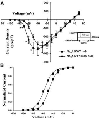
Fig. 3. The family of whole-cell sodium current traces from (A) Nav1.5/WT and from (B) Nav1.5/F1344S expressed in the tsA201 cell line. Currents were
generated from a holding potential of 140 mV from 80 to + 50 mV during 50 ms in 10-mV increments as shown in inset.
Fig. 4. (A) The mean current – voltage (I/V) relationships expressed as current density, where the amplitude of Na+currents were normalized to the cell capacitance and plotted versus the applied voltage. Similar protocol used as shown inFig. 1, were used here (see inset);*P < 0.05 and **P < 0.01. (B) The voltage dependence of steady-state activation ( G/V) of
Nav1.5/WT compared to the one obtained from Nav1.5/F1344S,
con-structed from the I/V shown on panel (A). The data points of the G/V curve were fitted using a Boltzmann equation with V1/2 and Kv
representing, respectively, the slope factor and the half-maximum voltage of activation: I/Imax= 1/(1 + exp(V V1/2/Kv)), Imax represents the
maxi-mum current.
D.I. Keller et al. / Cardiovascular Research 70 (2006) 521 – 529 524
3.2. Identification of the F1344S mutation in SCN5A and family screening
We identified a novel missense mutation, F1344S, in SCN5A in the index patient II-1 by direct DNA sequencing (Fig. 2A). A heterozygous T to C base change at position 4031 in exon 23 resulted in the change of Phenylalanine (TTC) to Serine (TCC) in residue 1344. The mutation was located in the DIII-S5 transmembrane region of the Nav1.5
sodium channel, a region highly conserved in many Na channels from different species (data not shown). In the index patient, we found the following common polymor-phisms in SCN5A (all at the homozygous state): C280C (T840C), R552G (C1654G), Q1027R (A3080G) and D1819D (C5457T). The H558R polymorphism, which is supposed to modulate the biophysical properties and the trafficking of SCN5A in the presence of other mutations, was absent in our patient[22,23].
The four daughters of the patient were screened clinically and genetically (Fig. 2B). Subject III-1 is a 15-year-old girl with a normal baseline ECG and normal echocardiography. She suffers from recurrent syncope occurring several times per year usually after getting up in the morning or related to periods of sleep deprivation. She never had a syncope during fever. Genetic testing revealed the F1344S mutation. Subject III-2 is a 12-year-old girl without history of syncope, with normal ECG and normal echocardiography. She does not carry the F1344S mutation. Subject III-3 is a 9-year-old girl with a history of one syncope during school class, and with normal ECG and echocardiography. She is also a carrier of the F1344S mutation. The fourth child is a 4-year-old girl (III-4) who never had syncope and has normal ECG. She also carries the mutation. Unfortunately, the parents declined flecainide testing in the children carrying the mutation. Family history revealed that four
brothers of the index patient died between the age of 6 months and 3 years. The cause of death is not clear as the family lives abroad. The mother of the index patient died from lung disease; the father lives abroad and could not be tested.
3.3. Biophysical characteristics of Nav1.5/F1344S mutant
Na+channels
3.3.1. Macroscopic Nav1.5 recordings and current densities
Macroscopic Na+ currents were recorded first at room
temperature from tsA201 cells expressing either WT (Nav1.5/WT) or mutant channels (Nav1.5/F1344S)
co-trans-fected with the h1-subunit. The resulting Nav1.5/WT and
Nav1.5/F1344S Na+ currents showed fast activation and
inactivation kinetics (Fig. 3). Current densities in pA/pF for
the WT and Nav1.5/F1344S mutant channels measured from
cells that were transfected with equal amount of DNA were not statistically different (Fig. 4A). However, the potential at which Na current appears was shifted to more positive potentials. This resulted in a shift of the G/V curve (steady-state activation) of about 10 mV to more positive potentials with no statistical changes in the slope factor for F1344S mutation (see Table 1for parameters).
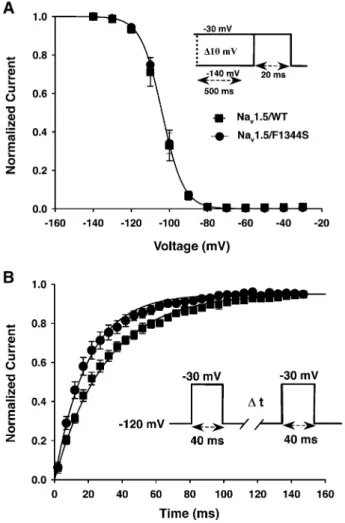
Steady-state inactivation was also studied using a double pulse protocol (Fig. 5A). No significant shifts in either half inactivation (V1/2) or slope factor were observed on mutant
channels compared to the WT channels. The recovery from inactivation studied at 120 mV using a two-pulse protocol
shows a rapid recovery (Fig. 5B) (see Table 1 for
parameters).
Entry to slow inactivation was shown to be affected in BS mutant channels, however, the slow inactivation of Nav1.5/F1344S mutation was not statistically significant
from WT (Fig. 6A).
Table 1
Biophysical parameters of Nav1.5/WT and Nav1.5/F1344S
23-C 40.5-C Nav1.5/WT Nav1.5/F1344S Nav1.5/WT Nav1.5/F1344S Steady-state activation V1/2(mV) 60.16T 1.42 (n = 10) 50.43T 2.03**n = 9) 60.69T 2.60 (n = 9) 42.44T 2.7** (n = 5) Kv(mV) 6.33T 0.33 (n = 10) 6.99T 0.52 (n = 9) 6.49T 0.67 (n = 9) 10.27T 0.80** (n = 5) Steady-state inactivation V1/2(mV) 103.82T 0.64 (n = 8) 104.47T 0.93 (n = 9) Kv(mV) 5.61T 0.07 (n = 8) 4.65T 0.06 (n = 9) Slow inactivation s (ms) 251.25T 32.37 (n = 8) 220.11T 29.89 (n = 9)
Recovery from inactivation
s (ms) 22.43T 1.60 (n = 8) 17.60T 1.88* (n = 9) V1/2= Mid point for activation or inactivation.
Kv= slope factor.
s = time constant. * P < 0.05. ** P < 0.01.
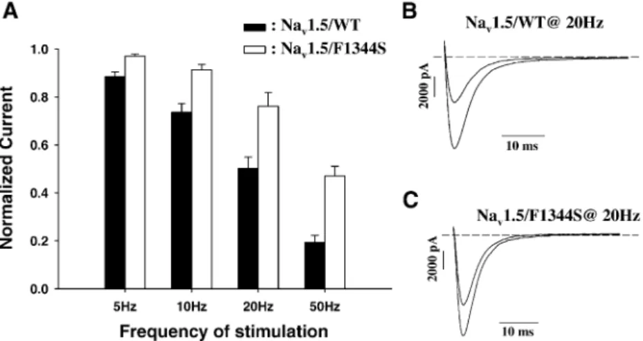
The frequency-dependent effect of Na+ current ampli-tudes was studied at frequencies found during fever in comparison with a normal frequency of stimulation. As expected since the recovery from inactivation was faster in the presence of the mutation, the currents were larger at higher frequency of stimulation, exhibiting a resistant to block (Figs. 6B and 7).
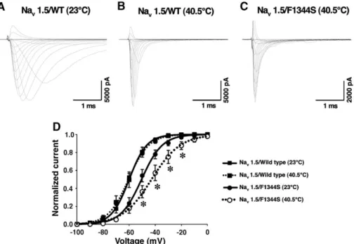
3.3.2. Effect of temperature on channel properties
When macroscopic Na+ currents were recorded at
temperatures up to 40.5-C, no effect was seen on mutant
Nav1.5/F1344S channels compared to WT channels (Fig. 8,
upper panel).
As expected, rising the temperature from 22 to 40.5-C markedly increased the rate of inactivation for WT and F1344S mutant channels (Fig. 8B and C). There was also no
effect on activation of WT channels at 23-C and 40.5-C
(parameters not shown). In contrast activation curves showed an 8 mV shift of activation towards more positive
potential with a significant change in the slope factor for mutant Nav1.5/F1344S channels measured at 40.5-C versus
23-C (Fig. 8D). Indeed, changes in temperature resulted in a further shift of the voltage-dependent activation curves towards more positive voltages with a significant effect on the slope factor (Fig. 8D) for F1344S mutation at 40.5-C (see Table 1for parameters). Although the figure shows a slight increase in current amplitudes, the maximum current densities at 40.5-C were not statistically significant from the one recorded at room temperature (data not shown).
4. Discussion
It is well recognized that the ECG patterns of ST segment elevation in the right precordial leads in patients with BS are inconstant and can be revealed by external factors such as medication and fever [14,24 – 26]. Although the effect of drugs such as class 1 antiarrhythmic’s, cocaine and antidepressant is probably related to the blockade of sodium channels, the molecular and the ionic mechanisms related to the effect of fever remain elusive.
We report a novel SCN5A mutation, F1344S, in a family with BS phenotype, associated in the index patient with fever-triggered VF. This mutation results in a loss of Na+ channel function by shifting the steady-state activation towards more positive potentials. The analysis of the mutant channels in heterologous expression system (tsA201 cell line) reveals normal kinetics of inactivation but a shift in activation that could potentially reduce the availability of sodium channels. A shift of the activation curve was found to underlay several BS mutations[27]. As the index patient developed the BS phenotype only during fever we studied the effect of the mutant Nav1.5/F1344S channel properties
at different temperatures. Activation curves showed the 10 mV shift of activation towards more positive potential at 40.5-C as noted at normal baseline experimental tempera-ture of 23-C. Interestingly, in addition to the shift of activation, we found a significant change in the slope factor.
The slope was less steep for mutant Nav1.5/F1344S
channels at 40.5-C compared to the one measured at
23-C. Apart from the BS, fever was identified as a
precipitant of idiopathic VF in a series of patients with structurally normal hearts[28]. In the study of Pasquie et al. temperature-dependent modification of ion channel proper-ties or expression were proposed to facilitate spontaneous activity within the right ventricular outflow tract or the Purkinje system as a potential mechanism to initiate VF.
In the present study, for the first time we show that the effect of temperature can be related to a further shift in tivation with a reduced slope factor, suggesting that the ac-tivation energies are markedly reduced in the presence of this specific mutation. Moreover, the F to S mutation may have a large effect on the structure of channel proteins since F is com-pletely hydrophobic and S it may H-bond with neighboring amino acid residues because of the presence of an OH group.
Fig. 5. (A) Steady-state availability curve was determined by using 500 ms conditioning pulses to voltages between 140 and 30 mV to a standard test pulse to either 30 mV (see inset). Test currents were normalized and plotted versus conditioning voltage. (B) Recovery from fast inactivation obtained using a double pulse protocol (see inset). A standard test pulse was used to monitor recovery, and the normalized test currents were plotted versus the recovery interval.
D.I. Keller et al. / Cardiovascular Research 70 (2006) 521 – 529 526
Fever is usually associated with sinus tachycardia, which is a confounding factor. Indeed, the index patient had a heart rate of 103 bpm. Therefore, we studied the effect of varying the frequency of stimulation on Na+current amplitudes. In contrast to WT channels, Na currents show even a resistant to block at high frequency of stimulation, probably due to
the fast recovery from inactivation. We found that the currents were increased at higher frequency of stimulation. Thus, a raise in heart rate seems not to be contributing to the risk for developing VT in patients carrying this particular mutation. This apparent gain of function will not result in a rescue of the loss of function due to the shift of activation.
Fig. 7. (A) Ratio of the currents elicited by the 50th and first pulses ( P50/P1) versus the pulsing frequency. (B and C) Representative raw current traces of
Nav1.5/WT (B) and Nav1.5/F1344S (C) stimulated at 20 Hz.
Fig. 6. (A) Development of slow inactivation of Nav1.5/WT and Nav1.5/F1344S sodium channels. Time course of entry into the slow inactivated state was
measured by using a triple-pulse protocol consisting of a variable duration conditioning pulse (2 ms to 1 s) to 30 mV to inactivate the channels. A 20-ms inter-pulse to 140 mV was applied to promote the rapid recovery of inactivated channels, and a standard test pulse was used to assay availability (see inset). The test currents were normalized and plotted versus the conditioning pulse interval (Nav1.5/WT,
˝
; Nav1.5/F1344S,&
). (B) Frequency-dependent inhibition ofNav1.5/WT and Nav1.5/F1344S sodium currents. A train of 50 pulses was applied to 10 mV (Nav1.5; n = 9) or 10 mV (Nav1.5/F1344S; n = 11) at
frequencies between 5 and 50 Hz. The peak currents elicited by each test pulse were normalized to the current of the first pulse ( Pn/P1, where n = 1 – 50) and
The F1344S mutation segregated to three of the four daughters. Four brothers of the index patient died suddenly at a very young age. It is possible that they were carriers of the F1344S mutation, suggesting a malignant course. Unfortunately no data on their phenotypes are available.
Several antibiotics and antidepressant such as tricyclic antidepressants are known to induce torsades de pointes by QT-interval prolongation or BS, respectively. One could speculate that the antibiotic agent may have influenced the repolarization of the mutant channels.
Piperacillin/tazobac-tam is not known to prolong the QT-interval (
www.torsa-des.org) and we do not further followed this hypothesis, also because the drug was administered after the initiation of VF. Before hospital admission, the patient took phenylephrin p.o. It is not known whether phenylephrin has the potential to unmask the BS phenotype and might have contributed to trigger VF.
In this patient and in his daughters carrying the F1344S SCN5A mutation, future episodes of fever should be aggressively treated with antipyretic agents whereas beta-blockers and phenylephrin should be completely avoided.
5. Conclusion
We identified a novel mutation in SCN5A, F1344S, in a patient with the BS and fever-induced VF. In our in vitro model we found that the mutation resulted in a loss of sodium channel function, as previously described for other SCN5A mutations leading to the BS. For the F1344S mutation, the mechanism was most probably a loss of function due to the shift of activation. Temperature
simulating fever resulted in a further shift of activation and a reduction in the slope factor. Taken together, our findings indicate that the shift in activation and change in the slope factor at higher temperature mimicking fever could reduce sodium currents availability and trigger the manifes-tation of the BS phenotype. Fever should be vigorously treated with antipyretics in patients with fever-induced BS.
Acknowledgements
We are grateful to Dr. Ve´ronique Fressart and Dr. Pascale Guicheney, INSERM U582 in Paris, France, for the verification of the mutation.
Dr. D.I. Keller was supported by grants from the University Hospital Basel, Switzerland, grants from the Swiss Electrophysiology/Pacemaker Foundation and the L. and Th. La Roche Foundation, Switzerland. The study was supported by the ‘‘Fondation Leducq’’ and grants from the Heart and Stroke Foundation of Que´bec (HSFQ), the Canadian Institutes of Health Research (CIHR) MT-13181. Dr. M. Chahine is an Edwards Senior Investigator (Joseph C. Edwards Foundation).
References
[1] Brugada P, Brugada J. Right bundle branch block, persistent ST segment elevation and sudden cardiac death: a distinct clinical and electrocardiographic syndrome. J Am Coll Cardiol 1992;20:1391 – 6. [2] Miyazaki T, Mitamura H, Miyoshi S, Soejima K, Aizawa Y, Ogawa S. Autonomic and antiarrhythmic drug modulation of ST segment elevation in patients with Brugada syndrome. J Am Coll Cardiol 1996;27:1061 – 70.
Fig. 8. Effect of temperature on Nav1.5/WT and Nav1.5/F1344S current and G/V curves. No differences in macroscopic Na+currents recordings for WT and
mutant currents at 40.5-C temperature (B – C). (D) Further shift of activation curves toward more positive voltages with a significant (*P < 0.05) effect on the slope factor in mutant currents at 40.5-C. The data point of the G/V curve were fitted using a Boltzmann equation.
D.I. Keller et al. / Cardiovascular Research 70 (2006) 521 – 529 528
[3] Brugada R, Brugada J, Antzelevitch C, Kirsch GE, Potenza D, Towbin JA, et al. Sodium channel blockers identify risk for sudden death in patients with ST-segment elevation and right bundle branch block but structurally normal hearts. Circulation 2000;101:510 – 5.
[4] Rolf S, Bruns HJ, Wichter T, Kirchhof P, Ribbing M, Wasmer K, et al. The ajmaline challenge in Brugada syndrome: diagnostic impact, safety, and recommended protocol. Eur Heart J 2003;24:1104 – 12. [5] Wilde AAM, Antzelevitch C, Borggrefe M, Brugada J, Brugada R, Brugada P, et al. Proposed diagnostic criteria for the Brugada syndrome: consensus report. Circulation 2002;106:2514 – 9. [6] Weiss R, Barmada MM, Nguyen T, Seibel JS, Cavlovich D, Kornblit
CA, et al. Clinical and molecular heterogeneity in the Brugada syndrome: a novel gene locus on chromosome 3. Circulation 2002; 105:707 – 13.
[7] Antzelevitch C. The Brugada syndrome. J Cardiovasc Electrophysiol 1998;9:513 – 6.
[8] Gussak I, Antzelevitch C, Bjerregaard P, Towbin JA, Chaitman BR. The Brugada syndrome: clinical, electrophysiologic and genetic aspects. J Am Coll Cardiol 1999;33:5 – 15.
[9] Baroudi G, Pouliot V, Denjoy I, Guicheney P, Shrier A, Chahine M. Novel mechanism for Brugada syndrome: defective surface localiza-tion of an SCN5A mutant (R1432G). Circ Res 2001;88:E78 – 83. [10] Baroudi G, Acharfi S, Larouche C, Chahine M. Expression and intracellular localization of an SCN5A double mutant R1232W/ T1620M implicated in Brugada syndrome. Circ Res 2002;90:E11 – 6. [11] Baroudi G, Napolitano C, Priori SG, Del Bufalo A, Chahine M. Loss of function associated with novel mutations of the SCN5A gene in patients with Brugada syndrome. Can J Cardiol 2004;20:425 – 30. [12] Antzelevitch C, Brugada R. Fever and Brugada syndrome. Pacing Clin
Electrophysiol 2002;25:1537 – 9.
[13] Saura D, Garcia-Alberola A, Carrillo P, Pascual D, Martinez-Sanchez J, Valdes M. Brugada-like electrocardiographic pattern induced by fever. Pacing Clin Electrophysiol 2002;25:856 – 9.
[14] Mok NS, Priori SG, Napolitano C, Chan NY, Chahine M, Baroudi G. A newly characterized SCN5A mutation underlying Brugada syn-drome unmasked by hyperthermia. J Cardiovasc Electrophysiol 2003; 14:407 – 11.
[15] Keller DI, Rougier JS, Kucera JP, Benammar N, Fressart V, Guicheney P, et al. Brugada syndrome and fever: genetic and molecular characterization of patients carrying SCN5A mutations. Cardiovasc Res 2005;67:510 – 9.
[16] Wang Q, Li Z, Shen J, Keating MT. Genomic organization of the human SCN5A gene encoding the cardiac sodium channel. Genomics 1996;34:9 – 16.
[17] Margolskee RF, McHendry-Rinde B, Horn R. Panning transfected cells for electrophysiological studies. Biotechniques 1993;15:906 – 11. [18] Jurman ME, Boland LM, Liu Y, Yellen G. Visual identification of individual transfected cells for electrophysiology using antibody-coated beads. Biotechniques 1994;17:876 – 81.
[19] Hamill OP, Marty A, Neher E, Sakmann B, Sigworth FJ. Improved patch-clamp techniques for high-resolution current record-ing from cells and cell-free membrane patches. Pflugers Arch 1981; 391:85 – 100.
[20] Wang DW, George Jr AL, Bennett PB. Comparison of heterologously expressed human cardiac and skeletal muscle sodium channels. Biophys J 1996;70:238 – 45.
[21] Wilde AAM, Antzelevitch C, Borggrefe M, Brugada J, Brugada R, Brugada P, et al. Proposed diagnostic criteria for the Brugada syndrome. Eur Heart J 2002;23:1648 – 54.
[22] Yang P, Kanki H, Drolet B, Yang T, Wei J, Viswanathan PC, et al. Allelic variants in long-QT disease genes in patients with drug-associated torsades de pointes. Circulation 2002;105:1943 – 8. [23] Makielski JC, Ye B, Valdivia CR, Pagel MD, Pu J, Tester DJ, et al. A
ubiquitous splice variant and a common polymorphism affect heterologous expression of recombinant human SCN5A heart sodium channels. Circ Res 2003;93:821 – 8.
[24] Darbar D, Yang T, Churchwell K, Wilde AA, Roden DM. Unmasking of Brugada syndrome by lithium. Circulation 2005;112:1527 – 31. [25] Bigwood B, Galler D, Amir N, Smith W. Brugada syndrome following tricyclic antidepressant overdose. Anaesth Intensive Care 2005;33: 266 – 70.
[26] Aouate P, Clerc J, Viard P, Seoud J. Propranolol intoxication revealing a Brugada syndrome. J Cardiovasc Electrophysiol 2005;16:348 – 51. [27] Smits JPP, Koopmann TT, Wilders R, Veldkamp MW, Opthof T, Bhuiyan ZA, et al. A mutation in the human cardiac sodium channel (E161K) contributes to sick sinus syndrome, conduction disease and Brugada syndrome in two families. J Mol Cell Cardiol 2005;38: 969 – 81.
[28] Pasquie´ JL, Sanders P, Hocini M, Hsu LF, Scave´e C, Jais P, et al. Fever as a precipitant of idiopathic ventricular fibrillation in patients with normal hearts. J Cardiovasc Electrophysiol 2004;15:1271 – 6.