HAL Id: tel-01490272
https://tel.archives-ouvertes.fr/tel-01490272
Submitted on 15 Mar 2017
HAL is a multi-disciplinary open access
archive for the deposit and dissemination of sci-entific research documents, whether they are pub-lished or not. The documents may come from teaching and research institutions in France or abroad, or from public or private research centers.
L’archive ouverte pluridisciplinaire HAL, est destinée au dépôt et à la diffusion de documents scientifiques de niveau recherche, publiés ou non, émanant des établissements d’enseignement et de recherche français ou étrangers, des laboratoires publics ou privés.
Impact of Wnt signalling on multipotent stem cell
dynamics during Clytia hemisphaerica embryonic and
larval development
Antonella Ruggiero
To cite this version:
Antonella Ruggiero. Impact of Wnt signalling on multipotent stem cell dynamics during Clytia hemis-phaerica embryonic and larval development. Development Biology. Université Pierre et Marie Curie - Paris VI, 2015. English. �NNT : 2015PA066561�. �tel-01490272�
Université Pierre et Marie Curie
Ecole Doctorale Complexité du Vivant
Par Antonella RUGGIERO
Pour obtenir le grade deDocteur de l’Université Pierre et Marie Curie
Impact of Wnt signalling on multipotent stem cell dynamics
during Clytia hemisphaerica embryonic and larval development
Dirigée par Evelyn Houliston
Co-encadrant Carine Barreau
Soutenue publiquement le 24-11-2015 devant la commission d’examen constituée de: Prof Agnes Audibert Professor - UPMC
Université Pierre et Marie Curie President Dr Peter Ladurner Assistant Professor
Institute of Zoology, University of Innsbruck Rapporteur Dr Fabian Rentzsch Group Leader
Sars Centre, Bergen Rapporteur Prof Paola Furla Professor - UNS
Université Nice Sophia Antipolis Examinateur Dr Evelyn Houliston Directeur de Recherche - CNRS UMR 7009 Directrice de thèse Université Pierre et Marie Curie
“Chaque homme dans sa nuit s’en va vers sa lumière” “Ciò che oggi fa notte dentro di noi, domani ci lascerà le stelle”
Acknowledgement
First of all I would like to thank Evelyn Houliston for giving me the possibility to do my PhD in her Laboratory and to Carine Barreau for being present in everyday lab life.
Thank you for giving me the freedom to develop my style and my ideas and for taking care of me.
In particular I would like to say merci to Remi who offered me a helping hand.
I would like to say thanks to all the present and the past Lab members: Patrick, Pascal, Luca, Chiara, Sandra, Gonzalo, Tsuyo and Sophie. It has been a pleasure to work with you, thank you for the support (technical and moral!).
A special thanks to Pascal that was my big-brother (I would prefer to say fratm) and to Lucas and Chiara those were always there for me.
I am also very grateful to all the people in the unit that I have met over these four years, in particular to Janet for the hospitality and to Stefano who was the first to welcome me here. Merci Sophie, Justine et Carole pour être toujours là dans cette longue marche dans le désert.
Vorrei ringraziare Stefania per essere stata allo stesso tempo: ricercatrice, capo, psicologa, mamma, amica... grazie per essere stata tutto questo!
Grazie di cuore alla mia famiglia e ai miei amici piu cari che mi hanno guardato da lontano!
General Summary
The aim of this work was to extend our understanding of the mechanisms regulating stem cell formation, specification and differentiation by studies in the non-bilaterian metazoan model Clytia hemisphaerica. Clytia, like other hydrozoan cnidarians, possess a particular population of multipotent stem cells called interstitial cells (i-cells), present during larval development and in the adult medusa, which are able to give rise both to somatic cell types and to gametes.
In bilaterian animals Wnt/β-catenin signalling regulates fundamental developmental processes such as germ layer segregation and primary body axis specification, but also regulates stem cell proliferation, lineage specification and differentiation.
I investigated the role of Wnt/β-catenin signalling in i-cell specification and differentiation. I established a set of markers for known i-cell derivatives: neural cells, nematocytes, gland cells and germ cells, which I used to monitor the presence of these cell types in Wnt3 morphant embryos. The results obtained suggest that Wnt/β-catenin signalling is involved in the last step of differentiation for certain neuronal cell types including nematocytes, but not for somatic cell fate choice from the multicellular i-cell pool. I found no evidence for germ cell fate specification from the multipotent i- cell pool prior to adult stages.
In the second part of my study I investigated the role of Wnt/β-catenin signalling in i-cell formation during embryogenesis. The results indicated that during normal development i-cell formation is Wnt/β-catenin independent and probably driven by inheritance of germ plasm containing localised mRNAs from the egg animal pole. In contrast in embryo re-patterning following embryo bisection, Wnt/β-catenin signalling appears to be necessary for de novo i-cell formation in the absence of germ plasm. Thus two distinct mechanisms can lead to i-cell formation during embryogenesis.
Overall the results obtained provided a better picture of how i-cells and their derivatives arise during embryogenesis and larval development.
TABLE OF THE CONTENTS
GENERAL SUMMARY ... II
INTRODUCTION ... 1
1)A GENERAL OVERVIEW OF STEM CELL FEATURES AND REGULATION ... 3
1.1 Stem cell properties ... 3
1.2 Stem cell potential ... 3
1.3 Generating cell type progenitors ... 5
1.3.1. Generating progenitors via unequal determinant segregation ... 7
1.3.2 Generating progenitors via environmental cues: the “Niche” ... 7
1.4 Maintaining the undifferentiated stem cell state ... 8
1.4.1 Transcriptional control ... 8
1.4.2 Epigenetic mechanisms ... 9
1.4.3 Regulatory RNAs ... 10
1.5 A shared gene toolkit for stem cells and germ line ... 11
1.5.2 Nanos regulates transcription and translation repression ... 14
1.6 Wnt signalling: a candidate for stem cell regulation ... 16
1.6.1 The Wnt/ β- catenin pathway ... 17
1.6.2 The Wnt/ Planar cell polarity pathway ... 19
1.6.3 Developmental roles of Wnt/β- catenin pathway: Germ layers and body axis ... 22
1.6.4 Wnt/β-catenin signalling and stem cell regulation ... 25
2)CNIDARIAN MODELS IN DEVELOPMENTAL BIOLOGY ... 27
2.1 Clytia life cycle and development ... 29
2.1.1 Clytia embryogenesis and larval development ... 33
2.1.2 Clytia medusa anatomy ... 34
2.2 Hydrozoan cell types ... 35
2.2.1 I-cells: a hydrozoan multipotent stem cell system ... 35
2.2.2 Nerve cells ... 42 2.2.3 Nematocytes ... 45 2.2.4 Gland cells ... 46 2.2.5 Gametes ... 46 2.2.6 Epithelial cells ... 47 2.3 I-cell dynamics ... 48
2.3.1 Analysis of i-cell multipotency ... 48
2.3.2 Generating i-cell progeny: I-cell derivatives specification and differentiation ... 49
2.3.3 Wnt signalling regulates i-cell differentiation ... 57
2.4 The embryological origin of i-cells ... 57
MATERIAL & METHODS ... 61
RESULTS I: ... 65
MONITORING THE SPATIAL AND TEMPORAL DISTRIBUTION OF I-CELL DERIVATIVES DURING CLYTIA LARVAL DEVELOPMENT. ... 65
1.BACKGROUND AND QUESTIONS ... 66
1.1 Nematogenesis progression during Clytia larval development. ... 67
1.1.1 Distribution of nematocytes during embryonic development ... 68
1.1.2 Mcol3-4a-expressing nematoblasts are largely restricted to the endodermal region ... 70
1.1.3 Sox15 is expressed during an extended period of nematogenesis ... 71
1.2 Neurogenesis during larval development ... 73
1.2.1 Clytia Prdl-a as putative neuronal marker ... 75
1.2.3 Clytia Asc-b is a expressed in the endoderm in both planulae and medusae ... 76
1.2.4 Zic-C is expressed in a sub-type of neural cells specific to medusa tentacles. ... 79
1.2.5 Neuropeptide expression defines mature neural subpopulations ... 86
1.3 Gland cell formation in Clytia planulae and in the medusae ... 88
1.4 Germ cell genes in Clytia ... 89
WNT/Β-CATENIN SIGNALLING IN EMBRYONIC PATTERNING, I-CELL FORMATION AND I-CELL DIFFERENTIATION DURING CLYTIA EMBRYIONIC AND LARVAL
DEVELOPMENT ... 97
2.BACKGROUND AND QUESTIONS ... 98
2.1 PAPER 1: Summary of the results ... 99
2.1.1 Investigating Wnt/β-catenin signalling in i-cell formation and differentiation during Clytia embryonic development. ... 99
PAPER1: ... 106
WNT SIGNALLING IN MULTIPOTENT STEM CELL FORMATION AND DIFFERENTIATION IN CLYTIA HEMISPHAERICA LARVAL DEVELOPMENT ... 106
2.2 PAPER 2: Summary of the Results ... 154
2.2.1 Identification of novel Clytia embryos patterning genes ... 154
2.2.2 Polarized expression pattern of Wnt3 target genes ... 155
2.2.3 IE genes show an i-cell like expression pattern ... 157
2.2.4 Different responses of Wnt3-MO responsive genes to Fz1-MO ... 157
PAPER2: ... 159
DIFFERENTIAL RESPONSES TO WNT AND PCPDISRUPTION PREDICT EXPRESSION AND DEVELOPMENTAL FUNCTION OF CONSERVED AND NOVEL GENES IN A CNIDARIAN. ... 159
2.3 Additional Results: Characterisation of putative novel i-cell genes under-expressed in Wnt3 morphant early gastrulae. ... 183
DISCUSSION & PERSPECTIVES ... 193
ANNEXE 1:COMPARISON OF EXPRESSION LEVELS OF SELECTED HYDRA GENES IN SEPARATED CELL POPULATIONS ... 209
List of the Figures
Figure 1 : Stem cell potential.
Figure 2 : Asymmetrical cell divisions. Figure 3 : Wnt signalling pathways.
Figure 4 : Planar cell polarity coordinates the orientation of cilia and hairs in metazoan tissues.
Figure 5 : Embryonic axis and Wnt β-catenin signalling.
Figure 6 : One hypothesis for the phylogenetic relationships between major metazoan clades.
Figure 7 : Cnidarian model in developmental biology. Figure 8 : Clytia life cycle.
Figure 9 : Clytia embryonic development. Figure 10 : Clytia medusa anatomy.
Figure 11 : I-cell and i-cell derivatives localisation i the Hydra polyp and in Clytia developmental stages and adult.
Figure 12 : Distribution of i-cell expressing Piwi, Nanos1, Vasa during larval development and in the medusa.
Figure 13 : RFamide immunoreactivity in hydrozoans nervous system. Figure 14 : Nematocytes structures.
Figure 15 : Structure of Clytia female gonad.
Figure 16 : Genetic circuitry involved in nematocytes and neuronal cell specification. Figure 17 : Molecular markers for Clytia Nematogenesis.
Figure 18 : Morphological and molecular evidence for perinuclear germ plasm localisation in Clytia eggs.
Figure 19 : Piwi expressing i-cells can form from both vegetal and animal regions.
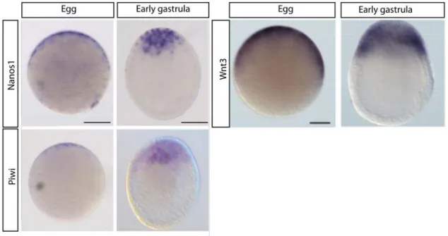
Figure 20 : Nanos1, Piwi and Wnt3 expression pattern comparison in eggs and at the early gastrula stage.
Figure 21 : Co-localisation of Piwi and Nanos1 transcripts.
Figure 22 : Appearance of nematocytes during larval development.
Figure 23 : Mcol3-4a expression allows tracking of the nematoblast population during larval development.
Figure 24 : Sox15 expression pattern in nematoblasts and in differentiating nematocytes. Figure 25 : Gene orthology of the metazoan paired- class homeobox transcription factors. Figure 26 : Prdl-a expression during larval development and in the adult medusa.
Figure 27 : Gene orthology of the metazoan bHLH family.
Figure 28 : Asc-b expression during larval development and in the adult medusa. Figure 29 : Gene orthology of the metazoan Gli/Zic.
Figure 30 : Zic-C expression during larval development and in the adult medusa. Figure 31 : RFamide expression during larval development and in the adult medusa. Figure 32 : GLWamide expression during larval development and in the adult medusa. Figure 33 : Antistasin expression during larval development and in the adult medusa. Figure 34 : Gene orthology of the metazoan Boule/DAZ family members.
Figure 35 : Boule/DAZ family gene expression during larval development and in the adult medusa.
Figure 36 : Tudor domain gene expression during larval development and in the adult medusa.
Figure 38 : Thyn1 expression during larval development and in the adult medusa. Figure 39 : Nanos1 and Piwi expression is not inhibited in Wnt3 morphants larvae. Figure 40 : Wnt3 is required for differentiation of RFamide neurons.
Figure 41 : A Wnt gradient is required for neural fate acquisition. Figure 42 : Wnt signalling participates in de novo i-cell formation.
Figure 43 : Comparative transcriptomic approach to unravel new genes involved in Clytia embryonic patterning .
Figure 44 : Nanos1,Piwi,Mos3 and Zfn845 expression pattern during larval development in young medusa and in adult jellyfish.
Figure 45 : Co-localisation of Piwi with Mos3 and Znf845.
Figure 46 : Znf845 is not expressed in gland cells or in nematocytes. Figure 47 : Znf845 is not expressed in Sox15 positive cells.
Figure 48 : I-cell cartography.
INTRODUCTION
Our current understanding of stem cell biology comes mainly from studies in classical vertebrate and invertebrate model systems, however in light of the very strong evolutionary conservation of the signalling pathways involved (e.g. Wnt), non canonical model organisms can contribute extensively to extend our knowledge and unravel novel aspects of stem cell regulation.
Hydrozoan cnidarians were among the first non-bilaterian metazoan animals in which stem cells were described. A particular population of multipotent stem cells termed interstitial cells (i-cells) can differentiate into somatic cells such as nerve cells, gland cells and nematocytes, as well as into gametes.
My thesis work addressed the regulation of the i-cell population in the hydrozoan Clytia
hemisphaerica. Various aspects of stem cell regulation were addressed, namely their
formation during embryogenesis, proliferation, specification and differentiation of derivative fates. The main aspects treated in this manuscript will be specification (how the i-cells chose their fate), and differentiation (how once specified they reach their terminal state), as well as their embryological origin.
In the first part of this Introduction (1), I will describe general stem cell features and the mechanisms that regulate their formation and differentiation in various taxa and tissues. The second part (2) is focussed on the cnidarian biology, in particular on i-cell biology. I will describe in detail the known i-cell derivative cell types: their morphology and gene expression characteristics, and what is known about the molecular mechanisms controlling their specification and differentiation.
1) A general overview of stem cell features and regulation
1.1 Stem cell properties
A general definition of a stem cell, as commonly found in cell biology textbooks (e.g. Molecular Biology of the Cell, Alberts 2002), is as an undifferentiated cell capable of both self-renewal and multi-lineage differentiation, displaying an unlimited proliferative activity that lasts (in vivo) the lifetime of the animal. Based on this definition a stem cell should have the following characteristics:
• No particular morphological distinctive features.
• Be able to generate at least one daughter cell with the same potential as the mother cell.
• Unlimited mitotic proliferation.
• Potential to generate large numbers of differentiated functional progeny.
1.2 Stem cell potential
Based on their potential to differentiate into any particular type of cell, stem cells can be classified into 4 different categories: totipotent stem cells, pluripotent stem cells, multipotent stem cells and unipotent stem cells (Figure 1).
1) Totipotent stem cells: a totipotent cell can give rise to various differentiated cell types including those of extra-embryonic tissues. The zygote is considered by some to be totipotent stem cells, although it doesn’t have the property of self-renewal, at least not directly.
2) Pluripotent stem cells: animal pluripotent stem cells are able to differentiate into all cell types of the three embryonic germ layers, i.e., ectoderm, mesoderm, and endoderm. The best-know example is the embryonic stem cell (ES cell) (Martin 1981). In mammals, ES cells derive from the inner cell mass of the blastocyst. The inner cell mass comprises a group of cells arising very early during pre-implantation development, which give rise to the embryo proper. The outer cell layer of the blastocyst instead gives rise only to extra-embryonic tissues.
In all the animals that lack extra embryonic tissues, the terms totipotent and pluripotent are equivalent.
3) Multipotent cells: multipotent stem cells show a restricted pattern of differentiation into a limited number of cell types. A good example of a multipotent stem cell system is provided by the hematopoietic system, responsible for producing mature blood cells. Hematopoietic stem cells called hemocytoblasts give rise to “transit amplifying” progenitors of the myeloid and lymphoid lineages. Transit-amplifying cells are fated cells that proliferate and generate fully differentiated cells. The myeloid progenitor cells will then generate erythrocytes, megakaryocytes and myeloid cells, which in turn proliferate and differentiate into eosinophil, basophil, neutrophil and monocytes. The lymphoid cells produce T lymphocytes, B-lymphocytes and NK lymphocytes.
Multipotent stem cells are defined as adult stem cells if they are found in adult stage and can generate all the cell types of the tissue from which they originate, as is the case for the hematocytoblasts.
4) Unipotent stem cells: unipotent stem cells have a very restricted cell differentiation capacity, and can generate only one single type of cell. Melanocyte stem cells provide one example of unipotent stem cells, producing the pigmented cells (melanocytes) present in hair follicles and in epidermis. Melanocyte stem cell systems were characterised for the first time in mouse hair follicles (Nishimura et al. 2002). During each cycle of hair regeneration and regression, melanoblasts proliferate and differentiate into fully mature melanocytes, which enter into the developing hair.
As mentioned in the hematopoiesis example above, stem cells divide to generate intermediate cells, termed transit-amplifying cells. This type of temporal and developmental hierarchical organisation reduces the number of cell divisions that stem cells must undergo (Morrison & Spradling 2008). However this ordered progression, from stem cells to differentiated cells is not rigid. Indeed in particular conditions committed, differentiating or differentiated cells can revert to the self-renewing stem cells state. Thus tissue stem cells have the potential to revert (to dedifferentiate in the most extreme examples) to stem cells under certain circumstances. An example of this cellular plasticity is provided by studies of mouse spermatogonia (Nakagawa et al. 2010). Spermatogenic cells are organised within a compartment populated by germ cells and by somatic cells with support functions. The germ cell precursors, the spermatogonia give rise to the spermatocytes. Several subpopulations of spermatogonia, can be distinguished by morphology and by gene expression patterns reflecting different degrees of differentiation.
During regeneration after injury, spermatogonia at the most advanced differentiating stages contribute to the population of the spermatogenetic compartment by re-converting into stem cells.
1.3 Generating cell type progenitors
The progenitors of particular cell types are generated when stem cell progeny loses their stem cell properties (proliferation and self renewal) and moves on the pathway towards differentiation. In order to provide progenitors of particular differentiated cell types while Figure 1: Stem cell potential.
The cartoon shows how stem cells gradually commit towards a differentiation pathway. The cells acquire a restricted fate, decreasing in potential. Pluripotent cells (in red) are able to give rise to multipotent stem cell here represented in three different colours (green, pink and blue). Multipotent stem cells have a reduced potential. Curved arrows illustrate the self-renewal ability of these stem cells. On the right, an illustration of the zygote that in mammals is a totipotent cell.
also renewing the stem cell population, the stem cells divide asymmetrically giving rise to two sister cells that have different fates, a feature that can be recognised by differences in size, morphology, gene expression pattern, or the number of subsequent cell divisions undergone by the two daughter cells (Horvitz & Herskowitz 1992). The asymmetric choice between self-renewal and differentiation can be regulated by extracellular cues coming from a “niche” acting preferentially on one of the daughter cell (a process termed environmental asymmetric cell division), or by intrinsic programs within the cell, which is known as divisional asymmetric cell division (Wilson & Trumpp 2006) or unequal segregation of localised cytoplasmic determinants (Figure 2).
In particular conditions such as during the formation of the stem cells pool, stem cell can divide symmetrically.
Figure 2: Asymmetric cell divisions.
A) Localised determinants direct the fate of the terminal differentiated cell, activating an intrinsic genetic program. B) Extrinsic cues determine the fate of one of the two daughter cells inducing the specification of cell precursor (CP) that will move into the differentiation pathway. Modified from Wilson & Trumpp (2006).
1.3.1. Generating progenitors via unequal determinant segregation
Two main events are necessary for setting up divisional asymmetric cell division: cell fate determinants have to be asymmetrically localised and the mitotic spindle has to be oriented with respect to the determinant axis (Betschinger & Knoblich 2004).
One example of asymmetric cell division of a stem cell involving unequal determinant distribution is seen in Drosophila neuroblasts (Knoblich 2001), which are central nervous system progenitor cells. Neuroblasts derive from the neuroectoderm tissue. Committed neuroblasts migrate into the sub neuroectodermal layer in a process termed delamination, and then generate by asymmetric cell division a small basal daughter cell called the ganglion mother (GMC) and a large apical daughter cell that retain a stem cell nature. The GMC is the neural progenitor as it will divide one more time and gives rise to two post mitotic neurons. In contrast the large apical cell retain a neuroblasts identity and continue to divide in a stem cell manner. The apical-basal polarity is set up by the PAR protein complex, which is localised apically. The PAR complex comprises a core of the three proteins: Par3, Par6 and aPKC (atypical Protein Kinase C), which have a role in asymmetric cell division across species and in different cell types (Guo & Kemphues 1995). The PAR complex regulates the localisation of the neural fate determinants in the GMC cells such as the membrane-associated Notch pathway suppressor Numb, the transcription factor Prospero, and specific adaptor proteins responsible for their basal localisation Miranda and Pon (Partner of Numb). Miranda is a coiled-coiled protein that binds Prospero and translocates it to the cell cortex, from where it is later released in the daughter cell to activate neural cell fate gene transcription.
1.3.2 Generating progenitors via environmental cues: the “Niche”
Stem cell “niches” are spatially distinct microenvironments that can include neighbouring cells, signalling molecules and other extracellular materials (Scadden 2006). These have been studied extensively in Drosophila and Caenorhabditis elegans gonads, which can be conveniently manipulated through genetic and molecular approaches. The C. elegans adult gonad shows an ordered progression in germ line formation from germ stem cells (GSCs) occupying the most distal extremity, to mature gametes positioned in the proximal part. A key population of undifferentiated somatic mitotic cells called “distal tip cells” (DTCs) covers the distal extremity of the gonad. DTCs are the major regulator of germ line
proliferation and are the main component of the gonad stem cell niche (Kimble & White 1981). The stem cell properties of the GSCs depend upon their contact with the DTCs. As GSCs move further away from the distal tip, they terminate their mitotic activity and enter into meiosis. DTCs cells control GSC differentiation through a Notch-like signal. DTCs express on the surface the delta-like ligand LAG-2, which activates the Notch-like receptor GLP-1, localised on the mitotic germ cells. In the cell contacting the DTCs, GLP-1 is activated and translocates to the nucleus, where it downregulates meiosis promoting genes like gld-1, gld-2 and nos3, required to maintain the stem cell state (Hansen 2004).
1.4 Maintaining the undifferentiated stem cell state
Maintenance of the undifferentiated stem cell state relies on a variety of regulatory mechanisms acting at different levels including transcriptional regulation, translational regulation and epigenetic modification of chromatin. During my thesis I used extensively as stem cell markers two genes involved in maintaining pluripotency through regulation of mRNA metabolism and translational control, Piwi and Nanos. Interestingly these two genes along with other mRNA binding proteins such as Vasa and PL10 which have traditionally been considered as “germ line genes” because of their highly conserved expression in germ cell precursors (PGC), are now emerging as more widely functioning stem cell regulators with functions outside the germ line.
Before introducing Piwi, Nanos in more detail, I will mention briefly in this section the main actors in transcriptional regulation and epigenetic regulation of stem cell identity.
1.4.1 Transcriptional control
A set of transcriptional factors (TFs) have been well characterised in vertebrate embryonic stem cells, including Nanog, Oct4, Klf4 and Sox2, which act to repress somatic differentiation programs (Chambers et al. 2003; Niwa et al. 2000; Boyer et al. 2005). Discovered in 1998 Oct4 was characterised as an essential TF in maintaining the pluripotent state (Nichols et al. 1998). Oct4 belongs to the POU transcription factor family and can either activate or repress target gene expression by binding to the octameric sequence consensus motif AGTCAAAT. This consensus motif can be found in regulatory
sequences including those in introns. Although POU domains that bind this sequence are conserved across metazoans (Gold et al. 2014), Oct4 orthologs have not been identified outside vertebrates.
Nanog is required for the maintenance of a pluripotent state and to prevent the differentiation. It is a tripartite protein with a central homeodomain. Transcriptional regulation is mediated by the C-terminal domain, which is characterised by two subdomains, WR and CRD2 (Pan & Pei 2005). Recent phylogenetic analysis shows that Nanog like Oct4 is not present in C. elegans or in Drosophila or in non vertebrate-chordates like Ciona and Amphiouxus (Satou et al. 2008) but is present only in vertebrates (Ma et al. 2013). Sox2 belongs to the SR-HMG protein family. It is needed for the maintenance of a pluripotent state. Indeed loss of Sox2 causes the activation of differentiation pathways (Niwa 2007). Sox2 can cooperate with Oct3/4 in the control of target gene transcription. Kfl4 is a member of the KFL (Krüppel-like factor) transcription factor family. It is a zinc finger protein, which can activate or repress target genes involved in cell proliferation and or differentiation.
1.4.2 Epigenetic mechanisms
Stem cell regulation mechanisms involve epigenetic mechanisms and non-coding RNA-mediated gene silencing. Epigenetic in this context refers to changes in DNA structure without modifications of the DNA sequence. Epigenetic mechanisms include histone modifications and DNA methylation (chemical modifications to the cytosine residues of DNA). The tri-methylation of particular residues on the DNA sequence can identify the state of the chromatin. The methylation of the Lysine 4 on the Histone 3 (metK4H3), is correlated with transcriptional activity, while compact chromatin, not accessible for transcription, is marked by methylation of the Lysine 27 on Histone 3 (metK27H3). A protein complex involved in chromatin modification is the Polycomb group complex (PcG). PcG proteins function as a repressive complex, is required for the long-term silencing of chromatin and has an important role in the differentiation of stem cells as well as in early embryonic development (Simon & Kingston 2013).
Three proteins form the core complex: enhancer of zeste (Ezh2), embryonic ectoderm development (Eed) and suppressor of zeste 12 (Suz12). Eed binds Suz12, which is a histone
methyltransferase while EZh2 maintains the silent tri-methylated state of the Lysine 27 residue of the target DNA.
PcG proteins are highly conserved across animal species and have been studied also in two hydrozoans: Podocoryne (Lichtneckert et al. 2002) and Hydra (Khalturin et al. 2007; Genikhovich et al. 2006). The Hydra Eed ortholog HyEED is expressed in hydrozoan multipotent stem cells termed interstitial cells (i-cells) and in nematoblasts (stinging cells peculiar to cnidarians) (Khalturin et al. 2007; Genikhovich et al. 2006), which will be introduced in detail respectively in section 2.2.1 and 2.2.3 of this Introduction chapter. As HyEDD is expressed in the early-stage nematoblasts located in Hydra gastric region, but never detected in differentiated nematocytes located in the head and in the foot. Overexpression of a construct in which HyEED was fused to eGFP under a ubiquitous actin promoter, revealed degradation of the fusion protein, and thus probably of the endogenous protein, during nematocyte differentiation. Furthermore, following treatment with an inhibitor of the proteasome machinery (MG132), HyEED expressing cells were detected in mature nematocytes located at the extremity of the Hydra polyps. This study points to a role for EED in Hydra in preventing nematocyte differentiation from nematoblasts by polycomb type chromatin remodelling.
1.4.3 Regulatory RNAs
Small non-coding regulatory RNAs, along with epigenetic mechanisms, facilitate the continued expression of stem cell TFs. Stem cell TFs contribute to maintaining pluripotency by preventing stochastic and aberrant induction of differentiation. Thus regulatory RNAs, such as microRNAs (miRNA), endogenous small interfering RNAs (siRNA), long non-coding RNAs (LncRNA) and Piwi-interacting RNA (piRNA) play an important role in the control of pluripotency.
In the last few years the role of the LncRNAs has been widely investigated (Flynn & Chang 2014). LncRNAs are non-coding transcripts greater then 200 base pairs in length. A specific LncRNA has been identified that participates in the regulation of pluripotent ES cells (Guttman et al. 2011). Knockdown of these LncRNAs in mouse ES cells results in downregulation of the expression of pluripotency markers including Oct4, Nanog, Sox2 and Klf4.
siRNAs and miRNAs bind to Argonaute/Piwi family proteins of the Ago sub-type which are widely expressed in different tissue types (Wutz 2013). They are characterised by three highly conserved domains: the PAZ, MID and PIWI domains. The N-terminal PAZ domain and the MID domain together bind small RNAs (Song et al. 2005), while the C-terminal PIWI domain, whose structure is similar to that of RNase H enzymes, cleaves the RNA/DNA hybrids formed when the regulatory RNAs hybridise to complementary sequences in the genomic DNA (Okamura et al. 2004). Piwi proteins bind to a specific type of regulatory RNAs called piRNAs, which are mostly restricted to germ line and stem cells (Thomson & Lin 2009), as described in more detail in the following sections. piRNAs have sequence complementary to those of transposable elements : mobile DNA sequences that can insert themselves into new positions in the genome where they can cause genome instability and loss of long-term fitness. Piwi-piRNA interaction leads to the degradation of piRNA/transposable elements hybrids, thus preventing specific transposons from integrating into the genome (Brennecke et al. 2007). A major function of Piwi and of pi-RNAs is thus thought to be to protect the genome from transposable mobile elements. More generally, RNA-binding proteins regulate every aspect of RNA metabolism, including pre-mRNA splicing, mRNA trafficking, stability, and translation. Among these, Nanos is an RNA binding protein with translational repressor function. In the next section I will give a more detailed introduction to the classical germ line genes, Nanos and Piwi that I used to track the multipotent stem cell population in Clytia.
1.5 A shared gene toolkit for stem cells and germ line
In the last few year molecular studies conducted in non-bilaterian organisms such as sponges and cnidarians, and in some invertebrates like planarians, have indicated the presence of a common molecular signature shared between some stem cell populations and the germ cells. These genes have been referred to as Germline Multipotency Program (GMP) genes (Juliano et al. 2010). The GMP gene set includes Nanos, Piwi, Vasa and PL10, all coding for RNA binding proteins. The GMP gene products have frequently been described as associated in granule-like structures in germ line cells variously termed nuage, chromatid bodies, germ plasm, and pole granules. These structures extensively described in primordial germ cells and developing oocytes, are composed of ribonucleoprotein
complexes located in the perinuclear region in PGCs which often translocate to subcortical regions of the oocyte during oogenesis (Kloc et al. 2004). Germ plasm presence in oocytes of different species is rather patchy across the animal phylogenetic tree leading to speculation that it may have arisen several times during evolution (Extavour & Akam 2003). Traditionally its role has been considered to be as a germ line determinant, which causes the cells of the embryo that inherit it to adopt a PGC fate. This determinant role for germ plasm has been shown by ablation or transplantation experiments in various insect species and in frogs (Kloc et al. 2004).
1.5.1 Piwi proteins in the germ line and stem cells
Piwi, Nanos and Vasa proteins and mRNAs, have repeatedly been found to be enriched in germ plasm in species where the germ plasm is present. More generally, across all species examined whether they have germ plasm in their oocytes or not, expression of the GMP genes is characteristic of the germ cell developmental lineage (‘germ line’). In non-bilaterian animals, including sponges and cnidarians, as well as in some non-bilaterian species, expression of Nanos, Piwi and other “germ line” genes has been documented also in various populations of non-germ line multipotent stem cells. This has led to the idea that the presence of these proteins may be a molecular signature of pluripotent/multipotent stem cells rather than exclusively a germ cell marker. The findings relating to this conclusion are described in more detail in the following sections.
• Piwi, a universal marker of germ line
Historically Piwi (p-element induced wimpy testis) was identified in Drosophila from genetic screening for mutants defective in stem cell division. Characterisation of the Piwi phenotype revealed a role in germline stem cell (GSC) maintenance via self-renewal (Lin & Spradling 1997). In Drosophila, Piwi is expressed in the germ line and in a somatic population of cells located apically with respect to the GSCs (Cox et al. 1998). In C. elegans prg-1 and prg-2 (Piwi related genes) are required for the maintenance of mitotic germ cells. The zebrafish Piwi homolog Ziwi is also required for maintenance and development of germ cells (Houwing et al. 2007), while Miwi expression in mouse is restricted to the male germ line, and affects spermatogenetic progression (Deng et al. 2002). In the polychaete
Capitella teleta, two Piwi orthologs were found, Ct-Piwi1 and Ct-Piwi2 (Giani et al. 2011).
Expression pattern analysis detected these two transcripts in overlapping domains of proliferating somatic stem cell in the posterior growth zone and in the male and female PGCs, localised in the mature gonads.
• Piwi a marker of stem cell in non-bilaterian metazoans
After the discovery of Piwi, in 1998 Cox and colleagues were the first to hypothesise an extended role of the Piwi family protein. They proposed that Piwi represents the first of a group of genes that acts both in germ line and in somatic stem cells to regulate stem cell maintenance, proliferation and differentiation. This hypothesis has now been confirmed thanks to the increasing number of studies showing the presence of Piwi protein in somatic stem cells in a variety of animal species.
One example of multipotent stem cells expressing Piwi is sponge archeocytes, which display morphological and biological stem cells characteristics (Simpson 1984). Recent studies on the fresh water sponge (demonsponge) Ehirava fluviatilis demonstrate that archeocytes self-renewal, proliferate and possess the capacity to differentiate into several cell types including gametes (Funayama 2010). Archeocytes expressed two Piwi orthologs
EflPiwiA and EflPiwiB. EflPiwiA is also expressed in another cell type specific to the sponge
called choanocytes, flagellated cells whose main function is nutrient uptake. Upon spontaneous disorganisation and reorganisation of the cells in Ehirava, choanocytes can trans-differentiate into archeocytes and then re-differentiate into spermatocytes (Gaino et al. 1984).
In demosponges, it was recently proposed that a stem cell system based on two stem cells populations exist: the archeocytes and the choanocytes (Funayama 2013). The archeocytes give rise to all cell types (Funayama 2010) and the choanocytes manifest their stem cell potential in special conditions, during sexual reproduction or during regeneration. In the latter case choanocytes could accelerate the processes of sponge body recovery (Funayama 2013).
In the ctenophore Pleurobrachia pileus, two Piwi orthologs are found to be expressed in somatic stem cells and in germ cells (Alié et al. 2011). In particular Piwi proteins are
expressed in undifferentiated cells in tentacle roots, in the stem cells that give rise to neuronal types at the aboral pole, and in the ciliated polster cells in the combs.
Piwi gene expression is also characteristic of the i-cell population in Hydra, Hydractinia and Clytia, (Nishimiya-Fujisawa and Kobayashi 2012, Kanska and Frank 2013; Leclère et al. 2012; Denker et al. 2008).
A further example of Piwi expression in somatic stem cells is seen in planarian neoblasts. Neoblasts are totipotent stem cells that can generate all somatic cell types as well as the germ line (Baguna 1981). In the planarian Schmidtea mediterranea two Piwi orthologs have been identified: Smedwi1 and Smedwi2. Functional studies by RNAi treatments show that Smedwi2 is necessary for the production of neoblast progeny (Reddien et al. 2005).
Despite the reported expression of Piwi genes in a wide range of somatic stem cell types, their function in these cells remains unclear. They may be involved both in maintenance of the stem cell pool, as seen in the Smedwi2 case, and in protection of the genome from transposons, which may compromise regeneration capacity or the ongoing tissue renewal seen in hydrozoans, ctenophore tentacles and sponges.
1.5.2 Nanos regulates transcription and translation repression
Nanos is an RNA binding protein that contains a CCHC zinc finger domain with well- conserved functions involved both transcriptional regulation and translational repression. Nanos was first identified in Drosophila as an essential factor in early embryonic patterning (Wharton & Struhl 1991); however it has since become clear that this role is particular to
Drosophila, while the conserved function of Nanos is associated with germ line.
• Nanos is associated with germ line
Nanos is expressed in primordial germ cells (PGCs) in widely divergent metazoans and its function is required for PGCs maintenance and migration in different organisms including zebrafish (Koprunner et al. 2001), C. elegans (Kraemer et al. 1999) and Xenopus (Lai et al. 2012). To execute its role as a translational repressor, Nanos acts together with a protein partner called Pumilio. mRNAs identified as Nanos/Pumilio targets have been characterised in several organisms and include the transcription factor Hunchback,
involved in the establishment of anterior-posterior polarity in Drosophila (Murata & Wharton 1995), and VegT an endoderm fate TF in Xenopus (Lai et al. 2012).
A role for Nanos in transcriptional repression in the germ line has also been shown in
Drosophila where somatic gene transcription is prematurely activated in mutants lacking Nanos (Kobayashi et al. 1996).
• Nanos somatic expression
In Drosophila, Nanos also acts outside the germ line directing dendrite morphogenesis (Brechbiel & Gavis 2008) and some data also available suggest a possible function for Nanos in somatic stem cell maintenance. In the sponge Sycon ciliatum, Nanos is expressed in oocytes and in macromeres that give rise to the pynacoderm, the external layer of the adult sponge (Leininger et al. 2014). Thus sponge Nanos is expressed in both germ line and somatic cells. In several hydrozoan species examined, the Nanos1 genes are expressed in i-cells and germ i-cells. In contrast the paralog Nanos2 genes are additionally expressed at other sites, including the epithelial endodermal hypostome cells in Hydra. Hydra Nanos2 expression increases during head regeneration, suggesting a role in head morphogenesis (Mochizuki et al. 2000). Clytia and Hydractinia Nanos2 paralogs, beside i-cells and germ line, are expressed in differentiating nematoblasts (Leclère et al. 2012; Kanska & Frank 2013), indicating presumably a role for Nanos2 in nematocyte formation. In the anthozoan cnidarian Nematostella Nanos orthologs have been characterised in two different studies. However the 2 reports show some discrepancies concerning the expression pattern of
Nanos1 and Nanos2. Torras and colleagues show a localised maternal distribution of Nanos1 mRNA(Torras & González-Crespo 2005), while the results of Extavour and
colleagues suggest that Nanos1 is only expressed zygotically from the early gastrula stage, in a population of ectodermal scattered cells (Extavour et al. 2005). Torras and colleagues also claim that Nanos2 is first detected at gastrula stage, then it localised in the posterior region of the planula larva and in developing tentacles, suggesting that Nanos2 could regulate the formation of posterior structures (Torras & González-Crespo 2005). In contrast Extavour and colleagues provide a detailed Nanos2 expression pattern showing for the first time the transcript at late blastula in a population of cells that in later stages will participate in the formation of the presumptive endoderm. In primary polyps Nanos2 expression is restricted to distinguishable patches of cells localised along the length of the mesenteries. These cells
have the typical PGCs morphology (with large nucleus and Nanos cytoplasmic localisation). Moreover these cells are Vasa immunoreactive, suggesting that they might correspond to Nematostella PGCs. In Nematostella it remains to be resolved whether Nanos proteins have a somatic stem cell and/or germ line function.
In planarians expression of Nanos, unlike Piwi, is restricted to germ line precursors and its function is linked to gonad development and in gonad regeneration condition (Wang et al. 2007) while in ctenophores the Nanos expression pattern has not yet been described.
1.6 Wnt signalling: a candidate for stem cell regulation
Extrinsic factors, notably acting via intercellular signalling pathways, play a major role in regulating the balance between stem cell differentiation and maintenance (section 1.3.2). Many different signalling pathways including Wnt, ERK-MAPK, BMP-TFGβ-Smad and Notch (Nusse et al. 2008; Zhang and Li 2005; Campos et al. 2004; Androutsellis-Theotokis et al. 2006) have been shown to regulate stem cell proliferation and/or differentiation in
vivo and in vitro, in different species and in different contexts of development and tissue
homeostasis. Ongoing studies in our group are focussed on the key participation of Wnt signalling in embryonic and larval development in Clytia, and so I addressed, during my thesis work the role of this pathway in i-cell development. In this part of the Introduction I will give an overview of the various Wnt signalling pathways.
• Overview of Wnt signalling
Wnt signalling regulates crucial aspects of cell fate determination, cell migration, cell polarity, organogenesis and primary body axis polarity during embryonic development. Wnt ligands and their Frizzled (Fz) receptors are evolutionary conserved during metazoan evolution. Indeed they are present throughout the animal kingdom (Croce & McClay 2008), including in organisms that diverged early from the bilaterians such as sponges and cnidarians, implying their presence in the metazoan common ancestor.
Wnt signalling can occur via β-catenin mediated regulation of gene transcription, described as the “canonical” or Wnt/β-catenin pathway (Figure 3A). Wnt signalling can also occur through β-catenin-independent pathways, including the Wnt/PCP (Figure 3B) and Wnt/Ca2+ (Figure 3C) pathways. The core proteins of the Wnt/PCP pathway regulate two different phenomena: coordination of individual cell polarity in the plane of the tissue
(Planar cell polarity), which can occur without Wnt ligands, and morphogenetic movements (Lapébie et al. 2011).
Wnt/Ca2+ pathway activation causes the release of intracellular Ca2+ with consequent activation of Cam Kinase II and Protein Kinase C which in turn drive the activation of nuclear factors like CREB and NFAT that promote the transcription of specific genes, including ones involved in organogenesis and establishment of the dorso-ventral axis (De 2011).
1.6.1 The Wnt/ β- catenin pathway
Wnt/β-catenin pathway core components include: § Wnts Ligand.
§ Receptors: Frizzled (Fz) family seven-pass-transmembrane proteins, and LRP5/6 co-receptors.
§ The multi-domain protein Dishevelled (Dsh), which binds to the cytoplasmic domain of Fz.
§ The transcriptional co-regulator β-catenin.
§ A cytoplasmic destruction complex including the multi-domain protein adenomatous polyposis coli tumor suppressor (APC), the multifunction enzyme glycogen synthase kinase 3β (GSK3β), and the multidomain protein Axin.
§ The transcription factor T-cell factor/lymphoid enhancing factor (TCF/Lef).
The Wnt/β-catenin pathway is activated by the binding of Wnt ligands to the Fz-LRP5/6 complex (Figure 3A). In the absence of Wnt ligands newly synthetized β-catenin accumulates in the cytosol and is phosphorylated by GSK3β. Phosphorylated β-catenin binds the APC-Axin destruction complex for degradation by the proteasome machinery. Upon Wnt/Fz interaction Dsh is phosphorylated and translocates to the plasma membrane (Rothbächer et al. 2000).
Phosphorylated Dsh recruits the APC-Axin destruction complex to the plasma membrane, thereby preventing destruction of β-catenin and resulting in its stabilisation and accumulation in the nucleus. Once in the nucleus, β-catenin interacts with the TCF/Lef transcription factor.
In the Wnt-off state the TCF/Lef complex binds the transcription repressor Groucho, forming a repressive complex which blocks transcription of Wnt target genes. In the Wnt on state, Groucho is replaced by the β-catenin converting the TCF complex from a transcriptional repressor to a transcriptional activator.
Figure 3: Wnt signalling pathways.
Diagram illustrating Wnt β-catenin dependent and independent signalling pathways.
A) The “canonical”, β-catenin dependent pathway starts with the binding of the wnt ligands to the Frizzled (Fz) and LRP5/6 receptor complex. This complex induces the stabilisation of β-catenin via the bound protein Dishevelled (Dsh). Increased β-catenin stabilisation is accompanied by its translocation to the nucleus. In the nucleus, β-catenin bound to the transcriptional activator TCF/LEF induces the transcription of the genes normally suppressed by TCF/LEF in the absence of Wnt. In the absence of Wnt signalling, β-catenin associates with APC and AXIN in destruction complex.
B) Interactions between the Fz/PCP core components (Fz, Dsh, Strabismus and Prickle) to assure planar cell polarity do not require the Wnt ligand (see Figure 4). Wnt binding can, however, regulate downstream cell behaviours via the so-called Wnt-PCP pathway through Fz and Dsh, which activates the small GTPase RhoA and its effector ROK (Rho-associated kinase) in order to regulate the actin cytoskeleton. Another downstream effector of Dsh is JNK which can be activated by RhoA.
C) The Wnt-Ca2+ pathway is activated through wnt ligand binding to Fz receptors. Fz induces the activation
of heterotrimeric G-proteins, which regulates calcium-calmodulin kinase 2 (CamK2) and protein kinase C (PKC). Modified from Habas & Dawid (2005).
The Wnt/β-catenin pathway can be negatively or positively stimulated by pharmacological treatments with molecules that target different components of the pathway, resulting in either its inhibition or activation. Commonly GSK3β has been targeted by treatments with LiCl or by paullone derivatives such as Alsterpaullone and BIO, resulting in artificial stabilisation of β-catenin and ectopic activation of the pathway.
1.6.2 The Wnt/ Planar cell polarity pathway
The use of the term “Wnt/PCP pathway” is rather confusing since Fz-mediated Planar Cell Polarity (PCP) does not necessarily involve Wnt. It was recently proposed to separate the effects mediated by the core components Fz, Dsh, Strabismus and Prickle into two pathways (Lapébie et al. 2011). One set of interactions termed Fz/PCP would coordinate planar cell polarity, while a second Wnt/Fz/Rho pathway involved in activating morphogenetic movements. These two pathways would act through different downstream modules.
• Fz/PCP and tissue polarity
In multicellular organisms, epithelial cells and other tissue sheets are not only polarized along the apicobasal axis, but also within the epithelial plane, a phenomenon known as planar cell polarity (PCP). In Drosophila PCP core components coordinate polarity between adjacent cells along the anterior-posterior axis of the embryo or the proximal-distal axis of the wings (Figure 4A). The PCP core components are Frizzled (Fz), Dishevelled (Dsh), the Lim domain protein Prickle (Pk), the four-pass-transmembrane protein Vang Gogh/Strabismus (Vang/Stbm), the ankyrin repeat protein Diego (Dgo) and the seven-pass-transmembrane atypical cadherin Flamingo/Starry night (Fmi/Stan) (Cho & Irvine 2004). One of the key features of this pathway is the asymmetric distribution of certain components between the two sides of the cell with respect to the direction of planar polarity. This asymmetrical distribution allows the propagation and coordination of the signalling from cell-to-cell (Figure 4B). The Stbm/Pk complex is localized on one side, while, Fz/Dsh/Dg complex is localised on the opposite side, such that Stbm and Fz interact extracellularly in neighbouring cells (Devenport 2014).
cuticular hairs and bristles that point towards the posterior pole (Gubb & Garcia-Bellido 1982) (Figure 4A). In vertebrates, PCP underlies the organisation and orientation of stereo-cilia in the sensory epithelium of the inner ear and the organisation of hair follicles (Figure 4A). PCP core components are extremely conserved throughout the animal kingdom. Their function in cilia orientation is conserved in hydrozoans, as is the case of Clytia. The
Clytia Strabismus (Stbm) ortholog is required for the coordination of ectodermal cilia
along the oral-aboral axis in the larva (Momose et al. 2012) (Figure 4A). Disrupting PCP in
Clytia embryo by Stbm morpholino injection impairs cilia orientation.
• Wnt/Fz/Rho: the Morphogenetic module
In chordates the Wnt/Fz/Rho module has been shown to regulate different morphogenetic movements in the embryo, notably convergent extension in the so-called Keller explants. The Keller explant is a piece of tissue including dorsal mesoderm and neural ectoderm from Xenopus early gastrula embryo. Two explants derived from two different embryos are cultured together. In wild type condition the neural ectoderm and the dorsal mesoderm undergo convergent extension (narrowing and elongating) forming a stereotyped morphology with two different domains (an elongated domain and a collar region interface). Keller explants are used to characterise the function of conserved heterologous proteins during gastrulation. Using this technique, it was shown that Nematostella Wnt5 and Wnt11 proteins impair convergent extension in Xenopus Keller explants (Rigo-Watermeier et al. 2012). Ectopic expression of Nematostella Wnt5 and Wnt11 induce opposite phenotypes. Wnt5 overexpression induces the formation of explants with a more elongating domain compared to the stereotyped morphology, whereas Wnt11 overexpression caused shorter explants, suggesting that these ligands affect cell convergent extension movements differently. It is not clear if Wnt5 and Wnt11 affect the establishment of the mesodermal cell polarity directly. Interestingly the overexpression of these two genes in Xenopus ventral blastomeres at the four-cell stage does not produce a secondary axis, unlike Nematostella Wnt1 overexpression. This suggests that Wnt5 and Wnt11 do not trigger the Wnt/β-catenin dependent pathway and that a β-catenin independent pathway was already present in the last common metazoan ancestor.
Figure 4: Planar cell polarity coordinates the orientation of cilia and hairs in metazoan tissues.
A) Examples of PCP coordination in the Drosophila wing blade, mammalian epidermis and Clytia planula ectodermal cilia. Cilia and hairs point in a single direction along the tissue axis, as indicated in the scheme. Ectodermal cilia in Clytia planula are oriented along the oral/aboral axis. Blue lines refer to a schematic illustrating that PCP components are localised at plasma membrane and are segregated along the epithelial plane. Red and green lines indicate the distribution of PCP core component at proximal/anterior to distal/posterior side. B) Asymmetrical distribution of PCP pathway components within neighbouring cells in the Drosophila wing blade. In blue Pk and Vang (Stbm) at anterior pole, in red Dg, Dsh and Fz localised at posterior side. Modified from Devenport (2014), Momose et al. (2012).
The Wnt/Fz/Rho module not only regulates morphognetic movements in vertebrates but also in cnidarians. In Hydra inhibition of JNK, one of the of the downstream elements of the Wnt/Fz/Rho module, by a chemical compound that specifically acts on JNK (not on p38 or ERK), impairs Hydra bud evagination (Philipp et al. 2009).In Clytia, it remains to be seen whether Wnt/Fz/Rho dependent movements, oriented by Fz/PCP, are responsible for elongation of the Clytia embryo during gastrulation, which is disrupted when Stbm is inhibited (Momose et al. 2012).
1.6.3 Developmental roles of Wnt/β- catenin pathway: Germ layers and body axis
Wnt/β-catenin signalling is widely deployed during early embryonic development to regulate germ layer formation and body axis (Clevers 2006; Amerongen & Nusse 2009), suggesting ancestral and highly conserved roles of this pathway. Wnt pathway via β-catenin stabilisation leads to endoderm/endomesoderm specification. This process has been demonstrated in a wide range of phyla including cnidarians, echinoderm, hemichordates and ascidians (Logan et al. 1999; Imai et al. 2000; Wikramanayake et al. 2003; Darras et al. 2011; Hudson et al. 2013). Studies in the solitary ascidians Phallusia
mammillata and Ciona intestinalis revealed that the fate choice between endoderm and
mesoderm is mediated through a β-catenin binary ON-OFF switch (Hudson et al. 2013). Hyperactivation or downregulation in isolated blastomere can convert the fate choice. For example it was shown that if an isolated blastomere retains an ON β-catenin state from 8 to 32 cell stage it will have an endoderm fate while in a OFF state the cell will give rise to the ectoderm (Hudson et al. 2013).
More widely discussed is the role of β-catenin nuclearisation as the key step in the establishment of the antero-posterior axis during bilaterian embryogenesis and the oral-aboral axis in non-bilaterian animals (Petersen & Reddien 2009; Marlow et al. 2013). During gastrulation β-catenin nuclearisation occurs and specifies oral-aboral identity, at the posterior pole in bilateria and the oral pole in cnidarians (Figure 5A). This correlation suggests that regionalised nuclear localisation of the β-catenin to establish embryo polarity was already a feature of embryogenesis in the common ancestor of metazoans.
Figure 5: Embryonic axis and Wnt/β-catenin signalling.
A) Comparison of the axial localisation of Wnt transcripts and β-catenin nuclearisation in metazoans. Wnt expression domains are represented with green lines, while β-catenin localisation is illustrated with sky blue dots.
In cnidarians Hydra, Clytia and
Nematostella (shaded in red),
β-catenin nuclearisation occurs at the oral pole. (Modified from Petersen & Reddien 2009). B) Model of axis determination in Clytia. Wnt/β-catenin pathway core components are distributed asymmetrically in the egg. On the animal side Wnt3 mRNA (in red), Fz1 mRNA (in green); on the vegetal side Fz3 mRNA (in blue). In Clytia embryos, β-catenin nuclearisation is restricted to the animal side and coincides with Wnt3 and Fz1 activity. Wnt3 and Fz1 via β-catenin drive the establishment of the oral identity. In contrast Fz3 on the aboral side inhibits β-catenin localisation. From Momose et al. (2008).
• Wnt signalling in Cnidarians
Genome and transcriptome analyses in cnidarian species have identified all the core components of Wnt and Fz/PCP signalling pathways, including Wnt ligands and Fz receptors (Kusserow et al. 2005; Guder et al. 2006; Lee et al. 2006). In cnidarians Wnt/β-catenin signalling has a key role in regulating axis formation, as well as other features of embryonic patterning, notably germ layer formation. This regulation of the cnidarian oral-aboral axis by Wnt signalling continues throughout the life cycle. In the anthozoan
Nematostella vectensis, pharmacological activation of the Wnt/β-catenin pathway during
development induces ectopic endoderm formation (Wikramanayake et al. 2003; Röttinger et al. 2012) while β-catenin knockdown, by morpholino injection into 1 cell stage embryos prevents endoderm formation and gastrulation (Leclère et. al under revision). Similarly in
Clytia, downregulation of Wnt3 (to abolish β-catenin stabilisation) severely delays
gastrulation, although endoderm formation recovers by the larva stage.
In Hydra and in Hydractinia polyps, hyperactivation of Wnt signalling, via pharmacological stabilisation of GSK3b, induces ectopic “oral identity” in the polyp body column (Hobmayer et al. 2000; Broun et al. 2005; Muller et al. 2007). Furthermore the transplantation of pieces of the “oralised” body column into a wild type polyps induce the formation of a second axis (Broun et al. 2005).
In Clytia developing embryos Wnt3 ligand is expressed on the future oral side, derived from the egg animal pole along with the Frizzled receptor, Fz1, while a second Frizzled, Fz3 is expressed at the opposite pole, (Figure 5B). Wnt β-catenin signalling through Fz1 specifies oral identity, while Fz3 acts as a Wnt/β-catenin pathway inhibitor and determines the aboral identity (Figure 5B). Wnt3 is expressed earlier than any other Wnt ligand during
Clytia embryogenesis, although 4 other ligands are later expressed in nested domains in the
oral ectoderm (Momose et al. 2008). Moreover Wnt3 disruption via morpholino injections abolishes the expression of the other Wnt ligands consistent with its predominant role in axis formation. In Clytia that Wnt/β-catenin (via Wnt3) thus controls aboral-axis determination but is not essential for endodermal formation (Momose et al. 2008).
1.6.4 Wnt/β-catenin signalling and stem cell regulation
Wnt/β-catenin signalling not only specifies the anterior-posterior axis in many metazoan species (Petersen & Reddien 2009) but also has other developmental roles including the regulation of stem cell dynamics. Within different stem cell systems (e.g. hematopoietic, intestinal stem cell system), Wnt signalling has been found to act to maintain tissue cell homeostasis by the regulation of cell proliferation and differentiation, promoting lineage specification and maintaining a pluripotency state (Sokol 2011, Nusse et al. 2008).
One of the well-studied systems in which Wnt/β-catenin signalling controls cell homeostasis by regulating cell proliferation and differentiation is the mouse crypt-villus axis in the intestinal stem cell system. At the base of the villi are small invaginations termed crypts, which represent the functional unit of the intestinal epithelium. Stem cells of the crypt-villus system are located at the bottom of each crypt. These cells divide and produce transit-amplifying progenitors that will finally differentiate in the villus. In contrast Paneth cells, which are differentiated cells that control the intestinal microbial environment, are located below the stem cells. Wnt signalling can promote both the proliferation of the stem cell and the differentiation of the Paneth cells. In vivo studies using mouse transgenic lines showed that abolition of Wnt/β-catenin signalling has dramatic effects on the crypt compartments. Mice mutants lacking the transcription factor TCF4 show complete depletion of the crypt stem cells (Pinto et al. 2003), while the hyperactivation of Wnt/β-catenin promotes de novo formation of the Paneth cells (Andreu et al. 2008).
Wnt/β-catenin also controls neuronal lineage specification from neural crest cells. During the development of the vertebrate nervous system, neural crest cells generate peripheral nervous system neurons and several non-neural derivatives such as melanocytes (Dorsky et al. 2000). The activation of the β-catenin pathway in neural crest cells promotes neural fate specification, such that ablation of the β-catenin gene in mouse neural crest cells causes the loss of sensory cells and melanocytes (Hari et al. 2002). Conversely when β-catenin is constitutively activated the number of neural crest derivatives increases.
Going back to a stem cell system, Wnt/β-catenin signalling has been proposed to maintain self-renewal and pluripotency in mouse and human ES cells (Sato et al. 2004). The pharmacological hyperactivation of the Wnt/β-catenin pathway in ES cells promotes a state of pluripotency as suggested by morphological and molecular markers (i.e. increased levels of the pluripotent stem cell marker Oct3/4, as described in Introduction section 1.4).
The role of Wnt/β-catenin in stem cell regulation is not restricted to vertebrates. Studies on i-cells, in hydrozoans indicate that the Wnt/β-catenin pathway regulates the i-cells differentiation (Teo et al. 2006; Khalturin et al. 2007). In Hydra and in Hydractinia adult polyps, increasing Wnt/β-catenin signalling, via GSK3 inhibition induces ectopic differentiation of the nematocytes and nerve cells (only in Hydractinia). In Hydra the activation of Wnt/β-catenin, promotes the ectopic differentiation of the nematoblasts (see Introdution section 2.3.3). In Hydra activation of Wnt/β-catenin promotes the supernumerary differentiation of nematoblasts (see Introduction section 2.3.3). In
Hydractinia nematogenesis is also stimulated, and it appears that pharmacological
stabilisation of Wnt/β-catenin first causes a transient increase in proliferating cells as shown by BrdU labelling assays, followed by an increase in the number of differentiated nerve cells and nematocytes.
How are the functions of Wnt signalling in regulating body axis formation and maintenance, germ layer segregation, stem cell multipotency and fate commitment related and coordinated? This remains an important question to address.
2) Cnidarian models in developmental biology
Most studies in developmental biology involve a relatively small number of “model“ organisms such as mouse, C. elegans, Drosophila and Xenopus. These are not sufficient to represent the wide diversity of life. It is becoming increasingly apparent that organisms outside these classical laboratory systems can make important contributions to understanding biological processes both evolutionary conserved and unique to particular animal groups.
In the last decade or so, the analysis of the increasing amount of genome and transcriptome data collected from non-conventional model species has revealed that most of the components of key signalling pathways involved in the regulation of fundamental developmental and cellular mechanisms are highly conserved across metazoans. For example genome analysis of the sponge Amphimedon queenslandica revealed the conservation of several gene families involved in fundamental mechanisms such as cell adhesion, body plan specification, and cell type differentiation (Srivastava et al. 2010). More specifically in cnidarians, a large and diverse phylum positioned as sister group to the main animal clade of Bilateria (Figure 6), genome sequences analysis has revealed that their genomes share from many gene families with bilaterians (Putnam et al. 2007; Chapman et al. 2010; Hwang et al. 2007; Soza-Ried et al. 2010), even though cnidarians differ from bilaterian animals by the lack of key features such as mesoderm and a central nervous system. Cnidarians possess a “complete” Wnt gene repertoire (Lee et al. 2006; Kusserow et al. 2005) whose function in the establishment of the embryonic axis seems to be conserved as discussed above (see Introduction section 1.6.3).
Interest in using cnidarians as experimental models in developmental biology has grown, partly because of their morphological simplicity, high regenerative capabilities, and phylogenetic position but also because of their ontogenic plasticity. For example some cnidarians can rest in low metabolic states to survive a critical period, while in particular circumstances such as after an injury they can reactivate genetic cell proliferation and morphogenesis (Boero et al. 1992; Piraino et al. 2004). The addition of genomic data in recent years provides an opportunity to study these processes at molecular level in cnidarians.