HAL Id: hal-03125791
https://hal.sorbonne-universite.fr/hal-03125791
Submitted on 29 Jan 2021
HAL is a multi-disciplinary open access
archive for the deposit and dissemination of
sci-entific research documents, whether they are
pub-lished or not. The documents may come from
teaching and research institutions in France or
abroad, or from public or private research centers.
L’archive ouverte pluridisciplinaire HAL, est
destinée au dépôt et à la diffusion de documents
scientifiques de niveau recherche, publiés ou non,
émanant des établissements d’enseignement et de
recherche français ou étrangers, des laboratoires
publics ou privés.
Karinna Rubio-Peña, Sara Al Rawi, Fanny Husson, France Lam, Jorge Merlet,
Vincent Galy
To cite this version:
Karinna Rubio-Peña, Sara Al Rawi, Fanny Husson, France Lam, Jorge Merlet, et al.. Mitophagy
of polarized sperm-derived mitochondria after fertilization. iScience, Elsevier, 2021, 24, pp.102029.
�10.1016/j.isci.2020.102029�. �hal-03125791�
Article
Mitophagy of polarized sperm-derived
mitochondria after fertilization
Karinna
Rubio-Pen˜a, Sara Al Rawi,
Fanny Husson,
France Lam, Jorge
Merlet, Vincent
Galy
vincent.galy@ sorbonne-universite.fr HIGHLIGHTS Oocyte-derived mitochondria become tubular only during the first zygotic divisionSperm mitochondria can not fuse but associate with the oocyte-derived mitochondria
Sperm mitochondria are still polarized when engulfed for autophagy degradation
Rubio-Pen˜a et al., iScience24, 102029 January 22, 2021ª 2020 The Authors. https://doi.org/10.1016/ j.isci.2020.102029
OPEN ACCESS
Article
Mitophagy of polarized sperm-derived
mitochondria after fertilization
Karinna Rubio-Pen˜a,
1Sara Al Rawi,
1Fanny Husson,
1France Lam,
2Jorge Merlet,
1and Vincent Galy
1,3,*
SUMMARY
Loss of membrane potential of sperm mitochondria has been regarded as the first
step preceding mitophagy degradation after their entry into the C. elegans
oocyte at fertilization. This is in line with the classical view of mitophagy of
defec-tive or abnormal mitochondria and could serve as a recognition signal for their
specific and quick autophagy degradation. Here, using TMRE
(tetramethylrhod-amine ethyl ester) and live imaging we show that this is not the case. Instead,
sperm inherited mitochondria show a stable labeling with TMRE before and at
the time of autophagosomes formation. Interestingly, this labeling remains in
late-stage-embryos of autophagy-defective-mutants suggesting that the loss of
membrane potential occurs upon the entry of the mitochondria into the
auto-phagy pathway. These stabilized and still polarized sperm mitochondria remain
distinct but associated with the maternal-derived mitochondrial network
sug-gesting a mechanism that prevents their fusion and represents an efficient
addi-tional protective system against fertilization-induced heteroplasmy.
INTRODUCTION
For years, the maternal inheritance of the mitochondrial genome was considered as the result of a large dilution of paternal mitochondria contribution with respect to maternal load (Song et al., 2016b;Sutovsky and Song, 2018). Now we know that it relies on a combination of various active mechanisms that prevent the transmission of sperm mitochondrial genome. Among these, the degradation of sperm mitochondria and their genome after their entry into the oocyte has been observed in various vertebrate and invertebrate species (DeLuca and O’Far-rell, 2012;Politi et al., 2014;Al Rawi et al., 2011;Sato and Sato, 2011;Song et al., 2016a). In C. elegans, the degra-dation of the sperm-inherited mitochondria by the autophagy machinery starts around 20 min after embryonic fertilization and completes within 2 hr, before the embryo reaches the 16-cell stage (Al Rawi et al., 2011;Sato and Sato, 2011). The recruitment of the LC3/ATG8 homologs LGG-1 and LGG-2 membrane associated ubiquitin like proteins around sperm mitochondria and the golgi-derived nematode-specific membranous organelles starts at the end of the female’s first meiotic division, 15-20 minutes after fertilization. The autophagosome formation oc-curs around 50 individualized globular sperm mitochondria ensuring maternal mitochondrial genome inheri-tance. Recently, the observation of a rapid loss of membrane potential, which was based on the loss of TMRE (tetramethylrhodamine ethyl ester), a cationic mitochondrial fluorescent marker from pre-loaded sperm-mito-chondria, was described as the first event occurring between sperm entry and autophagosome formation (Sato et al., 2018;Wang et al., 2016;Zhou et al., 2016). Interestingly, this premature loss of mitochondria mem-brane potential in sperm-derived mitochondria was observed even in mutants stabilizing sperm mitochondria in which the autophagosome machinery was impaired. Therefore, this has been suggested as a potential trig-gering signal for mitophagy in the paternal mitochondria elimination process.
RESULTS
Sperm-derived mitochondria are polarized after their entry in the embryo
CMXRos labeling of males to stain sperm mitochondria and track them after fertilization allowed us to observe the fate of these organelles after their entry into the embryo (Al Rawi et al., 2011;Lim et al., 2019;Sato and Sato, 2011;Wang et al., 2016;Zhou et al., 2011,2016). CMXRos represents a convenient mitochondrial marker but not a good sensor for mitochondrial membrane potential since it binds to internal mitochondrial components. TMRE instead is regarded as a reliable marker of polarized mitochondria since it stains only mitochondria with a mem-brane potential (Zorova et al., 2018). Intriguingly, experiments crossing TMRE labeled males with unlabeled her-maphrodites did not allow the tracking of sperm-derived mitochondria in the embryos even when isolated
1Sorbonne Universite´, CNRS,
Institut de Biologie Paris Seine, IBPS, Developmental Biology Laboratory, UMR7622, Paris, France
2Sorbonne Universite´, CNRS,
Institut de Biologie Paris Seine, IBPS, IBPS Imaging Facility, FRE3631 Paris, France 3Lead contact *Correspondence: vincent.galy@ sorbonne-universite.fr https://doi.org/10.1016/j.isci. 2020.102029
sperm mitochondria could be labeled (Wang et al., 2016;Zhou et al., 2016). This result was interpreted as a rapid loss of membrane potential of sperm mitochondria after their entry into the oocyte’s cytoplasm, however, the loss of the dye was never directly documented by live microscopy (Wang et al., 2016;Zhou et al., 2016). Alterna-tively, we reasoned that the small number of labeled sperm mitochondria entering an unlabeled oocyte com-bined with a property of TMRE to translocate in and out of the mitochondria, could lead to the dilution of the TMRE brought by sperm mitochondria. To test this hypothesis and avoid this potential dilution effect we pro-vided TMRE to both sperm- and oocyte-derived mitochondria and monitor fertilization cycles in TMRE labeled animals. 3D-multichannel time-lapse recordings of immobilized young hermaphrodites crossed in presence of TMRE to males with HSP-6::GFP green fluorescent mitochondria allowed the tracking of the sperm mitochondria from their entry into the oocyte (Figure 1A andVideo S1). No significant TMRE loss was observed in these con-ditions. This was confirmed on dissected embryos in Meiosis I and II (Figure 1B). While a complete overlap of TMRE and HSP-6::GFP was observed in meiotic embryos (n = 10) a gradual reduction of the fraction of TMRE labeled sperm mitochondria at the 2-cell and 4-cell stage was observed and quantified with 40,5% (n = 14) and 8% (n = 16) respectively (Figure 1C). Furthermore, we measured a 13 s half-recovery time of more than 80% of TMRE fluorescence after photobleaching of sperm-mitochondria (n = 11), consistent with polarized mitochondria and the dynamic exchange of the fluorescent marker in and out of sperm-derived mitochondria (Figure S1). Finally, addition of CCCP (carbonyl cyanide m-chlorophenylhydrazone) on early permeable embryos leads to the complete loss of TMRE from all mitochondria including sperm-derived mitochondria in 100% of the treated embryos but never in DMSO (dimethyl sulfoxide) alone controls (Figure 1D, n = 24 and n = 25, respec-tively), demonstrating that TMRE labeling of sperm-derived mitochondria was due to their membrane potential. The polarization of sperm-derived mitochondria was confirmed in 100% of meiotic embryos (n = 14) using TMRM (tetramethylrhodamine methyl ester), another cationic fluorescent dye accumulating in polarized mitochondria (Figure S2).
Autophagy targets polarized globular sperm-derived mitochondria
C. elegans sperm mitochondria were shown to have a globular shape in the fertilized embryo (Wang et al., 2016), raising the hypothesis that this morphology may be important for their entry into the autophagy pathway. Inter-estingly, interference with the maternal mitochondria dynamics had a modest impact on the kinetic of sperm-derived mitochondria clearance (Wang et al., 2016). The fragmentation of maternal mitochondria in a fusion mutant was suggested to distract the autophagy machinery from the sperm-derived mitochondria (Wang et al., 2016). Our morphological analysis by non-invasive gentle imaging of TMRE labeled mitochondria at the time of autophagosome formation around the sperm mitochondria (i.e. anaphase of meiosis I) revealed that the maternal mitochondria are fragmented in globular and rod shape mitochondria and acquire their tubular shape only later, during the first mitotic division. Indeed, TMRE labeling of all mitochondria showed that maternal mitochondria are organized as long tubes from the first mitosis (Figure 2A) to later stages (Figure 4
andVideo S2). The differential morphology of the sperm-versus oocyte-derived mitochondria is therefore not likely critical for their specific targeting of sperm mitochondria.
The observation that sperm mitochondria remain polarized at the one cell stage (Figure 1B) suggested that the formation of the autophagosomes would occur around polarized sperm mitochondria. Indeed, as anticipated, based on the TMRE labeling of all sperm mitochondria at the end of the second meiotic division (Figure 1B) plus the known timing of allophagosomes formation (Al Rawi et al., 2011;Djeddi et al., 2015;Sato and Sato, 2011), we observed that, in living embryos, LGG-1::GFP recruitment occurred around TMRE labeled sperm mitochondria at the end of the first female meiotic division and during the second meiotic division (Figure 2B). We have pre-viously demonstrated that there is a complete overlap of TMRE signal and sperm mitochondria until the end of meiosis II (Figure 1B). Therefore, we can safely assume that all sperm mitochondria are TMRE positive at this point. This showed that the loss of membrane potential of sperm-inherited mitochondria is not an early step in autophagosomes formation and is not required to initiate the process like in other mitophagy pathways (Ding and Yin, 2012).
Loss of membrane potential of sperm mitochondria requires autophagy
Since the fraction of sperm mitochondria labeled with TMRE strongly decreases between the 2- and 4-cell stage after their entry in autophagosomes, we wondered whether the loss of membrane potential requires their entry in the autophagy pathway. Thus, we tested if the mitochondrial membrane potential of sperm mitochondria would be maintained when autophagosome formation is impaired and sperm mitochondria remains present in late stage embryos. Sperm mitochondria stabilization is observed when key factors of the allophagy machinery such LGG-1 and ALLO-1 are impaired. ALLO-1 is an autophagy receptor that directly binds to the worm’s LC3
homolog LGG-1 through its LC3-interacting region motif (Sato et al., 2018). Homozygote allo-1(tm4756) mutant worms are viable and show stabilized sperm-derived mitochondria until late embryonic stage and even in L1 stage worms (Sato et al., 2018). Since homozygote lgg-1 loss-of-function is lethal, we performed lgg-1(RNAi) to stabilized sperm mitochondria in the progeny of heterozygous mutants as previously described (Djeddi et al., 2015). To assess the membrane potential of stabilized sperm-derived mitochondria in late stage embryos,
Figure 1. Sperm mitochondria remain polarized after fertilization
Males expressing mitochondrial marker HSP-6::GFP in the germline were crossed on TMRE plates overnight with N2 (A and C) or histone H2B::GFP expressing hermaphrodites to follow the transition from oocyte nuclear breakdown to meiotic divisions (B and D). Maternal and sperm derived mitochondria are labeled with TMRE (red) and sperm-derived mitochondria are tracked with the HSP-6::GFP marker (green). Chromatin is also marked with histone H2B::GFP (green) to identify the embryonic stage (B and D), unless otherwise stated.
(A) Still images of time-lapse of in-utero fluorescence spinning disk and DIC imaging of the fertilization process. Crossed hermaphrodites were immobilized with 15mm beads and 30 mM tetramizole on 2% agar pads before imaging. Maximum intensity Z-projections of 5 selected plans containing the tracked sperm mitochondria (arrow) through the process of fertilization (t = 0 min) and every 5 min. During the 15 min covered by these still images, sperm mitochondria remain labeled with TMRE in the newly fertilized oocyte (dash line). Scale bar: 10mm.
(B) Hermaphrodites were dissected in meiosis buffer with 15mm beads and early embryos were carefully released and mounted for imaging between a slide and a coverslip. Maximum intensity Z-projections of 25 consecutives deconvolved spinning disk images (400 nm apart) of Meiosis I (MI) (left panel) and II (MII) (right panel) of dissected embryos showed TMRE labeled sperm mitochondria. Magnified views of merged TMRE and HSP-6::GFP or TMRE channels highlighted areas are shown next to each panel. Note TMRE-labeled sperm mitochondria. Scale bars: 10mm and 2 mm (overviews and insets, respectively). (C) Sperm-derived mitochondria gradually lose TMRE fluorescence during early zygotic cell divisions. Maximum intensity Z-projections of 16 consecutives deconvolved spinning disk images (400 nm apart) of 2 and 4 cells stage dissected embryos (left panels). Magnified views of merged TMRE and HSP-6::GFP or TMRE channels highlighted areas are shown next to each panel. Percentage of TMRE-labeled sperm derived mitochondria in 2 and 4 cells embryos (n = 14 and n = 16, respectively) are significantly different (unpaired t test, p value <0.0001). Scale bars: 10mm and 2 mm (overviews and insets, respectively) and error bars represent s.e.m. (D) TMRE labeling of sperm-derived mitochondria reflects their membrane potential. Maximum intensity Z-projections of 23 consecutives deconvolved spinning disk images (250 nm apart) of MI TMRE labeled embryos prior (t = 0) (top) and 3 min after (bottom) treatment with CCCP (n = 24, left) or DMSO (n = 25, right). Magnified views of merged TMRE and HSP-6::GFP or TMRE channels highlighted areas are shown below each panel. TMRE fluorescence is lost upon CCCP treatment. The maternal meiotic DNA (H2B::GFP) allows to establish embryonic stage (dashed-line box). Arrowheads and asterisk indicate sperm-derived mitochondria and DNA, respectively. Scale bars: 10mm and 2 mm (overviews and insets, respectively).
we crossed LGG-1 depleted or allo-1(tm4756) mutant hermaphrodites with HSP-6::GFP males in the presence of TMRE. Using GFP as a marker we observed that only a small fraction of the total sperm mitochondria lost the TMRE signal and we quantified that 88% and 84% of the total sperm mitochondria remains polarized in lgg-1
Figure 2. The autophagy machinery targets polarized and globular sperm mitochondria at the end of the female first meiotic division
During the first 30 min of embryonic development, Meiosis I (MI) and II (MII), pronuclear formation (PNF) and the first mitosis take place. Recruitment of autophagy machinery starts during late MI. Oocyte and sperm-derived mitochondria are labeled with TMRE (red) in all panels.
(A) Maternal mitochondria change their shape and organization during the first hour after fertilization. Maximum intensity Z-projections of 21 consecutives deconvolved spinning disk images (400 nm apart) of TMRE-labeled embryos expressing Histone H2B::GFP (green). Sperm mitochondria (arrowhead) show unique spherical shape while oocyte-derived mitochondria change from fragmented and rod shape (MI) and become tubular (prometaphase of the first mitosis). Scale bar: 10mm (top) and 1 mm (bottom).
(B) Autophagy marker LGG-1 is recruited around polarized sperm mitochondria. Maximum intensity Z-projections of 61 deconvolved spinning disk images (250 nm apart) of labeled embryos expressing LGG-1::GFP (green). TMRE-labeled sperm mitochondria are surrounded by LGG-1 forming the autophagosome while theDJm is maintained until the PNF stage. Scale bars: 2mm (top) and 10 mm (bottom).
and allo-1 depleted 24-cell embryos, respectively (Figure 3A). Additionally, after CCCP treatment, 100% of allo-1(tm4756) permeabilized (perm-1(RNAi)) TMRE labeled late embryos completely lost TMRE in all mitochondria including sperm-derived mitochondria (Figure 3B, n = 33) while no loss was observed in DMSO-treated control embryos (Figure 3B, n = 31). All together our results demonstrate that TMRE labeling of sperm-derived mito-chondria was due to their membrane potential.
Stabilized sperm-derived mitochondria remain polarized and distinct from oocyte-derived mitochondria
While studying the membrane potential of stabilized sperm mitochondria, we noticed that these mitochondria looked to be in close proximity and closely associated with maternal mitochondria. Using live time-lapse record-ings of TMRE-labeled allo-1(tm4756) embryos with the GFP sperm mitochondria marker (HSP-6::GFP), we observed that sperm mitochondria remained as individualized spherical structures laying in closed contact with the tubular shaped maternal mitochondria (Figure 4A) at all times (Video S3). HSP-6 is a mitochondrial matrix protein. Interestingly, our observation suggested that there is no transfer of HSP-6::GFP protein from the sperm to the maternal mitochondria. To further test the ability or not of sperm mitochondria to fuse with the maternal mito-chondria, we tested if we could observe some protein exchange between both sperm and maternal mitochon-drial outer membranes. Thus, we used two reporter strains for mitochondria outer membrane (MOM) proteins. For the maternal outer membrane, we used CRISPR-Cas9 fusion (Paix et al., 2017) between gfp and tomm-20 to generate a reporter strain. Tom20/TOMM-20 is part of the translocase of the outer membrane complex (TOM complex) and is a receptor protein encoded in the nucleus (Becker et al., 2009;Neupert and Herrmann, 2007). As expected the TOMM-20::GFP fusion protein was broadly expressed in somatic tissues as well as in the germline oocytes and sperm. To be able to study stabilized sperm mitochondria, this fusion protein was intro-duced into the allo-1(tm4756) mutant strain. The fusion protein mRuby3::FNDC-1 was used as an MOM marker in sperm mitochondria. FUNDC1 (FUN14 domain containing 1), FNDC-1 homolog in C. elegans, is an MOM pro-tein involved in the clearance of mitochondria damaged by exposure to hypoxia and has being recently involved in paternal mitochondria elimination (Lim et al., 2019;Liu et al., 2012). Interestingly, we did not observe any ex-change of MOM proteins between the maternal and paternal mitochondria in late embryos (Figure 4B).
DISCUSSION
Transmission electron microscopy analysis in the 1-cell stage C. elegans embryo revealed that sperm mito-chondria can be distinguished from the maternal ones as they have circular sections, while the oocyte-derived mitochondria show sections of elongated shapes ((Zhou et al., 2016), our unpublished results). Furthermore, the average diameter of the wild-type sperm mitochondria is around 460 nm while for the more tubular maternal ones the average short diameter is around 240 nm (Zhou et al., 2016). Here, we used live imaging of mitotracker labeled embryos and revealed that the oocyte-derived mitochondria are fragmented during meiotic division and the tubular shape is evident only from the first mitotic division. Despite this modest morphological difference between sperm- and oocyte-derived mitochondria in the first minutes after fertiliza-tion, the embryo specifically recognizes and targets the sperm-derived population. Therefore, this result sug-gests that the fragmented property of the sperm mitochondria is not likely a major criterion for the specificity of their autophagy targeting. Another suspected triggering property of the sperm mitochondria was the loss of their membrane potential after their entry into the ooplasm. Rosamine-based MitoTracker dyes like CMXRos are sequestrated inside the mitochondria where they bind to intramitochondrial components allow-ing us to track mitochondria in fixed samples; however, this makes them poor reporters of changallow-ing mitochon-drial membrane potential (DJm)(Zorova et al., 2018). In contrast, the lipophilic cationic dye TMRE accumu-lates in the mitochondrial matrix proportionally to the magnitude of the membrane potential electronegativity (Ehrenberg et al., 1988) which makes it a suitable tool to estimate mitochondrial membrane potential. Importantly, TMRE is not retained in fixed samples and its distribution is highly sensitive to the experimental conditions and therefore it must be used carefully and in proper experimental conditions. The C. elegans embryo is fragile and prone to irreversible damage during the first minutes of development, especially prior egg-shell formation. Thus, the absence of TMRE signal previously reported (Sato et al., 2018;
Wang et al., 2016;Zhou et al., 2016) could have been caused by the observation of damaged embryos due to osmotic change, photo toxicity or even physical damage. We showed that sperm-derived mitochondria are TMRE and TMRM positive and only lose their membrane potential once inside the autophagosomes. We also tested the ability of TMRE to go in-and-out of sperm mitochondria within the embryo. Using FRAP (fluores-cence recovery after photobleaching) in TMRE labeled embryos, we observed that once a single sperm mito-chondrion was targeted and photobleached, fluorescence was recovered within less than a minute (Figure S1). This observation indicates that (i) uptake of new TMRE by sperm mitochondria is possible suggesting a
Figure 3. Loss of membrane potential of sperm mitochondria requires autophagy
Sperm mitochondria were stabilized in allo-1 or lgg-1 loss-of-function. Males expressing the mitochondrial HSP-6::GFP marker in the germline were crossed with allo-1(tm4756) and lgg-1(tm3489); lgg-1(RNAi) (lgg-1(lf)) mutants
hermaphrodites. Oocyte and sperm mitochondria were labeled with TMRE (red) and sperm-derived mitochondria tracked with the HSP-6::GFP marker (green).
(A) Maximum intensity Z-projections of spinning disk images of 20-24 cell embryos. Two-fold magnification of a single plane of the highlighted areas are shown below each panel. Sperm mitochondria GFP signal co-localizes with TMRE fluorescence in allo-1(tm4756) and lgg-1(lf) (in circles). Fraction of TMRE-labeled sperm mitochondria is 0.84 (n = 11) and 0.88 (n = 18), respectively. Scale bars: 10mm (top) and 2 mm (bottom). Error bars represent the s.e.m. In both cases the fraction of TMRE labeled and non-labeled sperm mitochondria are significantly different (paired t test – p value > 0.0001). (B) Maximum intensity Z-projections of spinning disk images of 20-24-cell stage allo-1(tm4756), perm-1(RNAi) embryos before (t = 0) and after treatment by CCCP or DMSO alone (t = 0 +3min). perm-1(RNAi) treatment is used for the permeabilization of the embryo’s eggshell to permit CCCP incorporation. Two-fold magnification of a single plane of the highlighted areas are shown below each panel, sperm mitochondria GFP signal co-localizes with TMRE fluorescence prior treatment. TMRE signal is lost upon CCCP treatment but not affected by DMSO treatment. Scale bars: 10mm (top) and 2mm (bottom) and error bars represent the s.e.m.
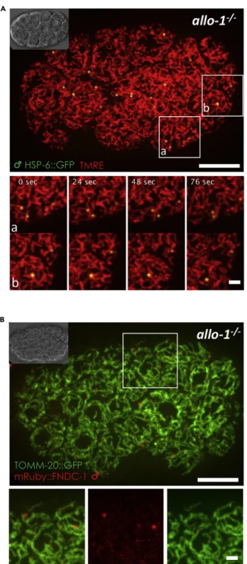
Figure 4. Stabilized sperm mitochondria are associated with maternal mitochondria
Sperm mitochondria are stabilized in allo-1(tm4756) mutant worms and move along with maternal mitochondria in the embryos, without exchange of mitochondrial outer membrane (MOM) proteins between them.
(A) allo-1(tm4756) hermaphrodites were crossed with HSP-6::GFP males in the presence of TMRE. Maternal and paternal mitochondria are both labeled with TMRE in the embryo (red) where sperm-derived mitochondria can be tracked with the HSP-6GFP marker (green). Maximum intensity Z-projections of 13 spinning disk deconvolved live images of a 24-cell stage
dynamic exchange of the dye within the embryo, and (ii) sperm mitochondria membrane potential is not lost before autophagosome formation.
Additionally, the loss of function of the autophagy factors, such as ALLO-1 and LGG-1, revealed that sta-bilized sperm-derived-mitochondria can maintain their membrane potential even in late embryonic stages in the absence of the autophagy machinery. Our results suggest that loss of membrane potential requires the entry of the mitochondria into the autophagosome, eliminating the possibility of it being the triggering signal for autophagy factors recruitment.
Intriguingly, in later embryonic stages, sperm mitochondria remain in spherical shape, and even when they seem to be closely associated with the surrounding maternal mitochondria, we did not observe them fused with the maternal mitochondria nor any exchange of mitochondrial proteins between sperm and maternal mitochondria. It is becoming clearer that sperm mitochondria have function during early steps of embryonic development (De Henau et al., 2020), but after this role is fulfilled, a delicate and precise process of elimination by autophagy is carried out to insure a strict maternal inheritance. Our results suggest that if this process fails, a backup system that prevents sperm mitochondria integration into the mitochondrial network could be acting as a last resort. More work is required to identify the triggering and selective mechanism for sperm mitochondria removal and the additional mechanism(s) preventing sperm mitochondrial DNA leakage.
Limitations of the study
The fact that sperm mitochondria remain polarized and separated from the maternal mitochondrial network un-til their entry into the autophagosomes in the C. elegans embryo argues for a late loss of integrity of the mito-chondria and a mechanism preventing their fusion. All the known mechanisms involved in the clearance of sperm-mitochondrial DNA are also at work in C. elegans but our discoveries may highlight a remarkable stability (like for the late involvement of the endonuclease G only after fertilization) and suggests that C. elegans sperm-mitochondria clearance may happen later in this organism compared to other organisms including vertebrates. These two properties may also exist in mammalian embryos but are technically challenging to address and beyond the scope of this work. The complete process of sperm mitochondria clearance takes several days in mammals compared to few hours in the C. elegans embryo, and mitochondria are particularly sensitive to experimental conditions.
Resource availability Lead contact
Further request and information for resources and reagents used in this published article should be directed and will be fulfilled by the lead contact, Vincent Galy,[email protected].
Materials availability
All data analyzed and generated in this research are included in this published article andSupplemental information.
Data and code availability
The published article includes all data generated in this study.
METHODS
All methods can be found in the accompanyingTransparent methods supplemental file.
Figure 4. Continued
embryo. z stack images were acquired every 0.24mm on a spinning disk microscope every 4 s. Magnifications of 2 highlighted areas are shown below. Scale bars: 10mm (top) and 2 mm (bottom).
(B) allo-1(tm4756); tomm-20::GFP young adult hermaphrodites were crossed with mRuby::fndc-1 males. Maximum intensity Z-projections of 7 spinning disk live images of a 60-cell stage embryo. Maternal MOM TOMM-20::GFP protein (green) and sperm MOM mRuby::FNDC-1protein (red) are both still visible and do not overlap suggesting that there is no exchange between the membranes of the two types of mitochondria. Magnifications of the merge and individual channels of the highlighted area are shown below. Scale bars: 10mm (top) and 2 mm (bottom).
SUPPLEMENTAL INFORMATION
Supplemental information can be found online athttps://doi.org/10.1016/j.isci.2020.102029.
ACKNOWLEDGMENTS
We are very grateful to Dr M. Labouesse for giving us access to his FRAP microscope, to V. Peletier, V. Par-rales, and R. Zamy for valuable technical assistance during all stages of this project. Some strains were provided by the Caenorhabditis Genetic Center, which is funded by NIH Office of Research Infrastructure Programs, USA (P40 OD010440). This work was supported by the Bettencourt Schueller Foundation, France (Coups d’e´lans pour la recherche to V.G.); the Agence Nationale de la Recherche, France (ANR 12-BSV2-0018-01 to V.G.); the Fondation pour la Recherche Me´dicale, France (Equipe FRM DEQ20160334874 to V.G. and K.R.) and by the ARC Foundation, France (fellowship to S.A.).
AUTHOR CONTRIBUTIONS
K.R., S.A., F.H., and J.M. conducted the experiments; F.L. deconvolved the images; K.R. and V.G. designed and analyzed the experiments and wrote the paper.
DECLARATION OF INTERESTS
The authors declare no competing interests.
Received: September 16, 2020 Revised: December 17, 2020 Accepted: December 29, 2020 Published: January 22, 2021 REFERENCES
Al Rawi, S., Louvet-Vallee, S., Djeddi, A., Sachse, M., Culetto, E., Hajjar, C., Boyd, L., Legouis, R., and Galy, V. (2011). Postfertilization autophagy of sperm organelles prevents paternal
mitochondrial DNA transmission. Science 334, 1144–1147.
Becker, T., Gebert, M., Pfanner, N., and van der Laan, M. (2009). Biogenesis of mitochondrial membrane proteins. Curr. Opin. Cell Biol. 21, 484–493.
De Henau, S., Page`s-Gallego, M., Pannekoek, W.J., and Dansen, T.B. (2020). Mitochondria-derived H2O2 promotes symmetry breaking of the C. elegans zygote. Dev. Cell 53, 263–271.
DeLuca, S.Z., and O’Farrell, P.H. (2012). Barriers to male transmission of mitochondrial DNA in sperm development. Dev. Cell 22, 660–668.
Ding, W.X., and Yin, X.M. (2012). Mitophagy: mechanisms, pathophysiological roles, and analysis. Biol. Chem. 393, 547–564.
Djeddi, A., Al Rawi, S., Deuve, J.L., Perrois, C., Liu, Y.Y., Russeau, M., Sachse, M., and Galy, V. (2015). Sperm-inherited organelle clearance in C. elegans relies on LC3-dependent autophagosome targeting to the
pericentrosomal area. Development 142, 1705– 1716.
Ehrenberg, B., Montana, V., Wei, M.D., Wuskell, J.P., and Loew, L.M. (1988). Membrane potential can be determined in individual cells from the nernstian distribution of cationic dyes. Biophys. J. 53, 785–794.
Lim, Y., Rubio-Pen˜a, K., Sobraske, P.J., Molina, P.A., Brookes, P.S., Galy, V., and Nehrke, K. (2019). Fndc-1 contributes to paternal
mitochondria elimination in C. elegans. Dev. Biol. 454, 15–20.
Liu, L., Feng, D., Chen, G., Chen, M., Zheng, Q., Song, P., Ma, Q., Zhu, C., Wang, R., Qi, et al.. (2012). Mitochondrial outer-membrane protein FUNDC1 mediates hypoxia-induced mitophagy in mammalian cells. Nat. Cell Biol. 14, 177–185.
Neupert, W., and Herrmann, J.M. (2007). Translocation of proteins into mitochondria. Annu. Rev. Biochem. 76, 723–749.
Paix, A., Folkmann, A., and Seydoux, G. (2017). Precision genome editing using CRISPR-Cas9 and linear repair templates in C. elegans. Methods 121–122, 86–93.
Politi, Y., Gal, L., Kalifa, Y., Ravid, L., Elazar, Z., and Arama, E. (2014). Paternal mitochondrial destruction after fertilization is mediated by a common endocytic and autophagic pathway in Drosophila. Dev. Cell 29, 305–320.
Sato, M., and Sato, K. (2011). Degradation of paternal mitochondria by fertilization-triggered autophagy in C. elegans embryos. Science 334, 1141–1144.
Sato, M., Sato, K., Tomura, K., Kosako, H., and Sato, K. (2018). The autophagy receptor ALLO-1 and the IKKE-1 kinase control clearance of paternal mitochondria in Caenorhabditis elegans. Nat. Cell Biol. 20, 81–91.
Song, W.-H., Yi, Y.-J., Sutovsky, M., Meyers, S., and Sutovsky, P. (2016a). Autophagy and ubiquitin–proteasome system contribute to sperm mitophagy after mammalian fertilization. Proc. Natl. Acad. Sci. U S A 113, E5261–E5270.
Song, W.H., Yi, Y.J., Sutovsky, M., Meyers, S., and Sutovsky, P. (2016b). The ART and science of sperm mitophagy. Autophagy 12, 2510–2511.
Sutovsky, P., and Song, W.H. (2018). Post-fertilisation sperm mitophagy: the tale of mitochondrial Eve and Steve. Reprod. Fertil. Dev. 30, 56–63.
Wang, Y., Zhang, Y., Chen, L., Liang, Q., Yin, X.M., Miao, L., Kang, B.H., and Xue, Di. (2016). Kinetics and specificity of paternal mitochondrial elimination in Caenorhabditis elegans. Nat. Commun. 7, 12569.
Zhou, Q., Li, H., Li, H., Nakagawa, A., Lin, J.L.J., Lee, E.-S., Harry, B.L., Skeen-Gaar, R.R., Suehiro, Y., William, et al.. (2016). Mitochondrial endonuclease G mediates breakdown of paternal mitochondria upon fertilization. Science 353, 394–399.
Zhou, Q., Li, H., and Xue, D. (2011). Elimination of paternal mitochondria through the lysosomal degradation pathway in C. elegans. Cell Res 21, 1662–1669.
Zorova, L.D., Popkov, V.A., Plotnikov, E.Y., Silachev, D.N., Pevzner, I.B., Jankauskas, S.S., Babenko, V.A., Zorov, S.D., Balakireva, A.V., Juhaszova, et al.. (2018). Mitochondrial membrane potential. Anal. Biochem. 552, 50–59.