HAL Id: hal-02025890
https://hal.archives-ouvertes.fr/hal-02025890
Submitted on 19 Feb 2019
HAL is a multi-disciplinary open access
archive for the deposit and dissemination of
sci-entific research documents, whether they are
pub-lished or not. The documents may come from
teaching and research institutions in France or
abroad, or from public or private research centers.
L’archive ouverte pluridisciplinaire HAL, est
destinée au dépôt et à la diffusion de documents
scientifiques de niveau recherche, publiés ou non,
émanant des établissements d’enseignement et de
recherche français ou étrangers, des laboratoires
publics ou privés.
Reflection and transmission of visible light by sugi
wood: effects of cellular structure and densification
Hiroyuki Sugimoto, Sakiko Kawabuchi, Masatoshi Sugimori, Joseph Gril
To cite this version:
Hiroyuki Sugimoto, Sakiko Kawabuchi, Masatoshi Sugimori, Joseph Gril. Reflection and transmission
of visible light by sugi wood: effects of cellular structure and densification. Journal of Wood Science,
Springer Verlag, 2018, 64 (6), pp.738-744. �10.1007/s10086-018-1751-7�. �hal-02025890�
https://doi.org/10.1007/s10086-018-1751-7 ORIGINAL ARTICLE
Reflection and transmission of visible light by sugi wood: effects
of cellular structure and densification
Hiroyuki Sugimoto1,2 · Sakiko Kawabuchi3 · Masatoshi Sugimori1 · Joseph Gril2,4
Received: 9 February 2018 / Accepted: 29 June 2018 © The Japan Wood Research Society 2018
Abstract
Transmittance and reflectance of visible light by sugi wood (Cryptomeria japonica) were investigated in the longitudinal (L) and tangential (T) directions. Transmittance was the highest in the L direction and reflectance was the highest in the T direction, suggesting that structural anisotropy influences transmittance and reflectance. Intra-ring variations observed with a microspectrometer indicated that T transmittance was higher for latewood than for earlywood, but there was no such trend in for L transmittance in which the highest levels occurred near the annual ring boundaries, on either the earlywood or late-wood side, and the lowest at the transition from earlylate-wood to latelate-wood. Dependence of L transmittance on wavelength also showed variations according to the intra-ring position. The increasing of transmittance of earlywood at wavelengths < 500 nm with increasing wavelength was observed, but this was not confirmed for latewood because of absorption by lignin. These observations supported a previously published finding, which was based on measurements in the radial direction, that the number of internal cell wall reflections, rather than density, determines wood lightness. Indeed, in the L direction, most of the incident light passes through lumens in earlywood and through cell walls in latewood, while it is subjected to numer-ous internal reflections at the interface between lumens and cell walls. This was further confirmed by the transmittance of earlywood being greatly decreased by radial compression.
Keywords Visible light · Cell structure · Transmittance · Reflection · Compression
Introduction
Owing to its esthetic characteristics, wood is used for the manufacture of many high-value products [1]. Therefore, the appearance of wood, such as its texture, grain patterns, or color, has been studied. Studies investigating the light-ness of the wood surface have also been conducted by many
researchers [2–5], and in general, it is believed that wood with higher density tends to be darker in terms of light absorption. However, this could not always be explained well by the density dependence of wood lightness. Palvi-ainen et al. [6] investigated the influence of density on dif-fraction using 30 wood species. They showed that samples with lower densities are likely to produce more scatter-ing. However, such samples of different species also have different anatomical features. In a previous study [7], we measured total light transmittance and total reflectance of sugi (Cryptomeria japonica) compressed at various levels. The results showed that in medium- and long-wavelength regions, the relationship between transmission and reflec-tion is strong because the influence of cell wall absorpreflec-tion is small. That is, the effect of reflection on lightness in the long-wavelength region is as comparable to that of absorp-tion by the cell wall.
The Kubelka– Munk (KM) theory based on specific scat-tering coefficient and specific absorption coefficient is fre-quently used for paper or powder [8] to understand the spec-troscopic characterization. However, KM theory only deals
* Hiroyuki Sugimoto
sugimoto.hiroyuki.rw@ehime-u.ac.jp
1 Ehime University Graduate School of Agriculture, Tarumi,
Matsuyama 790-8566, Japan
2 LMGC, Univ. Montpellier, CNRS, CC4048, 163 rue Auguste
Broussonnet, 34090 Montpellier, France
3 Abashiri Nambu District Forest Office, Forest Agency,
Ministry of Agriculture, Forestry and Fisheries, 656-3, Koshimizu, Koshimizu-cho, Shari-gun, Hokkaido 099-3632, Japan
4 CNRS, Université Clermont Auvergne, Sigma Clermont,
Institut Pascal, Campus des Cezeaux, 2 av. Blaise Pascal, TSA 60206, CS 60026, 63178 Aubière Cedex, France
Journal of Wood Science
1 3
with the propagating energy of incident light from the front to the rear, parallel to the direction of propagation in the case of a material that is homogeneous along its thickness; it may thus be difficult to apply to wood given its inhomoge-neous structure [9, 10]. Moreover, this theory supposes that the incident light mainly pass the substance. In wood, the volume ratio of air to cell wall varies greatly depending on the species and tree part, so that light might pass through air in lumen as much as through the cell wall, in a proportion depending on that ratio. Therefore, we need a model for light transmission and reflection considering not only the cell wall material [11] but also cell structure.
In a previous study, focused on the influence of density on the transmittance/reflectance of visible light, the compres-sion and measurement directions were fixed in the radial direction. In this study, to examine the influence of cell structure, the transmittance and reflectance of light were measured in both longitudinal and tangential directions. Light properties in earlywood and latewood were compared using a microspectrometer. Moreover, to examine the effect of compression treatment, measurements in a direction dif-ferent from that of the compression were performed, along with observation using microtomography.
Materials and methods
Sample preparationWood blocks were taken from sugi (Cryptomeria
japon-ica) sapwood. The sample dimensions were 64 mm in the
tangential direction (T) × 47 mm in the radial direction (R) × 142 mm in the longitudinal direction (L). To measure density, one portion of 10 mm in the L direction was cut from each of these blocks, and the remaining parts were used for visible light spectroscopy measurements. Density was measured by the double-weight method. Prior to vis-ible light spectroscopy, some specimens were subjected to compression treatment: after being steamed at 100 °C for 2 h, they were compressed in the R direction using a press machine (Denki Netsuban; SHINTO Metal Industries Corp.) equipped with a flat heating metallic die polished to a mirror finish. Neglecting the deformation in the T direction result-ing from the compression, we defined a compression ratio (CR) varying from zero for an uncompressed state to unity for a fully compressed state as follows:
where d0, d, and dL are the original, compressed, and limit heights of the specimen, respectively. The limit height is calculated by dry weight divided by cell wall density of 1.5 and by base area.
CR = (d0− d)∕(d0− dL) × 100,
After being compressed to the predefined thickness, each sample was left for 24 h and then left to stand in a desiccator. After 1 week, the dimensions and weight were measured.
For visible light spectroscopy measurement, compressed samples were cut by a rim saw into 10 specimens of various thicknesses of 0.3– 1.5 mm in the L direction; the remaining parts were also cut into 10 specimens of various thicknesses of 0.3– 1.5 mm in the T direction.
Visible light spectroscopy measurement
The total light reflectance and total light transmittance in the wavelength range from 350 to 800 nm were measured at 20 °C using a UV– Vis spectrophotometer (V-670; JASCO Co. Ltd.) equipped with an integrating sphere.
Irradiation light energy within a bandwidth of 5 nm of this device in the visible light region was about 1 µW and the spot diameter was about 5 mm. The samples were cut into 15-mm squares to fit the cell. A barium sulfate white plate was used as the reference sample.
Image analysis by X-ray CT
The images were observed using an X-ray CT scanner (Sky-scan 1272 Bruker MicroCT). After spectroscopy measure-ment, the samples were cut into sections of 5 mm in the T direction. The observation conditions were 50 kV, 200 µA, 0.2° rotation, and 1.2 µm pixel size at a temperature of 20 °C and relative humidity of 60%. The obtained data were recon-structed using NRecon software. CTVO software was used for 3D visualization.
Transmittance measurement using UV–Visible/NIR micro-spectrometers
The transmittance at the micro-scale in the visible light region was measured by UV– Visible/NIR micro-spectrome-ters (MSV-5200; JASCO Co. Ltd.). The measurement diam-eter was 100 µm. Four points in earlywood and four points in latewood were measured for each specimen. Further, for the uncompressed L sample, 20 points were measured along two annual rings.
Results and discussion
Difference of transmittance and reflectance between L and T directions
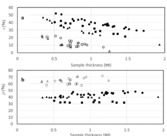
Figure 1 shows the influence of sample thickness on total light transmittance (τ) and reflectance (ρ) of sugi wood in the
L and T directions at the wavelength of 750 nm. Although
τ decreased and ρ increased when the thickness increased,
as reported previously. Moreover, τ of the L sample was larger than that of the T sample. ρ showed the opposite trend. These results suggest that τ and ρ properties are affected by the anisotropy of wood cell structure.
The annual ring structure is the most clearly recognized wood feature involving a contrast of brightness. Therefore, we started by comparing the difference of τ between early- and latewood, as observed using micro-spectrometers. τ of all samples was small at short wavelengths up to 400 nm. Moreover, τ of latewood in all T samples was larger than that of earlywood. Typical results of the T sample with a thickness of 5 mm are shown in Fig. 2. In previous paper [7], we predicted the τ by the expression of the number of the cell wall/lumen interfaces. In T samples, the incident light will pass through a series of cell wall/lumen reflection interfaces, especially in earlywood, as can be predicted by the expression. However, in latewood, where cells have a thick cell wall and a small lumen diameter, in addition to
being well aligned in both R and T directions, the incident light often passes through the cell wall without encounter-ing the interface. This explains why τ of latewood is higher than that of earlywood. On the other hand, this trend was not observed in the L sample. This suggests the possibility that other mechanisms are involved in the L direction.
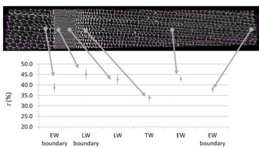
Figure 3 compares the cell structure image obtained by micro-CT scanning and the corresponding τ measured by a microspectrometer, at the wavelength of 750 nm. The trans-mittance greatly varied depending on the measurement loca-tion. From the pith (right) side, τ of the earlywood (EW) was over 35%; it dropped to < 35% in the transition zone (TW), increased up to 45% in the latewood (LW) toward the bound-ary of the annual ring (LWb), and then recovered to the same level of > 35% in the next EW zone. τ most likely depends on the location even within the same LW.
Figure 4 shows the dependence of τ in the L direction on wavelength in sugi wood at various locations and its loga-rithmic curves. For short wavelengths up to 500 nm, there was an increase of τ in EW with increasing wavelength in the curves, but this was not confirmed for TW and LW. There-fore, the light path differs between EW and LW; higher val-ues of EW transmittance are possibly because of lower light absorption. It is assumed that absorbance at wavelengths up to 600 nm is because of the existence of lignin [12]. Lower
τ of TW and LW than that of EW suggests that the light in
TW and LW passes through the cell wall more than that in EW. In contrast, for long wavelengths, the τ of LW is greater than that of EW. In TW, at the transition from EW to LW, τ is the lowest. Tsuchikawa and Tsutsumi showed that, for short wavelengths in the near-infrared region, in other words, for long-wavelength visible light, scattering occurs Fig. 1 Influence of sample thickness on total light transmittance (τ)
and reflectance (ρ) of sugi wood in the longitudinal direction (filled circles) and tangential direction (open filled circles)
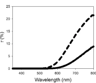
Fig. 2 Comparison of wavelength dependence of total light transmit-tance (τ) between earlywood (solid line) and latewood (dashed line) in the T direction for a thickness of 0.52 mm
Journal of Wood Science
1 3
in a direction perpendicular to that of incident light [13]. Therefore, the light path in this part is longer than that in other parts due to the occurrence of substantial scattering.
For long wavelengths, the τ of LW is greater than that of EW because the light passes through the cell wall with less numerous interfacial reflections. Furthermore, light passing through the cell wall may hardly diffuse because of total reflection at the cell wall/lumen interface.
This can be qualitatively confirmed by image evaluation. Figure 5 shows an image with the light from the photo-graphing side (left) and from the back of the sample (right). The former corresponds to reflected light and the latter to transmitted light. In the left image, LW is darker than EW, whereas in the right image it is lighter and more scattered; in particular, the light is most visible close to annual ring
boundaries. Moreover, the darkest part is TW. These trends agree well with the microspectrometer results.
If the above-mentioned assumption that TW is the dark-est owing to numerous interfaces is correct, the increase in density owing to compression should not necessarily lead to an increase in τ because compression treatment in a direction different from that of the light path does not decrease the number of interfaces between cell wall and lumen.
Change in transmittance by compression
To quantify the effect of compression, the relationship between τ and ρ and the thickness in the L and T directions of radially compressed samples with various compression ratios is shown in Fig. 6. Both τ and ρ of compressed samples were lower than those of uncompressed wood. These results are contrary to those in a previous paper [7]. The light in the compressed part may have been scat-tered toward the outside of the integrating sphere. For a given thickness, the interfaces of refraction and the con-sequent increasing of light pass are increased by compres-sion treatment. To investigate the relationship between the change of cell structure resulting from the compression and the light properties, images of cell structure obtained by X-ray CT scanning are shown in Fig. 7 and τ in T direc-tion of earlywood of compressed samples measured by microspectrometer is shown in Fig. 8. Calculated values are shown for comparison, based on the following model [7]. We considered the case of a regular arrangement of cells with a square cross section of width 30 µm and thin cell wall (which is the case of sugi wood). We neglect the direct transmission of light through the thin cell walls disposed parallel to the light direction, and only consider the reflection occurring at the interface between lumens Fig. 3 Cell structure image by
X-ray CT of L sample with a thickness of 0.60 mm and total light transmittance (τ) at given positions of the same sample
Fig. 4 Dependence of total light transmittance (τ) in the L direction on wavelength at the positions shown in Fig. 3
and transverse cell walls. The sample thickness being D (µm), the number of such reflections equals D/15 and the total transmittance (τ) can be described by
where the transmittance of each interface (τi) is given by
assuming a refractive index for the cell wall (n1) of 1.6 [14] and for air (n0) of 1.
Comparing the compressed and uncompressed samples in terms of end grain, the cell wall of earlywood collapsed in an intricate way. (1) 𝜏 = 𝜏 i D 15, (2) 𝜏 i= 4n0n1 (n0+ n1)2
This change of cell structure may have increased the num-ber of diffused reflections in the earlywood per measurement area because of more numerous interfaces resulting from the intricate cell wall even so steaming treatment affects τ [15]. The lower ρ values of compressed samples than that of uncompressed samples are in good agreement with practical appearance of lightness of earlywood (Fig. 9).
Conclusion
To investigate the effects of cellular structure and densi-fication on total light transmission and reflection in the visible light region, the anisotropy of total light transmit-tance (τ) and reflectransmit-tance (ρ), as well as the micro-local τ of Fig. 5 Comparison of an image
of L sample with a thickness of 0.76 mm with the light from the photographing side (left) and from the back of the sample (right)
Journal of Wood Science
1 3
uncompressed and compressed wood, was measured. Based on the obtained results, the following conclusions can be drawn:
• τ in the L direction is higher than that in the T direction.
In all T direction samples, τ of latewood was higher than that of earlywood. Conversely, in the L direction, a simi-lar trend was not observed.
• Although τ of late- and earlywood near the annual ring boundaries was high, in the transition from early- to late-wood, it was the lowest. These facts suggest that in the
L direction, most of the incident light passed through
lumens in earlywood and through cell walls in latewood, while in the transition part, diffusion due to light passing
lumen/wall interfaces occurs more often, resulting in a lower τ.
• For short wavelengths up to 500 nm, there is an increase of τ of EW with increasing wavelength in the curves, but this was not confirmed for TW and LW. Absorbance up to 600 nm caused by lignin, and the thinner cell walls of EW could explain that observation. At longer wave-lengths, τ of latewood is greater than that of earlywood. These results support the previous deduction that differ-ences of τ among the parts in rings are because of the different light paths.
• The transmittance of the earlywood was greatly reduced by compression treatment, probably because of numerous internal reflections at the cellular level. The lower ρ of compressed samples than that of uncompressed samples are in good agreement with practical appearance of light-ness of earlywood.
Fig. 6 Relationship between a total light transmittance (τ) or b reflec-tance (ρ) and the thickness in L (close) and T (open) directions of compressed samples with various compression ratios (circle: uncom-pressed, triangle: 81%, square: 87%)
Fig. 7 Image of the cell
struc-ture of compressed wood using X-ray CT scanning
Fig. 8 The relationship between total light transmittance (τ) in T
direction of earlywood of compressed samples measured by micro-spectrometer with various compression ratios and sample thicknesses; dashed line, calculation based on model/equation
References
1. NTT docomo HP (in Japanese). https ://www.nttdo como.co.jp/ info/news_relea se/2011/03/09_00.html. Accessed 20 July 2018 2. Kitamura Y (1987) Measured colour values of important domestic
and imported wood (in Japanese). Bull For Forest Prod Res Inst 347:203– 239
3. Kataoka Y, Kiguchi M, Williams RS, Evans PD (2007) Vio-let light causes photo degradation of wood beyond the zone affected by ultraviolet radiation. Holzforschung 61:23– 27 4. Mitsui K, Takada H, Sugiyama M, Hasegawa R (2005) Changes
in the properties of light-irradiated wood with heat treatment. Part 1. Effect of treatment conditions on the change in color. Holzforschung 55:601– 605
5. Ban M, Inagaki T, Ma T, Tsuchikawa S (2018) Effect of cellu-lar structure on the optical properties of wood. J Near Infrared Spectrosc 26:53– 60
6. Palviainen J, Silvennoinen R (2001) Inspection of wood density by spectrophotometry and a diffractive optical element based sensor. Meas Sci Technol 12:345– 352
7. Sugimoto H, Rikitake T, Sugimori M (2018) Optical reflection and transmission of sugi wood in visible light (in Japanese). Mokuzai Gakkaishi 64:66– 71
8. Scallan AM, Borch J (1972) An interpretation of paper reflectance based upon morphology I. Initial considerations. Tappi 55:583– 588
9. Tolvaj L, Mitsui K, Varga D (2011) Validity limits of Kubelka-Munk theory for DRIFT spectra of photodegraded solid wood. Wood Sci Techol 45:135– 146
10. Tsuchikawa S, Tsutsumi S (1996) Application of near infrared spectrophotometry to wood III. Behavior of reflected and trans-mitted light for parallel beam of incident light (in Japanese). Mokuzai Gakkaishi 42:733– 742
11. Saarinen K, Muinonen K (2001) Light scattering by wood fib-ers. Appl Opt 40:5064– 5077
12. Mononen K, Jääskeläinen AS, Alvila L, Pakkanen TT, Vuori-nen T (2005) Chemical changes in silver birch (Betula pendula
Roth) wood caused by hydrogen peroxide bleaching and
moni-tored by color measurement (CIELab) and UV– Vis, FTIR and UVRR spectroscopy. Holzforschung 59:381– 388
13. Tsuchikawa S, Tsutsumi S (1996) Application of near infrared spec-trophotometry of wood II. Behavior of irradiated light in an aggre-gate of tracheids and directional characteristics of diffuse reflected and transmitted light (in Japanese). Mokuzai Gakkaishi 42:343– 353 14. Fink S (1992) Transparent wood—a new approach in the
func-tional study of wood structure. Holzforschung 46(5):403– 408 15. Bourgois P, Janin G, Guyonnet R (1991) The color measurement:
a fast method to study and to optimize the chemical transforma-tions undergone in the thermically treated wood (in French). Holz-forschung 45:377
Fig. 9 Images of the L samples
(upper) and the T samples (lower) with compressed treat-ment (right) and uncompressed (left). Measured light properties at framed red squares