HAL Id: hal-00360818
https://hal.archives-ouvertes.fr/hal-00360818v2
Submitted on 9 Mar 2009HAL is a multi-disciplinary open access
archive for the deposit and dissemination of sci-entific research documents, whether they are pub-lished or not. The documents may come from teaching and research institutions in France or abroad, or from public or private research centers.
L’archive ouverte pluridisciplinaire HAL, est destinée au dépôt et à la diffusion de documents scientifiques de niveau recherche, publiés ou non, émanant des établissements d’enseignement et de recherche français ou étrangers, des laboratoires publics ou privés.
Cell wall component and mycotoxin moieties involved in
binding of fumonisin B1 and B2 by lactic acid bacteria.
Vincent Niderkorn, Diego Morgavi, Bettina Aboab, Marielle Lemaire, Hamid
Boudra
To cite this version:
Vincent Niderkorn, Diego Morgavi, Bettina Aboab, Marielle Lemaire, Hamid Boudra. Cell wall com-ponent and mycotoxin moieties involved in binding of fumonisin B1 and B2 by lactic acid bacteria.. Journal of Applied Microbiology, Wiley, 2009, pp.977-985. �hal-00360818v2�
Cell wall component and mycotoxin moieties involved in the binding of fumonisin B
1and B
2by lactic acid bacteria
V. Niderkorn 1,2 , D.P. Morgavi 1 , B. Aboab 3 , M. Lemaire 3 and H. Boudra 1
1INRA, UR1213 Herbivores, Saint-Genès Champanelle, France 2Lallemand S.A.S, 19, rue des briquetiers, Blagnac, France 3CNRS, UMR 6504, SEESIB, University Blaise Pascal, Aubiere, France
Correspondence to Hamid Boudra, INRA, UR1213 Herbivores, Site de Theix, F-63122 Saint-Genès Champanelle, France. E-mail: hboudra@clermont.inra.fr
KEYWORDS
binding ; bioavailability ; detoxification ; fumonisins ; lactic acid bacteria ; peptidoglycan ; probiotics ABSTRACT
Aims: The ability of lactic acid bacteria (LAB) to bind fumonisins B1 and B2 (FB1, FB2) in fermented
foods and feeds and in the gastrointestinal tract could contribute to decrease their bioavailability and toxic effects on farm animals and humans. The aim of this work was to identify the bacterial cell wall component(s) and the functional group(s) of FB involved in the LAB–FB interaction.
Methods and Results: The effect of physicochemical, enzymatic and genetic treatments of bacteria and the removal/inactivation of the functional groups of FB on toxin binding were evaluated. Treatments affecting the bacterial wall polysaccharides, lipids and proteins increased binding, while those degrading peptidoglycan (PG) partially decreased it. In addition, purified PG from Gram-positive bacteria bound FB in a manner analogue to that of intact LAB. For FB, tricarballylic acid (TCA) chains play a significant role in binding as hydrolysed FB had less affinity for LAB.
Conclusions: Peptidoglycan and TCA are important components of LAB and FB, respectively, involved in the binding interaction.
Significance and Impact of the Study: Lactic acid bacteria binding efficiency seems related to the peptide moiety structure of the PG. This information can be used to select probiotics with increased FB binding efficiency.
Introduction
Fumonisins, a structurally related mycotoxin group produced by Fusarium verticillioides and Fusarium
proliferatum, are common contaminants of corn and corn-based products worldwide (Shephard et al.
1996). There are several identified fumonisins, but fumonisin B1 (FB1) and B2 (FB2) are the most
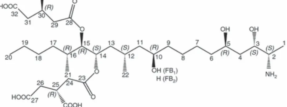
important and constitute up to 70% of the fumonisins found in naturally contaminated foods and feeds. FB1 is the diester of propane-1,2,3-tricarboxylic acid (tricarballylic acid, TCA) and
2-amino-12,16-dimethyl-3,5,10,14,15-pentahydroxyeicosane, in which the C14 and C15 hydroxyl groups are esterified
with the terminal carboxyl group of TCA. FB2 is the C10 deoxy analogue of FB1, in which the
corresponding stereogenic units on the icosane backbone have the same configurations (Fig. 1).
Fumonisins B1 and B2 are phytotoxic to corn (Lamprecht et al. 1994), cytotoxic to various mammalian
cell lines (Abbas et al. 1993) and FB1 is a carcinogen in rat liver and kidney (IARC 2002). The
occurrence of these analogues in home-grown corn has been associated with an increased risk of esophageal cancer in humans (Shephard et al. 2000). FB1 is considered possible carcinogens to human
and classified as class 2B (IARC 2002). These mycotoxins are the causal agent of two well described diseases in domestic animals: equine leukoencephalomacia (Riley et al. 1997) and porcine pulmonary edema syndrome (Harrison et al. 1990). In addition, they have also been associated with nephrotoxic, hepatotoxic and immunosupressing effects in various animal species (Morgavi and Riley 2007). The mechanism of action appears to involve mainly disruption of sphingolipid biosynthesis by the inhibition of the enzyme sphingosine N-acetyltransferase (ceramide synthase) (reviewed by Voss et al. 2007). FB are more toxic than their hydrolysed or N-acetylated derivatives (Gelderblom et al. 1993). The free amino group appears to play a specific role in the biological activity of fumonisins.
Binding of FB by lactic acid bacteria (LAB) from fermented foods and feeds, and by LAB present in the gastrointestinal tract (GIT) could contribute to decrease the toxin bioavailability. This property could also decrease the exposure of intestinal mucosa to FB. Gut tissues exposed to FB have a diminished immune response and an altered barrier function against colonization by pathogenic Escherichia coli (Bouhet et al. 2004). Viable and nonviable LAB are able to bind FB in a pH, genus, bacterial density and analogue (FB2 > FB1) dependent manner in vitro (Niderkorn et al. 2006). FB binding is rapid and particularly
effective in acidic conditions, forming a stable complex in the range of pH present in the GIT. This activity is probably present in a variety of fermented foods and feeds (Mokoena et al. 2005; Niderkorn
et al. 2007) and might also operate in the stomach. Binding of other major mycotoxins: aflatoxin B1
(Haskard et al. 2000), zearalenone (El-Nezami et al. 2002a) and certain trichothecenes (El-Nezami et al. 2002b) by some probiotic LAB has also been shown in vitro. In the absence of a simple detoxification method for foods and feeds contaminated by FB, the use of selected strains of LAB appears as a promising approach to reduce their toxicological effects. However, an understanding of the binding mechanism is required to allow the optimization and safe dietary application of this technology. The aim of this work was to identify the component of the bacterial cell wall and the chemical structure of FB involved in the mechanism of binding.
Materials and methods
Bacteria and bacteria-derived materials
Strains Lactobacillus paraplantarum CNRZ 1885 (CNRS, FRE2326 Strasbourg, France) and
Streptococcus thermophilus RAR1 (LAB collection of the Research Unit for Food Process Engineering
and Microbiology, INRA, Thivernal-Grignon, France) were used in most experiments. Streptococcus
thermophilus CNRZ 1066 and its non-capsular, non-exopolysaccharide (EPS) producing mutant Strep. thermophilus JIM 8752 (delta epsE) were obtained from the Microbial Genetics Unit, INRA,
Jouy-en-Josas, France. Lactococcus lactis subsp. cremoris MG1363 and mutants, in which the synthesis of certain cell wall components and adhesion properties are affected, were from the LAB and Opportunistic Pathogens Laboratory, INRA, Jouy-en-Josas, France. Bacterial strains were grown at optimal temperature (30 or 37°C) in De Man, Rogosa, Sharpe broth for lactobacilli and M17 broth (Oxoïd Ltd., Basingstoke, UK), supplemented with 0·5% of glucose for lactococci or 10% of lactose for streptococci. Commercial purified peptidoglycans (PG) from Gram-positive bacteria Micrococcus luteus and Bacillus subtilis were purchased from Sigma, Steinheim, Germany.
Determination of the bacterial cell wall component involved in binding
To identify the binding site, bacteria were subjected to different physicochemical and enzymatic treatments. Bacteria (Lact. paraplantarum CNRZ 1885 and Strep. thermophilus RAR1) were prepared in advance and stored at −18°C until use. Optimization tests showed that freezing did not negatively affected the binding ability of these strains (shown in results). For experiments, bacteria were thawed at room temperature, washed twice with 0·01 mol l−1 phosphate-buffered saline (PBS), pH 7·4 and treated by one of the following methods: water (25 or 100°C, 15 min), hydrochloric acid (1 mol l−1 HCl, 100°C, 15 min), sodium dodecyl sulphate (SDS, 2% w/v, 100°C, 15 min) or trichloracetic acid (10% w/v, 100°C, 15 min). After treatment, suspensions were centrifuged (3000 g, 10 min, 5°C). For enzymatic treatments, washed
bacteria were resuspended in 1 ml lysozyme (Sigma; 45 000 U ml−1 in phosphate buffer, pH 6), mutanolysin (Sigma; 5000 U ml−1 in phosphate buffer, pH 6), pronase E (Sigma; 1 mg ml−1 in 0·01 mol l−1 PBS, pH 7·4), lipase (Sigma; 1 mg ml−1 in 0·01 mol l−1 PBS, pH 7·4) or trypsin (Sigma; 1 mg ml−1 in Tris-HCl buffer, pH 8, 10 mmol l−1 CaCl
2). Suspensions were incubated at 37°C for 2 h with
shaking (240 rev min−1) and centrifuged (12 000 g, 10 min, 5°C). All bacterial pellets from both the physicochemical and enzymatic treatments were washed three times with 4 ml of PBS and used for the binding assay. Non-treated controls were added at each experimental run. All experiments were performed in triplicate.
Determination of the functional group of fumonisins involved in binding
To identify which functional group of FB can interact with bacteria, different chemical reactions were applied at different sites of FB derivatives. FB1 and FB2, purchased from Sigma and Promec (Tygerberg,
South Africa), respectively, were dissolved in an exact volume of acetonitrile–water in a 1 : 1 (v/v) ratio to achieve the desired concentration of stock solutions. Hydrolysed FB1 (HFB1) and FB2 (HFB2) were
obtained according to Pagliuca et al. (2005). Total hydrolysis of pure FB1 and FB2 was checked by
HPLC. The chromatograms showed absence of FB peak and presence of a single peak with retention times corresponding to the expected HFB product (Pagliuca et al. 2005). An optimized procedure was also used to determine the effect of the amine group in binding. Free amine of both FB was hidden by
reaction with ortho-phthalaldehyde (OPA). This option was chosen because the fumonisins of the group A in which the free amine is naturally absent are not commercially available.
In vitro binding assay
Treated and non-treated bacteria (109 or 1010 CFU ml−1 for certain experiments, see footnotes of tables) were tested as previously described (Niderkorn et al. 2007). Briefly, bacterial material was suspended in 1 ml of corn infusion adjusted to pH 4 with lactic acid and containing FB1 and FB2 (5 µg ml−1 each) or
their derivative compounds. The corn infusion was prepared by steeping dry whole-plant corn in water and filtering as described by Niderkorn et al. (2007). For each experiment, positive controls containing no bacterial material and a negative control containing no toxin were included. Assays and controls were incubated at 25°C for 1 h and centrifuged (3000 g, 10 min, 5°C). Supernatants and bacterial pellets were analysed for FB by reversed-phase HPLC to determine free and bound fractions respectively. Because of the instability of the FB-OPA derivative (Williams et al. 2004), assays with the free amine hidden were performed following an exact timing: At t = 0, a pure FB solution (800 µg ml−1) and reagent with (or without) OPA were mixed (1 : 1 v/v). At t = 2 min, 50 µl of this mixture was mixed to 950 µl of acidified corn infusion containing bacteria (1010 CFU ml−1), then incubated for 9·25 min at 25°C. At t = 12 min,
tubes were centrifuged (4500 g, 3 min, 4°C). At t = 20 min, supernatants containing free FB were derivatized with OPA. All samples were injected at t = 22 min. In these conditions, preliminary assays have shown that the complex FB-OPA remains sufficiently stable to carry out measurements. For this experiment, pellets were not analysed.
Fumonisins analysis
Supernatants from all samples and pellets extracts were fourfold diluted in acetonitrile-water (1 : 1 v/v), then 40 µl were added to 60 µl 0·1 mol l−1 borate buffer at pH 10 and 100 µl of OPA reagent were added. The preparation was mixed and allowed to react for 2 min before injection of 20 µl into the HPLC system. For FB extraction, 1 ml acetonitrile–water (1 : 1 v/v) was added to the bacterial pellets and this mixture was vigorously vortexed, placed in an ultrasonic bath for 6 min, then centrifuged (4500 g, 3 min, 5°C). Analysis of FB and their hydrolysed derivatives were done at room temperature by HPLC, using fluorimetric detection. The HPLC system consisted of a GOLD 126 solvent module (Beckman Coulter, Fullerton, CA, USA), an automatic sampler (Spectra-Physics, San Jose, CA, USA) equipped with a 100-µl loop and a fluorescence detector FL3000 (Spectra-System, San Jose, CA, USA). Separation of FB1,
FB2, HFB1 and HFB2 was performed on a C18 reversed-phase column (Prontosil, 150 × 4·6 mm, 3 µm,
Bishoff Chromatography) with a gradient elution using acetonitrile (A) and water–methanol (1 : 1 v/v) acidified at pH 3·35 with pure acetic acid (B). The gradient was started at 10% of solvent A, which increased to 60% in 6 min, then maintained at 60% for 7 min, before returned to the initial condition in 1 min. The flow rate was 1 ml min−1 and detection was set at 336 nm excitation and 440 nm emission.
The retention times of FB1, FB2, HFB1 and HFB2 were 9·9, 12·2, 10·2, 13·4 min respectively. The
percentage of free (or bound) mycotoxin was calculated as 100× [Peak area of mycotoxin in the supernatant (or pellet extract)/Peak area of mycotoxin in the positive control].
Statistical analysis
Data was subjected to the analysis of variance (ANOVA). A significant difference between means of
controls and assays (P < 0·05) was determined by Dunett's test using the STATISTICAL ANALYSIS SYSTEM
(SAS) software package, ver. 8 (SAS Institute Inc., Cary, NC, USA). Results
Bacterial cell wall components affecting binding
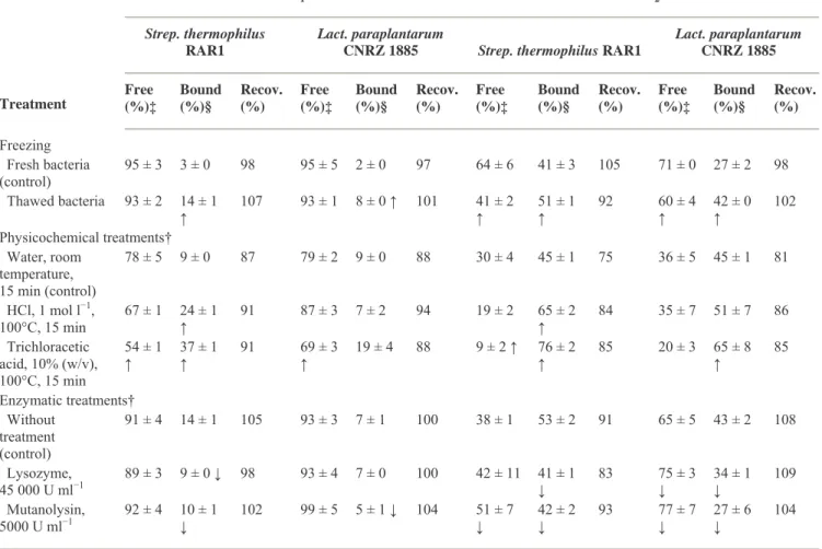
None of the physicochemical treatments applied to bacteria decreased binding of FB1 or FB2. On the
contrary, freezing/thawing and thermal treatments of bacteria increased the bound fractions of FB1 and
FB2 in both tested strains (P < 0·05) (Table 1). Among the chemical treatments, trichloracetic acid caused
a large increase in bound FB proportion (P < 0·05). HCl also produced the same effect although it was only significant on Streptococcus cells (P < 0·05). For the enzymatic treatments, lysozyme and mutanolysin were the only treatments which caused a partial, but significant decrease of this activity (P < 0·05) (Table 1). In contrast, lipase, trypsin and pronase E, an unspecific protease from Streptomyces
griseus, had no effect on binding (P > 0·05) (data not shown).
Table 1 Effect of freezing, chemical and enzymatic treatments of bacteria on binding of fumonisin B1 and B2 by
Streptococcus thermophilus RAR1 and Lactobacillus paraplantarum CNRZ 1885*
FB1 FB2
Strep. thermophilus RAR1
Lact. paraplantarum
CNRZ 1885 Strep. thermophilus RAR1
Lact. paraplantarum CNRZ 1885 Treatment Free (%)‡ Bound (%)§ Recov. (%) Free (%)‡ Bound (%)§ Recov. (%) Free (%)‡ Bound (%)§ Recov. (%) Free (%)‡ Bound (%)§ Recov. (%) Freezing Fresh bacteria (control) 95 ± 3 3 ± 0 98 95 ± 5 2 ± 0 97 64 ± 6 41 ± 3 105 71 ± 0 27 ± 2 98 Thawed bacteria 93 ± 2 14 ± 1 ↑ 107 93 ± 1 8 ± 0 ↑ 101 41 ± 2 ↑ 51 ± 1 ↑ 92 60 ± 4 ↑ 42 ± 0 ↑ 102 Physicochemical treatments† Water, room temperature, 15 min (control) 78 ± 5 9 ± 0 87 79 ± 2 9 ± 0 88 30 ± 4 45 ± 1 75 36 ± 5 45 ± 1 81 HCl, 1 mol l−1, 100°C, 15 min 67 ± 1 24 ± 1 ↑ 91 87 ± 3 7 ± 2 94 19 ± 2 65 ± 2 ↑ 84 35 ± 7 51 ± 7 86 Trichloracetic acid, 10% (w/v), 100°C, 15 min 54 ± 1 ↑ 37 ± 1 ↑ 91 69 ± 3 ↑ 19 ± 4 88 9 ± 2 ↑ 76 ± 2 ↑ 85 20 ± 3 65 ± 8 ↑ 85 Enzymatic treatments† Without treatment (control) 91 ± 4 14 ± 1 105 93 ± 3 7 ± 1 100 38 ± 1 53 ± 2 91 65 ± 5 43 ± 2 108 Lysozyme, 45 000 U ml−1 89 ± 3 9 ± 0 ↓ 98 93 ± 4 7 ± 0 100 42 ± 11 41 ± 1 ↓ 83 75 ± 3 ↓ 34 ± 1 ↓ 109 Mutanolysin, 5000 U ml−1 92 ± 4 10 ± 1 ↓ 102 99 ± 5 5 ± 1 ↓ 104 51 ± 7 ↓ 42 ± 2 ↓ 93 77 ± 7 ↓ 27 ± 6 ↓ 104
Data shown are means ± SD of triplicates.
↓ and ↑ within the same column, indicate that treatment decreased or increased binding (P < 0·05) compared with corresponding control. *For all experiments, treated or not treated bacteria (109 CFU ml−1) were incubated in acidified corn infusion containing FB
1 and FB2
(5 µg ml−1 each) for 1 h at 25°C.
†Experiments were performed with thawed bacteria. Enzymatic treatments were done at 37°C for 2 h. ‡Free fraction of fumonisin remaining in supernatant (vs control without bacteria).
Table 2 Effect of mutations affecting lipoteichoic acids and peptidoglycan biosynthesis in Lactococcus lactis subsp.
cremoris on binding of fumonisin B1 and B2*
FB1 FB2
Genotype Protein, function affected or phenotype Free (%) Bound (%) Recov. (%) Free (%) Bound (%) Recov. (%)
Derivatives from L. lactis subsp. cremoris MG1363
MG1363 Wild type 92 ± 6 4 ± 1 96 25 ± 2 65 ± 3 90
dltD LTA synthesis 88 ± 1 ↑ 11 ± 1 ↑ 99 30 ± 0 69 ± 4 99
pbp 2A− PBP 2A (PG transpeptidase) 94 ± 1 4 ± 1 98 48 ± 8 ↓ 47 ± 1 ↓ 95 pbp 2B− PBP 2B (PG transpeptidase) 95 ± 5 5 ± 0 100 52 ± 6 ↓ 45 ± 3 ↓ 97 Data shown are means ± SD of triplicates.
↓ and ↑ within the same column, indicate that treatment decreased or increased binding (P < 0·05) compared with the corresponding control.
PG, peptidoglycan; LTA, lipoteichoic acids; PBP, Penicillin binding protein.
*Bacteria (1010 CFU ml−1) were incubated in corn infusion containing FB
1 and FB2 (5 µg ml−1 each) for 1 h at 25°C.
Role of peptidoglycan
We observed decrease of FB2 binding with mutants of L. lactis that had an altered PG structure because of
perturbed transpeptidase functions (pbp 2A− and 2B−) (Shohayeb and Chopra 1987) (P < 0·05) (Table 2). However, mutants acmA and ponA from L. lactis, in which the immobilization property (phenomenon of adhesion, chain and biofilm formation) was modified (Mercier et al. 2002), had no effect on binding as compared with wild type L. lactis (P > 0·05) (data not shown). To confirm the role of PG component, purified PG from Gram-positive bacteria M. luteus and B. subtilis at different concentrations (0, 0·1, 0·5, 1 and 1·5 mg ml−1) were tested in a similar way. Results showed that these polymers can bind FB in an analogue dependent manner (FB2 > FB1) (Fig. 2). Significant bound fractions were observed even with
the lowest concentration tested (0·1 mg PG ml−1). However, the binding efficiency varied between the two purified PG tested (B. subtilis > M. luteus).
Figure 2 Fractions of fumonisin B1 (□, ) and B2 (○, •) bound to purified peptidoglycans from Bacillus subtilis (open
symbols) and Micrococcus luteus (closed symbols). Data shown are the mean and standard deviations (error bars) of triplicates.
Fumonisins structural component affecting binding
To identify the role of the main functional groups of FB in the formation of the mycotoxin–cell wall complex, the free amine and TCA arms were alternatively hidden or removed. When the free-amine group was hidden by derivatization with OPA, the proportions of FB1 and FB2 bound by Lact. paraplantarum CNRZ 1885 and Strep. thermophilus RAR1 were higher than those observed with
unmodified toxins (Table 3). This effect was more pronounced for FB1 (P < 0·05) than FB2 (P > 0·05).
Inversely, the binding rates of HFB1 and HFB2 for both strains was lower than those of FB1 and FB2
with FB1. To explain the different binding behaviour between FB1 and FB2, we investigated their
three-dimensional structure by molecular modelling in conditions simulating those of the binding tests. For that, conformations were carried out in aqueous conditions applying ionized states of carboxyl and amine groups of FB in acidic conditions to generate the most stable conformer using MACROMODEL 8.0
(Shroedinger Inc, Portland, OR, USA). Results showed that a hydrogen bond in FB1 structure is formed
between the hydrogen of the hydroxyl group at C10 and the oxygen of the carbonyl group of the TCA at
C15 (Fig. 3).
Table 3 Effect of hydrolysis of fumonisin's tricarballylic acid chains and free amine group inactivation on binding of fumonisin B1 and B2 by Lactobacillus paraplantarum and Streptococcus thermophilus*
Toxin Lact. paraplantarum CNRZ 1885 Strep. thermophilus RAR1
Free (%) Bound (%) Recov. (%) Free (%) Bound (%) Recov. (%)
FB1 73 ± 7 27 ± 2 100 59 ± 1 40 ± 1 99 HFB1 80 ± 6 14 ± 0 ↓ 94 85 ± 10 ↓ 22 ± 1 ↓ 107 FB2 19 ± 1 74 ± 1 93 27 ± 1 71 ± 3 98 HFB2 43 ± 4 ↓ 46 ± 11 ↓ 89 48 ± 4 ↓ 45 ± 3 ↓ 93 FB1† 89 ± 1 ND‡ 81 ± 1 ND FB1-OPA 68 ± 2 ↑ ND 66 ± 8 ↑ ND FB2† 56 ± 4 ND 31 ± 4 ND FB2-OPA 48 ± 0 ND 26 ± 0 ND
Data shown are means ± SD of triplicates.
↓ and ↑ within the same column, indicate that treatment decreased or increased binding (P < 0·05) compared with the corresponding control.
*Bacterial density = 1010 CFU ml−1.
†Treated in the same way as FB-OPA derivatives (see M&M for details). ‡Not determined.
Figure 3 Molecular conformations of FB1 and FB2 in aqueous solution. Conformational analysis of molecules in water
solution was performed using Monte-Carlo Multiple Method (Chang et al. 1989) with AMBER force field (Weiner et al. 1984; Cornell et al. 1995) and GB/SA solvation model (Still et al. 1990) of MACROMODEL 8.0 programme (Shroedinger Inc, Portland, OR, USA). To take account of pKas of fumonisins, conformations were carried out applying ionized states
of carboxyl and amine groups. In this case, four Na+ and one Cl− were added in the solution to maintain neutral charge of
the molecular system.
Discussion
The ability of LAB to bind fumonisins might contribute to decrease the bioavailability and toxic effects of FB1 and FB2 in human and farm animals. The binding activity of LAB could be integrated in the criteria
of selection of probiotics and starters used for the acidification of fermented corn meals and corn silage. However, the mechanism of binding is unknown and the LAB–FB interaction needs to be better understood to optimize the selection of strains. In this paper, we provided some insight into the interaction between FB and LAB that can explain different binding behaviour of FB1 and FB2.
Determination of the bacterial cell wall binding site
The cell wall of LAB has the typical Gram-positive structure made of a thick, multilayered PG sacculus in which proteins, teichoic acid (TA) and LTA and polysaccharides are associated (Delcour et al. 1999). We previously reported that all genera of LAB are capable to bind FB1 and FB2 (Niderkorn et al. 2007)
suggesting that the binding site is a component largely conserved in the cell wall of these bacteria. This component is synthesized early in the bacterial growth cycle since binding was observed in the latency phase. Binding was observed throughout the growth cycle with a maximum at the end of the exponential phase (data not shown).
The increase in binding observed with heat- and acid-treated bacteria (Table 1) was also reported for other mycotoxins such as aflatoxin B1 (El-Nezami et al. 1998) and zearalenone (El-Nezami et al. 2002a).
It is known that these treatments degrade the surface of the cell wall. The trichloracetic acid treatment to extract PG-associated cell wall polymers from Gram-positive bacteria is well established (Heckels and Virji 1988). Polysaccharides and TA are known targets of HCl and trichloracetic acid treatments (Quiberoni et al. 2000). Our results suggest that binding takes place in the subsurface of the cell wall in sites exposed by the heat or acid treatments.
The results obtained with mutant strains are in agreement with the physicochemical and enzymatic treatments of bacteria. Taken together, results indicate that the binding site of FB are not surface polysaccharides, lipids or proteins, but may be rather the PG or compounds tightly associated to it, as it was suggested for the binding of aflatoxin B1 (Lahtinen et al. 2004). It is worth mentioning that LTA are
the main components responsible for the hydrophobicity of the cell wall and thus, for the adhesion properties of bacteria (Dahlback et al. 1981). However, these adhesion properties appear not to have any
function in FB binding. Similarly, the mechanism of immobilization of bacteria, characterized by natural
PG modifications consisting of little breaks in the PG structure (Ibrahim et al. 2004), seems not to be associated with FB binding.
Binding by commercially available purified PG from two strains of Gram-positive bacteria (Fig. 2) was consistent with our results obtained with intact LAB, thus, supporting the hypothesis that PG is likely the binding site of FB. The PG backbone is a conserved structure composed of linear glycan chains alternating N-acetyl glucosamine (GlcNAc) and N-acetyl muramic acid (MurNAc) in a β (1→4) linkage. These chains are crosslinked by means of short peptides. The specific amino acid sequence of peptide bridges and consequently, the molecular structure of PG vary with the bacterial species (Schleifer and Kandler 1972). As the bound fraction of FB varied between the PG tested, but also among genera of LAB (Niderkorn et al. 2007), it seems that the amino acid sequence play an important role in the efficiency of the mechanism. The PG structure vary mainly in the amino acid in position 3 (AA3) of the peptide bridge
and in the cross-linking amino acids. B. subtilis and M. luteus differ in both the AA3 and cross-linking
amino acids (Schleifer and Kandler 1972). This difference could explain their dissimilar efficiency in binding FB. The higher binding efficiency of the Streptococcus genus compared with the Lactobacillus genus could be because of the amino acid sequence of the cross-bridge that is two to three molecules of L
-Ala in the former and D-Asp in the latter (Schleifer and Kandler 1972; Bouhss et al. 2002).
Relationship between fumonisins structure and binding
The higher binding rate of the FB-OPA derivative compared with unmodified FB suggest that the free-amine group possessing nucleophilic properties is not involved in FB1 and FB2 interaction with bacteria.
In addition, in acidic conditions, the ionized state of this function could even decrease binding, in particular for FB1. Controls containing reagents other than OPA, e.g. mercaptoethanol, used in the
derivatization reaction were done to exclude possible interferences on binding. However, the exact function that OPA may have on the FB derivative and/or on the bacterial cell wall that could modify the interaction was unknown. The use of natural fumonisin derivatives such as N-acetylated FB1 might be a
better alternative to test the exact role of the free-amino group. Notwithstanding the reservations associated to the derivatization methodology, masking the free-amino group increased rather than decreased binding suggesting that this chemical function has not a positive effect on FB-bacteria interaction. Inversely, the lower binding rate of hydrolysed FB compared with the intact FB indicates that one or both TCA arms play a positive role in the mechanism.
In spite of their similar structure, FB2 was in all experiments more bound than FB1 (Tables 1 and 2 and
Fig. 2). The same tendency between FB was recorded for OPA-treated or hydrolysed derivatives (FB2
-OPA > FB1-OPA, HFB2 > HFB1) (Table 3). These results are in agreement with those reported for other
bacteria and other experimental conditions (Niderkorn et al. 2006, 2007). The only structural variation between FB1 and FB2 consists in an additional hydroxyl group in C10 for FB1 (Fig. 1). Thus, it is
reasonable to postulate that this hydroxyl group plays directly or indirectly a negative role in binding. The spatial conformation induced by the hydrogen bond of FB1 makes the molecule more coiled and
apparently less favourable to binding by the bacterial cell wall. This conformation could disturb the interactions with the PG. These molecular conformations were conserved through pH variation as the addition of charges on functional groups of FB1 and FB2 did not affect the results of modelling. However,
as HFB2 was more bound than HFB1, it seems that the hydroxyl group in C10 continues to be
unfavourable to binding after TCA removal.
The objective of this work was to improve our understanding of the LAB–FB binding interaction. However, further work is needed before this methodology can be used to treat contaminated feeds. It is important to note that, differently from normal probiotic strains, LAB used to bind FB should have a low capacity to adhere to intestinal mucus and enterocytes to reduce the risk of toxin release in the GIT. The efficiency of bacterial strains to modulate intestinal toxin absorption and toxicity was already
demonstrated in vivo for aflatoxin B1 in both rats (Gratz et al. 2006) and humans (El-Nezami et al. 2006).
Conclusions
In this work, we demonstrated that PG of LAB and more generally PG of Gram-positive bacteria, are the most likely site of FB binding. This result helps to explain the widespread binding of fumonisins by LAB. Existing differences in binding capacity of different bacterial species can be rationally explained by the
variation in PG structure. This observation should allow to select efficient strains in terms of FB binding, as fermentation starters and/or probiotic mixtures on the base of their PG-type. We also showed that at least one TCA arm of FB play an important role in their binding to bacteria. As it was reported that TCA arms also play a favourable role in the intestinal absorption of FB1 (Dantzer et al. 1999; De Angelis et al.
2005), binding of FB1 and FB2 could decrease even more their absorption and their toxic effects on the
intestinal mucosal cells. However, further quantitative in vitro and in vivo studies are needed to evaluate the real impact of LAB binding activity on the bioavailability of FB in higher organisms.
References
Abbas, H.K., Gelderblom, W.C., Cawood, M.E. and Shier, W.T. (1993) Biological activities of fumonisins, mycotoxins from
Fusarium moniliforme, in jimsonweed (Datura stramonium L.) and mammalian cell cultures. Toxicon 31, 345–353.
Bouhet, S., Hourcade, E., Loiseau, N., Fikry, A., Martinez, S., Roselli, M., Galtier, P., Mengheri, E. et al. (2004) The mycotoxin fumonisin B1 alters the proliferation and the barrier function of porcine intestinal epithelial cells. Toxicol Sci 77, 165–171.Bouhss, A., Josseaume, N., Severin, A., Tabei, K., Hugonnet, J.E., Shlaes, D., Mengin-Lecreulx, D., van Heijenoort, J. et al. (2002) Synthesis of the L-alanyl-L-alanine cross-bridge of Enterococcus faecalis peptidoglycan. J Biol Chem 277, 45935–45941.
Chang, G., Guida, W.C. and Still, W.C. (1989) An internal-coordinate Monte Carlo method for searching conformational space. J Am Chem Soc 111, 4379–4386.
Cornell, W.D., Cieplak, P., Bayly, C.I., Gould, I.R., Merz, K.M., Ferguson, D.M., Spellmeyer, D.C., Fox, T. et al. (1995) A second generation force field for the simulation of proteins, nucleic acids, and organic molecules. J Am Chem Soc 117, 5179– 5197.
Dahlback, B., Hermansson, M., Kjelleberg, S. and Norkrans, B. (1981) The hydrophobicity of bacteria – an important factor in their initial adhesion at the air–water inteface. Arch Microbiol 128, 267–270.
Dantzer, W.R., Hopper, J., Mullin, K., Hendrich, S. and Murphy, P.A. (1999) Excretion of 14 C-fumonisin B, 14 C-hydrolyzed fumonisin B, and 14 C-fumonisin B-fructose in rats. J Agric Food Chem 47, 4291–4296.
De Angelis, I., Friggè, G., Raimondi, F., Stammati, A., Zucco, F. and Caloni, F. (2005) Absorption of fumonisin B1 and aminopentol on an in vitro model of intestinal epithelium; the role of P-glycoprotein. Toxicon 45, 285–291.
Delcour, J., Ferain, T., Deghorain, M., Palumbo, E. and Hots, P. (1999) The biosynthesis and functionality of the cell-wall of lactic acid bacteria. Antonie Van Leeuwenhoek 76, 159–184.
El-Nezami, H., Kankaanpaa, P., Salminen, S. and Ahokas, J. (1998) Physicochemical alterations enhance the ability of dairy strains of lactic acid bacteria to remove aflatoxin from contaminated media. J Food Prot 61, 466–468.
El-Nezami, H., Polychronaki, N., Salminen, S. and Mykkanen, H. (2002a) Binding rather than metabolism may explain the interaction of two food-grade Lactobacillus strains with zearalenone and its derivative α-zearalenol. Appl Environ Microbiol 68, 3545–3549.
El-Nezami, H.S., Chrevatidis, A., Auriola, S., Salminen, S. and Mykkanen, H. (2002b) Removal of common Fusarium toxins
in vitro by strains of Lactobacillus and Propionibacterium. Food Addit Contam 19, 680–686.
El-Nezami, H.S., Polychronaki, N.N., Ma, J., Zhu, H., Ling, W., Salminen, E.K., Juvonen, R.O., Salminen, S.J. et al. (2006) Probiotic supplementation reduces a biomarker for increased risk of liver cancer in young men from Southern China. Am J
Clin Nutr 83, 1199–1203.
Gelderblom, W.C., Cawood, M.E., Snyman, S.D., Vleggaar, R. and Marasas, W.F. (1993) Structure-activity relationships of fumonisins in short-term carcinogenesis and cytotoxicity assays. Food Chem Toxicol 31, 407–414.
Gratz, S., Taubel, M., Juvonen, R.O., Viluksela, M., Turner, P.C., Mykkanen, H. and El-Nezami, H. (2006) Lactobacillus
rhamnosus strain GG modulates intestinal absorption of aflatoxin B1 and its fecal excretion and toxicity in rats. Appl Environ Microbiol 72, 7398–7400.
Harrison, L.R., Colvin, B.M., Greene, J.T., Newman, L.E. and Cole, J.R. Jr (1990) Pulmonary edema and hydrothorax in swine produced by fumonisin B1, a toxic metabolite of Fusarium moniliforme. J Vet Diagn Invest 2, 217–221.
Haskard, C., Binnion, C. and Ahokas, J. (2000) Factors affecting the sequestration of aflatoxin by Lactobacillus rhamnosus strain GG. Chem Biol Interact 128, 39–49.
Heckels, J.E. and Virji, M. (1988) Separation and purification of surface components. In Bacterial Cell Surface Techniques ed. Hancock, I.C. and Poxton, I.R. pp. 67–135. New York: John Wiley and Sons.
Ibrahim, M., Briandet, R., Mistou, M.Y., Chretien, A., Tremblay, J. and Kulakauskas, S. (2004) Immobilization of lactococci.
Lait 84, 103–114.
International Agency for Research on Cancer. (2002) IARC Monographs on the Evaluation of the Carcinogenic Risks to
Humans: Some Traditional Herbal Medicines, some Mycotoxins, Naphthalene and Styrene. Lyon, France: International
Agency for Research on Cancer 82, 301–366.
Lahtinen, S.J., Haskard, C.A., Ouwehand, A.C., Salminen, S.J. and Ahokas, J.T. (2004) Binding of aflatoxin B1 to cell wall components of Lactobacillus rhamnosus strain GG. Food Addit Contam 21, 158–164.
Lamprecht, S.C., Marasas, W.F.O., Alberts, J.F., Cawood, M.E., Gelderblom, W.C.A., Shephard, G.S., Thiel, P.G. and Calitz, F.J. (1994) Phytotoxicity of fumonisins and TA-toxin to corn and tomato. Phytopathology 84, 393–391.
Mercier, C., Durrieu, C., Briandet, R., Domakova, E., Tremblay, J., Buist, G. and Kulakauskas, S. (2002) Positive role of peptidoglycan breaks in lactococcal biofilm formation. Mol Microbiol 46, 235–243.
Mokoena, M.P., Chelule, P.K. and Gqaleni, N. (2005) Reduction of fumonisin B1 and zearalenone by lactic acid bacteria in fermented maize meal. J Food Prot 68, 2095–2099.
Morgavi, D.P. and Riley, R.T. (2007) An historical overview of field disease outbreaks known or suspected to be caused by consumption of feeds contaminated with Fusarium toxins. Anim Feed Sci Technol 137, 201–212.
Niderkorn, V., Boudra, H. and Morgavi, D.P. (2006) Binding of Fusarium mycotoxins by fermentative bacteria in vitro. J Appl
Microbiol 101, 849–856.
Niderkorn, V., Morgavi, D.P., Pujos, E., Tissandier, A. and Boudra, H. (2007) Screening of fermentative bacteria for their ability to bind and biotransform deoxynivalenol, zearalenone and fumonisins in an in vitro simulated corn silage model. Food
Addit Contam 24, 406–415.
Pagliuca, G., Zironi, E., Ceccolini, A., Matera, R., Serrazanetti, G.P. and Piva, A. (2005) Simple method for the simultaneous isolation and determination of fumonisin B1 and its metabolite aminopentol-1 in swine liver by liquid chromatography-fluorescence detection. J Chromatogr B 819, 97–103.
Quiberoni, A., Stiefel, J.I. and Reinheimer, J.A. (2000) Characterization of phage receptors in Streptococcus thermophilus using purified cell walls obtained by a simple protocol. J Appl Microbiol 89, 1059–1065.
Riley, R.T., Showker, J.L., Owens, D.L. and Ross, P.F. (1997) Disruption of sphingolipid metabolism and induction of equine leukoencephalomalacia by Fusarium proliferatum culture material containing fumonisin B2 or B3. Environ Toxicol Pharmacol 3, 221–228.
Schleifer, K.H. and Kandler, O. (1972) Peptidoglycan types of bacterial cell walls and their taxonomic implications. Bacteriol
Rev 36, 407–477.
Shephard, G.S., Thiel, P.G., Stockenstrom, S. and Sydenham, E.W. (1996) Worldwide survey of fumonisin contamination of corn and corn-based products. J AOAC Int 79, 671–687.
Shephard, G.S., Marasas, W.F.O., Leggott, N.L., Yazdanpanah, H., Rahimian, H. and Safavi, N. (2000) Natural occurrence of fumonisins in corn from Iran. J Agric Food Chem 48, 1860–1864.
Shohayeb, M. and Chopra, I. (1987) Mutations affecting penicillin-binding proteins 2 a, 2 b and 3 in Bacillus subtilis alter cell shape and peptidoglycan metabolism. Microbiology 133, 1733–1742.
Still, W.C., Tempczyk, A., Hawley, R.C. and Hendrickson, T. (1990) Semianalytical treatment of solvation for molecular mechanics and dynamics. J Am Chem Soc 112, 6127–6129.
Voss, K.A., Smith, G.W. and Haschek, W.M. (2007) Fumonisins: toxicokinetics, mechanism of action and toxicity. Anim Feed
Sci Technol 137, 299–325.
Weiner, S.J., Kollman, P.A., Case, D.A., Singh, U.C., Ghio, C., Alagona, G., Profeta, S. and Weiner, P. (1984) A new force field for molecular mechanical simulation of nucleic acids and proteins. J Am Chem Soc 106, 765–784.
Williams, L.D., Meredith, F.I. and Riley, R.T. (2004) Fumonisin-ortho-phthalaldehyde derivative is stabilized at low temperature. J Chromatogr B 806, 311–314.