HAL Id: inserm-00422257
https://www.hal.inserm.fr/inserm-00422257
Submitted on 6 Oct 2009HAL is a multi-disciplinary open access archive for the deposit and dissemination of sci-entific research documents, whether they are pub-lished or not. The documents may come from teaching and research institutions in France or abroad, or from public or private research centers.
L’archive ouverte pluridisciplinaire HAL, est destinée au dépôt et à la diffusion de documents scientifiques de niveau recherche, publiés ou non, émanant des établissements d’enseignement et de recherche français ou étrangers, des laboratoires publics ou privés.
Level with a Laminar Flow Chamber.
Pierre Bongrand, Anne-Marie Benoliel, Anne Pierres
To cite this version:
Pierre Bongrand, Anne-Marie Benoliel, Anne Pierres. Studying Molecular Interactions at the Single Bond Level with a Laminar Flow Chamber.. Cellular and Molecular Bioengineering, Spinger (en ligne) / BMES (imprimé), 2008, 1 (4), pp.247-262. �10.1007/s12195-008-0031-9�. �inserm-00422257�
(This author-edited paper was accepted for publication in Cellular and Molecular Bioengineering and published in vol 1: 247-262, 2008)
Studying Molecular Interactions at the Single Bond Level with a
Laminar Flow Chamber
Anne Pierres, Anne-Marie Benoliel and Pierre Bongrand1
Lab. Adhesion and Inflammation, INSERM UMR600, CNRS UMR6212, Aix-Marseille Universités. Parc Scientifique de Luminy, Case 937, 13288 Marseille Cedex 09 France
Abstract
During the last decade, many investigators developed new methodologies allowing to study ligand-receptor interactions with unprecedented accuracy, up to the single bond level. Reported results include information on bond mechanical properties, association behaviour of surface-attached molecules, and dissection of energy landscapes and reaction pathways. The purpose of the present review is to discuss the potential and limitations of laminar flow chambers operated at low shear rates. This includes a brief review of basic principles, practical tips and problems associated with data interpretation. It is concluded that flow chambers are ideally suited to analyze weak interactions between a number of biomolecules, including the main families of adhesion receptors such as selectins, integrins, cadherins and members of the immunoglobulin superfamily. The sensitivity of the method is limited by the quality of surfaces and efficiency of the studied ligand-receptor couple rather than the hardware. Analyzing interactions with a resolution of a piconewton and a few milliseconds shows that ligand-receptor complexes may experience a number of intermediate binding states, making it necessary to examine the definition of association and dissociation rates. Finally, it is emphasized that association rates measured on surface-bound molecules are highly dependent on parameters unrelated to binding surfaces.
1 - Introduction.
1.1 - Purpose of the review.
It is now well recognized that living cells are endowed with hundreds of membrane receptors that they continuously use to interact with their environment. Also, cells do not only perceive the specificity of receptor ligands they encounter. Indeed, the mechanical properties and topography of surfaces exposing cognate ligands are important determinants of cell behaviour1. Unravelling underlying phenomena requires an accurate knowledge of many receptor properties that have long been ignored, including mechanical sensitivity of bonds, or dependence of the kinetics of bond formation on the molecular properties of ligand- bearing molecules as well as their spatial distribution and molecular environment.
While these properties could not be studied with conventional physical-chemical methods of measuring soluble molecule association, a number of new methods were devised during the last two decades to explore specific interactions between surface-attached molecules up to the single bond level. The exquisite sensitivity of these methods generated a dramatic breakthrough in the accuracy of our analysis of molecular interactions. Indeed, monitoring single bond formation and dissociation allowed us to bypass difficult problems such as force sharing between multiple bonds or assessing the effect of partial geometrical match on the kinetics of bond formation. Most popular methods were based on surface forces apparatuses2, parallel plate flow chambers3, atomic force microscopy4, biomembrane force probes5,6 or optical tweeezers7. The purpose of the present review is to provide a reasonably concise description of the potential and limitations of the flow chamber, and emphasize a few practical difficulties. General principles will be illustrated with a specific example. The reader is referred to previous reviews for more information on ligand-receptor interactions8, interpretative issues9 or biological relevance of experimental data10.
1
Author for correspondence, Phone : (33) 491 82 88 52 ; Fax : (33) 491 82 88 51 ; Email : [email protected]
1.2 - Which kind of information do we need ?
Before presenting the details of a technique, it is certainly warranted to discuss the kind of information it is expected to provide. Indeed, ligand-receptor interactions were studied with a variety of techniques long before laminar flow chambers were largely used.
It has long been considered that affinity accounted for most properties of ligand-receptor interactions. A strong bond was considered to be a high affinity bond. Also, there was definite experimental support to the concept that bond lifetime was positively correlated to affinity as well as receptor efficiency11.
However, several key experiments performed fifteen years ago made it clear to the biological community that in some important situations the efficiency of an adhesion receptor was linked to its capacity to bind its ligand rapidly12. Thus, the capacity of selectins to tether leukocytes flowing with high velocity was felt to require a high rate of bond formation13. Further, a quantitative analysis of the kinetic properties of secondary antibodies led to the conclusion that “there is a premium on binding target antigens rapidly”14. Also, since living cells may sense their environment through continuous elongation and retraction of pseudopods with a period or order of 10 seconds15 or even through surface undulations with higher than 1 Hz frequency16, it is important that surface membrane receptors be able to recognize rapidly surrounding ligands.
In addition to the importance of kinetic parameters, the capacity of adhesion receptors to allow cells to resist or exert mechanical forces was recognized as an important functional parameter. Indeed, it has long been shown that adherent cells might pull underlying substrata17 and the need for endothelial cell receptors to resist high hydrodynamic forces during leukocyte capture as an initial phase of inflammation was also well recognized13.
Thus, it seems reasonable to conclude that a satisfactory description of the behaviour of a given ligand-receptor couple would require a tractable way of measuring i) the rate of bond formation
kon(d) between a ligand and a receptor molecule maintained at distance d and ii) the rate koff(F) of
bond rupture as a function of applied force F. Note that a simple form of the function koff has long been
suggested by G. Bell in a seminal paper18 :
koff(F) = koff(0) exp(F/F°) [1]
The force parameter F° was approximated by Bell as kBT/δ, where kB is Boltzmann’s constant, T is the
absolute temperature and δ is the bond range. This formula, that remains in wide use as a first approximation19, should yield a workable description of bond rupture with two independent parameters.
2 - The flow chamber: history and basic principles.
2.1 - Hydrodynamic flow have long been used to study biological systems.
Hydrodynamic flow were used by cell biologists for several decades to quantify the efficiency of bond formation between cells and other cells or surfaces20,21 or the force required to detach adherent cells
21-23
. However, only indirect reasoning could relate experimental data to the molecular properties of adhesion receptors24. The problem was that cell detachment was highly dependent on the number of bonds and cell mechanical properties as well as ligand-receptor interaction parameters. Similarly, the efficiency of cell capture by a surface is highly dependent on the molecular environment of cell surface receptors as well as membrane micro- and nanotopography.
A definite progress was achieved by H. Goldsmith25 who used a mobile capillary tube to monitor antibody-mediated interaction between osmotically sphered erythrocytes subjected to a very weak Poiseuille flow. The experimental device combined several key features : i) the use of sphered particles allowed accurate determination of hydrodynamic forces, ii) the choice of a low flow allowed the experimenters to subject cells to forces low enough to be resisted by single bonds, and iii) erythrocytes could thus be agglutinated with low enough amounts of antibodies to generate single-bond interactions. In this way, Goldsmith and colleagues could monitor the rupture of cell doublets probably linked by single bonds, yielding an average unbinding force of several tens of piconewtons, fully consistent with Bell’s prediction. A difficulty with this methodology was that the motion was fairly complex. Cells were indeed subjected to time-dependent forces that were alternatively
compressive and disruptive, and bound doublets could only be followed during a limited period of time.
Several years later, Kaplanski et al.3 used a laminar flow chamber to monitor the motion of blood neutrophils driven by piconewton forces along activated endothelial cell monolayers, thus allowing to monitor the formation and dissociation of single molecular bonds between endothelial E-selectin molecules and neutrophil surface ligands such as PSGL-1. It was thus possible to visualize the kinetics of bond rupture, leading to a bond lifetime on the order of one second. In the following years, many authors used laminar flow chambers to monitor ligand-receptor interaction at the single bond level. This approach became more and more popular. Indeed, it was acknowledged a few years later that most of the published two-dimensional off-rates were measured by the flow chamber method26. In addition to off-rate and force-dependence determinations27,28, flow chambers yielded some information on association rates29, dissection of energy landscapes30,31 and they allowed two independent teams to evidence the existence of the elusive catch bonds32, 33 which, as predicted on theoretical grounds34, displayed increased lifetime in presence of a tensile force.
2.2 - The flow chamber : basic principles.
Figure 1 : The flow chamber : Basic principle and output. A Receptor-bearing particles or cells are driven
along a ligand-coated surfaces by a laminar flow. A freely flowing particle displays a translation velocity U together with rotation ω. A single bond maintaining a particle at rest is subjected to a tensile force generated by the hydrodynamic force and torque, depending on the geometry of interacting surfaces. The force on the bond is usually less than 10 times the force on the particle. B Direct or computer-assisted monitoring of particles yields displacement curves usually appearing as sequences of line segment corresponding to periods of free flow interspersed with arrests (arrows) of varying duration. Arrests are considered as representative of binding
events. C A record of arrest durations may be used to build an unbinding curve by plotting the logarithm of the
number of particles remaining bound after a period of time t following arrest. If binding events are due to identical monophasic attachments, unbinding curves are expected to be close to straight lines, the slope of which is the detachment rate. Usually, experimental curves display upward concavity as shown on the figure, provided the observation period is long enough.
As shown on Fig. 1, the basic principle of the flow chamber is quite simple : receptor-bearing cells or particles are introduced in a parallelepipedal chamber the floor of which is derivatized with ligand molecules. A laminar flow is generated with a pump resulting in a force parallel to the chamber floor. This may be superimposed on the sedimentation force. The wall shear rate G usually ranges between a few second-1 and several hundreds of second-1. The velocity of a cell-size particle close to the floor thus ranges between several µm/s and more than 1 mm/s. The position of flowing particles is monitored and trajectories are recorded, usually revealing arrests interspersed with periods of fairly constant velocity (Fig. 1B). Two independent pieces of information may thus be obtained : the binding frequency, i. e. the number of binding events per second or per millimeter of particle displacement,
and the detachment kinetics, i.e. the fraction of particles remaining bound as a function of time after arrest.
2.3 - The flow chamber : typical orders of magnitude.
For practical reasons that will be clarified below, many experiments were performed in our laboratory with microspheres of 1.4 µm radius, markedly smaller than typical cells of 5-10 µm radius. The position of the particle centroid is easily determined with 50 nm accuracy when the pixel size is about 25 nm. Further, 20 ms temporal resolution is readily obtained with standard videocameras (provided images are disinterlaced). Also, while many authors operated flow chambers under wall shear rates of 100 s-1 or more in order to mimic leukocyte-endothelium interaction in blood vessels, we used about tenfold lower shear rates, which allowed us to monitor binding events mediated by most cell adhesion receptors, such as cadherins, integrins or members of the immunoglobulin superfamily, while few receptors other than selectins can initiate detectable single-bond-mediated cell arrests under conditions resembling blood flow.
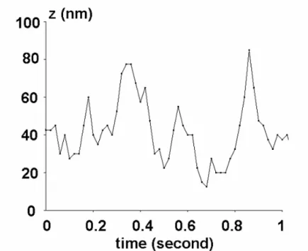
Figure 2 : Typical fluctuations of the distance between small microspheres and the chamber surface. The
curves shows a typical set of distances between the chamber floor and a microsphere of 2.8 µm diameter as used in many experiences. Data were obtained by computer simulation47. The range of distances spanned by the particle is of comparable magnitude as the length of many ligand-receptor couples.
As a rule of thumb, the velocity of a freely flowing sphere close to the chamber floor (in µm/s) is comparable to the wall shear rate G (in s-1) times the sphere radius. Note that this velocity is sometimes called the hydrodynamic velocity. Thus, a wall shear rate of a few s-1 should be sufficient to make detectable a binding event lasting several tens of milliseconds. Note however that brownian motion may reduce the performance of the system. It is noteworthy that the properties of the motion of a sphere near a plane in a laminar shear flow have long been determined and tabulated35. An important result is that the sphere displays both translation and rotation. The relative velocity between the plane and the closest region on the sphere is on the order of 47 % of the translation velocity. Thus, the contact duration between a ligand and a receptor molecule of total length 40 nm is expected to be on the order of 10 ms when the wall shear rate G is 10 s-1.
When a sphere of 1.4 µm radius is maintained at rest by a single bond with a typical length of a few tens of nanometers, the force on the bond (in pN) is about 0.5 G. Note that the force is only weakly dependent on the bond length30.
It may thus be concluded that the laminar flow chamber operated a low shear rate is well suited to monitor the formation and dissociation of single bonds provided that their lifetime is higher than 0.1 s.
A particular feature of small microspheres is that they display substantial brownian motion. As shown on Fig. 2 , a vertical motion of about 100 nm amplitude is well suited to explore the binding
range of typical ligand-receptor couples. Indeed, the total length of LFA-1/ICAM-1 and
P-selectin/PSGL-1 couples that are involved in leukocyte-endothelium interaction is about 40 nm and 80 nm respectively. Also, the CD2/CD58 couple involved in T-lymphocyte interaction with many cells is about 16 nm long.
When cells are used rather than microspheres, several differences are expected. Brownian motion is less important. The force experienced by a bond maintaining a cell at rest is more difficult to estimate since is depends on cell nanotopography and rheological properties. An extreme case may be the formation of a membrane tether of several tens of micrometers length linking a flowing cell to an anchoring point36. Also, cell position is more difficult to determine accurately (due to the fairly irregular shape) and the position of cell image centroid is necessarily superimposed on the position of cell-to-substratum contact.
3 - Data processing.
3.1 - Quality check.
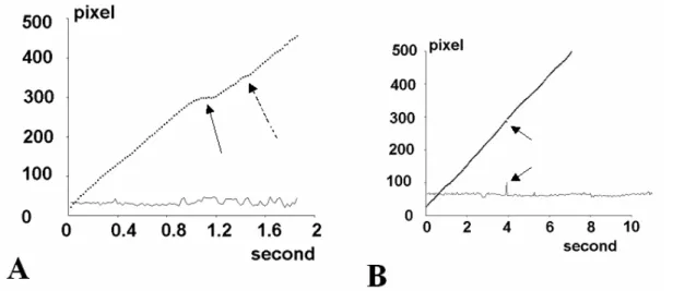
A single observation of particle flow through a microscope field during a typical period of time of 10 minutes may yield several hundreds of trajectories with hundreds of particle positions each, amounting to several tens of thousands of positions. A representative example is shown on Fig. 3. Thus, a careful quality check is required to eliminate artefactual binding events or undesirable trajectories. The following two points may be considered :
- A typical problem is the possibility of a collision between a flowing particle and a bound one, resulting in false binding event. This may be detected by checking that the area of tracked object falls within preselected limits.
Figure 3 : Difficulty of delimiting binding events. Two representative displacement curves are shown. In each
case, the set of particle positions is shown (dots) together with the variations of particle area as calculated by the tracking system. A a short (full arrow) and an ultrashort (broken arrow) arrests are shown. This emphasizes the need for a careful choice of threshold parameters used to define arrests. B An artefactual arrest detected by the transient area increase. This is probably due to the passage of a rapid particle.
- Erroneously low binding frequencies can be found when incompletely sedimented particles are monitored. Sedimentation time may not be a problem when the flow rate is low enough to allow particles to reach the bottom between their entry into the chamber and passage through the microscope observation field. When this condition is not fulfilled, it may be useful to eliminate particles with a velocity higher than a predetermined threshold based on the wall shear rate. When cells or large particles are studied, it may be a good idea to divide the chamber floor into a ligand-coated and a
ligand-free region. Cells may thus be allowed to sediment on the nonadhesive part of the chamber floor before starting the flow37.
3.2 - Arrest definition.
When high shear rates are used, there is no difficulty in delimiting binding events. This may explain why arrest definition was not considered as an important issues when selectin-mediated attachment of fast particles was studied, which is probably the model that was first studied most often with flow chambers, in order to achieve a molecular interpretation of the seminal experiments reported by Lawrence and Springer13. However, when flow chambers are operated under low shear rate to monitor weak bonds of short duration, the definition of binding events may not be straightforward. As shown on Fig. 4, a particle may be defined as arrested when it moves by less than a threshold distance ξ during a time interval of duration τ. It is important to know that the minimum duration of detectable binding events is31 :
dm = τ - ξ/u [2]
(where u is the velocity of the freely moving particle). It is also seen that the true duration of a binding event is different from the apparent duration defined as the period of time during which the particle is considered as arrested :
dt = da – 2 ξ/u [3]
It is obviously essential to correct apparent values when the effect of the shear rate on binding frequency and duration is studied (see e.g. 38 for an illustration of the importance of this correction).
Figure 4 : Calculated arrest duration is dependent on arrest definition. The thick lines represent binding
events of equal length separating two periods of more rapid (A) or slower (B) displacement. The broken lines show the periods of time where particles are defined as arrested, using threshold parameters ξ and τ as defined. Clearly, a correction is required to obtain "true" arrest duration.
3.3 - Unbinding plots.
The primary output of data processing is a record of all binding events together with their duration. This allows straightforward derivation of unbinding plots (Fig. 1C) displaying the logarithm of the proportion of binding events lasting at last time t as a function of t. If binding events are mediated by
single bonds with monophasic detachment kinetics, unbinding curves appear as straight lines, the
slope of which is equal to the dissociation rate koff. However, non linearity is a frequent occurrence as
i) Additional bond formation may occur after initial particle arrest, resulting in progressive strengthening of attachment3,38. A practical way of ruling out this possibility consists of using sufficiently low surface densities of attachment receptors on surfaces.
ii) A single bond may display multiphasic behaviour with a time-dependent strengthening due to the passage of sequential barriers on the energy landscape. This phenomenon was reported very early30 and may be responsible for the so-called history-dependence of molecular bonds 40,41.
iii) Particle-to-surface attachment may be mediated by several bond species with different dissociation rates. Note the intriguing possibility that a given molecular pair may form different bond types, as was cleverly demonstrated with the surface forces apparatus42. Note also that it may be difficult to discriminate between (ii) and (iii) on the sole basis of unbinding curves.
iv) Finally, if the initial bond number between particles and surfaces is higher than one, a time-dependent increase of detachment rate resulting in downward concavity of unbinding curves is expected. This situation was not frequently reported.
3.4 - Binding frequency.
While most reports that appeared to date deal with bond rupture, the flow chamber may provide valuable information on bond formation29. Indeed, the efficiency of cell adhesion receptors often relies on their capacity to form bonds during a short period of time 12. However, it must be emphasized that single bond formation proved more difficult to study than bond dissociation for at least two reasons : i) Bond dissociation may in principle be studied with a conceptually simple experiment consisting of allowing surface-bound molecules to bind for a sufficient period of time, then subjecting them to a disruptive force that may be either constant3,27 or steadily increasing 4,6 and recording the rupture time as easily evidenced by a detectable separation of bound surfaces. Studying bond formation requires to generate multiple intermolecular contacts and perform multiple checks to determine the proportion of contacts conducive to attachment.
ii) Bond rupture is less dependent on molecular environment than bond formation. Indeed, when a molecular bond is subjected to a disruptive force, the force exerted on the bond is fairly well controlled provided binding molecules are flexible enough to allow for a single relative orientation of the force and the bond. This condition may be assumed to be fulfilled since bond formation between rigid surface-attached structures is quite difficult. Therefore, the bonds that are usually monitored involve molecules with suitable conformation. However, determining the intrinsic rate of bond formation between surface-attached molecules would require a nanometer-scale knowledge of the topography and conformation of molecules brought into molecular contact as well as the distribution of molecules with a potential to interfere with molecular contacts such as sterically repulsive structures43,44. In other words, an exact knowledge of the conditions of bond formation is usually out of reach and severe approximations are required to derive intrinsic molecular association rates from binding frequencies measured on surface-attached molecules.
Laminar flow chambers are well suited to study receptors with moderate binding efficiency or low surface density since flowing particles can scan an extensive contact area. It seems fairly easy to determine the average frequency of particle or cell arrest per unit length of trajectory or per unit time of observation. Some points of caution are warranted concerning the interpretation of results.
i)There are two limiting cases allowing simple interpretation of results45. If binding efficiency is high (usually when the flow is very slow), the binding frequency represents the number of encounters between active molecules, i.e. adhesion molecules with a conformation compatible with bond formation. Thus, what is measured is a geometrical rather than a kinetic parameter. In this case, the binding frequency per unit length should be weakly affected by limited variations of the flow. Conversely, if binding is a rather inefficient process and many molecular encounters occur before bond formation, binding frequency will depend on the total encounter time between active molecules. If binding probability is proportional to the encounter time (which is the basis of on-rate definition, although it may require some discussion) binding frequency per unit time should be weakly affected by limited variations of the flow. As recently reported, both limiting cases may be observed with a given experimental device, depending on the assayed ligand-receptor couple45.
ii) Clearly, substantial information can be drawn from the experimental dependence of binding frequencies on the flow rate. However, it must be emphasized that binding frequencies are highly dependent on the definition of binding events, i.e. on the choice of arbitrary threshold parameters τ and ξ defined above as shown by Eq. [2]. Indeed, if bond rupture follows monophasic kinetics with rate constant koff, only a proportion exp(-koffdm) will be recorded if only binding events lasting more
than dm are detected. However, this proportion is dependent on the flow rate in a complex way since
koff is usually increased (according to Bell's law) whereas dm may be decreased (according to Eq. 2)
when the wall shear rate is increased. Thus, any detailed analysis of binding frequencies requires a suitable correction to make arrest detection independent of the shear rate38.
3.5 - Significance of binding frequency and connection with molecular parameters.
The use of brownian particles (Fig. 2) makes it possible to sample a range of distances between receptors and ligands, which should in principle yield information on the relationship between bond molecular separation and binding probability29,46. The problem remains to achieve an accurate determination of the distribution of particle-to-surface distances as well as binding frequency. While limited information could be derived from the particle velocity measurement through hydrodynamic laws47, it would be desirable to achieve simultaneously particle height measurement and distance detection. Simple optical techniques such as RICM/IRM should in principle yield this kind of information, but real-time determination of particle-to-surface distance remains currently challenging48.
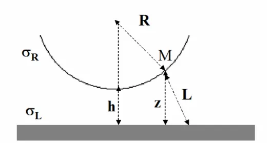
Figure 5 : Counting the number of ligand-receptor couples interacting in the flow chamber. Defining as L
the range of the interaction, a point M of the microsphere surface can interact with ligand molecules located on a disk of area π (L2 - z 2) on the chamber floor.
While an extensive discussion of the connection between binding frequencies measured with flow chambers and 2-dimensional or 3-dimensional association constants would not fall into the scope of the present review, a brief qualitative discussion may be useful to delineate the current challenge. For this purpose, we shall assume that the association frequency of surface-bound receptors and ligands whose anchoring points are separated by a distance d is a constant konMol if d is lower than a threshold
L (corresponding to the binding range) and 0 otherwise. Thus, the attachment frequency of a receptor-coated sphere separated from a ligand-receptor-coated plane by a distance h (Fig. 5) is simply :
Rdz z R k h F R L h L onMol (
πσ
[ ])2πσ
) ( =∫
2 − 2 [4]where σL and σR are respectively the surface densities of receptor and ligand molecules on interacting
efficiency (i.e. reaction-limited) regime. Now, the binding frequency per unit of time is easily obtained by averaging F(h) with Boltzmann's law. Applying [4] to microspheres of 2.8 µm radius used in our laboratory and expressing surface densities as numbers of molecules per square micrometer, we find :
F = <F(h)> = 0.32 konMolσR σL [5]
where the association rate is in s-1 and surface densities in molecule/µm2. Now, an important challenge is to relate 2-dimensional and 3-dimensional association rates. It must be understood that this attempt may be meaningful only if both 2D and 3D reactions are reaction-limited, not diffusion-limited. If this assumption is fulfilled, we may suggest an approximate way of relating the conventional 3-dimensional association rate we shall denote as Kon3D to a molecular association frequency we shall
denote as konMol. Assuming that binding may occur with frequency konMol when molecules are separated
by a distance lower than L, the binding frequency of a given receptor molecule is simply konMol times
the probability of finding a ligand in the sphere of radius L surrounding the receptor, i.e. about 4/3πL3 [L], where [L] is the (3-dimensional) ligand concentration. Thus, the relationship between konMol and
Kon3D is simply :
Kon3D = 4/3πL 3
konMol [6]
Since Kon3Dis usually expressed in Mole -1
s-1 rather than µm3molecule-1s-1, Eq. [6] reads : Kon3D = 2.5 10
9
L3 konMol [7]
Where L is expressed in µm and Kon3D in Mole -1
s-1. Combining Eqs [5] and [7] should in principle allow us to relate 3-D association rates to experimental data obtained with the flow chamber. According to our experience, 3D constants derived with Eqs 5 and 7 are significantly lower than estimates obtained of free molecules with surface plasmon resonance. Thus, in a study of cadherin moieties, L and konMol were estimated at about 0.01 µm and 1.2 10
-3
s-1 respectively leading to an estimated value of 25 M-1s-1 for Kon3D, which is about one thousandfold lower than expected
29,46
. There are at least three possible reasons for this discrepancy : i) only a small proportion of surface-bound receptors and ligands might have a conformation compatible with binding, ii) the mobility of surface-bound receptors and ligands might be low enough to result in a decrease of konMol as compared to
soluble molecules, and iii) the definition of bound states is different in 2D and 3D experiments, which may lead to measurable consequences due to the complexity of energy landscapes. Thus, more work is certainly required to clarify the significance of 2D and 3D association rates as well as molecular binding frequencies.
In contrast with model particles, the monitoring of cell-surface adhesion under flow yields quantitative information with high physiological relevance3,13,27, but translating binding frequencies into molecular parameters relies on many assumptions relative to cell surface topography and molecular structure.
4 - Interpretative issues
While the principle of the flow chamber and significance of yielded information might seem quite straightforward, it is useful to emphasize some difficulties that may strongly affect data interpretation (see also9).
4.1 - Are single bonds actually detected ?
An essential point to achieve molecular interpretation of experimental data is to demonstrate that most binding events studied are actually mediated by single bonds. Indeed, the force dependence of multivalent attachments is heavily influenced by the mode of force sharing between bonds and the possibility of rebinding49. Also, if only multivalent attachments are capable of generating detectable binding events, binding frequency may be more representative of molecular clustering than bona fide association rate, which might explain discrepancies between force parameters obtained on L-selectins
with flow chambers27 and biomembrane force probes50. Unfortunately, as explained below, there is no single definitive way of ensuring that single bonds are actually monitored.
A useful check may consist of performing sequential dilutions of receptors and ligands, which should result in proportional decrease of binding frequency without any alteration of unbinding curves if single bonds are actually studied. However, similar conclusions could be attained if a proportion of binding molecules clustered and only aggregates could generate detectable binding events.
Note that other techniques, such as atomic force microscopy or biomembrane force probe, are subject to the same difficulty. The usual argument that a low proportion of cycles being conducive to bond formation should ensure that most binding events be mediated by single bonds is not fully rigorous, since this argument does not rule out the possibility that only multivalent attachment might be detected.
A strong argument would consist of checking that binding event frequency and rupture behaviour monitored in a given set of experiments are indeed similar to single bond-mediated events. However, this requires us to assume that single bond have actually been formally identified by at least some investigators.
4.2 - Can a bound state be rigorously defined ?
Most experiments done on molecular association are designed and performed with the underlying assumption that it is in principle possible to define a “free” and a “bound” state of a ligand-receptor couple. This seemed quite reasonable and successful until experimental dissection of energy landscapes led to results that were viewed as paradoxical40. Thus, some discussion is warranted.
A ligand-receptor couple may be described as a point in a multidimensional space (thus, a static description of a couple of rigid and asymmetric molecules will require a total of N=12 parameters). The energy landscape may be viewed as an hypersurface in a space of (N+1) dimension, representing the variations of the complex free energy as a function of its N-dimensional state. The bound state may thus be defined as a region of the N-dimensional space. In order to simplify the discussion, we shall assume that a single coordinate is sufficient to describe the complex formation and dissociation along a one-dimensional reaction path. This simple view, first used by Eyring51, was often deemed sufficient although is was recently questioned52.
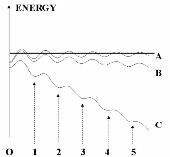
Figure 6 : Difficulty of defining binding states. A unidimensional energy landscape is shown, revealing a
series of binding states of decreasing depth (numbered 0, 1 ...5), as illustrated by curve A. The "overall" bound state must be defined somewhat arbitrarily. Applying a growing disruptive forces (curves B and C) will flatten the energy barriers, and the weakest complexes will disappear (see e.g. 5 on C).
A simplified one-dimensional energy landscape is shown on Fig. 6. Since it is now well established that ligand-receptor association may involves many different states, there is some arbitrariness in defining the bound state. We must be aware that this raises several problems.
i) if internal barriers are low enough to allow transition between different bound states within a period of time much shorter than - say - a millisecond, all complexes that will be studied experimentally will be similar. However, if transition requires minutes or more, the complex state may depend on its mode of preparation or, in short, on its history40,41.
ii) the bound state may be "reasonably" defined by deciding that complex dissociation will require a detectable time (say a millisecond) when subjected to a "reasonable" force (say less than several tens of piconewtons). This may exclude "ultraweak" associations that may influence experimental data38. Worse, as shown on Fig. 6, since the energy landscape is modified by energy forces, threshold distances that fitted energy barriers in absence of force may become meaningless in presence of force. iii) Finally, since the representation of energy landscapes as a sequence of well-defined barriers and basins is only an approximation53,54, the boundaries of defined states may not be as clearcut as might be required to measure accurate kinetic constants.
5 - Technical points
While technical details were more fully described in a number of primary reports, it may be useful to present a brief description of experimental problems that must be considered when planning experiments with the flow chamber.
5.1 - Coupling molecules to surfaces
The success of experiments is highly dependent on the preparation of receptor coated surfaces.
It is often most convenient to use glass coverslips as removable floors for flow chambers. Indeed, this provides optimal optical conditions for microscopic observation. Also, proteins may be easily coupled to glass. We often used polylysine adsorption followed by glutaraldehyde activation, covalent coupling of a first protein layer and inactivation of unreacted aldehyde groups with glycine or ethanolamine28. In some cases, coating glass with an aminosilane layer proved an efficient way of reducing nonspecific interactions1,55. Glass coverslips were also used as substrates for cell monolayers3.
Polystyrene surfaces were also used successfully after coating with adsorbed proteins such as
fibronectin56.
Finally, we also used freshly cleaved mica that is often used as a smooth surface as the subnanometer scale57. In addition to its capacity to adsorb proteins, this may be treated with nickel chloride in order to bind genetically engineered proteins bearing an hexahistidine tag 29,31. The advantage of this technique is that it allows accurate control of the orientation of surface-bound proteins.
It is well known that the capacity of surface-bound receptors to bind ligands is usually heavily dependent on the presence between the surface and the receptor site of a spacer with sufficient length and flexibility. A very convenient way of achieving this goal was to use several layers of antibodies, which may also select suitable orientation. As an example, chemical coupling of an anti-mouse immunoglobulin antibody, followed by addition of a suitable murine monoclonal antibody and then tested receptor will provide a flexible linker of more than 40 nm length, allowing very efficient interaction with free or surface-bound molecules29.
Note in this respect that the use of multiple layers of reagents is often rewarding. Thus, a molecule X of interest may be coupled to a surface with a sequence of protein A followed by a suitable anti-X monoclonal antibody. Alternatively, adsorbed biotin (e.g. used as biotinylated albumin) may be used to bind avidin or streptavidin, which will bind to biotinylated X. This sandwich technique usually works fairly well, possibly because the four binding sites of the streptavidin molecules are exposed on opposed sides. The interest of these multiple layer combinations is threefold : firstly, they may allow for proper orientation of the binding molecule. Secondly, they reduce the quantitative need for rare products, since the first adsorption step is usually the most expensive in terms of needed amount of reagents. Thirdly, the last step is ideally suited to prepare serial dilution of active molecules.
5.2 - Surface density
Quantitative interpretation of experimental data usually requires at least approximate determination of the surface density of ligand and receptor molecules. While radioactive labelling remains the most
sensitive technique, it is often difficult to use. Fluorescence labelling is easily amenable to simple calibration procedures31, but it is difficult to detect surface densities lower than several tens of molecules per squared micrometer. New fluorescent probes such as Alexa or quantum dots may at least partly raise this limitation. Finally, some authors were happy with enzymatic58 or luminescence-based59 immunoassays.
An important point that must be borne in mind is that the density of "active" sites may be substantially lower than the total density of molecules. Thus, it is probably a good idea to assay site density with labelled ligands or specific antibodies rather than labelling receptor or ligand molecules before coupling to surfaces.
5.3 - Passivation
As will be emphasized below, the performance of the flow chamber (and probably many other methods used for studying molecular interactions) is essentially set by the ratio between specific binding events and non specific particle arrests. Therefore, an important part of an experimental scheme may consist of determining optimal ways of minimizing nonspecific interactions. The most common way of achieving this goal may consist of coating test surfaces with a "non specific" blocker such as bovine albumin or casein. Another possibility that is commonly used by living cells consists of coating surfaces with repeller molecules such as polyethyleneglycol or polysaccharides mimicking pericellular matrices48. However, it must be borne in mind that the molecular environment of receptors may alter rate constants, particularly kinetic association parameters.
5.4 - Cells and Particles
While flow chambers were successfully used to study the attachment of cells and artificial particles, it must be emphasized that both models yielded different kinds of information.
Flow chambers were first applied to cellular models to obtain physiologically relevant information. Indeed, it seemed obvious that the conventional way of centrifuging cell mixtures or even to deposit cell supensions on suitably adhesive substrates or cell monolayers might not suitably represent situations met by cells in their natural environment. However, the molecular interpretation of experimental data is hampered by several problems : i) the cell surface exposes a variety of molecules with a potential adhesive or anti-adhesive role in many situations, ii) the distribution and density of membrane adhesion receptors are difficult to manipulate freely, which may makes it difficult to ensure that single bonds are actually observed. iii) the topography and molecular properties of cell surfaces make it difficult to control and event to estimate accurately the kinetics and mechanics of interactions between cell surface molecules and substrates. iv) due to the irregularity of cell shape, it is often difficult to determine the duration of cell-substrate molecular contact with high accuracy.
In view of aforementioned difficulties, it is not surprising that model systems were often used to study biomolecule interactions, and comparison between these and model particles and cellular systems revealed similarities60 or differences45 depending on tested receptors and parameters. The interest of studying the interaction between microspheres and artificial surfaces is that the hydrodynamics of interaction can be determined with high accuracy based on standard results from fluid mechanics, surface structure is relatively well controlled (although many suppliers may used incompletely documented procedures to avoid non specific interactions). Also, it is possible to control the surface density of receptor molecules, which is a key requirement to study single molecule interactions.
The size of microspheres may deserve some discussion. Maximum sensitivity may be obtained when the ratio between the velocity of freely flowing particles and the force exerted on a bond maintaining a particle at rest is maximum. This ratio approximately scales as R-1.5, where R is the particle radius30, thus supporting the use of small particles. However, using small particles also involves some limitations : firstly, the accuracy of particle localization is dependent on the particle size. Secondly, brownian motion strongly alters the significance of experiments performed with smaller particles47. In our laboratory, microspheres of 2.8 µm radius proved well suited to study with reasonable sensitivity transient interactions generated between ligands and receptors. Recent studies performed with larger particles48 allowed better optical monitoring of particle position. Indeed, combining reflection interference contrast microscopy with the flow chamber allowed simultaneous observation of particle vertical and horizontal motion. Also, adhesive interactions were more efficient
than expected. Thus, it is possible that complementary information might be obtained by using a range of particle sizes to study a given ligand-receptor couple.
5.5 - Image processing
While valuable information can be drawn from flow chamber experiment with a mere stopwatch, only a computer-based tracking system can allow satisfactory use of all provided pieces of information.
Early experiments were done with standard3,27 or fast61 videocameras. Videorecorders were used to archive images on videotapes for delayed analysis. Images were digitized and particle position was determined with a custom made27 or commercial tracking software. In our laboratory, cell or microsphere position was readily determined with a custom-made program : particle contour determination was based on intensity contrast, that was obtained by properly focusing the microscope. The centroid of the particle image was then determined with an accuracy that may be estimated as the pixel size divided by the square root of the image area (in squared pixel). Thus, we routinely determined the position of 2.8 µm diameter microspheres with about 50 nm accuracy. Assuming the substratum was sufficiently smooth, the sphere-to-particle contact might be considered as superimposed on the image center. As emphasized above, it is essential to use control procedures to rule out artefactual binding events due to particle collision or change of image contrast. Monitoring particle area was felt satisfactory for this purpose.
While fast cameras have been more and more often used to analyze selectin-mediated interactions in presence of a flow rate comparable to that found in blood vessels, other membrane receptors can form adhesion only under low shear rate. Under these conditions, general purpose cameras and videotape recorders proved fully satisfactory. However, recent generalization of high efficiency data compression (with codecs such as DivX) in addition to the replacement of conventional videocameras with CCD videocameras resulted in a progressive replacement of videotapes with numeric storage devices such as hard disks or DVDs. Basic processing software is now included in standard operating systems (e.g. DirectX as a standard Windows component). While this technology makes available highly cost effective image processing equipment, it must be borne in mind that present-day image compression algorithms, efficient as they may be, result in partial information loss and less accurate control of data processing that previous generation equipment.
6 - Limitations of the method
It would be useful and appealing to indicate clear limitations of the resolutive power of the laminar flow chamber, such as the minimum duration of detectable interactions. Also, it is certainly important to identify the limiting parameters, i.e. the parameters that should be improved in order to increase the power of the method. Although a detailed discussion of all relevant parameters would not fall into the scope of this brief review, three important points will be discussed below.
6.1 - Non specific versus specific arrests
Quantitative analyses of “specific” interactions mediated by well-defined ligand-receptor couples are plagued by the inescapable occurrence of so-called nonspecific interactions involving a variety of ill-defined molecular structures on interacting surfaces. A frequent problem is that there is no absolute way of discriminating between specific and nonspecific interactions62. Indeed, these interactions involve similar physical forces and may not exhibit strikingly different orders of magnitude. As a consequence, it is difficult to discriminate between specific and nonspecific interactions on the basis of quantitative parameters such as lifetime or force constant. Worse, it is difficult to exclude the possibility that a specific interaction be strengthened by additional nonspecific forces, or that a nonspecific binding event be reinforced by subsequent specific bonds. Thus, the significance of an experimental study is strongly dependent on the preparation of surfaces minimizing the frequency of nonspecific events, in order that the ratio between specific and nonspecific events be as high as possible.
Obviously, surface quality must be checked, and it is important to chose adequate controls : thus, it is certainly incorrect to use as a control for a receptor-coated surface (say, glass) the same uncoated surface since nonspecific interactions are expected to be quite different. Thus, control
surfaces must closely mimic the systems that are investigated. For example, an antibody may be replaced with a control antibody of similar class and different specificity, or an active molecule may be replaced with a mutant differing from the wild type species by a few aminoacids63. Also, binding molecules such as streptavidin might be blocked by small ligand molecules (i.e. biotin) that are supposed to modify binding sites with a minimal effect on surrounding surfaces31.
As a consequence, the sensitivity of the method is often set by the efficiency of the binding system that is studied : indeed, if bond formation occurs every 100 moving step of a particle, nonspecific events occurring with 1/1,000 frequency should not invalidate the analysis, while they would severely impair attempts at quantifying specific interactions occurring every 10,000 step.
6.2 - Brownian motion
As previously indicated, small size particles often proved most suitable to detect transient and weak molecular interactions at the single bond level. It is important to note that in this case the limits of the flow chamber analysis are set by brownian motion rather that the accuracy of determination of particle position or time resolution47. Indeed, the random brownian displacement of a particle during a time interval of duration t scales as t1/2, while the flow-induced displacement scales as t. Thus, brief displacements can be analyzed only when the flow rate is increased, which increases the force on the bonds and usually decreases bond lifetime. Note that this limitation is less important when binding can occur in presence of high flow rates, as exemplified by selectin-mediated interactions. Thus, different setups may be advantageous when flow chambers are used to quantify the binding properties or receptors with highly different binding parameters such as e.g. selectins and cadherins. This situation is illustrated on Fig. 7.
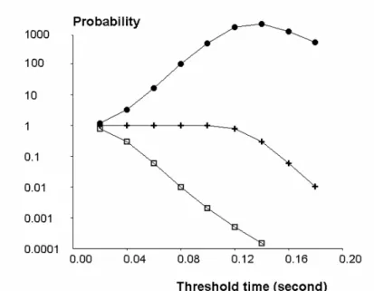
Figure 7 : Performance of the flow chamber. Considering a model system made of microspheres exposed to a
wall shear rate of 10 s-1 and forming adhesive bonds of 100 ms duration, we estimated the probability of detecting a binding event (crosses) and the probability of counting an artefactual arrest (squares) during a monitoring step of 20 ms. The ratio between detection probability and artefact probability is also shown (full circles). As expected, the probability of detecting a short arrest fell when the threshold time was too high (crosses), while the probability of observing an artefactual arrest was high when the threshold time was too low (squares). Thus, there is an optimal threshold time (dots) that depends on the duration of arrests that are deemed significant.
6.3 - Surface topography
As already mentioned, an accurate knowledge of the topography of interacting surfaces and molecules is required to translate experimental results into intrinsic molecular parameters. While this remark is relevant to both association and dissociation rate determination, association rates are particularly sensitive to the structure of interacting surfaces. Here are some possible mechanisms.
- It is a general finding that molecular clustering may strongly affect binding efficiency. Thus, if a significant fraction of surface-bound molecules are clustered, it is conceivable the that majority of binding events might involve multiple bonds. The only way to formally rule out this possibility would be to check that the number of binding events is significantly higher than the number of encounters between clusters of ligands and receptors. Only high resolution methods such as atomic force microscopy might provide sufficient information to consider this possibility quantitatively.
- The disruptive force experienced by a molecular bond maintaining a particle at rest in presence of a hydrodynamic force is obviously dependent on the geometrical (and rheological) properties of interacting surfaces.
- The frequency of molecular encounters between ligand and receptors is also highly dependent on nanometer-scale topographical features. This phenomenon was cleverly demonstrated with cellular models by demonstrating that adhesive interaction under flow may require that cell membrane receptors be located on the tip of microvilli64. Also, bulky molecules may prevent contact between smaller receptors and ligands43,44.
- Also, the orientation of rigidly bound surface molecules is an important parameter of binding efficiency. Clearly, a receptor with binding site turned towards the surface where it is bound would be quite inefficient in a flow chamber. Thus, deriving molecular association rates from binding frequencies requires at least approximate determination of the fraction of exposed sites. This is not a trivial problem since a surface-bound site may actively bind soluble ligand and remain unable to interact with surface-bound structures.
These difficulties were an incentive to try and prepare fully adhesive surface by coupling well oriented receptors to mica surfaces known to be smooth at the subnanometer level29,31,63. More work is required to present the effect of surface irregularities, as detected with methods such as atomic force microscopy, on binding parameters obtained with flow chambers.
Conclusion
Currently available systems allows us in principle to detect binding events lasting but a few milliseconds in presence of piconewton forces. This is probably sufficient to reveal most biologically relevant interactions. However, this limit is rarely obtained in view of a variety of technical difficulties hampering complete control of surface topography, ligand and receptor distribution and environment, and nonspecific interactions.
7 - Comparison with other techniques
7.1 - Particular features of the flow chamber.
As compared with other tools such as atomic force microscope, biomembrane force probe or optical trap, the laminar flow chamber displays some specific properties :
- Contact between ligands and receptors is usually quite short, on the order of a few milliseconds under low shear rate, as compared to standard contact times of order of 100 ms with other techniques. Interestingly, it was found that a short contact time might favor single-bond interactions65.
- The disrupting force generated by the flow chamber may be considered as constant, as compared to an adjustable loading rate available with atomic force microscopes and biomembrane force probes. While conceptually simpler to analyze, a constant force yields less information on energy landscapes. Further, varying the shear rate will alter at the same time the contact time and disruptive force, thus impairing data interpretation. Indeed, increased dissociation rate in presence of higher flow rate may be accounted for by force sensitivity of a given bound state, according to Bell's law, or different initial state when binding is multiphasic.
- The flow chambers allow cells or particles to scan an extended area, which allows to decrease the surface density of binding molecules and helps ruling out multivalent interactions.
- Finally, the flow chamber is well suited to detect transient binding events since bond formation can be detected within milliseconds. This is well suited to study the kinetics of bond formation, and also to study weak bonds.
7.2 - Comparison between different results.
Indeed, flow chambers were first used to study selectin-mediated bonds 3,27 as well as fairly weak interactions formed by the lymphocyte CD2 receptor28 or cadherins29. Strong interactions such as streptavidin-biotin association were analyzed only several years later with this technique31 and reported information concerned only transient complexes formed by molecules. Conversely, atomic force microscopy4 and biomembrane force probe6 were first used to study the remarkably strong streptavidin-biotin interaction, and the much weaker homotypic bonds formed by cadherin molecules were analyzed only later66,67. Bonds of intermediate strength formed by antigen and antibodies were successfully studied quite early with both flow chambers30 and atomic force microscopy68.
Thus, only recently results obtained on a similar ligand-receptor couple with flow chambers and AFM or BFP could be compared. We shall only mention two examples.
The interaction between L-selectin and specific ligands, which is thought to play an important role in tethering flowing leucocytes to endothelial walls during inflammation or lymphocyte circulation, was studied with flow chambers69 and biomembrane force probe50. The wall shear rate used with the former approach ranged between about 40 s-1 and 180 s-1. While the zero force dissociation rate was comparable (respectively 6.6 s-1 and 3 s-1), the force dependence was quite difference since a disruptive force of about 200 pN was found to increase the dissociation rate by a factor of less than 3 with the flow chamber, and more than 1,000 with the BFP method. A possible explanation50 would be that binding events observed in the flow chamber be mediated by a few bonds.
Interactions between recombinant cadherin moieties bound to microspheres were studied with the flow chamber operated at low shear rate, ranging between about 5 s-1 and 20 s-129,38,63, atomic force microscopy70, biomembrane force probe60,61. Interactions formed by the outer two domains of E-cadherin (EC12) revealed a binding state with a lifetime on the order of 1 second and a Bell force constant of a few piconewtons. However, the significance of this match was partially hampered by the demonstration of a multiplicity of binding states with fairly different lifetime and force constant.
The example of streptavidin-biotin interaction clearly illustrates the difficulty of comparing different methods. Streptavidin-biotin bond is very high with an affinity constant on the order of 1015Mole-1. The equilibrium binding state is stable enough to resist all hydrodynamic forces generated with a flow chamber when the shear rate is low enough to allow bond formation. Thus, stable bonds will result in permanent arrests. The flow chamber only allowed to study the rupture of transient binding states corresponding to the outer part of energy/distance plots31. Experiments done with atomic force microscopes or biomembrane force probes revealed inner barriers4,6. Thus, complementary results were obtained with different methods.
8 - Conclusion
Laminar flow chambers were first developed to mimic adhesion events occurring in flowing blood, in presence of a wall shear rate ranging between a few hundreds of s-1 and several thousands of s-1. Experimental data were of high biological relevance but they were arguably difficult to connect with intrinsic molecular interaction parameters. Using wall shear rates ranging between a few s-1 and a few tens of s-1 proved an efficient way of analyzing a part of the energy landscapes formed between most adhesion receptors and their ligands. This may bring specific information on biomolecule interactions, in combination with information brought by other devices such as atomic force microscopes68,71 , biomembrane force probes6,72 or optical traps73,74.
In addition to its intrinsic value un helping us to understand the role of different cell membrane receptors, the highly detailed information obtained at the single bond level should prove particularly useful to check the predictions of molecular dynamic simulations, that are made possible by the increasing availability of accurate structural information of molecular complexes as provided with X-ray diffraction. This should bring us nearer the ultimate goal of relating the structure and function of biomolecules75.
References
1 – Cretel E., A. Pierres, A. M. Benoliel, and P. Bongrand. How cells feel their environment : a focus on early dynamic events. Cell. Mol. Bioeng. 1:5-14, 2008.
2 - Helm C. A., W. Knoll, and J. N. Israelachvili. Measurement of ligand-receptor interactions. Proc. Natl. Acad. Sci. USA 88 : 8169-8173, 1991.
3 - Kaplanski G., C. Farnarier, O. Tissot, A. Pierres, A. M. Benoliel, M.C. Alessi, S. Kaplanski, and P. Bongrand. Granulocyte-endothelium initial adhesion. Analysis of transient binding events mediated by E-selectin in a laminar shear flow. Biophys. J., 64 : 1922-1933, 1993.
4 - Florin E. L., V. T. Moy, and H. E. Gaub. Adhesion forces between individual ligand-receptor pairs. Science 264 : 415-417, 1994.
5 - Evans E., R. Merkel, K. Ritchie, S. Tha, and A. Zilker. "Picoforce method to probe submicroscopic actions in biomembrane adhesion". In: Studying cell adhesion, edited by P. Bongrand, P. M. Claesson, and A. S. G. Curtis, New York : Springer Verlag. 1994, pp 125--139.
6 - Merkel R., P. Nassoy, A. Leung, K. Ritchie, and E. Evans. Energy landscapes of receptor-ligand bonds explored with dynamic force spectroscopy. Nature 397:50-53, 1999.
7 - Miyata H., R. Yasuda, and K. Kinosita Jr. Strength and lifetime of the bond between actin and skeletal muscle alpha-actinin studied with an optical trapping technique. Biochem. Biophys. Res. Com. 1290 : 83-88, 1996.
8 - Bongrand P. Ligand-receptor Interactions. Rep. Prog. Phys. 62 : 921-968, 1999.
9 - Zhu C., M. Long M, S. E. Chesla, and P. Bongrand. Measuring receptor/ligand interaction at the single-bond level : experimental and interpretative issues. Ann. Biomed. Engineering 30: 305-314, 2002.
10 - Robert P., A. M. Benoliel, A. Pierres, and P. Bongrand. What is the biological relevance of the specific bond properties revealed by single molecule studies ? J. Mol. Recognition. 20:432-447, 2007.
11 - Matsui K., J. J. Boniface, P. Steffner, P. A. Reay, and M. M. Davis. Kinetics of T-cell receptor binding to peptide/I-Ek complexes : correlation of the dissociation rate with T-cell responsiveness. Proc. Natl. Acad. Sci. USA 91:12862-12866, 1994.
12 - Williams A. F. Out of equilibrium. Nature 352 : 473-474, 1991.
13 - Lawrence M. B., and T. A. Springer. Leukocytes roll on a selectin at physiologic flow rates : distinction from and prerequisite for adhesion through integrins. Cell, 65 : 859-873, 1991.
14 - Foote J., and C. Milstein. Kinetic maturation of an immune response. Nature 352:530-532, 1991. 15 - Giannone G, B. J. Dubin-Thaler, H. G. Döbereiner, N. Kieffer, A. R. Bresnick, and M. P. Sheetz. Periodic lamellipodial contractions correlate with rearward actin waves. Cell 116:431-443, 2004.
16 - Pierres A., A. M. Benoliel, D. Touchard, and P. Bongrand. How cells tiptoe on adhesive surfaces before sticking. Biophys. J. 94:4114-4122, 2008.
17 - Harris A. K., P. Wild, and D. Stopak. Silicone rubber substrata : a new wrinkle in the study of cell locomotion. Science 208 : 177-179, 1980.
18 - Bell G. I. Models for the specific adhesion of cells to cells. Science 200 : 618-627, 1978.
19 - Dudko O. K., G. Hummer, and A. Szabo. Intrinsic rates and activation free energies from single-molecule pulling experiments. Phys. Rev. Letters 96: 108101, 2006.
20 - Curtis A. S. G. The measurement of cell adhesiveness by an absolute method. J. Embryol. Exp. Morph. 22:305-325, 1969.
21 - Mège J. L., C. Capo, A. M. Benoliel, and P. Bongrand. Determination of binding strength and kinetics of binding initiation. A model study made on the adhesive properties of P388D1 macrophage-like cells. Cell Biophys. 8 : 141-160, 1986.
22 - Weiss L. The measurement of Cell Adhesion. Exp. Cell Res. Suppl 8 : 141-153, 1961.
23 - Bongrand P., C. Capo, A. M. Benoliel, and R. Depieds. Evaluation of intercellular adhesion with a very simple technique. J. Immunological Methods 28 : 133-141, 1979.
24 - Capo C., F. Garrouste, A. M. Benoliel, P. Bongrand, A. Ryter, and G. I. Bell, Concanavalin A-mediated thymocyte agglutination : a model for a quantitative study of cell adhesion. J. Cell Sci. 26 : 21-48, 1982.
25 - Tha S. P., J. Shuster, and H. L. Goldsmith. Interaction forces between red cells agglutinated by antibody. IV Time and force dependence of break-up. Biophys. J. 50 : 1117-1126, 1986.
26 - Dustin M. L., S. K. Bromley, M. M. Davis, and C. Zhu. Identification of self through two-dimensional chemistry and synapses. Ann. Rev. Cell Dev. Biol. 17 : 133-157, 2001.
27 - Alon, R., D. A. Hammer, and T. A. Springer. Lifetime of P-selectin-carbohydrate bond and its response to tensile force in hydrodynamic flow. Nature, 374 : 539-542, 1995.
28 - Pierres A., A. M. Benoliel, P. Bongrand, and P. A. van der Merwe. Determination of the lifetime and force dependence of interactions of single bonds between surface-attached CD2 and CD48 adhesion molecules. Proc. Natl. Acad. Sci. USA 93 : 15114-15118, 1996.