HAL Id: hal-01657963
https://hal.archives-ouvertes.fr/hal-01657963
Submitted on 17 May 2018
HAL is a multi-disciplinary open access
archive for the deposit and dissemination of
sci-entific research documents, whether they are
pub-lished or not. The documents may come from
teaching and research institutions in France or
abroad, or from public or private research centers.
L’archive ouverte pluridisciplinaire HAL, est
destinée au dépôt et à la diffusion de documents
scientifiques de niveau recherche, publiés ou non,
émanant des établissements d’enseignement et de
recherche français ou étrangers, des laboratoires
publics ou privés.
female type 2 diabetic goto-kakizaki rat heart
Martine Desrois, Carole Lan, Jamileh Movassat, Monique Bernard
To cite this version:
Martine Desrois, Carole Lan, Jamileh Movassat, Monique Bernard. Reduced up-regulation of the nitric
oxide pathway and impaired endothelial and smooth muscle functions in the female type 2 diabetic
goto-kakizaki rat heart. Nutrition and Metabolism, BioMed Central, 2017, 14, pp.6.
�10.1186/s12986-016-0157-z�. �hal-01657963�
R E S E A R C H
Open Access
Reduced up-regulation of the nitric oxide
pathway and impaired endothelial and
smooth muscle functions in the female
type 2 diabetic goto-kakizaki rat heart
Martine Desrois
1,3*, Carole Lan
1, Jamileh Movassat
2and Monique Bernard
1Abstract
Background: Type 2 diabetes is associated with greater relative risk of cardiovascular diseases in women than in men, which is not well understood. Consequently, we have investigated if male and female displayed differences in cardiac function, energy metabolism, and endothelial function which could contribute to increased cardiovascular complications in type 2 diabetic female.
Methods: Male and female Control and type 2 diabetic Goto-Kakizaki (GK) isolated rat hearts were perfused during 28 min with a physiological buffer before freeze-clamping for biochemical assays. High energy phosphate compounds and intracellular pH were followed using31P magnetic resonance spectroscopy with simultaneous measurement of contractile function. Nitric oxide (NO) pathway and endothelium-dependent and independent vasodilatations were measured as indexes of endothelial function. Results were analyzed via two-way ANOVA,p < 0.05 was considered as statistically significant.
Results: Myocardial function was impaired in male and female diabetic versus Control groups (p < 0.05) without modification of energy metabolism. Coronary flow was decreased in both diabetic versus Control groups but to a higher extent in female GK versus male GK rat hearts (p < 0.05). NO production was up-regulated in diabetic groups but to a less extent in female GK rat hearts (p < 0.05). Endothelium-dependent and independent vasodilatations were impaired in female GK rat compared with male GK (p < 0.05) and female Control (p < 0.05) rat hearts.
Conclusions: We reported here an endothelial damage characterized by a reduced up-regulation of the NO pathway and impaired endothelial and smooth muscle functions, and coronary flow rates in the female GK rat hearts while energy metabolism was normal. Whether these results are related to the higher risk of cardiovascular complications among type 2 diabetic female needs to be further elicited in the future.
Keywords: Type 2 diabetic heart, Gender differences, Cardiac function, Energy metabolism, Endothelial function Background
Cardiovascular diseases (CVDs) are the major causes of morbidity and mortality in patients with diabetes mellitus. CVDs are long-term complications of type 2 dia-betes mellitus, with a two-fold increased risk of heart fail-ure and greater mortality after myocardial infarction [1].
Diabetic women have a greater relative risk for CVDs than diabetic men, and newly diagnosed diabetic women showed higher relative risk for cardiovascular death than diabetic men in the large DECODE study [2]. Women with type 2 diabetes may undergo even more adverse changes in coagulation, inflammation and vascular func-tion than men [3, 4]. Stronger associafunc-tions have been reported between insulin resistance/metabolic syndrome and inflammation and endothelial dysfunction in women than in men [5]. Interestingly, it is proposed that women have to undergo greater metabolic deterioration than men
* Correspondence:martine.desrois@univ-amu.fr
1
Aix-Marseille Université, CNRS, CRMBM, Marseille, France
3Centre de Résonance Magnétique Biologique et Médicale (CRMBM), UMR n°
7339, Aix-Marseille Université, CNRS, Faculté de Medecine, 27 Bd Jean Moulin, Marseille Cedex 05 13385, France
Full list of author information is available at the end of the article
© The Author(s). 2017 Open Access This article is distributed under the terms of the Creative Commons Attribution 4.0 International License (http://creativecommons.org/licenses/by/4.0/), which permits unrestricted use, distribution, and reproduction in any medium, provided you give appropriate credit to the original author(s) and the source, provide a link to the Creative Commons license, and indicate if changes were made. The Creative Commons Public Domain Dedication waiver (http://creativecommons.org/publicdomain/zero/1.0/) applies to the data made available in this article, unless otherwise stated.
to develop type 2 diabetes and as such many insulin resist-ance risk factors must change to a greater extent [6]. However, few studies have explored the sex differences in the emerging risk factor profile in individuals with or without type 2 diabetes. Consequently, we have investi-gated if male and female without ischemic insult already displayed differences in cardiac function, energy metabol-ism, and endothelial function which could contribute to increased cardiovascular complications in type 2 diabetic female. This study was performed in male and female type 2 Goto-Kakizaki (GK) rats.
Six independent genetic loci are responsible for the defects in glucose and insulin metabolism in the Goto-Kakizaki (GK) rat, a highly inbred strain derived from outbred, glucose-intolerant Wistar rats that spontan-eously develop type 2 diabetes within the first few weeks of age [7]. The GK is one of the best-characterized ani-mal models of spontaneous type 2 diabetes mellitus [8]. In this model, cardiac insulin resistance was associated with impaired insulin signaling pathway [9]. We have previously shown greater hypertrophy, lower insulin-stimulated glucose uptake rates and increased ischemic injury in the female compared with the male GK rat hearts [10]. In addition, modification of the NO pathway was involved in increased susceptibility of the type 2 dia-betic GK rat heart to ischemic injury [11]. Significantly, this model allows one to study the effect of diabetes on the heart without other complications such as obesity.
Here, the effect of gender on type 2 diabetic heart was assessed in male and female Control Wistar and GK iso-lated rat hearts perfused with a physiological buffer as described previously [11]. High-energy phosphates and intracellular pH were measured during the experimental time course by 31P magnetic resonance spectroscopy with simultaneous measurement of contractile function. Energy compounds and oxidative stress in cardiac tis-sues were evaluated by High Performance Liquid Chromatography (HPLC). Myocardial tissue content of creatine kinase and lactate dehydrogenase were also used as markers of cellular damage. Total nitrate concen-tration as well as expression of endothelial nitric oxide synthase, AKT and Phospho-Akt were deter-mined as markers of the NO pathway. Endothelium-dependent and inEndothelium-dependent vasodilatations were also measured in separate experiments in order to assess, respectively, endothelial and smooth muscle functions. A preliminary form of this study has been published as an abstract [12].
Methods
Animals
Age-matched Control Wistar (male,n = 19; female, n = 24) and Goto-Kakizaki rats (GK/Par subline [8]) (male,n = 22; female,n = 23) (7–8 mo, weight 265–512 g) were used in
the experiments. Animals were fed ad libitum with a com-mercial pelleted chow (diet 113, SAFE, Augy, France).
Heart perfusion and experimental protocol
Rats were anaesthetized by intraperitoneal injection of 35 mg/kg pentobarbital sodium. After removal of the heart, blood samples were taken from the chest cavity and immediately centrifuged, and the supernatant was kept on ice for determination of plasma glucose and free fatty acids (FFAs). Hearts were cannulated and perfused in the Langendorff mode at constant pressure as described previously [11]. The end-diastolic pressure was set to 10 mmHg for all groups at the beginning of perfusion. Left ventricular developed pressure and heart rate were monitored as previously reported [13]. The rate pressure product (RPP) (product of left ventricular developed pressure and heart rate) was used as an index of cardiac function [11]. Coronary flow was measured by time collection of the coronary venous effluent.
Experimental protocol for31P magnetic resonance spectroscopy and biochemical analyses
Control (male n = 10, female n = 14) and GK (male n = 13, female n = 12) hearts were perfused during 4 min with non-recirculating phosphate-free Krebs-Henseleit bicarbonate buffer which had the following composition (mM): NaCl (118), KCl (4.7), CaCl2(1.75), MgSO4(1.2),
ethylenediaminetetraacetate tetrasodium (0.5), NaHCO3
(25) and glucose (11). After stabilization, hearts were perfused for 28 min with a physiological recirculating Krebs-Henseleit buffer containing 0.4 mM palmitate, 3% albumin, 11 mM glucose, 3U/L insulin, 0.8 mM lactate and 0.2 mM pyruvate. The perfusion solutions were gassed with a mixture of 95% oxygen and 5% carbon dioxide to give a pH of 7.4 and the temperature was maintained at 37 °C.
31
P magnetic resonance spectroscopy on isolated perfused rat heart
Perfused rat hearts were placed in a 20-mm magnetic resonance sample tube and inserted into a 31P probe that was seated in the bore of a superconducting wide-bore (89-mm) 4.7 Tesla magnet (Oxford Instruments, Oxford, UK) interfaced with a Bruker-Nicolet WP-200 spectrometer (Bruker, Karlsruhe, Germany). 31P spectra were obtained by the accumulation of 328 free induction decay signals acquired during 4 min (flip angle, 45°; time resolution = 0.7 s; spectral width, 4500 Hz; 2048 data points). Prior to Fourier transformation, the free induc-tion decay was multiplied by an exponential funcinduc-tion which generated a 20-Hz line broadening. The appropri-ate conditions for acquiring 31P magnetic resonance spectra, the quantification of phosphorus metabolites and the determination of intracellular pH have been
detailed previously [11]. Quantification of the signal in-tegrals was carried out using an external reference con-taining an aqueous solution of 0.6 M phenylphosphonic acid. A series of eight 31P NMR spectra were recorded during the perfusion protocol.
Collection of data
Heart function and31P magnetic resonance spectra were simultaneously monitored during the perfusion protocol. Blood samples were collected immediately after excising the heart. For biochemical analyses, hearts were rapidly freeze-clamped with a Wollenberger clamp precooled in liquid nitrogen at the end of the experimental protocol and kept at−80 °C before analysis.
Biochemical analyses in plasma
Plasma glucose and free fatty acids (FFAs) were deter-mined as described previously [11].
Biochemical analyses in freeze-clamped hearts Creatine, phosphocreatine, adenine nucleotides and malondialdehyde (MDA)
Determination of creatine, phosphocreatine, adenine nu-cleotides and MDA was performed as described previ-ously [11]. MDA was used to evaluate lipid peroxidation as a measure of oxidative stress [14].
Lactate dehydrogenase (LDH) and creatine kinase (CK) activities and water content
LDH, CK and water content were measured as previ-ously described [15].
NO pathway
Total nitrate concentration as well as total protein expression of Akt, Phospho-Akt and endothelial NOS (eNOS) were measured to assess the NO pathway. Total nitrate concentration (NOx) NOx was deter-mined according to the method described by Cross et al. [16].
Protein expression of Akt, Phospho-Akt and eNOS A portion of cardiac tissue was homogenized as described by Ye et al. [17]. Protein samples (50 μg for Akt, Phospho-Akt and 90 μg for eNOS) were run with 4–20% Tris–HCl ready gel (Thermo Scientific) or 6% SDS-PAGE, respectively and transferred to pure nitro-cellulose membrane. The membranes were incubated overnight at 4 °C with primary antibodies against eNOS (1/1000, Becton Dickinson (DB) Transduction Laboratories, USA), Akt (1/1000, Cell Signaling Technology, Inc.), Phospho-Akt (Ser473)(1/1000, Cell Signaling Technology, Inc.) and β-actin (1/5000, Sigma) and secondarily with HRP-conjugated anti-mouse or anti-rabbit antibodies
(Santa Cruz Biotechnology, Inc.). The immunoblots were developed using ECLTM Western Blotting Detec-tion Reagent (AmershamTM). The protein signals were quantified using the MicroChemi 4.2 system (DNR Bio-Imaging Systems Ltd., Israel). The intensity of each protein signal was normalized to the corre-sponding β-actin stain signal. Data are expressed as ratios between the protein and the corresponding β-actin signal density.
Endothelium dependent and independent vasodilatations
In separate experiments, endothelium-dependent and independent vasodilatations were measured using 5-hydroxytryptamine (5-HT, 10−7M) and papaverine (5*10
−6M) to assess endothelial and smooth muscle functions
respectively, as previously described [15] in Control (malen = 9; female, n = 10) and GK (male, n = 9; female, n = 11) isolated perfused rat hearts with Krebs-Henseleit buffer. The 5-HT and papaverine hydrochloride were dissolved in the buffer to give the desired concentration and were obtained from Sigma Chemical Co. (St. Louis; Missouri). The coronary flow was recorded during the perfusion with the Krebs-Henseleit buffer and during the infusion of 5-HT or papaverine. The increase in cor-onary flow during drug infusion was calculated and expressed as a percentage of the basal value.
Expression of results and statistical analyses
Data are expressed as means ± SEM. Function and 31P magnetic resonance spectroscopy data are presented as absolute values. Significant differences between groups were determined using two-way ANOVA with repeated measures over time for the time-dependent variables (function and31P magnetic resonance spectroscopy data) followed by Bonferroni post-hoc test with Graphpad Prism software (Graphpad prism 5.0, La Jolla, CA). For biochemical data, the effects of time and group were an-alyzed with two-way ANOVA followed by Bonferroni post-hoc test. Unpaired Student’s t-test was used for other parameters. A p value of less than or equal to 0.05 was considered to indicate significant difference.
Results
Physiological parameters of male and female Control and GK rats
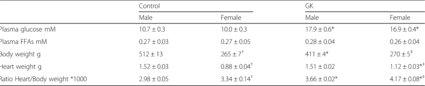
Plasma glucose was 67% and 69% higher in male and female GK rats versus their respective Controls (Table 1, p < 0.05). Plasma FFAs were similar in the four groups (Table 1). Heart to body weight ratio was 23% higher in male GK compared with male Control (Table 1, p < 0.0001) due to a significantly lower body weight of male GK compared with male Control (p < 0.05) and simi-lar heart wet weights in both groups. In female GK, heart to body weight ratio was 25% higher compared with
female Control with a similar body weight in both groups but a significantly higher heart weight in female GK versus female Control (p < 0.0001).
Gender effect on myocardial function
Myocardial function as represented by the rate pressure product (RPP), was significantly decreased in male and female diabetic animals compared with the respective Controls (p < 0.0001, Fig. 1a) due to a lower heart rate in
male and female GK rat hearts (p < 0.0001, Fig. 1b) vs Controls. End diastolic pressure (EDP, mmHg) was not different between groups (Control, male 10 ± 2, female 7 ± 2; GK male 8 ± 1, female 9 ± 2).
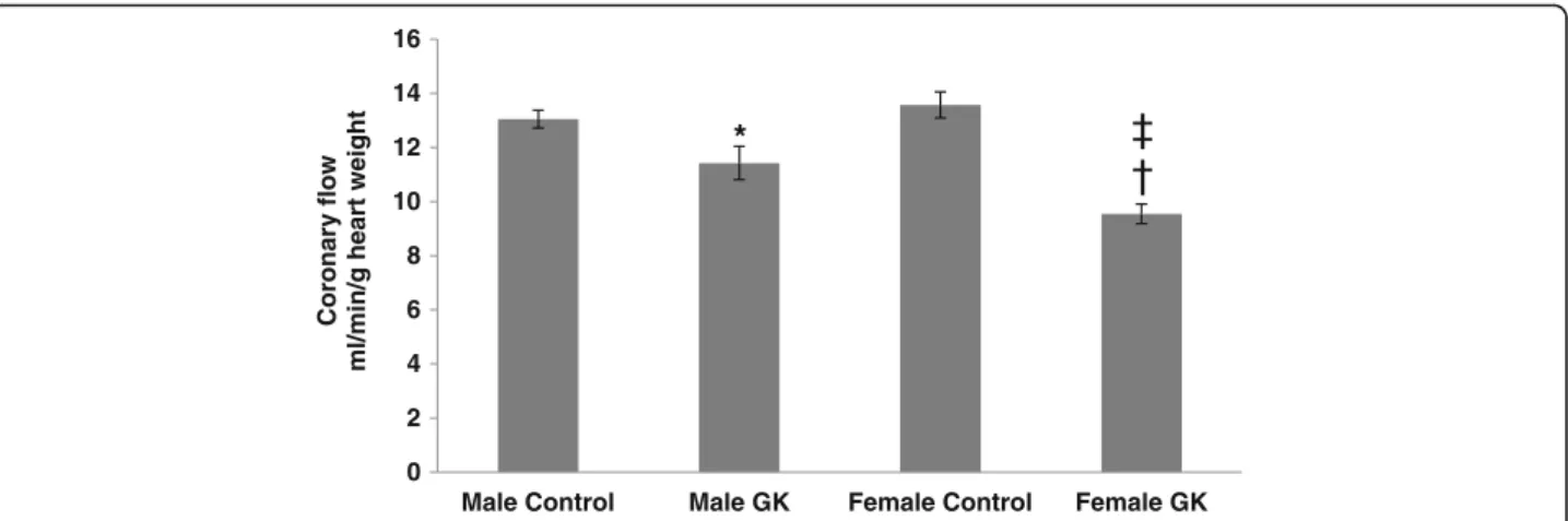
Coronary flow (CF) in Control and diabetic rat hearts was shown in Fig. 2. CF was decreased in male and female GK rat hearts compared with their respective Controls (p = 0.0420 and p < 0.0001 respectively). Inter-estingly, CF was significantly lower in female GK
Table 1 Physiological parameters in Control (malen = 10, female n = 14) and GK (male n = 13, female n = 12) rats
Control GK
Male Female Male Female Plasma glucose mM 10.7 ± 0.3 10.0 ± 0.3 17.9 ± 0.6* 16.9 ± 0.4* Plasma FFAs mM 0.27 ± 0.03 0.27 ± 0.05 0.28 ± 0.04 0.26 ± 0.04 Body weight g 512 ± 13 265 ± 7† 411 ± 4* 270 ± 5‡ Heart weight g 1.52 ± 0.03 0.88 ± 0.04† 1.51 ± 0.02 1.12 ± 0.03*‡ Ratio Heart/Body weight *1000 2.98 ± 0.05 3.34 ± 0.14† 3.66 ± 0.02* 4.17 ± 0.08*‡
*versus respective Controls,p < 0.0001.†versus Male Control,p < 0.05.‡versus Male GK,p < 0.0001
* Versus Controls, p < 0.0001. 0 10000 20000 30000 40000 50000 4 8 12 16 20 24 28 32
t
c
u
d
or
P
er
u
s
s
er
P
et
a
R
mmHg
/min
Time (min)
Male Control Male GK Female Control Female GK*
* Versus Controls, p < 0.05. 0 100 200 300 400 4 8 12 16 20 24 28 32tr
a
e
He
t
a
R
ni
m/
st
a
e
b
Time (min)
*
a
b
Fig. 1 Rate pressure product (RPP, mmHg/min) (a) and Heart Rate (mmHg) (b) in Control (malen = 10, female n = 14) and GK (male n = 13, female n = 12) rat hearts. Results are means ± SEM. * versus respective Controls,p < 0.0001
compared with male GK rat hearts (p = 0.0137). No difference was shown between male and female Control rat hearts.
Gender effect on energy metabolism and intracellular pH (pHi)
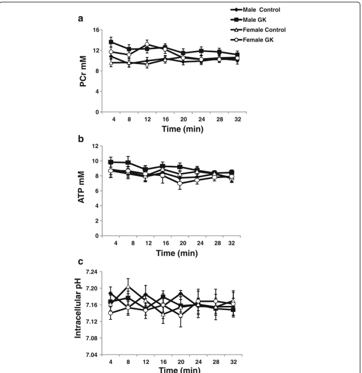
Kinetics of PCr (A), ATP (B) and intracellular pH (C) as measured by 31P magnetic resonance spectroscopy are shown in Fig. 3. No significant differences were found in PCr and ATP contents in male and female diabetic and Control rat hearts. pHi was the same in all groups with-out any significant differences (Fig. 3c). Kinetics of phos-phomonoesters (PME) and inorganic phosphate (Pi) were shown in an (Additional file 1: Figure S3d and S3e, respectively). PME and Pi were similar in all groups.
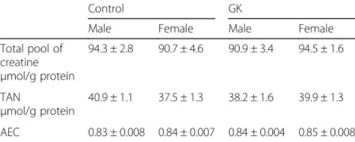
Gender effect on creatine, adenine nucleotide compounds and oxidative stress
The total pool of creatine (creatine and phosphocrea-tine) was similar in all groups (Table 2). No significant difference was found in total adenine nucleotides and adenylate energy charge between male and female con-trol and diabetic rat hearts (Table 2). MDA content (expressed in μmol/g protein), as an index of oxidative stress, was not different in male and female GK rat hearts (0.06 ± 0.01 and 0.05 ± 0.00 respectively) com-pared with male and female Control rat hearts (0.06 ± 0.00 and 0.06 ± 0.00).
Gender effect on cellular damage and water content
LDH and CK activities (expressed in U/mg protein) were similar in male (2.39 ± 0.10 and 5.96 ± 0.30) and female (2.32 ± 0.11 and 5.62 ± 0.28) Controls, and male (2.40 ± 0.17 and 6.02 ± 0.35) and female (2.40 ± 0.23 and 5.89 ± 0.27) GK rat hearts. Water content, expressed as a percentage, was not significantly different in male and
female Control (83.65 ± 0.75 and 83.10 ± 1.80) and GK (83.62 ± 1.79 and 84.18 ± 0.91) rat hearts.
Gender effect on NO pathway Total nitrate concentration (NOx)
Tissue NOx content in male and female Control and GK rats was shown in Table 3. We found an increased NOx in both diabetic groups compared with their respective Controls, indicating up-regulation of the NO pathway, but to a less extent in female GK rat hearts with a lower NOx content in female compared with male GK rat hearts (p = 0.0004).
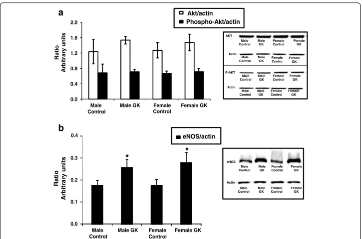
Protein expression of Akt, Phospho-Akt and eNOS
Protein expressions of Akt, Phospho-Akt (A) and eNOS (B) were given as ratios relative to actin protein content and were shown in Fig. 4. We found similar protein expressions of Akt and Phospho-Akt in the four groups (Fig. 4a). Interestingly, eNOS expression (Fig. 4b) was significantly increased in both male and female GK rat hearts compared with their respective Controls without any effect of gender (p < 0.05).
Gender effect on endothelial and smooth muscle functions
Endothelium-dependent and independent vasodilatations were shown in Table 4. Endothelium-dependent and in-dependent vasodilatations were not different in male Control and GK rat hearts. By contrast, endothelium-dependent and inendothelium-dependent vasodilatations were signifi-cantly impaired in female GK compared with male GK (p < 0.05) and female Control (p < 0.05) rat hearts.
Discussion
The aim of the study was to investigate if male and female without ischemic injury displayed differences in cardiac function, energy metabolism, and endothelial function which could contribute to increased cardiovascular
0 2 4 6 8 10 12 14 16
Male Control Male GK Female Control Female GK
Coronary flow
ml/min/g heart weight
*
‡
†
Fig. 2 Coronary flow (CF), expressed in ml/min/g heart weight, in Control (malen = 10, female n = 14) and GK (male n = 13, female n = 12) rat hearts. Results are means ± SEM. *p = 0.0420 and † p < 0.0001, versus respective Controls; ‡ p = 0.0137, versus male GK rat hearts
complications in type 2 diabetic female. Myocardial function was impaired similarly in both male and female diabetic GK rats. Cardiac energy metabolism was normal in both diabetic groups compared with their respective Controls. Conversely, coronary flow was decreased in both diabetic groups but to a higher level in female GK rat hearts. Total nitrate concentra-tion was up-regulated in both diabetic groups but to
a less extent in female GK rat hearts. eNOS/actine was similarly increased in both male and female GK groups without modification of Akt pathway in all groups. Endothelium-dependent and independent va-sodilatations were impaired only in female GK rat hearts. Together, these results could be related to higher risk of cardiovascular complications in type 2 diabetic female.
PCr
m
M
0 4 8 12 16 4 8 12 16 20 24 28 32Time (min)
Male Control Male GK Female Control Female GK 0 2 4 6 8 10 12 4 8 12 16 20 24 28 32P
T
Am
M
Time (min)
7.04 7.08 7.12 7.16 7.20 7.24 4 8 12 16 20 24 28 32r
al
ull
e
c
ar
t
nIp
H
Time (min)
a
b
c
Fig. 3 Kinetics of phosphocreatine (PCr) (a), adenosine triphosphate (ATP) (b) and intracellular pH (pHi) (c) in Control (malen = 10, female n = 14) and GK (malen = 13, female n = 12) rat hearts, measured by31P magnetic resonance spectroscopy. Results are expressed in mM except pHi, and are means ± SEM
It is known than non-diabetic men are at more risk of developing cardiovascular disease than non-diabetic women. Interestingly, here the non-diabetic male and female rats do not show any difference in coronary flow or endothelial function which could be explained by the lack of stress conditions such as ischaemic insult. How-ever the relative risk of cardiovascular disease incidence and mortality associated with type 2 diabetes compared with non-diabetes is stronger in women than in men [18, 19]. There are well characterized differences in trad-itional risk factors among diabetic men and women although these do not fully explain the gender differ-ences observed. The reasons why diabetes in women in-creases the relative risk of CHD more than in men compared with their non-diabetic counterpart is not clear, but a possible explanation may be that diabetes has a greater adverse effect on CVD risk factors in women than in men. Previous studies have shown differ-ences in lipid abnormalities to be more pronounced between diabetic and non-diabetic women than between diabetic and nondiabetic men [20] but this dyslipidemia appears insufficient to explain the differences in clinical risk [21]. Wannamethee et al. [6] have reported that the greater adverse influence of diabetes per se on abdom-inal adiposity and insulin resistance, and down-stream blood pressure, lipids (low HDL-cholesterol), endothelial dysfunction (t-PA), and systemic inflammation (WBC) in women compared with men may contribute to their greater relative risk of coronary heart disease. Interest-ingly, we have also previously reported higher insulin
resistance in the female than in the male GK rat hearts [10]. Another possible explanation may be due to a need for women to undergo much larger metabolic pertur-bances to transit from non-diabetes to diabetes, ie in general women may have to “deteriorate” more to get diabetes, they need to put on more weight, and deterior-ate their insulin sensitivity and reldeterior-ated risk factors to a greater extent than men [6]. On the other hand, the rea-son for the greater relative risk of CVD associated with diabetes in women compared with men may be also due to the difference in the treatment of cardiovascular heart disease risk factors between men and women or gender response to therapy [22, 23].
Endothelial dysfunction is an early sign of diabetic vas-cular disease and reduced endothelium-dependent vaso-relaxation (EDV) to vasodilators is generally used as a reproducible parameter to investigate the endothelial function under various pathological conditions. Here, the endothelial function was evaluated by a panel of markers, including the NO pathway (NOx production and AKT, Phospho-AKT and eNOS expression) com-bined to endothelial and smooth muscle vasodilatations and to the measurement of the coronary flow. NO pro-duction was increased in both GK rat groups but was less pronounced in female GK rat hearts. Total eNOS protein expression was similarly increased in both diabetic groups as reported before [11, 24] without any effect of gender. AKT protein expression and phosphor-ylation were similar in diabetic groups indicating that AKT did not play a major role in gender effect on the NO regulation. It has been hypothesized that upregula-tion of eNOS in diabetes was a consequence of the enhanced oxidative stress induced by hyperglycemia [24–26] and inactivation of NO by the production of re-active oxygen intermediates. MDA production, an index of oxidative stress, was similar in both male and female Control and GK rat hearts suggesting a normal oxidative stress, by contrast to our previous study showing in-creased MDA content in older male GK rat hearts [11]. Consequently, it would be interesting to evaluate both reactive oxygen species production and the anti-oxidant defence in male and female GK rat hearts in order to ac-curately rule out a role of oxidative stress on NO modu-lation in male and female type 2 diabetic GK rat hearts. Finally, measuring the state of eNOS phosphorylation, which is critical for NO synthesis, should be performed to further investigate the NO pathway. Lower NO up-regulation in female GK hearts is difficult to explain. Decreased NO availability in female GK rat hearts may be linked to a decrease in NOS activity due to increased NOS uncoupling [24] and/or impaired intracellular BH4/BH2 [27].
Decreased coronary flow and lower NOx content in the female diabetic rat hearts were associated with
Table 2 Total pool of creatine, total adenine nucleotides and adenylate energy charge in Control (malen = 10, female n = 14) and GK (malen = 13, female n = 12) rat hearts
Control GK
Male Female Male Female Total pool of creatine μmol/g protein 94.3 ± 2.8 90.7 ± 4.6 90.9 ± 3.4 94.5 ± 1.6 TAN μmol/g protein 40.9 ± 1.1 37.5 ± 1.3 38.2 ± 1.6 39.9 ± 1.3 AEC 0.83 ± 0.008 0.84 ± 0.007 0.84 ± 0.004 0.85 ± 0.008
TAN total adenine nucleotides (ATP + ADP + AMP), AEC adenylate energy charge ((ATP + 0.5ADP)/(ATP + ADP + AMP) * 10)
Table 3 Total nitrate concentration (NOx) in Control (malen = 10, femalen = 14) and GK (male n = 13, female n = 12) rat hearts
Control GK
Male Female Male Female NOx
nmol/mg protein
0.18 ± 0.02 0.19 ± 0.01 0.39 ± 0.03* 0.24 ± 0.01†‡
*p < 0.0001 versus Male Control rat hearts†p = 0.0113 versus Female Control
rat hearts
impaired endothelium-dependent and independent vaso-dilatations in the female GK rat hearts. By contrast, up-regulation of the NO pathway in the male GK rat hearts was probably involved in normal endothelial and smooth muscle functions but, nevertheless, this was insufficient for maintaining a normal coronary flow. Interestingly, we have reported higher decrease in basal coronary flow with higher increase in NOx production in male Control and GK older animals (9–14 months) [11]. Impaired endothelium-dependent vasorelaxation (EDV) has been observed in both type 1 and type 2 diabetes [28], wherever some studies have shown enhanced EDV in diabetes [29]. Interestingly, Kobayashi et al. [30] reported enhanced acetylcholine-induced relaxation and impaired
norepinephrine-induced contraction, due to NO over-production via eNOS and increased α2D-adrenoceptor
expression in early-stage GK rats. Impaired acetylcholine-induced relaxation in later-stage GK rats is due to reduc-tions in both NO production and NO responsiveness. Conflicting data were also obtained when responses to vasoconstricting agents were studied [30, 31]. The reason for these discrepancies is not clear. However, the duration of the disease, among other factors, may play an important role in the extent of the alteration of vascular reactivity to vasodilating or vasoconstricting agents in diabetes [32]. Zhang et al. [33] demonstrated that Ach-induced relaxa-tions were significantly impaired in mesenteric arteries from both male and female diabetic rats at one and eight weeks. Interestingly, the extent of impairment was signifi-cantly greater in diabetic females than in diabetic males at eight weeks suggesting a shift away from a putative endothelium-derived hyperpolarizing factor (EDHF) towards a greater reliance on NO. Several other re-ports [34, 35] suggest that hyperglycemia and diabetes affect male and female vascular beds differently. Clin-ically, these differences reveal a stronger association between CVD and diabetes in women than in men
Akt/actin Phospho-Akt/actin 0.0 0.4 0.8 1.2 1.6 2.0 Male Control Male GK Female Control Female GK Ra ti o yr ar ti br A units oi t a R yr ar ti br As ti n u 0.4 Male Control Male GK Female Control Female GK eNOS/actin 0.0 0.1 0.2 0.3
*
*
a
b
Male Control Male GK Female Control Female GK AKT Actin Male Control Male GK Female Contro Female GK Male Control Male GK Female Control Female GK P-AKT Actin Male Control Male GK Female Control Female GK eNOS Male Control Male GK Female Control Female GK Actin Male Control Male GK Female Control Female GKFig. 4 Protein expression of Akt, Phospho-Akt (a) and eNOS (b) in Control (malen = 10, female n = 14) and GK (male n = 13, female n = 12) rat hearts. Protein expression were measured by western blot assay and results are expressed as a ratio relatively to actin protein content and are means ± SEM. * versus respective Controls,p < 0.05
Table 4 Endothelium-dependent and independent vasodilatations in Control (malen = 9, female n = 10) and GK (male n = 9, female n = 11) rat hearts
Control GK
Male Female Male Female 5-HT % 32.6 ± 3.0 33.9 ± 3.3 30.5 ± 2.1 19.9 ± 2.6*† Papaverine % 28.0 ± 3.7 29.4 ± 3.2 32.3 ± 4.4 19.2 ± 2.5*†
[36, 37]. Interestingly, Goel et al. [34, 35] reported a predisposition of female rabbit aorta compared with male rabbit aorta toward impairment of endothelium-dependent vasodilation under hyperglycemic condi-tions, possibly via activation of PKCβ and superoxide production. Gender differences in sex hormones may be also one explanation for the differences in NO production/release in GK rats. Vascular strips from female rats were found to release more NO in response to acetylcholine than vascular strips from male rats [38]. These data suggest that estrogen may directly stimulate NO production/release in women. Conversely, the predominant male sex hormone tes-tosterone (or other androgens) may cause decreased NO production/release, as suggested by Herman [39]. Interestingly Al-Mulla et al. [40] reported reduced es-trogen and increased testosterone levels in the female GK rats and the possible roles of these hormones in inflammatory processes involved in wound healing impairment in type 2 diabetes. The independent con-tributions of estrogens and androgens to the control of endothelial function in normal and pathophysio-logical states, especially in type 2 diabetes, remain to be fully elucidated.
Myocardial function was significantly decreased in female GK rats and to the same extent than in the male GK rats. As suggested before [11], impaired cardiac function was probably related to a significantly lower heart rate in both GK rat hearts compared with their Controls, possibly caused by hyperglycemia which alters excitation-contraction coupling by lengthening the period of mechanical relaxation [41]. On the other hand, we have previously shown a 38% decreased protein level of IRS1, one of the major insulin-signaling component, in male GK rat hearts [9], which could be also involved in impaired cardiac function in diabetic rats as reported by Qi et al. [42]. Interestingly, Soliman et al. [43] re-ported that the RhoA/ROCK pathway contributes to contractile dysfunction in diabetic heart at least in part by sustaining PKCβ2 activation, iNOS activation and ROS production via a positive feedback loop that requires an intact cytoskeleton. Mitochondrial dysfunc-tion could be also involved in impaired cardiac funcdysfunc-tion in both GK rat heart groups as reported recently in high-fat diet mice [44]. On the other hand, peroxisome proliferator-activated receptors (PPARs) may also play a role in functional and metabolic abnormalities of the type 2 diabetic GK rat heart [45]. However, here cardiac energy metabolism was normal in female GK rat hearts and similar to the male GK rat heart suggesting normal mitochondrial respiration in GK rat hearts. We have also previously reported that increased susceptibility of older male type 2 diabetic GK rat heart to ischemic injury was not associated with impaired energy metabolism [11].
By contrast, reduced myocardial phosphocreatine/ATP ratio, indicating impaired high energy phosphate-metabolism and energy deficit [46, 47] has been reported in human diabetic cardiomyopathy. However Diamant et al. [48] found a decreased PCr/ATP in type 2 diabetic pa-tients but did not confirm this finding in a subsequent study with a group of well-controlled uncomplicated type 2 diabetic patients [49], probably due to differences in patient characteristics.
Limitations
The experiments were conducted here on isolated perfused hearts. In this model, we do not have the interactions with the other organs and with the whole body physiology and metabolism, which has both advantages and limitations. The advantage of this model is to be able to study the in-trinsic properties of the heart alone without the interactions with the other organs and whole physiology.
On the other hand, studying the heart in vivo using magnetic resonance imaging or echocardiography has an additional value by taking in account the whole physi-ology. In accordance with the results of the present study, using multiparametric magnetic resonance im-aging, we have previously shown that adult female GK rats had defective myocardial blood flow associated with altered left ventricular function in vivo [50], which is consistent with the ex vivo results reported here.
Conclusion
Here, we studied hemodynamic function, energy metab-olism, cellular integrity and endothelial function in male and female Control and GK rat without ischemic insult in order to check if gender differences already exist at basal state which could explain increased cardiovascular complications in type 2 diabetic female. We reported an endothelial damage characterized by reduced up-regulation of the NO pathway combined with impaired endothelial and smooth muscle functions and coronary flow rates in female diabetic rat hearts while energy me-tabolism was normal. Whether these results and in-volved molecular mechanisms are related to the higher risk of cardiovascular complications among type 2 dia-betic female waits to be further elicited in the future.
Additional file
Additional file 1: Figure S3. Kinetics of phosphomonoesters (PME) (D) and inorganic phosphate (Pi) (E) in Control (malen = 10, female n = 14) and GK (malen = 13, female n = 12) rat hearts, measured by31P magnetic
resonance spectroscopy. Results are expressed in mM and are means ± SEM. (DOCX 70 kb)
Abbreviations
CK:Creatine kinase; eNOS: Endothelial nitric oxide synthase; LDH: Lactate dehydrogenase; NO: Nitric oxide
Acknowledgements
We thank Christiane Dalmasso from CRMBM, Marseille. We thank Professor Bernard Portha and Danielle Bailbé from Laboratoire de Biologie et Pathologie du Pancréas Endocrine, Paris.
Funding
This work was supported by Aix-Marseille Université, CNRS (UMR 7339) and the French Program“Investissement d’Avenir” (grant “Infrastructure d’Avenir en Biologie Santé–ANR-11-INBS-0006”).
Availability of data and materials Not applicable.
Authors’ contributions
DM: contributed to design, experiments, data analysis and manuscript writing; LC: contributed to experiments and biochemical analysis; MJ: contributed to animal supply; MB: contributed to design, interpretation of the overall study and manuscript writing. All authors read and approved the final manuscript.
Competing interests
The authors declare that they have no competing interests.
Consent for publication Not applicable.
Ethics approval
All procedures involving animals were approved by the Institutional Ethic Committee for animal research of the Medical School La Timone of Marseille. All animals received humane care in compliance with the Principle of Laboratory Animal Care formulated by the National Society for Medical Research and the“Guide for the Care and Use of Laboratory Animals” prepared by the Institute of Laboratory Animal Resources and published by the National Institutes of Health (NIH Publication No. 85–23, updated 2011). All investigations in this project were conducted under a license for animal research (n° 10–18072011) granted by the French Ministry of Agriculture. Author details
1Aix-Marseille Université, CNRS, CRMBM, Marseille, France.2Université
Paris-Diderot, CNRS, UMR 8251, Laboratoire de Biologie et Pathologie du Pancréas Endocrine (B2PE), Unité BFA, Paris, France.3Centre de Résonance
Magnétique Biologique et Médicale (CRMBM), UMR n°7339, Aix-Marseille Université, CNRS, Faculté de Medecine, 27 Bd Jean Moulin, Marseille Cedex 05 13385, France.
Received: 11 August 2016 Accepted: 25 December 2016
References
1. Taegtmeyer H, McNulty P, Young ME. Adaptation and maladaptation of the heart in diabetes: Part I: general concepts. Circulation. 2002;105:1727–33. 2. Hu G. Gender difference in all-cause and cardiovascular mortality related to
hyperglycaemia and newly-diagnosed diabetes. Diabetologia. 2003;46(5):608–17. 3. Donahue RP, Rejman K, Rafalson LB, Dmochowski J, Stranges S, Trevisan M.
Sex differences in endothelial function markers before conversion to pre-diabetes: does the clock start ticking earlier among women? The Western New York Study. Diabetes Care. 2007;30(2):354–9.
4. Steinberg HO, Paradisi G, Cronin J, Crowde K, Hempfling A, Hook G, Baron AD. Type II diabetes abrogates sex differences in endothelial function in premenopausal women. Circulation. 2000;101:2040–6.
5. Regitz-Zagrosek V, Lehmkuhl E, Weickert MO. Gender differences in the metabolic syndrome and their role for cardiovascular disease. Clin Res Cardiol. 2006;95(3):136–47.
6. Wannamethee SG, Papacosta O, Lawlor DA, Whincup PH, Lowe GD, Ebrahim S, Sattar N. Do women exhibit greater differences in established and novel risk factors between diabetes and non-diabetes than men? The British regional heart study and British Women’s heart health study. Diabetologia. 2012;55(1):80–7. 7. Bisbis S, Bailbe D, Tormo MA, Picarel-Blanchot F, Derouet M, Simon J, Portha
B. Insulin resistance in the GK rat: decreased receptor number but normal kinase activity in liver. Am J Physiol. 1993;265(5 Pt 1):E807–813.
8. Portha B. Programmed disorders of beta-cell development and function as one cause for type 2 diabetes? The GK rat paradigm. Diabetes Metab Res Rev. 2005;21(6):495–504.
9. Desrois M, Sidell RJ, Gauguier D, King LM, Radda GK, Clarke K. Initial steps of insulin signaling and glucose transport are defective in the type 2 diabetic rat heart. Cardiovasc Res. 2004;61(2):288–96.
10. Desrois M, Sidell RJ, Gauguier D, Davey CL, Radda GK, Clarke K. Gender differences in hypertrophy, insulin resistance and ischemic injury in the aging type 2 diabetic rat heart. J Mol Cell Cardiol. 2004;37:547–55.
11. Desrois M, Clarke K, Lan C, Dalmasso C, Cole M, Portha B, Cozzone PJ, Bernard M. Upregulation of eNOS and unchanged energy metabolism in increased susceptibility of the aging type 2 diabetic GK rat heart to ischemic injury. Am J Physiol Heart Circ Physiol. 2010;299(5):H1679–1686. 12. Desrois M, Lan C, Dalmasso C, Portha B, Bailbé D, Cozzone PJ, Bernard M. Endothelial and smooth muscle damage but normal energy metabolism in the female type 2 diabetic Goto-Kakizaki (GK) rat heart. Magn Reson Mater Phys Biol Med. 2013;26(1):p286.
13. Desrois M, Caus T, Belles PM, Dalmasso C, Lan C, Cozzone PJ, Bernard M. Limitation of myocardial and endothelial injury of the rat heart graft after preservation with centre de resonance magnetique biologique et medicale (CRMB) solution. Transpl Int. 2008;21(3):276–83.
14. Lefevre G, Beljean-Leymarie M, Beyerle F, Bonnefont-Rousselot D, Cristol JP, Therond P, Torreilles J. Evaluation of lipid peroxidation by measuring thiobarbituric acid reactive substances. Ann Biol Clin (Paris). 1998;56(3): 305–19.
15. Desrois M, Sciaky M, Lan C, Cozzone PJ, Bernard M. L-arginine during long-term ischemia: effects on cardiac function, energetic metabolism and endothelial damage. J Heart Lung Transplant. 2000;19:367–76.
16. Cross HR, Murphy E, Steenbergen C. Ca(2+) loading and adrenergic stimulation reveal male/female differences in susceptibility to ischemia-reperfusion injury. Am J Physiol Heart Circ Physiol. 2002;283(2):H481–489.
17. Ye Y, Lin Y, Manickavasagam S, Perez-Polo JR, Tieu BC, Birnbaum Y. Pioglitazone protects the myocardium against ischemia-reperfusion injury in eNOS and iNOS knockout mice. Am J Physiol Heart Circ Physiol. 2008;295(6): H2436–2446.
18. Huxley R, Barzi F, Woodward M. Excess risk of fatal coronary heart disease associated with diabetes in men and women: meta-analysis of 37 prospective cohort studies. BMJ. 2006;332(7533):73–8.
19. Lee WL, Cheung AM, Cape D, Zinman B. Impact of diabetes on coronary artery disease in women and men: a meta-analysis of prospective studies. Diabetes Care. 2000;23(7):962–8.
20. Williams K, Tchernof A, Hunt KJ, Wagenknecht LE, Haffner SM, Sniderman AD. Diabetes, abdominal adiposity, and atherogenic dyslipoproteinemia in women compared with men. Diabetes. 2008;57(12):3289–96.
21. Juutilainen A, Kortelainen S, Lehto S, Ronnemaa T, Pyorala K, Laakso M. Gender difference in the impact of type 2 diabetes on coronary heart disease risk. Diabetes Care. 2004;27(12):2898–904.
22. Lyons MR, Peterson LR, McGill JB, Herrero P, Coggan AR, Saeed IM, Recklein C, Schechtman KB, Gropler RJ. Impact of sex on the heart’s metabolic and functional responses to diabetic therapies. Am J Physiol Heart Circ Physiol. 2013;305(11):H1584–1591.
23. McGill JB, Vlajnic A, Knutsen PG, Recklein C, Rimler M, Fisher SJ. Effect of gender on treatment outcomes in type 2 diabetes mellitus. Diabetes Res Clin Pract. 2013;102(3):167–74.
24. Bitar MS, Wahid S, Mustafa S, Al-Saleh E, Dhaunsi GS, Al-Mulla F. Nitric oxide dynamics and endothelial dysfunction in type II model of genetic diabetes. Eur J Pharmacol. 2005;511(1):53–64.
25. Bulhak AA, Jung C, Ostenson CG, Lundberg JO, Sjoquist PO, Pernow J. PPAR-alpha activation protects the type 2 diabetic myocardium against ischemia-reperfusion injury: involvement of the PI3-Kinase/Akt and NO pathway. Am J Physiol Heart Circ Physiol. 2009;296(3):H719–727.
26. Jesmin S, Zaedi S, Maeda S, Yamaguchi I, Goto K, Miyauchi T. Effects of a selective endothelin a receptor antagonist on the expressions of iNOS and eNOS in the heart of early streptozotocin-induced diabetic rats. Exp Biol Med (Maywood). 2006;231(6):925–31.
27. Crabtree MJ, Smith CL, Lam G, Goligorsky MS, Gross SS. Ratio of 5,6,7,8-tetrahydrobiopterin to 7,8-dihydrobiopterin in endothelial cells determines glucose-elicited changes in NO vs. superoxide production by eNOS. Am J Physiol Heart Circ Physiol. 2008;294(4):H1530–1540.
28. De Vriese AS, Verbeuren TJ, Van de Voorde J, Lameire NH, Vanhoutte PM. Endothelial dysfunction in diabetes. Br J Pharmacol. 2000;130(5):963–74.
29. Shen B, Ye CL, Ye KH, Liu JJ. Mechanism underlying enhanced endothelium-dependent vasodilatation in thoracic aorta of early stage streptozotocin-induced diabetic mice. Acta Pharmacol Sin. 2003;24(5):422–8.
30. Kobayashi T, Matsumoto T, Ooishi K, Kamata K. Differential expression of alpha2D-adrenoceptor and eNOS in aortas from early and later stages of diabetes in Goto-Kakizaki rats. Am J Physiol Heart Circ Physiol. 2004;287(1):H135–143.
31. Taylor PD, Oon BB, Thomas CR, Poston L. Prevention by insulin treatment of endothelial dysfunction but not enhanced noradrenaline-induced contractility in mesenteric resistance arteries from streptozotocin-induced diabetic rats. Br J Pharmacol. 1994;111(1):35–41.
32. Abboud K, Bassila JC, Ghali-Ghoul R, Sabra R. Temporal changes in vascular reactivity in early diabetes mellitus in rats: role of changes in endothelial factors and in phosphodiesterase activity. Am J Physiol Heart Circ Physiol. 2009;297(2):H836–845.
33. Zhang R, Thor D, Han X, Anderson L, Rahimian R. Sex differences in mesenteric endothelial function of streptozotocin-induced diabetic rats: A shift in the relative importance of EDRFs. Am J Physiol Heart Circ Physiol. 2012;303(10):H1183–98.
34. Goel A, Thor D, Anderson L, Rahimian R. Sexual dimorphism in rabbit aortic endothelial function under acute hyperglycemic conditions and gender-specific responses to acute 17beta-estradiol. Am J Physiol Heart Circ Physiol. 2008;294(6):H2411–2420.
35. Goel A, Zhang Y, Anderson L, Rahimian R. Gender difference in rat aorta vasodilation after acute exposure to high glucose: involvement of protein kinase C beta and superoxide but not of Rho kinase. Cardiovasc Res. 2007;76(2):351–60.
36. Hu G, Jousilahti P, Qiao Q, Katoh S, Tuomilehto J. Sex differences in cardiovascular and total mortality among diabetic and non-diabetic individuals with or without history of myocardial infarction. Diabetologia. 2005;48(5):856–61.
37. Onat A, Hergenc G, Keles I, Dogan Y, Turkmen S, Sansoy V. Sex difference in development of diabetes and cardiovascular disease on the way from obesity and metabolic syndrome. Metabolism. 2005;54(6):800–8.
38. Huang A, Sun D, Koller A, Kaley G. Gender difference in flow-induced dilation and regulation of shear stress: role of estrogen and nitric oxide. Am J Physiol. 1998;275(5 Pt 2):R1571–1577.
39. Herman SM, Robinson JT, McCredie RJ, Adams MR, Boyer MJ, Celermajer DS. Androgen deprivation is associated with enhanced endothelium-dependent dilatation in adult men. Arterioscler Thromb Vasc Biol. 1997;17(10):2004–9. 40. Al-Mulla F, Leibovich SJ, Francis IM, Bitar MS. Impaired TGF-beta signaling and
a defect in resolution of inflammation contribute to delayed wound healing in a female rat model of type 2 diabetes. Mol Biosyst. 2011;7(11):3006–20. 41. Ren J, Gintant GA, Miller RE, Davidoff AJ. High extracellular glucose impairs
cardiac E-C coupling in a glycosylation-dependent manner. Am J Physiol. 1997;273:H2876–83.
42. Qi Y, Xu Z, Zhu Q, Thomas C, Kumar R, Feng H, Dostal DE, White MF, Baker KM, Guo S. Myocardial loss of IRS1 and IRS2 causes heart failure and is controlled by p38alpha MAPK during insulin resistance. Diabetes. 2013;62(11):3887–900. 43. Soliman H, Gador A, Lu YH, Lin G, Bankar G, MacLeod KM. Diabetes-induced
increased oxidative stress in cardiomyocytes is sustained by a positive feedback loop involving Rho kinase and PKCbeta2. Am J Physiol Heart Circ Physiol. 2012;303(8):H989–H1000.
44. Marciniak C, Marechal X, Montaigne D, Neviere R, Lancel S. Cardiac contractile function and mitochondrial respiration in diabetes-related mouse models. Cardiovasc Diabetol. 2014;13:118.
45. Lee WS, Kim J. Peroxisome proliferator-activated receptors and the heart: lessons from the past and future directions. PPAR Res. 2015;2015:271983. 46. Bugger H, Abel ED. Molecular mechanisms for myocardial mitochondrial
dysfunction in the metabolic syndrome. Clin Sci (Lond). 2008;114(3):195–210. 47. Scheuermann-Freestone M, Madsen PL, Manners D, Blamire AM,
Buckingham RE, Styles P, Radda GK, Neubauer S, Clarke K. Abnormal cardiac and skeletal muscle energy metabolism in patients with type 2 diabetes. Circulation. 2003;107(24):3040–6.
48. Diamant M, Lamb HJ, Groeneveld Y, Endert EL, Smit JW, Bax JJ, Romijn JA, de Roos A, Radder JK. Diastolic dysfunction is associated with altered myocardial metabolism in asymptomatic normotensive patients with well-controlled type 2 diabetes mellitus. J Am Coll Cardiol. 2003;42(2):328–35. 49. Rijzewijk LJ, van der Meer RW, Lamb HJ, de Jong HW, Lubberink M, Romijn
JA, Bax JJ, de Roos A, Twisk JW, Heine RJ, et al. Altered myocardial substrate metabolism and decreased diastolic function in nonischemic human diabetic cardiomyopathy: studies with cardiac positron emission
tomography and magnetic resonance imaging. J Am Coll Cardiol. 2009;54(16):1524–32.
50. Iltis I, Kober F, Desrois M, Dalmasso C, Lan C, Portha B, Cozzone PJ, Bernard M. Defective myocardial blood flow and altered function of the left ventricle in type 2 diabetic rats: a noninvasive in vivo study using perfusion and cine magnetic resonance imaging. Invest Radiol. 2005;40(1):19–26.
• We accept pre-submission inquiries
• Our selector tool helps you to find the most relevant journal
• We provide round the clock customer support
• Convenient online submission
• Thorough peer review
• Inclusion in PubMed and all major indexing services
• Maximum visibility for your research Submit your manuscript at
www.biomedcentral.com/submit