HAL Id: hal-01313666
https://hal.sorbonne-universite.fr/hal-01313666
Submitted on 10 May 2016
HAL is a multi-disciplinary open access
archive for the deposit and dissemination of
sci-entific research documents, whether they are
pub-lished or not. The documents may come from
teaching and research institutions in France or
abroad, or from public or private research centers.
L’archive ouverte pluridisciplinaire HAL, est
destinée au dépôt et à la diffusion de documents
scientifiques de niveau recherche, publiés ou non,
émanant des établissements d’enseignement et de
recherche français ou étrangers, des laboratoires
publics ou privés.
Distributed under a Creative Commons Attribution| 4.0 International License
Evaluation of the SLOMYCO Sensititre® panel for
testing the antimicrobial susceptibility of
Mycobacterium marinum isolates
Marion Chazel, Hélène Marchandin, Nicolas Keck, Dominique Terru,
Christian Carrière, Michael Ponsoda, Véronique Jacomo, Gilles Panteix,
Nicolas Bouzinbi, Anne-Laure Bañuls, et al.
To cite this version:
Marion Chazel, Hélène Marchandin, Nicolas Keck, Dominique Terru, Christian Carrière, et al..
Evalu-ation of the SLOMYCO Sensititre® panel for testing the antimicrobial susceptibility of Mycobacterium
marinum isolates. Annals of Clinical Microbiology and Antimicrobials, BioMed Central, 2016, 15 (1),
pp.30. �10.1186/s12941-016-0145-1�. �hal-01313666�
RESEARCH
Evaluation of the SLOMYCO Sensititre
®
panel for testing the antimicrobial susceptibility
of Mycobacterium marinum isolates
Marion Chazel
1, Hélène Marchandin
1,2, Nicolas Keck
3, Dominique Terru
1, Christian Carrière
1,5,6,
Michael Ponsoda
4, Véronique Jacomo
4, Gilles Panteix
4, Nicolas Bouzinbi
1, Anne‑Laure Bañuls
7, Marc Choisy
7,
Jérôme Solassol
6,8,9, Alexandra Aubry
10,11,12,13†and Sylvain Godreuil
1,5,6*†Abstract
Background: The agar dilution method is currently considered as the reference method for Mycobacterium marinum
drug susceptibility testing (DST). As it is time‑consuming, alternative methods, such as the E‑test, were evaluated for
M. marinum DST, but without success. The SLOMYCO Sensititre® panel, recently commercialized by TREK Diagnostic Systems (Cleveland, OH), can be used for DST in slow‑growing mycobacteria and for antimicrobial agents recom‑ mended by the Clinical and Laboratory Standards Institute (CLSI) for M. marinum DST. The main goal of this work was to evaluate the SLOMYCO Sensititre® panel method for DST in M. marinum isolates from human patients and fish rela‑ tive to the reference agar dilution method.
Methods/Results: The reproducibility of the minimum inhibitory concentration (MIC) determination (±1 log2
dilution) was very good for both the agar dilution method and SLOMYCO Sensititre® panel (>90 % agreement). The percentage essential agreement between methods varied, depending on the drug: between 97 and 75 % for cipro‑ floxacin, moxifloxacin, linezolid, isoniazid, clarithromycin, amikacin, rifabutin and rifampin, 74 % for trimethoprim, 72 % for doxycycline, 70 % for sulfamethoxazole, 59 % for streptomycin, 33 % for ethambutol and only 2.2 % for ethiona‑ mide. When the agar dilution and SLOMYCO Sensititre® panel results were converted into interpretive criteria, the category agreement was 100 % for amikacin, ciprofloxacin, clarithromycin, moxifloxacin, rifabutin, sulfamethoxazole and trimethoprim, 98 % for ethambutol and 96 % for rifampin and no agreement for doxycycline.
Conclusions: The SLOMYCO Sensititre® panel method could provide a potential alternative to the reference agar dilution method, when DST in M. marinum is required, except for doxycycline.
Keywords: Mycobacterium marinum, SLOMYCO Sensititre® panel, Agar dilution method, Antimicrobial susceptibility
testing, Human and fish isolates
© 2016 Chazel et al. This article is distributed under the terms of the Creative Commons Attribution 4.0 International License (http://creativecommons.org/licenses/by/4.0/), which permits unrestricted use, distribution, and reproduction in any medium, provided you give appropriate credit to the original author(s) and the source, provide a link to the Creative Commons license, and indicate if changes were made. The Creative Commons Public Domain Dedication waiver (http://creativecommons.org/ publicdomain/zero/1.0/) applies to the data made available in this article, unless otherwise stated.
Background
Mycobacterium marinum, a slow-growing
nontubercu-lous photochromogenic mycobacteria, is an ubiquitous waterborne organism [9, 10, 17] that causes diseases
in many fish species from cold or warm, fresh or salted water, and also in many other aquatic animals, such as amphibians, mammals and oysters [9, 10, 17]. In humans,
M. marinum infection is commonly limited to the skin,
but it can spread to deeper structures, resulting in teno-synovitis, arthritis and osteomyelitis [1, 8, 13] and, rarely, in disseminated infection in immunocompromised patients [13].
Mycobacterium marinum is naturally multi-drug
resist-ant and there is no standardized resist-antimicrobial treat-ment for M. marinum infections [7]. As the wild-type
Open Access
*Correspondence: s‑godreuil@chu‑montpellier.fr
†Alexandra Aubry and Sylvain Godreuil contributed equally to this work 5 Department of Bacteriology‑Virology, INSERM U1058 “Pathogenesis and Control of Chronic Infections”, Université Montpellier‑EFS, CHU Arnaud de Villeneuve 371 avenue du Doyen Gaston Giraud, 34295 Montpellier, France
Page 2 of 7 Chazel et al. Ann Clin Microbiol Antimicrob (2016) 15:30
susceptibility pattern of M. marinum is well known [2] and acquired resistance has not been described so far, antimicrobial susceptibility testing is not recommended except in the case of treatment failure and relapse [16].
The Clinical and Laboratory Standards Institute (CLSI) recommends microdilution for M. marinum drug suscep-tibility testing (DST) [16], but the agar dilution method is currently considered to be the reference method [2, 4,
19]. As this method is time-consuming, alternative meth-ods, such as the E-test, were evaluated, but showed poor agreement with the reference method and therefore are not suitable for DST in M. marinum [2, 11, 19]. Recently, the SLOMYCO Sensititre® panel was commercialized by TREK Diagnostic Systems (Cleveland, OH) [3]. This is a standard-order broth microdilution panel that can be used to evaluate the susceptibility of slow growing myco-bacteria to 14 antimicrobial agents, including those rec-ommended by CLSI for M. marinum DST [3].
The main goal of this work was to evaluate the SLO-MYCO Sensititre® panel method for DST in M. marinum strains from humans and fish relative to the reference agar dilution method.
Methods
Bacterial strains and growth conditions
The origin and other information concerning the tested
M. marinum isolates are described in Broutin et al. [5]. The 35 M. marinum isolates from human patients and nine from fish were collected in France between 1995 and 2007. None was from patients who experienced treat-ment failure or relapse. M. marinum identification was performed using GenoType Mycobacterium AS/CM, a commercial multiplex line-probe assay (Hain Lifesci-ence GmbH, Nehren, Germany). Isolates were stored at −80 °C in Middlebrook 7H9 broth (DIFCO, Detroit, MI, USA) containing 5 % OADC (DIFCO, Detroit, MI, USA) until determination of the Minimum inhibitory con-centrations (MICs). Mycobacteria were then cultured in Löwenstein-Jensen (LJ) slants (bioMérieux, Marcy l’Etoile, France) and in Middlebrook 7H10 agar (DIFCO, Detroit, MI, USA). The M. marinum ATCC 927 strain (from fish) and the M. marinum ATCC BAA-535/M strain isolated from an infected patient were used as con-trols for MIC determination.
Antimicrobial agents
Amikacin, ciprofloxacin, clarithromycin, doxycycline, ethambutol, ethionamide, isoniazid, rifampin, rifabu-tin, streptomycin and trimethoprim (Sigma-Aldrich, Lyon, France), linezolid (Pfizer, France) and moxifloxa-cin (Bayer, Wuppertal, Germany) were tested in this study. Stock solutions of each drug were prepared using the appropriate solvent and were filter-sterilized before
storage at −80 °C. To prior testing, each drug was freshly diluted in sterile deionized water. The concentration ranges of the tested antimicrobial agents are indicated in Table 1.
MIC determination by using the agar dilution method
The agar dilution method was performed on Müller-Hinton agar (Becton–Dickinson, France) supplemented with 5 % OADC according to CLSI [16]. Twofold dilu-tions of the antibiotics to be tested were added to obtain the required final concentrations. Confluent colonies of the different bacteria in LJ slants were swept with a loop and emulsified in sterile water and the concentration was adjusted to the McFarland n. 1 standard turbidity. A 1/100 dilution of each suspension adjusted to the McFar-land n. 1 standard was inoculated using a Steers replica-tor to deliver approximately 104 colony-forming units
(CFU) per spot. Plates were incubated at 30 °C [16]. The MICs (i.e., the lowest concentration of antibiotic result-ing in complete inhibition of growth) of the tested anti-biotics were determined after 7 and 14 days of growth of the different M. marinum isolates/controls.
MIC determination using the SLOMYCO Sensititre® panel
(broth micro‑dilution method)
Inocula for the SLOMYCO Sensititre® panel were pre-pared according to the CLSI and the manufacturer’s instructions [16]. SLOMYCO plates were incubated in a non-CO2 incubator at 30 °C until the controls showed
sufficient growth (7–14 days). The MICs were deter-mined visually using an inverted mirror and read as the lowest concentration of the antibiotic showing 100 % growth inhibition.
Analysis of the results
The reproducibility of both methods was evaluated by performing two independent tests for each method and for each of the 44 isolates (a total of 176 tests) and five independent tests for the M. marinum ATCC 927 and ATCC BAA-535/M strains (a total of 30 tests). Each test result was independently interpreted by two blinded readers. The reproducibility value was defined as the per-centage of strains with the same MIC value ±1 log2
dilu-tion at each test. The essential agreement between the agar dilution and SLOMYCO Sensititre® panel results was expressed as the percentage of isolates that showed the same MIC value ±1 log2 dilution with the two
meth-ods. Category agreement was evaluated using the break-points for determining the susceptibility and resistance categories recommended by CLSI [16]. For this study only very major errors (i.e., an isolate resistant [R] by the reference method, but susceptible [S] by the tested method) and major errors (S by the reference method
and R by the test method) were considered. Minor errors (intermediate [I] by one method and S or R by the other method) were not considered because the CLSI break-points allow only two characterization categories (R or S) [16].
Results
The reproducibility of the results obtained with the agar dilution method (reference method) was very good (Table 2) for all antibiotics. The MICs of the 14 antimicro-bial agents determined by using the agar dilution method were distributed in a narrow range (Table 1). Comparison of the MICs for the M. marinum isolates from infected humans and fish did not reveal any difference.
The MICs for the two reference M. marinum strains (ATCC 927 and ATCC BAA-535/M) were within 0–1 dilution of the MICs of the 44 tested isolates and within the expected range.
The MICs at which 90 % of isolates were inhibited (MIC90) of amikacin (4 μg/mL), clarithromycin (2 μg/mL)
and rifabutin (0.12 μg/mL) were below the CSLI break-point, whereas the MIC90 of ciprofloxacin (2 μg/mL),
doxycycline (4 μg/mL), ethambutol (4 μg/mL), moxiflox-acin (1 μg/mL), rifampin (1 μg/mL) and sulfamethoxazole (19 μg/mL) were close to the breakpoint [16]. The MIC90
of rifampin, rifabutin, moxifloxacin and trimethoprim
(between 0.1 and 1 μg/mL) were lower than those of the other tested antibiotics.
The relatively high MIC90 of ethionamide (10 μg/mL),
isoniazid (8 μg/mL) and streptomycin (16 μg/mL), for which breakpoints are not available, suggests that they are not good candidates for the treatment of M. marinum infection. In contrast, linezolid (MIC90: 2 μg/mL) was
very effective against the different M. marinum isolates with MIC values among the lowest in our study.
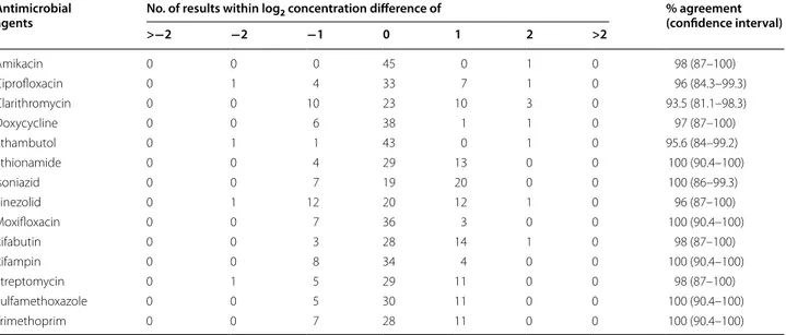
The SLOMYCO Sensititre® panel method (tested
method) produced remarkably consistent and reproduc-ible results (Table 3). Reproducibility with this method was 100 % in the case of ethionamide, isoniazid, moxi-floxacin, rifampin, trimethoprim and sulfamethoxazole, and between 98 and 87 % for amikacin, ciprofloxacin, clarithromycin, doxycycline, ethambutol, linezolid, rifabutin and streptomycin (Table 3). The percent-age essential agreement (±1 log2 dilution) between the
MICs obtained with the tested and the reference meth-ods (Table 4) greatly varied depending on the drug: from 98 % for ciprofloxacin and linezolid to 2.2 % for ethiona-mide. Good agreement percentages were obtained for moxifloxacin (91.3 %), isoniazid (87 %) and clarithromy-cin (85 %); whereas, agreement was lower for amikaclarithromy-cin (76.1 %), rifampin (76.1 %), rifampin (76 %), trimetho-prim (74 %), doxycycline (72 %), sulfamethoxazole
Table 1 MIC (μg/mL) of the 14 antibiotics tested in 46 M. marinum isolates (35 clinical, 9 fish and 2 references strains), determined by using the agar dilution method
a Concentration range of the tested drugs by using the agar dilution method (ADM) and the SLOMYCO Sensititre® panel method (SSPM): amikacin (0.25–32 mg/L, ADM; 1–64 µg/mL, SSPM); ciprofloxacin (0.12–16 µg/mL, ADM, SSPM); clarithromycin (0.06–32 µg/mL, ADM, SSPM); doxycycline (0.12–16 µg/mL, ADM, SSPM); ethambutol (0.5–16 µg/mL, ADM, SSPM); ethionamide (0.3–20 µg/mL, ADM, SSPM); isoniazid (0.25–16 µg/mL, ADM; 0.25–8 µg/mL, SSPM); linezolid (0.12–32 µg/mL, ADM; SSPM, 1–64 µg/mL); moxifloxacin (0.06–8 µg/mL, ADM; 0.12–8 µg/mL, SSPM); rifabutin (0.06–16 µg/mL, ADM; 0.25–8 mg/L, SSPM); rifampin (0.12–8 mg/L, ADM; 0.25–8 µg/mL, SSPM); streptomycin (0.5–32 µg/mL, ADM; 0.5–64 µg/mL, SSPM); sulfamethoxazole (2.38–152 µg/mL, ADM, SSPM); trimethoprim (0.12–8 µg/mL, ADM, SSPM); NA not available
Antimicrobial agent
[breakpoints (µg/mL)]a MIC50 MIC90 Range
Total (n = 46) Human (n = 35) Fish (n = 9) Total (n = 46) Human (n = 35) Fish (n = 9)
Amikacin (>32) 2 2 2 4 4 2 2–4 Ciprofloxacin (>2) 2 2 2 2 2 2 2 Clarithromycin (>16) 2 2 2 2 2 2 1–2 Doxycycline (>4) 4 4 4 4 4 4 1–4 Ethambutol (>4) 2 2 2 4 4 2 1–8 Ethionamide (NA) 5 5 5 10 10 10 2.5–10 Isoniazid (NA) 8 8 8 8 8 8 2–8 Linezolid (NA) 1 1 1 2 2 2 0.5–4 Moxifloxacin (>2) 1 1 1 1 1 1 0.12–1 Rifabutin (>2) 0.12 0.12 0.12 0.12 0.12 0.12 0.12 Rifampin (>1) 0.5 0.5 0.5 1 1 1 0.5–2 Streptomycin (NA) 16 16 16 16 16 16 4–32 Sulfamethoxazole (>38) 9.5 9.5 9.5 19 19 19 4.75–19 Trimethoprim (>2) 0.5 0.5 0.5 1 1 1 0.25–1
Page 4 of 7 Chazel et al. Ann Clin Microbiol Antimicrob (2016) 15:30
(70 %), streptomycin (59 %) and particularly ethambutol (33 %). In the case of ethambutol and ethionamide, the SLOMYCO Sensititre® panel method underestimated the MICs by 2–3 dilutions compared with the refer-ence method. When the agar dilution and SLOMYCO Sensititre® panel results were converted into inter-pretive categories (resistance/susceptibility) using the CLSI breakpoints, the category agreement was 100 % for amikacin, ciprofloxacin, clarithromycin, moxifloxa-cin, rifabutin and sulfamethoxazole–trimethoprim
(SMT-TMP), 98 % for ethambutol and 96 % for rifampin (Table 5). Two very major discrepancies were observed for rifampin (2/46) and one for ethambutol (1/46). No agreement was observed between the two methods for doxycycline, with 46 (100 %) major discrepancies. The SLOMYCO Sensititre® method overestimated the dox-ycycline MIC by 1 dilution; however, with this method, doxycycline MIC90 (8 μg/mL) was close to the
suscep-tibility breakpoint obtained by using the agar dilution method (4 μg/mL).
Table 2 Reproducibility of the results (i.e., MIC, expressed in µg/mL, of the tested antibiotics in the 46 M. marinum iso-lates) obtained with the agar dilution method
Antimicrobial
agents No. of results within log2 concentration difference of % agreement (confidence interval)
>−2 −2 −1 0 1 2 >2 Amikacin 0 0 0 45 1 0 0 100 (89–100) Ciprofloxacin 0 0 4 33 7 2 0 96 (84.3–99.3) Clarithromycin 0 0 10 23 13 0 0 100 (88–99) Doxycycline 0 0 6 38 1 1 0 98 (87–100) Ethambutol 0 0 2 43 0 1 0 98 (87–100) Ethionamide 0 0 7 29 10 0 0 100 (90.4–100) Isoniazid 0 0 5 21 20 0 0 100 (86–99.3) Linezolid 0 1 12 20 12 1 0 96 (87–100) Moxifloxacin 0 0 10 33 3 0 0 100 (90.4–100) Rifabutin 0 1 3 28 14 0 0 98 (87–100) Rifampin 0 0 12 30 4 0 0 100 (90.4–100) Streptomycin 0 1 5 29 11 0 0 98 (87–100) Sulfamethoxazole 0 0 10 25 11 0 0 100 (90.4–100) Trimethoprim 0 0 10 26 10 0 0 100 (90.4–100)
Table 3 Reproducibility of the results (i.e., MIC, expressed in µg/mL, of the tested antibiotics in the 46 M. marinum iso-lates) obtained with the SLOMYCO Sensititre® panel method
Antimicrobial
agents No. of results within log2 concentration difference of % agreement (confidence interval)
>−2 −2 −1 0 1 2 >2 Amikacin 0 0 0 45 0 1 0 98 (87–100) Ciprofloxacin 0 1 4 33 7 1 0 96 (84.3–99.3) Clarithromycin 0 0 10 23 10 3 0 93.5 (81.1–98.3) Doxycycline 0 0 6 38 1 1 0 97 (87–100) Ethambutol 0 1 1 43 0 1 0 95.6 (84–99.2) Ethionamide 0 0 4 29 13 0 0 100 (90.4–100) Isoniazid 0 0 7 19 20 0 0 100 (86–99.3) Linezolid 0 1 12 20 12 1 0 96 (87–100) Moxifloxacin 0 0 7 36 3 0 0 100 (90.4–100) Rifabutin 0 0 3 28 14 1 0 98 (87–100) Rifampin 0 0 8 34 4 0 0 100 (90.4–100) Streptomycin 0 1 5 29 11 0 0 98 (87–100) Sulfamethoxazole 0 0 5 30 11 0 0 100 (90.4–100) Trimethoprim 0 0 7 28 11 0 0 100 (90.4–100)
Discussion
The manufacturer’s guidelines (TREK Diagnostic Sys-tems, Cleveland, OH) recommend the SLOMYCO Sen-sititre® method for antibiotic susceptibility testing in M.
marinum, but studies evaluating the concordance with
the reference agar dilution method have not been pub-lished yet. As the SLOMYCO Sensititre® panel technique presents several advantages (commercial availability; standardization; easy to set up and to interpret; and
amenable to automation [2, 19] compared to the time-consuming and cumbersome agar dilution method, our objective was to compare the performance of these two methods.
The lack of difference between the MICs for the M.
marinum isolates from infected humans and fish could
be explained by the fact that most of the clinical sam-ples were from patients who handled infected fish (from aquarium tanks or fish-related work) and, therefore, the antibiotic susceptibility profiles of the human isolates reflected those of the fish isolates.
Our results show that the reproducibility of the SLO-MYCO Sensititre® results for the reference strains and
the M. marinum isolates (from human and fish) was very good. All results of the independent tests were within the ±1 log2 dilution acceptable level of
varia-tion. Moreover, the level of agreement (±1 log2 dilution)
between the results (MICs and interpretive categories) obtained with the SLOMYCO Sensititre® panel and the
agar dilution methods was good for most of the antibi-otics recommended by CLSI and ATS [12] for the treat-ment of (rifampin, rifabutin, amikacin, clarithromycin and sulfamethoxazole–trimethoprim) or with therapeu-tic potential (linezolid, isoniazid) for M. marinum infec-tions. It should be noted that two very major errors were observed for rifampin in isolates in which the MIC for rifampin was just above the breakpoint (2 μg/mL) and that still belong to the wild type population [2]. The clini-cal significance of these data is unclear. Moreover, the MIC of the only rifampin-resistant M. marinum reported until now [14] was clearly above the breakpoint (>16 µg/
Table 4 Comparison of the MIC values obtained by using the SLOMYCO Sensititre® panel and the agar dilution methods
for 46 M. marinum isolates
Antimicrobial
agents No. of results within log2 concentration difference of % essential agreement (confidence interval)
>−2 −2 −1 0 1 2 >2 Amikacin 0 11 34 1 0 0 0 76.1 (70–87) Ciprofloxacin 0 1 32 11 2 0 0 98 (87–100) Clarithromycin 4 3 23 15 1 0 0 85 (70.5–93.2) Doxycycline 0 0 0 0 33 12 1 72 (56.3–83.5) Ethambutol 12 19 14 1 0 0 0 33 (20–48.1) Ethionamide 36 9 1 0 0 0 0 2.2 (0.1–13) Isoniazid 0 6 27 11 2 0 0 87 (73–95) Linezolid 0 0 10 20 15 1 0 98 (87–100) Moxifloxacin 0 4 20 18 3 0 1 91.3 (87–100) Rifabutin 0 0 0 0 35 11 0 76.1 (61–87) Rifampin 0 11 20 15 0 0 0 76.1 (61–87) Streptomycin 2 17 22 5 0 0 0 59 (43.3–72.7) Sulfamethoxazole 2 10 15 13 4 1 1 70 (54.1–82) Trimethoprim 3 9 26 6 2 0 0 74 (59–85.2)
Table 5 Comparison of the susceptibility testing results (resistance/susceptibility) and category errors (very major or major error) by using the SLOMYCO Sensititre® panel
(SSPM) and the agar dilution method (ADM) for 46 M.
marinum isolates following the CLSI guidelines
SMT–TMP sulfamethoxazole–trimethoprim Antimicrobial agents [breakpoints (µg/mL)] % resistant
ADM % resistant SSPM Category errors (N°) % agree‑ment
Amikacin (32) 0 0 None 100
Ciprofloxacin (2) 0 0 None 100 Clarithromycin
(16) 0 0 None 100
Doxycycline (4) 0 100 Major (46) 0 Ethambutol (4) 2.2 0 Very major (1) 98 Moxifloxacin (2) 0 0 None 100
Rifabutin (2) 0 0 None 100
Rifampin (1) 4.3 0 Very major (2) 96
Page 6 of 7 Chazel et al. Ann Clin Microbiol Antimicrob (2016) 15:30
mL). Unfortunately, we could not evaluate the ability of the SLOMYCO Sensititre® panel to detect resistant strains, because none of the isolates tested in this study was considered as antibiotic-resistant.
Conversely, poor agreement was observed for etham-butol. The MICs for ethambutol obtained with the SLO-MYCO Sensititre® panel were 2–3 dilutions lower than those obtained with the reference method. However, this poor agreement resulted in only one very major error, when MICs were converted into interpretive categories. This discrepancy corresponds to a strain with a MIC just above the breakpoint (16 µg/mL) (Table 1).
In the case of doxycycline, no agreement was observed between methods resulting in 46 major errors. This could be explained by (i) the overestimation of doxycycline MICs by the SLOMYCO Sensititre® panel that could be due to the medium or pH, and (ii) the fact that the MIC50
and MIC90 were similar to the breakpoint value. Among
the antibiotics recommended by CLSI and on the basis of in vitro susceptibility testing, our results confirm that rifampin, rifabutin, amikacin, clarithromycin and sul-famethoxazole–trimethoprim are good options for the treatment of M. marinum infections, as reported in pre-vious studies [2, 4, 11, 14, 15, 19]. Sulfamethoxazole and trimethoprim showed good in vitro activity against M.
marinum and could be considered as an alternative
treat-ment [19]. The present study brings new data on M.
mari-num susceptibility pattern to ethionamide, streptomycin
and linezolid. The American Thoracic Society recom-mendations for the treatment of some nontuberculous mycobacteria (NTM) infections include the use of strep-tomycin (for rifampin-resistant M. kansassii infection) and ethionamide (for M. malmoense infection) [7, 12]. However, the lack of clinical experience in M. marinum infections and the absence of breakpoints for NTM sus-ceptibility and resistance to these two antibiotics did not allow predicting their potential efficiency in M. marinum infections [16]. Linezolid has been reported to be effective against mycobacteria (M. chelonae and M. marinum) [6,
7] and for treating skin and soft tissue infections [6, 7]. In our study, linezolid was one of the most active antimicro-bial agents in agreement with the low MIC determined in a previous study [4]. Despite the lack of breakpoint values for M. marinum, linezolid may be an interesting alterna-tive therapeutic agent due to its pharmacological proper-ties. This study confirmed that M. marinum is resistant to isoniazid and ethambutol [7] and the observed M.
mari-num susceptibility pattern corresponded to the wild type
phenotype, as previously reported [2, 7, 14, 18, 19]. As the ethambutol MIC90 was close to the breakpoint and clear
evidence on the clinical efficacy of this antibiotic are lack-ing, it should not be recommended as a first-line drug for
M. marinum infection treatment.
Conclusion
In this study, we evaluated the SLOMYCO Sensititre® panel
method for susceptibility testing in 44 M. marinum iso-lates (from humans and fish) relative to the reference agar dilution method. Our results indicate that the SLOMYCO Sensititre® panel method could provide a potential
alterna-tive to the reference agar dilution method, when DST in M.
marinum is required, except for doxycycline. If doxycycline
susceptibility needs to be tested, the use of another method (broth microdilution) is more appropriate.
Consent
Written informed consent was obtained from the patients for the publication of this report.
Authors’ contributions
MC, HM, NK, DT, SG designed this study and did most of the writing, sup‑ ported by CC, MP, VJ, GP, NB, ALB, MC, JS, AA and SG who have been involved in drafting the manuscript and have made substantial contributions to data acquisition. All authors read and approved the final manuscript.
Author details
1 Département de Bactériologie‑Virologie, CHRU Montpellier, Montpellier, France. 2 UMR 5119 ECOSYM, Equipe Pathogènes et Environnements, Labo‑ ratoire de Bactériologie, U.F.R. des Sciences Pharmaceutiques et Biologiques, Montpellier, France. 3 Laboratoire Départemental Vétérinaire de l’Hérault, Montpellier, France. 4 Laboratoire Biomnis, Lyon, France. 5 Department of Bacteriology‑Virology, INSERM U1058 “Pathogenesis and Control of Chronic Infections”, Université Montpellier‑EFS, CHU Arnaud de Villeneuve 371 avenue du Doyen Gaston Giraud, 34295 Montpellier, France. 6 Université Montpellier, Montpellier, France. 7 MIVEGEC (Laboratoire Maladies Infectieuses et Vecteurs, Ecologie, Génétique, Evolution et Contrôle), UMR CNRS 5290/IRD 224, Univer‑ sité de Montpellier, Montpellier, France. 8 Department of Biopathology, CHRU Montpellier, Montpellier, France. 9 Department of Clinical Oncoproteomic, Montpellier Cancer Institute, Montpellier, France. 10 Centre National de Réfé‑ rence des Mycobactéries et de la Résistance aux Antituberculeux, Paris, France. 11 Laboratoire de Bactériologie‑Hygiène Paris, AP‑HP, Hôpital Pitié‑Salpêtrière, 75013 Paris, France. 12 Centre for Immunology and Microbial Infections, team 13, Sorbonne Universités, UPMC Univ Paris 06, U1135, 75013 Paris, France. 13 INSERM, U1135, Paris, France.
Acknowledgements
This study was supported by the Montpellier University Teaching Hospital (CHU Montpellier, “Equipe Performante Recherche” contract). We would like to thank INSERM, IRD and CNRS for technical support. We thank Elisabetta Ander‑ marcher for assistance in preparing and editing the manuscript, and Nicolas Veziris for helpful discussions. The authors declare they have no conflict of interest.
Competing interests
The authors declare that they have no competing interests. Received: 4 December 2015 Accepted: 27 April 2016
References
1. Aubry A, Chosidow O, Caumes E, Robert J, Cambau E. Sixty‑three cases of Mycobacterium marinum infection: clinical features, treatment, and antibiotic susceptibility of causative isolates. Arch Intern Med. 2002;162:1746–52.
2. Aubry A, Jarlier V, Escolano S, Truffot‑Pernot C, Cambau E. Antibiotic susceptibility pattern of Mycobacterium marinum. Antimicrob Agents Chemother. 2000;44:3133–6.
• We accept pre-submission inquiries
• Our selector tool helps you to find the most relevant journal
• We provide round the clock customer support
• Convenient online submission
• Thorough peer review
• Inclusion in PubMed and all major indexing services
• Maximum visibility for your research Submit your manuscript at
www.biomedcentral.com/submit
Submit your next manuscript to BioMed Central
and we will help you at every step:
3. Babady NE, Hall L, Abbenyi AT, Eisberner JJ, Brown‑Elliott BA, Pratt CJ, McGlasson MC, Beierle KD, Wohlfiel SL, Deml SM, Wallace RJ Jr, Wen‑ genack NL. Evaluation of Mycobacterium avium complex clarithromycin susceptibility testing using SLOMYCO Sensititre panels and JustOne strips. J Clin Microbiol. 2010;48:1749–52.
4. Braback M, Riesbeck K, Forsgren A. Susceptibilities of Mycobacterium marinum to gatifloxacin, gemifloxacin, levofloxacin, linezolid, moxifloxa‑ cin, telithromycin, and quinupristin‑dalfopristin (Synercid) compared to its susceptibilities to reference macrolides and quinolones. Antimicrob Agents Chemother. 2002;46:1114–6.
5. Broutin V, Banuls AL, Aubry A, Keck N, Choisy M, Bernardet JF, Michel C, Raymond JC, Libert C, Barnaud A, Stragier P, Portaels F, Terru D, Belon C, Dereure O, Gutierrez C, Boschiroli ML, Van De Perre P, Cambau E, Godreuil S. Genetic diversity and population structure of Mycobacterium marinum: new insights into host and environmental specificities. J Clin Microbiol. 2012;50:3627–34.
6. Brown‑Elliott BA, Crist CJ, Mann LB, Wilson RW, Wallace RJ Jr. In vitro activ‑ ity of linezolid against slowly growing nontuberculous Mycobacteria. Antimicrob Agents Chemother. 2003;47:1736–8.
7. Brown‑Elliott BA, Nash KA, Wallace RJ Jr. Antimicrobial susceptibility testing, drug resistance mechanisms, and therapy of infections with nontuberculous mycobacteria. Clin Microbiol Rev. 2012;25:545–82. 8. Eberst E, Dereure O, Guillot B, Trento C, Terru D, van de Perre P, Godreuil S.
Epidemiological, clinical, and therapeutic pattern of Mycobacterium
mari-num infection: a retrospective series of 35 cases from southern France. J
Am Acad Dermatol. 2012;66:e15–6.
9. Falkinham JO 3rd. Epidemiology of infection by nontuberculous myco‑ bacteria. Clin Microbiol Rev. 1996;9:177–215.
10. Falkinham JO 3rd. Nontuberculous mycobacteria in the environment. Clin Chest Med. 2002;23:529–51.
11. Flynn CM, Kelley CM, Barrett MS, Jones RN. Application of the Etest to the antimicrobial susceptibility testing of Mycobacterium marinum clinical isolates. J Clin Microbiol. 1997;35:2083–6.
12. Griffith DE, Aksamit T, Brown‑Elliott BA, Catanzaro A, Daley C, Gordin F, Holland SM, Horsburgh R, Huitt G, Iademarco MF, Iseman M, Olivier K, Ruoss S, von Reyn CF, Wallace RJ Jr, Winthrop K. An official ATS/IDSA statement: diagnosis, treatment, and prevention of nontuberculous mycobacterial diseases. Am J Respir Crit Care Med. 2007;175:367–416. 13. Lahey T. Invasive Mycobacterium marinum infections. Emerg Infect Dis.
2003;9:1496–8.
14. Parrish N, Luethke R, Dionne K, Carroll K, Riedel S. Case of Mycobacterium
marinum infection with unusual patterns of susceptibility to commonly
used antibiotics. J Clin Microbiol. 2011;49:2056–8.
15. Rhomberg PR, Jones RN. In vitro activity of 11 antimicrobial agents, including gatifloxacin and GAR936, tested against clinical isolates of
Mycobacterium marinum. Diagn Microbiol Infect Dis. 2002;42:145–7.
16. Standards., N. C. C. C. L. S. Susceptibility testing of mycobacteria, nocardia, and other aerobic actinomycetes; approved standard‑ second edition. NCCLS documented M24‑A2. National Committee for Clinical Laboratory Standards, Wayne; 2011.
17. Vincent V, Gutierrez C. Mycobacterium marinum. Antibiotiques. 2005;7:133–7.
18. Wallace RJ Jr, Brown‑Elliott BA, Crist CJ, Mann L, Wilson RW. Comparison of the in vitro activity of the glycylcycline tigecycline (formerly GAR‑936) with those of tetracycline, minocycline, and doxycycline against isolates of nontuberculous mycobacteria. Antimicrob Agents Chemother. 2002;46:3164–7.
19. Werngren J, Olsson‑Liljequist B, Gezelius L, Hoffner SE. Antimicrobial susceptibility of Mycobacterium marinum determined by E‑test and agar dilution. Scand J Infect Dis. 2001;33:585–8.