HAL Id: hal-02390915
https://hal.archives-ouvertes.fr/hal-02390915
Submitted on 3 Dec 2019
HAL is a multi-disciplinary open access
archive for the deposit and dissemination of
sci-entific research documents, whether they are
pub-lished or not. The documents may come from
teaching and research institutions in France or
abroad, or from public or private research centers.
L’archive ouverte pluridisciplinaire HAL, est
destinée au dépôt et à la diffusion de documents
scientifiques de niveau recherche, publiés ou non,
émanant des établissements d’enseignement et de
recherche français ou étrangers, des laboratoires
publics ou privés.
Confirmation of the use of Latex IgM on cerebrospinal
fluid for improving stage determination of Human
African Trypanosomiasis
Philippe Truc, Vincent Jamonneau, Jean-François Guégan
To cite this version:
Philippe Truc, Vincent Jamonneau, Jean-François Guégan. Confirmation of the use of Latex IgM on
cerebrospinal fluid for improving stage determination of Human African Trypanosomiasis. African
Journal of Biotechnology, Academic Journals, 2005, 4 (6), pp.517 - 521. �10.5897/AJB2005.000-3094�.
�hal-02390915�
ISSN 1684–5315 © 2005 Academic Journals
Full Length Research Paper
Confirmation of the use of Latex IgM on cerebrospinal
fluid for improving stage determination of Human
African Trypanosomiasis
P. Truc
1*, Vincent Jamonneau
1, Jean-François Guegan
21
Institut de Recherche pour le Développement, UR177 « Trypanosomoses Africaines », International Campus of
Baillarguet, IRD / CIRAD, TA 207 / G 34 398 Montpellier cedex 5 France.
2
Institut de Recherche pour le Développement, Génétique et Evolution des Maladies Infectieuses, UMR 2724
IRD-CNRS, Equipe "Evolution des Systèmes Symbiotiques", 911 Avenue Agropolis B.P. 64501 34394 Montpellier Cedex
5 France.
Accepted 10 May, 2005
The clinical evolution of the chronic form of Human African Trypanosomiasis starts with the
haematolymphatic or first stage (P1). The meningoencephalitic or second stage (P2) begins when
trypanosomes reach the cerebrospinal fluid (CSF). The classical stage determination method is based
on CSF cell count, CSF protein concentration and/or the presence of trypanosomes detected in CSF.
However their cut-off values and the sensitivity of detection of trypanosomes in CSF remains doubtful
while the appropriate treatment depends on this determination of disease stage. Thus, the classical
stage determination is reconsidered using new serological tests, and results were compared to the
clinical data. Thirty-eight patients were classified into 4 clinical groups according to the observed
degree of severity of neuropsychiatric signs. Based on multivariate analysis to evaluate the relevance of
the new serological tests as compared with clinical groups, we confirm that Latex IgM CSF, cheap and
easy to perform under field conditions, may improve stage determination of the disease.
Key words: Human African Trypanosomiasis, stage determination, Latex IgM.
INTRODUCTION
The clinical evolution of the chronic form of Human
African Trypanosomiasis (HAT) starts with the
haematolymphatic or first stage (P1) with a few typical
clinical signs (such as the primary lesion or trypanosomal
chancre, or the posterior cervical swollen lymph node).
The meningoencephalitic or second stage (P2) begins
when trypanosomes reach the cerebrospinal fluid (CSF)
with the progressive appearance of neurological
disorders. The classical stage determination method is
*Corresponding author. E-mail: truc@ird.fr. Tel: +33 611 888 828.
based on CSF cell count (cut-off, 5 cells/µl), CSF protein
concentration (cut-off, 37 mg/100 ml by the dye-binding
protein assay) and/or the presence of trypanosomes
detected by simple or double centrifugation of CSF
(WHO, 1998). However the cut-off values and the
sensitivity of detection of trypanosomes in CSF remain
doubtful (Truc et al., 1999). On the other hand, clinical
examination for detection of neurological disorders may
contribute to detect a second stage of the disease but is
not satisfactory either (Bisser et al., 2000). However, the
appropriate treatment requires an accurate determination
of disease stage. Indeed, the treatment of patients in
stage P1 is usually pentamidine, while melarsoprol is
used for stage P2. Because of toxicity and severe,
518 Afr. J. Biotechnol.
potentially fatal side effects of melarsoprol and the
possibility to cure some P2 patients using pentamidine
(Doua et al., 1996), stage determination must be highly
accurate.
Recently, several new techniques for parasite detection
in the CSF have been discovered. For example, these
techniques include specific IgM antibodies (Lejon et al.,
1998,
2002 ;
Büscher
et
al.,
1999),
anti-galactocerebroside antibodies in CSF produced in
response to the progressive demyelinating process
triggered by the presence of trypanosomes in the cerebral
environment (Bisser et al., 2000), or blood-cerebrospinal
fluid barrier dysfunction and IgM detection (Bisser et al.,
2002).
Thus the classical stage determination might be
reconsidered using these new serological techniques, and
results could be compared to clinical data. Indeed,
patients could be classified into 4 clinical groups
according to the observed clinical syndroms. Similar
classification was already proposed (Bisser et al., 2000).
Based on multivariate analysis to evaluate the relevance
of the new serological tests as compared with clinical
groups, we confirmed that the Latex IGM could improve
stage determination.
PATIENTS AND METHODS
Patients, diagnostic and treatments
The consenting patients were diagnosed between 1996 and 1998 in Côte d’Ivoire either at Daloa (Projet de Recherche sur la Trypanosomiase), Bouaflé (Regional Treatment Centre), or at various sites during several medical surveys conducted by the National Control Program against HAT and the HAT team from the Institut Pierre Richet located in Bouaké.
Diagnosis was established according to the WHO protocol (WHO, 1998), starting with a serological test using whole blood and plasma
(Card Agglutination Test for Trypanosomiasis, CATT/T. T.b.
gambiense; Magnus et al., 1978). In case of positive result, the search for trypanosomes was done either in lymphatic liquid by punction of swollen lymph node, or in whole blood by the mini-anion exchange centrifugation technique (mAECT; Lumsden et al., 1979). Stage determination was done by the classical method recommended by WHO (WHO, 1998): protein content dosage in CSF (P2 if > 37 mg/100 ml of CSF by the dye binding protein assay), the lymphocyte count in CSF (P2 if >5 cells/µl), and by microscopic examination of CSF after double centrifugation for direct observation of trypanosomes. One of these criteria with or without the presence of trypanosomes, forms the basis for second stage of HAT.
Clinical examination
Clinical examination included palpation (search for hepatomegaly, splenomegaly, lateral cervical lymphadenopathy), cardiovascular examination, dermatological examination (chancre of inoculation, trypanids), and search for endocrinological disorders (impotence, facial oedema, amenorrhoea, spontaneous abortion). The neurological disorders were mainly the alteration of mental state
(psychiatric symptoms), abnormal reflexes, tone and co-ordination disorders, hyperaesthesia, sleep disturbances (alteration of circadian rythm), and the presence of archaic reflexes.
Patients were asked about other clinical symptoms such as asthenia, anorexia, fever, headache, nausea and pruritus. Several questions were asked also to patients or their family in order to estimate roughly the mode of evolution of the disease.
The presence or absence of each symptom, in particular these relevant to neurological disorders (Jamonneau et al., 2002), allowed the classification of patients into 4 groups, from group 0 corresponding to the absence of neurological signs to group 3 corresponding to the presence of a neurological syndrome. Groups 1 and 2 correspond to intermediate clinical patterns with some neurological disorder noted for patients from the group 2.
Laboratory examination
For all tests, venous samples were obtained by venepuncture, and CSF by lumbar puncture before treatment. Serum and CSF were aliquoted (2 tubes of 1.5 ml for each fluid) and stored in liquid nitrogen. These tubes were shipped to the Institute of Tropical Medicine in Antwerp (Belgium), the Institute of Tropical Neurology in Limoges (France), and the Laboratory of Parasitology, University of Bordeaux II (France). The following dosages were performed: (1) Determination of the end titre (ET) using serum for CATT/T. b. gambiense (Magnus et al., 1978). The test is positive if ET>¼. (2) Determination of the end titre (ET) using serum for Latex/T. b. gambiense (Büscher et al., 1991). The test is positive if ET>1/16. (3) Determination of the end titre (ET) using CSF for Latex/T. b. gambiense (Büscher et al., 1999). The test is positive if ET>1. (4) Determination of the end titre (ET) using CSF for Latex/IgM. The test is positive if ET>1/16 (Lejon et al., 2002). (5) CSF protein concentration using the Kit BCA (Pierce). The cut off-value considered is 750 mg/l to determine a positive result. (6) Dosage of albumin concentration in serum and CSF by nephelometry (Lejon et al., 2002). The functionality of the blood-CSF barrier was evaluated using the CSF/serum albumin ratio, Qalb. The age-related upper
reference limits for Qalb are 5x10⁻³ (up to 15 years), 6.5x10⁻³ (up to
40 years) and 8x10⁻³ (up to 60 years). If Q
alb>7.4, it might indicate a
blood-CSF barrier dysfunction. (7) Dosage of total IgM and IgG concentrations in serum and CSF by nephelometry (Bisser et al., 1997; Lejon et al., 1998). (8) Dosage of specific IgM and IgG concentrations in serum and CSF by ELISA (Lejon et al., 1998) using antibodies against 3 specific antigenic variants LiTat 1.3, 1.5 and 1.6. These dosages allow calculation of the Organism Specific Antibodies Index (OSAI), i.e. IgG OSAI and IgM OSAI. OSAI>1.5 indicates an infection in the central nervous system (CNS; Reiber, 1998). (9) Dosage of cysteine, NO-cysteine, anti-tryptophan and anti-NO-anti-tryptophan-like epitope antibodies in serum by ELISA (Boullerne et al., 1995). (10) Dosage of anti-galactocerebroside antibodies (anti-GalC) in serum and CSF by ELISA (Amevidge et al., 1992). (11) Dosage of anti-neurofilament antibodies in serum and CSF by ELISA (Ayed et al., 1997).
Statistical analysis
First we used a discriminant analysis to assign actual group membership, i.e. the 4 clinical groups as defined above, against predicted membership (Wilkinson et al., 1992). To compute this multivariate model, we used all the available discriminators to predict the 4 groups, and then we selected as much as possible for optimal minimal models using stepwise discriminant analysis. For clarity, we only illustrate the optimal minimal model we obtained for predicting actual group membership.
Table 1. Relevant biological and clinical results for each patient. The results of other laboratory examinations performed (see
Patients and Methods) were not statistically significant (p>0.05) between patients assigned to clinical groups.
Patient no. Age Stage (WHO) Clinical group Cell/µl CSF Tryps CSF Protein CSF (mg/l) Latex IgM CSF (end titre)
611 47 1 0 0 - 487 8 612 11 1 0 0 - 653 2 613 11 1 0 0 - 554 0 614 13 1 0 0 - 507 1 636 29 1 0 4 - 573 64 660 63 1 0 0 - 486 0 661 11 1 0 0 - 537 32 662 21 1 0 0 - 924 64 664 12 1 0 0 - 621 1 669 17 1 0 2 - 650 4 2597 56 1 0 4 - 706 2 2600 47 1 0 4 - 583 1 2604 22 1 0 0 - 522 2 665 55 1 3 0 - 1218 128 2561 14 2 0 28 - 594 8 2584 45 2 0 6 - 665 8 2598 35 2 0 6 - 627 0 2601 56 2 0 10 - 761 4 2603 38 2 0 12 - 514 4 602 23 2 0 45 - 1063 128 2569 48 2 0 304 + 1316 128 2587 65 2 0 87 - 679 32 2499 65 2 1 482 + 1332 128 2570 14 2 1 848 + 974 128 635 55 2 2 240 + 1198 128 648 40 2 2 15 - 757 128 2548 37 2 2 710 + 1179 64 2557 30 2 2 1058 + 1546 128 2582 41 2 2 6 + 774 16 603 15 2 3 140 + 1302 128 610 45 2 3 90 - 739 64 634 31 2 3 48 + 1466 256 638 9 2 3 113 + 776 128 655 38 2 3 120 + 1828 128 656 13 2 3 40 + 1474 256 668 20 2 3 22 - 1358 128 2562 29 2 3 888 + 1532 256 2574 42 2 3 50 + 549 64
Then a generalized linear model (GLIM) was built in order to assess simultaneously which explanatory variables and/or their interaction terms (2-way and 3-way interactions) better explained membership for the 4 clinical groups. Stepwise procedures were
used to retain the best models as described in S-Plus 2000 guide to
statistics (Mathsoft, Inc. 1999). When the order of entry of the
different retained predictors altered residual deviances and partial testing, we decided to select variables according to their Akaike criterion (from the lowest to the highest; Crawley, 1993). For further details about statistical methods, see Mc Cullagh & J. A. Nelder. Generalized linear Models. Second edition, Chapman & Hall Ltd. London, UK. 1989. As the response data corresponded to
520 Afr. J. Biotechnol.
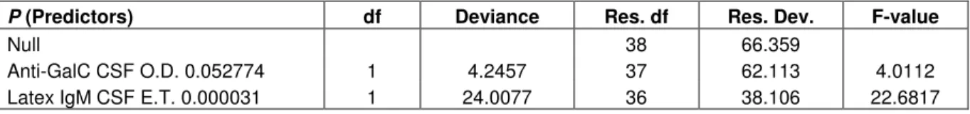
Table 2. Summary of a Generalized Linear Model (GLIM) using a quasi-likelihood error structure (Phi=1.058) and a logit
link function for explaining the 4 HAT clinical groups (see text) with the significant independent parameters retained after a stepwise selection procedure. Final model: Null deviance = 66.36, Residual deviance = 38.10, df = 2,36.
P (Predictors) df Deviance Res. df Res. Dev. F-value
Null 38 66.359
Anti-GalC CSF O.D. 0.052774 1 4.2457 37 62.113 4.0112
Latex IgM CSF E.T. 0.000031 1 24.0077 36 38.106 22.6817
proportions, we first used a Poisson error structure, and then we chose a Quasi-Likelihood estimation with a logit function since residuals tended to be overdispersed (McCullagh and Nelder, 1989). In each case, predictive models based on classification were compared to explanatory models using GLIM.
RESULTS AND DISCUSSION
Thirty-eight patients were included in the present study.
The results of laboratory examinations that are useful for
stage determination according to WHO, and those of
Latex IgM CSF which is the only one new highly
significant variable for the new stage determination are
summarised in Table 1. The best fit was obtained using a
minimal model with lymphocytes/µl, protein in CSF,
LatexIgM CSF, IgG OSAI and Q
alb, to predict assignment
to a given clinical group. According to the multivariate
modeling, 18 patients were classified as group 0, and 2
patients perfectly assigned to group 1. In addition, only 3
patients of group 2 were predicted to belong to this group
(60%), while 9 of those belonging to group 3 were
correctly classified (90%). The classification score, on the
average, was relatively high with 84% of patients correctly
assigned to their relevant group of clinical symptoms.
Results of GLIM to explain the classification of patients
into the 4 clinical groups indicate that the only significant
predictor variable was Latex IgM CSF (
p
=0.000031, Table
2). This finding shows a strong influence of the Latex IgM
CSF variable (85% of the total explained deviance) in
categorizing the level of pathogenesis in patients affected
by sleeping sickness.
Out of the 3 classical tests recommended for stage
determination by WHO (WHO, 1998), CSF protein
concentration is not usually performed because of lack of
reagent necessary for the traditional dye binding protein
assay. In the present study, we used also a more
sophisticated BCA technique (Pierce). However, routine
use of new kit on a large scale in Africa appears to be
unrealistic because of the specific and expensive devices
and reagents required. Likewise, the low sensitivity of the
search for trypanosomes in the CSF by the centrifugation
technique is known as compared to PCR-based methods
used for parasite DNA detection (Truc et al., 1999). Once
again and despite the improvement of its specificity (Truc
et al., 2002), the use of PCR based methods for routine
parasitological diagnosis appears unrealistic in the actual
context in Africa.
Some patients with trypanosomes in CSF were
successfully cured using pentamidine in a previous study
(Doua et al., 1996), probably because of a small amount
of pentamidine passing through the blood-brain barrier
(Van Nieuwenhove, 1999). The last classical test for
stage determination is the CSF cells (or lymphocytes)
count. The cut-off of 5 cells/µl is controversial (Bisser et
al., 1997; Doua and Boa, 1994). In addition, even the
conditions in which lumbar puncture is performed may
influence final cell count. For example, bloody puncture
and small volume of CSF may concentrate some cells
originating from blood mixed together with CSF (when
such a sample is not detectable by eye). Therefore, cell
count takes into account all cells whatever their origin,
leading to an overestimation of cells in CSF. In the
absence of neurological disorders, the relevance of these
3 classical tests remains important because it still leads to
the decision of the appropriate treatment, pentamidine for
P1 and melarsoprol for P2. The problem is not the
reliability of these tests but their doubtful cut-off value and
sensitivity. In the present study, statistical analysis
confirmed the Latex IgM CSF as major parameter for the
determination of CNS involvement (Lejon et al., 2002).
This is an important improvement because Latex IgM
CSF is a cheap and simple test, easy to perform under
field conditions. However, the cut-off value is still
controversial (Lejon et al., 2002). The interest of our
integrated approach is to consider all clinical and
biological results because it has been shown that the
cut-off value or sensitivity of only one technique is not
sufficient to allow an accurate stage determination. The
combination of several parameters appears to be more
discriminant. Although the other biological markers were
already described as specific of a CNS involvement and
most of them are antibodies in serum (cysteine,
anti-NO-cysteine, anti-tryptophan, anti-NO-tryptophan), our
statistical analysis found also that the combination of only
two parameters was much more relevant (Latex IgM CSF
and anti-GalC CSF). It has been mentioned that routine
use of a very sophisticated technique, such as O.D.
determination for anti-GalC CSF under field conditions,
appears unrealistic in the actual context of the control
against HAT in Africa. Furthermore, the high correlation
between IgM and anti-GalC concentrations in the CSF
and the CNS involvement has already been demonstrated
(Bisser et al., 2000).
Therefore, we recommend the use of Latex IgM CSF
only after a large-scale evaluation (in particular to define
an accurate cut-off value). The main advantage is its ease
of use under field conditions and its rapidity. However,
present results suggest that other biological markers still
be of value especially when they can be analysed in
serum. Indeed, this may avoid the lumbar puncture which
is often a traumatism for the patient. Unfortunatly, lumbar
puncture is still in use nowadays.
In the absence of new drug for HAT treatment and
because of the actual situation of sleeping sickness in
Africa, it is crucial to reconsider the stage determination
as soon as possible. Also, further investigations are
required to avoid if possible lumbar puncture in the future.
ACKNOWLEDGEMENTS
We thank the Projet de Recherche Clinique sur la
Trypanosomiase (PRCT, Daloa, Côte d’Ivoire) and the
Regional treatment Center of Bouaflé (Côte d’Ivoire) for
the clinical investigations. We thank also P. Vincendeau
(Faculty of Medicine, Bordeaux, France), M. Dumas and
B. Bouteille (Institut of Tropical Neurology, Limoges,
France), V. Lejon (Institut of Tropical Medicine, Antwerp,
Belgium) for kindly performing serological and CFS tests.
We warmly thank L. Basco and M. Boussinesq (Institut de
Recherche pour le Développement, Cameroon) for the
valuable discussions and critical reading of the
manuscript. This work was supported by Fonds d’Aide à
la Coopération, Ministère français des Affaires
Etrangères, and by the Institut de Recherche pour le
Développement (France).
REFERENCES
Amevidge M, Jauberteau-Marchan MO, Bouteille B, Doua F, Breton JC, Dumas M (1992). Human African trypanosomiasis : presence of antibodies to galactocerebrosides. Am. J. Trop. Med. Hyg. 47: 652-662.
Ayed Z, Brindel I, Bouteille B, Van Merveinne N, Doua F, Houinato D, Dumas M, Jauberteau MO (1997). Detection and characterization of autoantibodies directed against neurofilament proteins in human African trypanosomiasis. Am. J. Trop. Med. Hyg. 57: 1-6.
Bisser S, Ayed Z, Bouteille B, Stanghellini A, Breton JC, Dumas M, Jauberteau MO (2000). Central nervous system involvment in African trypanosomiasis : presence of anti-galactocerebroside antibodies in patients cerebrospinal fluid. Trans. R. Soc. Trop. Med. Hyg. 94: 225-226.
Bisser S, Bouteille B, Sarda J, Stanghellini A, Ricard D, Jauberteau MO, Marchan F, Dumas M, Breton JC (1997). Apport des examens biochimiques dans le diagnostic de la phase nerveuse de la Trypanosomiase Humaine Africaine. Bull. Soc. Path. Ex. 90 (5): 321-326.
Boullerne AI, Petry KG, Meynard M, Geffard M (1995). Indirect evidence for nitric oxide involvment in muliple sclerosis by characterization of
circulating antibodies directed against conjugated S-nitrocysteine. J. Neuroimm 60: 117-124.
Büscher P, Draelants E, Magnus E, Vervoort T, Van Meirvenne N (1991). An experimental latex agglutination test for antibody detection in human African trypanosomiasis. Ann. Soc. Belge Méd. Trop. 55: 559-569.
Büscher P, Lejon V, Magnus E, Van Merveinne N (1999). Improved latex agglutination test for detection of antibodies in serum and cerebrospinal fluid of Trypanosoma brucei gambiense infected patients. Acta Trop. 73: 11-20.
Crawley MJ (1993). GLIM for Ecologists. Blackwell Science Ltd. Oxford, UK.
Doua F, Boa Y (1994). Actualités thérapeutiques de la trypanosomiase. Bull. Soc. Path. Ex. 87: 337-340.
Doua F, Miézan TW, Sanon Singaro JR, Boa Y, Baltz T (1996). The efficacy of pentamidine in the treatment of earlylate stage
Trypanosoma brucei gambiense trypanosomiasis. Am. J. Trop. Med. Hyg. 54: 163-168.
Jamonneau V, Garcia A, Ravel S, Cuny G, Oury B, Solano P, N’Dri L, Sanon R, Frézil JL, Truc P (2002). Genetic characterization of
Trypanosoma brucei gambiense and clinical evolution of human african trypanosomiasis in Côte d’Ivoire. Trop. Med. Int. Health. 7, 7: 610-621.
Lejon V, Büscher P, Sema NH, Magnus E, Van Meirvenne N (1998). Human African trypanosomiasis a latex agglutination field test for quantifying IgM in cerebrospinal. Bull. World Health Organ. 76, 6: 553-558.
Lejon V, Legros D, Richer M, Ruiz JA, Jamonneau V, Truc P, Doua F, Djé N, N’Siesi FX, Bisser S, Magnus E, Wouters I, Konings J, Vervoort T, Sultan F, Büscher P, (2002). IgM quantification in the cerebrospinal fluid of sleeping sickness patients by a latex card agglutination test. Trop. Med. Int. Health. 7, 8: 685-692.
Lumsden WHR, Kimber CD, Evans DA, Doig SJ (1979). Trypanosoma
brucei: Miniature anion-exchange centrifugation technique for detection of low parasitemias; adaptation for field use.Trans. R. Soc. Trop. Med. Hyg. 73: 312-317.
Magnus E, Vervoort T, Van Meirvenne N (1978). A card-agglutination test with stained trypanosomes (CATT) for the serological diagnosis of T. gambiense trypanosomiasis. Ann. Soc. Belge Méd. Trop. 59: 169-176.
McCullagh P, Nelder JA (1989). Generalized linear Models. 2nd edition. Chapman & Hall Ltd. London, UK.
Okomo-Assoumou MC, Geffard M, Daoulouede C, Chaugier C, Lemesre JL, Vincendeau P (1995). Circulating antibodies directed against Tryptophan-like epitopes in sera of patients with Human African Trypanosomiasis. Am. J. Trop. Med. Hyg. 52 (5): 461-7. Reiber H (1998). Cerebrospinal fluid-physiology, analysis and
interpretation of protein patterns for diagnosis of neurological diseases. Mult. Sclerosis. 4: 99-107.
Reiber H, Felgenhauer K (1987). Protein transfer at the blood cerebrospinal fluid barrier and the quantitation of the humoral immune response within the central nervous system. Clin. Chim. Acta. 163: 319-28.
Truc P, Jamonneau V, Cuny G, Frézil JL (1999). Use of polymerase chain reaction in human African trypanosomiasis stage determination and follow-up. Bull. World Health Organ 77, 9: 745-48.
Truc P, Ravel S, Jamonneau V, N’Guessan P, Cuny G (2002). Genetic variability within Trypanosoma brucei gambiense and co-infections by different genotypes in Human African Trypanosomiasis patients in Côte d’Ivoire. Trans. R. Soc. Trop. Med. Hyg. 96 (1): 52-55.
Van Nieuwenhove S (1999). Present strategies in the treatment of human African trypanosomiasis. In : "Progress in Human African Trypanosomiasis sleeping sickness". Editors M Dumas, B Bouteille, A Buguet, Springer Publ. Paris. pp. 253-280.
Wilkinson L, Hill M, Vang E (1992). Sysqtat for the Macintosh. Version 5.2. Evanston, IL. USA.
World Health Organization (1998). Control and surveillance of African trypanosomiasis. WHO Technical Series. 881: 113.