HAL Id: inserm-00168635
https://www.hal.inserm.fr/inserm-00168635
Submitted on 29 Aug 2007HAL is a multi-disciplinary open access archive for the deposit and dissemination of sci-entific research documents, whether they are pub-lished or not. The documents may come from teaching and research institutions in France or abroad, or from public or private research centers.
L’archive ouverte pluridisciplinaire HAL, est destinée au dépôt et à la diffusion de documents scientifiques de niveau recherche, publiés ou non, émanant des établissements d’enseignement et de recherche français ou étrangers, des laboratoires publics ou privés.
Human placental development is impaired by abnormal
human chorionic gonadotropin signaling in trisomy 21
pregnancies.
Guillaume Pidoux, Pascale Gerbaud, Olivier Marpeau, Jean Guibourdenche,
Fatima Ferreira, Josette Badet, Danièle Evain-Brion, Jean-Louis Frendo
To cite this version:
Guillaume Pidoux, Pascale Gerbaud, Olivier Marpeau, Jean Guibourdenche, Fatima Ferreira, et al.. Human placental development is impaired by abnormal human chorionic gonadotropin signaling in trisomy 21 pregnancies.: hCG signaling is impaired in trisomy 21 pregnancy. Endocrinology, Endocrine Society, 2007, 148 (11), pp.5403-13. �10.1210/en.2007-0589�. �inserm-00168635�
Human placental development is impaired by abnormal hCG signaling in
trisomy 21 pregnancies
Guillaume Pidoux*1,2, Pascale Gerbaud*1,2, Olivier Marpeau1,2, Jean Guibourdenche1,3, Fatima Ferreira1,2, Josette Badet1,2, Danièle Evain-Brion1,2 and Jean-Louis Frendo1,2,4.
1INSERM, U767, Université Paris Descartes, Paris, F-75006 France.
2Faculté des Sciences Pharmaceutiques et Biologiques, Université Paris Descartes, Paris, F-75006 France.
3AP-HP, Hôpital Cochin, service de biochimie hormonale, Paris, F-75014 France 4CNRS, Université Paris Descartes, Paris, F-75006 France.
* these two authors contributed equally to the work.
Corresponding author: Dr. Jean-Louis FRENDO, INSERM U767, Faculté de Pharmacie, 4 Avenue de l'Observatoire, 75270 Paris, France. e-mail: jean-louis.frendo@univ-paris5.fr
Running title: HCG signaling is impaired in trisomy 21 pregnancies.
Key words: LH/CG-receptor, trophoblast, Down syndrome, cell-cell fusion, maternal serum markers
Reprint requests to corresponding author.
Disclosure statement: The authors of this manuscript have nothing to declare.
HAL author manuscript inserm-00168635, version 1
HAL author manuscript
Endocrinology (2007) epub ahead of print
HAL author manuscript inserm-00168635, version 1
HAL author manuscript
Endocrinology 2007;148(11):5403-13
HAL author manuscript inserm-00168635, version 1
HAL author manuscript
Abstract
Placental development is markedly abnormal in women bearing a fetus with trisomy 21, with defective syncytiotrophoblast (ST) formation and function. The ST arises from cytotrophoblast (CT) fusion and plays an essential role by secreting hCG, which is essential to placental development. In T21 pregnancies, CT do not fuse and differentiate properly into ST leading to the secretion of an abnormal and weakly bioactive hCG. In this study, we report for the first time, a marked decrease in the number of mature hCG receptor (LH/CG-R) molecules expressed at the surface of T21-affected CT. The LH/CG-R seems to be functional based on sequencing that revealed no mutations or deletions and binding of recombinant hCG as well as endogenous hCG. We hypothesize that weakly bioactive hCG and lower LH/CG-R expression may be involved in the defect of ST formation. Interestingly, the defective ST formation is mimicked in normal CT cultures by using LH/CG-R siRNA which result in a lower hCG secretion. Furthermore, treatment of T21-affected CT with recombinant hCG overcomes
in vitro the T21 phenotype, allowing CT to fuse and form a large ST. These results illustrate for the
first time in trisomy 21 pathology, how abnormal endogenous hCG signaling impairs human placental development.
Introduction
The human placenta is characterized by extensive invasion of the trophoblast in the maternal uterus, creating direct trophoblast contact with maternal blood (haemochorial placentation). In early pregnancy, cytotrophoblasts (CT) proliferate and invade the maternal endometrium to form the anchoring villi (1). Cytotrophoblasts also differentiate into a continuous multinucleated layer known as the syncytiotrophoblast (ST).
Human chorionic gonadotropin (hCG) is produced by the trophoblast, and especially by the ST covering the chorionic villi and bathing in maternal blood (2). The syncytiotrophoblast plays an essential role during pregnancy by allowing fetomaternal exchanges and by secreting placental hormones into the maternal blood. In vivo and in vitro, the syncytiotrophoblast arises from cytotrophoblastic cell fusion and differentiation. Numerous factors regulate ST formation, in an autocrine or paracrine manner (see for review (3)), including hCG (4, 5), and oxidative stress related to overexpression of copper/zinc superoxide dismutase located on chromosome 21 (6, 7). The molecular mechanisms underlying CT fusion and differentiation are poorly understood, but proteins involved in cell adhesion (cadherin 11)
(8) and cell-cell communication (connexin 43)
(9) are directly involved. We also recently demonstrated the direct involvement of syncytin 1, a human endogenous retroviral envelope glycoprotein (10).
Very few of the genes involved in human placental development and trophoblast differentiation have been identified. In contrast, with the increasing number of transgenic and knock-out mice and rats, many of the genes involved in murine placental development have been characterized (see for review (11)). However, results obtained in mice are difficult to extrapolate to humans, owing to the specific features of human placental development (3). For instance, hCG does not exist in mice and rats. Anomalies in CT differentiation and cell fusion may lead to severe placental abnormalities. In trisomy 21-affected pregnancies, the CT fuse poorly or tardily and the resulting defect in ST formation is associated with a decrease in hCG synthesis and secretion (12). We recently
demonstrated that hCG secreted by T21-affected CT is abnormally glycosylated (13), and Susan Fisher's group has described variable defects in CT differentiation along the invasive pathway
(14).
Trisomy of chromosome 21 (T21), which causes the phenotype known as Down’s syndrome, is the major known genetic cause of mental retardation, affecting about 1 in 800 live births. Screening strategies to identify women at an increased risk of bearing a T21 fetus are based on maternal age, ultrasound signs and maternal serum markers (15). Some of these markers, such as hCG, are of placental origin. The hCG level in maternal serum is abnormally elevated at 14–18 weeks in pregnancies with a T21 fetus, for reasons that are largely unknown.
HCG belongs to the family of gonadotrophin hormones, that also includes luteinizing hormone (LH), follicle-stimulating hormone (FSH) and thyroid-stimulating hormone (TSH) (16). These glycoprotein hormones are composed of two subunits, α and β. The α subunit, common to the other gonadotrophin hormones, is a 92-amino-acid polypeptide with two N-linked oligosaccharides. β-hCG is a 145-amino-acid polypeptide with two N-linked oligosaccharides and four O-linked oligosaccharides (17). The action of hCG in stimulating CT fusion and differentiation is primarily mediated via the chorionic gonadotropin receptor (LH/CG-R), which can also bind hLH (4, 5, 18). When engaged by these hormones, the LH/CG-R couples to a number of G-proteins and activates adenylate cyclase, phospholipase C and ion channels, thereby stimulating the cAMP and inositol phosphate signaling cascades (19, 20). LH/CG-R, which has seven transmembrane domains, belongs to a subfamily of G-protein-coupled receptors (21) also comprising the FSH receptor (FSH-R) and the TSH receptor (TSH-R). The human LH/CG-R gene has been assigned to chromosome 2p21 (22). Its coding region is over 60 kb long, and it has been cloned in pig, mouse, rat and also human, in whom it is composed of 11 exons and 10 introns (16, 21, 23, 24). LHR has been also cloned in fishes (25-28), monkeys (29), bears (30) and many other species. The presence of LH/CG-R in human placenta was first described by Alsat (31) and subsequently confirmed by other authors (32, 33). We recently showed that LH/CG-R
expression is modulated during normal CT fusion and differentiation (34).
To better understand the defective ST formation occurring in T21-affected pregnancies, we studied the involvement of the abnormal hCG by examining its function and receptor interaction. We found that T21-affected pregnancy is associated with a low LH/CG-R expression, and that the secreted abnormal hCG can bind to its receptor. This low LH/CG-R expression, together with the secretion of abnormal hCG, is involved in the defective ST formation, as specific inhibition of LH/CG-R expression by siRNA in normal CT mimics the T21 phenotype (defective ST formation). More interestingly, treatment of T21-affected CT in vitro with normal recombinant hCG overcome the T21 phenotype, allowing CT to fuse and form a large ST.
Materials and Methods
Placental tissue collection.
French law allows termination of pregnancy with no gestational age limit when severe fetal abnormalities are present. Placentas were collected at the time of termination, between 12 and 35 weeks of gestation (amenorrhea), in T21-affected pregnancies and gestational-age-matched control pregnancies. Gestational age was confirmed by sonographic measurement of crown-rump length at 8-12 wk of gestation. Control pregnancies were terminated because of severe bilateral or low obstructive uropathies or major cardiac abnormalities. The karyotype of placental cells was determined in all cases (free T21 or normal). The study was approved by our local ethics committee.
Trophoblast cell culture
Cytotrophoblasts from normal and trisomic placentas were isolated as previously described
(35). After sequential trypsin/DNase I digestion followed by Percoll gradient centrifugation, the cells were further purified by negative selection to obtain a trophoblast preparation not contaminated by other cells, by using monoclonal anti-human leukocytic antigen A, B and C antibodies (W6-32HL, Sera Lab, Crawley Down, UK) according to a published method
(36, 37). This antibody reacts with most cell types (e.g. macrophages, fibroblasts, extravillous trophoblasts) but not with villous cyto- or syncytiotrophoblasts. Cytokeratin 7 immunocytochemistry was used to confirm the
cytotrophoblastic nature of attached cells: 95-98% of the cells were positively stained.
Hormone assay
The hCG concentration was determined in culture medium at 24h and 72h by using an enzyme-linked fluorescence assay (Vidas System, BioMerieux, Marcy l’Etoile, France) with a detection limit of 2 mU/ml. All values are the mean ± SEM of triplicate determinations.
HCG biological activity assay
The biological activity of secreted hCG was tested on Leydig cells (MA-10 cells, a generous gift from Pr. M. Ascoli, University of Iowa, Iowa City, IA) as previously described (38). HCG levels were first assayed in trophoblast culture medium. Various amounts of culture medium were added as previously described (13). The results were expressed as the progesterone concentration per number of cells for each hCG concentration added to the control and T21 trophoblast culture medium. Progesterone was assayed with the ACS180SE instrument (Bayer), a polyclonal antibody against hCG (A0231, rabbit anti-human, Dako©, Glostrup, Denmark at 7µg/ml), and a polyclonal antibody against LH/CG-R (LHR H50, rabbit anti human, Santa Cruz biotechnology inc, CA, USA at 2µg/ml) to block the action of hCG on MA-10 cells.
Immunoblotting
Proteins (70µg) were solubilized in RIPA buffer, submitted to 7.5% SDS-PAGE and transferred to nitrocellulose membranes. The membranes were immunoblotted with a polyclonal antibody against human LH/CG-R (LHR-H50, rabbit anti-human, Santa-Cruz Biotechnology Inc, CA, USA) at 2µg/ml, and the specific band was revealed by chemiluminescence (West Pico Chemiluminescent, Pierce, Rockford, IL, USA) after incubation with an anti-rabbit peroxidase-coupled antibody (Jackson ImmunoResearch, Baltimore, USA). Actin was immunoblotted with a rabbit polyclonal anti-actin antibody (Sigma-Aldrich, MO, USA, at 1:1000).
Cross-linking and immunoprecipitation
DTSSP (3,3’-Dithiobis [sulfosuccinimidyl-propionate]) is a soluble, homobifunctional N-hydroxysuccinimide ester (NHS-ester). This cross-linker is thiol-clivable and primary amine-reactive. NHS-ester reactions with primary amines form covalent amide bonds that results in
the release of N-hydroxysuccinimide. To cleave the covalent bond, we used 10mM DTT at 37°C for 30min.
Protein G Plus-Agarose (Immunoprecipitation Reagent, Santa-Cruz biotechnology inc, CA, USA) was pre-mixed with a polyclonal antibody to human LHCG-R (LHR-H50). Cells (106/well) were seeded in six-well plates and cultured as previously described, except for overnight serum-free cultures. After 24h of culture, 2mM DTSSP was added to the culture medium for 30min at 25°C to cross-link hCG to LHCG-R. Stop solution (Tris/Glycine 20mM, pH 7.5) was then added at 25°C for 15min. The cells were then washed with PBS and scraped in ice-cold RIPA buffer. After sonication the cellular extract was transferred to the immuno-complex Protein G-antibody to human LHCG-R, incubated overnight at 4°C and washed four times in RIPA buffer. Proteins were reduced with 10mM DTT (sufficient to cleave the covalent bond), and eluted by heating at 60°C for 10min in 1x electrophoresis sample buffer (Bio-Rad laboratories, CA, USA). Aliquots were submitted to 7.5% SDS-PAGE and transferred to nitrocellulose membranes. Immunoprecipitates were treated with antibodies as described above.
RNA extraction and RT-polymerase chain reaction (RT-PCR)
Total RNA was extracted from trophoblastic cells after 24h of culture by using the Trizol® reagent (Invitrogen Life Technologies, CA, USA). RT-PCR was performed as previously described (34) using specific oligonucleotide primers based on the coding sequence of the LH/CG-R (NM 000233) (Fig. 3A): P1(+): 5'-CA GACTTTTGCATGGGGCTC-3', P1(-): 5'-GTG GCAGTGGTCATAGACTACAC-3', P2(+): 5'-G CATCTGTAACACAGGCATC-3', P2(-): 5'-CA TCTGGTTCAGGAGCACAT-3', P3(+): 5'-CA AGCTTTCAGAGGACTTAATGAGGTC-3', P3 (-): 5'-AAAGCACAGCAGTGG CTGGGGTA-3', Actin (NM 001101) (+): 5'-GTGGGGCGCC CCAGGCACCA-3' and Actin(-): 5'-CTCCTTA ATGTCACGACGATTTC-3'.
RNA samples pretreated with DNAse I were also amplified as controls. Amplified products were analyzed by electrophoresis on 1.8% agarose gel, visualized with ethidium bromide, and transferred to membranes (GeneScreen, NEN Life Science Products Inc, MA, USA). The amplified cDNA was hybridized with three LH/CG-R-specific cDNA probes (P1, P2 and P3) radiolabeled with 32P by using a random priming
method. P1 is specific for the LH/CG-R trans-membrane and intracellular domains and spans positions 1300 to 2099 (position 1 corresponds to the A of the ATG start codon of the LH/CG-R coding sequence); P2 and P3 were specific for the extracellular domain, located from positions 386 to 1055 and 199 to 845, respectively. After prehybridization for 4h at 60°C in 50% deionized formamide, 1% SDS, 2xSSC, 10% dextran sulfate, the membranes were hybridized at 60°C in the same buffer containing the specific LH/CG-R cDNA probes and were washed at 55°C, twice in 2xSSC, 1% SDS for 30min, 2xSSC containing 0.1% SDS for 30min, and 2xSSC for 5min. The membranes were analyzed by Cyclone (Storage Phosphor System, Hewlett Packard. France).
Cloning and DNA sequencing of LH/CG-R
PCR products were eluted from agarose gel, cloned into the pCRII-TOPO vector and sequenced as previously described (34).
Binding assay
Trophoblastic cells (106/well) were seeded in six-well plates and cultured as described above. After 24h of culture they were washed and placed 2h in DMEM without FCS, then placed in 1ml of DMEM, 0.1% BSA, 1mM HEPES. To determine the time of 125I-hCG incubation for maximum binding, we performed a time-course study at 25°C (from 10min to 2h) with 0.5nM 125I-hCG. Thirty minutes was the most effective time, corresponding to maximum binding of 125 I-hCG in trophoblastic cells (data not shown). For equilibrium binding experiments the cells were incubated for 30 min at 25°C with 0.5nM 125 I-hCG and with increasing concentrations of unlabeled hCG (from 10-12 M to 10-8 M, C6322, Sigma-Aldrich, MO, USA). At the end of the incubation period the cells were washed and scrapped, and bound radioactivity was counted. Assays were performed in triplicate. Data were analyzed by using the LIGAND fitting program (version 4.97) (39).
125I-labeled hCG was prepared as described by Hunter (40), using chloramine T as the oxidative reagent, as previously described (34).
Intracellular cAMP determination
At 24h of culture, cells (106/well) were stimulated with increasing concentrations of hCG (from 10-12 M to 10-6 M, C6322, Sigma-Aldrich, MO, USA) in the presence of IBMX (3-isobutyl-1-methylxanthine) to prevent cAMP
degradation. Cells were frozen on dry ice and cAMP was extracted from with ice-cold 65% ethanol. The extracts were dried and kept at -20°C until use. Cyclic AMP concentrations were determined with an assay kit (Amersham Biosciences, NJ, USA) as previously described
(34). Assays were performed in sextuplet. To determine the optimum time of cAMP accumulation under hCG stimulation, we performed a time-course study (from 5min to 1h) by stimulating trophoblasts with hCG (10-12M to 10-6M). The most effective stimulating time was 20min (data not shown). We used a polyclonal antibody against hCG (A0231, rabbit anti human, Dako, Glostrup, Denmark at 7µg/ml) to block the action of hCG on trophoblasts.
LH/CG-R siRNA protocol
LH/CG-R siRNA was a Smartpool mix (4 different LH/CG-R siRNAs pooled) purchased from Dharmacon (Lafayette, CO, USA). SiRNA transfection was performed using the DharmaFECT 2 siRNA transfection reagent (Dharmacon, Lafayette, CO, USA) according to the manufacturer’s protocol. Briefly, 5µl (20µM) of LH/CG-R siRNA (M-003681, Dharmacon, Lafayette, CO, USA) or scrambled siRNA (46-2629, Invitrogen Life Technologies, CA, USA) was diluted in 245µl of OPTI-MEM (Invitrogen Life Technologies, CA, USA) and 4µl of transfection reagent (DharmaFECT 2) was diluted in 246µl of OPTI-MEM. The two solutions were incubated for 5min at room temperature then combined and incubated for 20min at room temperature. The mixture was added to the cells (2.0x10⁶/well) and incubated for 48h at 37°C in air-5% CO2. After transfection the medium was removed and kept for hormone assay. Cells were collected and used for immunoblot analysis.
Transfection efficiency was determined by testing siRNA uptake by primary cytotrophoblast cultures. After 5 h of culture, cytotrophoblasts were incubated with a fluorescein-labeled dsRNA oligomer for 18 h, then washed three times in PBS, fixed at 24, 48 and 72 h of culture and analyzed by fluorescence microscopy. The dsRNA oligomer were taken up from the first 24 hours (60% of cells were labeled) and the proportion of labeled cells then increased progressively with the time (75% at 48 hours, 85% at 72 hours). Whereas the number of dead cells after transfection remains constant (∼10%) during the culture and at a very low rates. Nuclei
were stained blue by using the Hœchst 33342 reagent. A dead cell reagent (ethidium homodimer-I, staining dead cells red) was used to assed cell viability after transfection, visually or quantitatively. Both reagents are fluorescent compounds that bind to DNA; however, Hœchst 33342 binds to DNA in living cells, while the dead cell reagent binds only to the DNA of dying cells. Transfected cells can be visualized by fluorescence microscopy, as they integrate the fluorescein-labeled dsRNA oligomer. This experiment enabled us to determine the optimum concentrations of siRNA and transfection reagents.
Statistical tests
Statistical analysis was performed using the StatView F-4.5 software package (Abacus Concepts, Inc., CA, USA). Values are presented as mean ± SEM. Significant differences were identified by using ANOVA, and p<0.05 was considered significant.
Results
Defective ST formation in T21
cytotrophoblastic cells
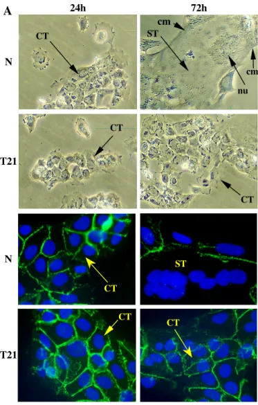
Mononucleated cytotrophoblasts isolated from normal placenta aggregate and fuse to form a syncytiotrophoblast at 72h of culture. In contrast, cytotrophoblasts isolated from T21-affected placentas aggregate but fuse poorly, forming a few small syncytiotrophoblasts after 3 days of culture (Fig.1A). With cells isolated from normal placenta, in vitro syncytiotrophoblast formation is associated with an increase in hCG secretion into the culture medium (Fig.1B), from 7.4±2.3 (in mUI/ml/106 cells) at 24h to 1089±61 at 72h. With cells isolated from T21-affected placentas, the defective syncytiotrophoblast formation is associated with a significant lower (p<0.0001) hCG secretion into the culture medium compared to normal cells (16.8±9.5 and 366±28 mIU/ml/106 cells at 24h and 72h respectively) (Fig.1B).
Defective LH/CG-R expression in T21 cytrophoblastic cells
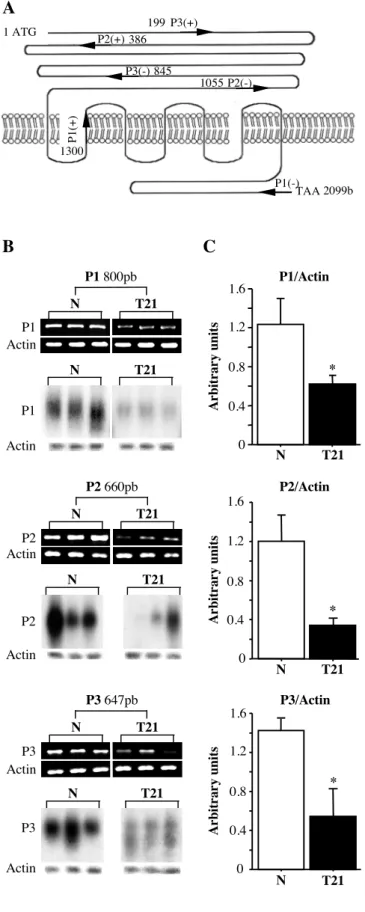
HCG secretion by the ST is lower in T21 than in normal cells. In order to evaluate the role of hCG and its receptor in ST formation, we first studied the expression of LH/CG-R mRNA in normal and T21-affected cytotrophoblasts at 24h of culture. We carried out semi-quantitative RT-PCR experiments with three sets of primers (P1, P2 and P3; positions indicated in Fig.2A). To
avoid contamination by genomic DNA, each RNA extract was pretreated with DNAse I. Moreover, the primers of pairs P2 and P3 were located on separated exons.
As shown in figure 2B, the 800-bp, 660-bp and 647-bp fragments obtained with P1, P2 and P3 respectively indicated that LH/CG-R mRNA was significantly less abundant in T21-affected cytotrophoblasts than in normal cytotrophoblasts. No significant difference was noted in the actin mRNA (control) level. To confirm the specificity of these RT-PCR results, amplification products were transferred to Nylon membranes and hybridized with 32P-radiolabelled probes. We used three probes specific for LH/CG-R, spanning the extracellular, transmembrane and intracellular domains (for positions see Materials and Methods). Hybridization confirmed the significant lower LH/CG-R mRNA expression in T21-affected cytotrophoblasts. No difference was noted with an actin-specific control probe. Normalization of LH/CG-R mRNA to actin mRNA confirmed the decrease in LH/CG-R mRNA levels in T21-affected cytotrophoblasts (Fig.2C). Using primer set P1, LH/CG-R mRNA levels were 1.23±0.26 (in arbitrary units) in normal CT compared to 0.6±0.1 in T21-affected CT (p<0.013). A similar decrease was observed with primer sets P2 (1.20±0.26 and 0.35±0.06; p<0.034) and P3 (1.42±0.12 and 0.54±0.28; p<0.043). Interestingly, the relative expression was similar with the three primer sets (respectively 2.0-, 3.4- and 2.6-fold).
We then used western blot to determine LHCG-R protein expression in extracts of normal and T21-affected CT, using the polyclonal antibody LHR-H50 (Fig.3A). Two major bands were observed: a 85-95 kDa band corresponding to the mature form of the LH/CG-R (noted m on Fig.3A) present at the cell surface, and a 65-75 kDa band which is the precursor (noted p) of the cell-surface receptor (34) (for review see (41)). As shown in Figure 3A, the mature form of LH/CG-R was far less abundant in T21-affected CT than in normal CT, whereas no significant difference in actin expression was observed. Normalization of mature LH/CG-R protein expression to actin expression showed a significant difference (at least 68%) between normal CT and T21-affected CT (9.2±0.7 and 2.9±1.4 arbitrary units; p<0.0038).
To confirm the decrease in LH/CG-R mRNA and protein levels, we performed binding experiments with 125I-hCG at 24h of culture of normal and T21-affected CT (Fig.3B). Scatchard analysis showed that the number of 125I-hCG molecules bound per normal CT (3511±693) was significantly higher (p<0.04) than that in T21-affected CT (1124±350). The difference in Kd values between normal CT (0.5±0.2nM) and T21-affected CT (0.4±0.2nM) was not statistically significant. These results indicate that T21 CT express 3 times fewer LH/CG cell-surface receptors less than normal CT. However, the LH/CG-R molecule expressed at the surface of T21 CT bound 125I-hCG with the same affinity as the LH/CG-R on normal CT.
The reduced level of functional mature LH/CG-R at the cell surface of T21-affected CT was confirmed by measuring cAMP production in response to increasing hCG concentrations, at 24h of culture. As shown in figure 3C, at 10-10M hCG, corresponding to maximum cAMP accumulation, the ability of hCG to stimulate cAMP production in trophoblastic cells was significantly higher (p<0.007) in normal CT (222±3 fmol/mg of protein) than in T21-affected CT (164±10 fmol/mg of protein). Stimulation with epinephrin (used as a positive control) induced similar accumulation of intracellular cAMP in T21-affected CT as in normal CT, showing that the T21-affected CT were viable and that the reduced cAMP production was not due to increased apoptosis of T21 cells or to a defect in the cAMP pathway.
These results clearly shown that LH/CG-R expression at the surface of trophoblastic cells is markedly reduced in T21-affected pregnancies. Specific inhibition of LH/CG-R expression by siRNA inhibits syncytium formation and hCG secretion by normal cytotrophoblastic cells We then tried to mimic with normal CT what we observed in T21-affected CT, by incubating normal CT with LH/CG-R siRNA. As shown by western-blot analysis (Fig.4B), LH/CG-R siRNA markedly reduced LH/CG-R protein expression. Normalization of LH/CG-R protein expression to actin expression showed 74% of inhibition compared to cells tranfected with scrambled siRNA (8.0±0.1 arbitrary units; p<0.002). A similar decrease (78% inhibition; p<0.002) was also found when we compared control non
transfected cells to cells transfected with LH/CG-R siLH/CG-RNA. No difference was seen between non transfected cells (control) and cells transfected with scrambled siRNA, indicating that transfection had no effect on the decreased receptor expression.
Specific inhibition of LH/CG-R expression by siRNA was associated with a strong decrease in CT fusion and differentiation. The histogram in Fig.4A shows that there were more mononuclear cells (58.5±0.2%) in cultures treated with LH/CG-R siRNA than in those incubated with scrambled siRNA (32.5±0.1%; p<0.005). Calculation of an apparent fusion index showed a 2.1-fold decrease in fusion (53.3±7.1% of scrambled-siRNA-treated cells fused, compared to 25.0±0.6% of LH/CG-R siRNA-treated cells; p<0.017). LH/CG-R siRNA treatment led to a decrease in the number and size of syncytia. Similar results were obtained when LH/CG-R siRNA-treated cells were compared to non transfected control cells (data not shown). Interestingly, the decrease in syncytium formation observed when normal cells were treated with LH/CG-R siRNA was associated with a decrease of syncytium function. As illustrated in figure 4C, hCG secretion into the culture medium was far lower with LH/CG-R siRNA-treated cells than with scrambled-siRNA-treated cells (59% reduction) or control cells (66% reduction). This result was not due to a difference in cell viability following transfection, as hCG secretion by control and scrambled-siRNA-treated cells was similar.
These results point to a direct role of LH-CG-R in CT fusion and differentiation during ST formation.
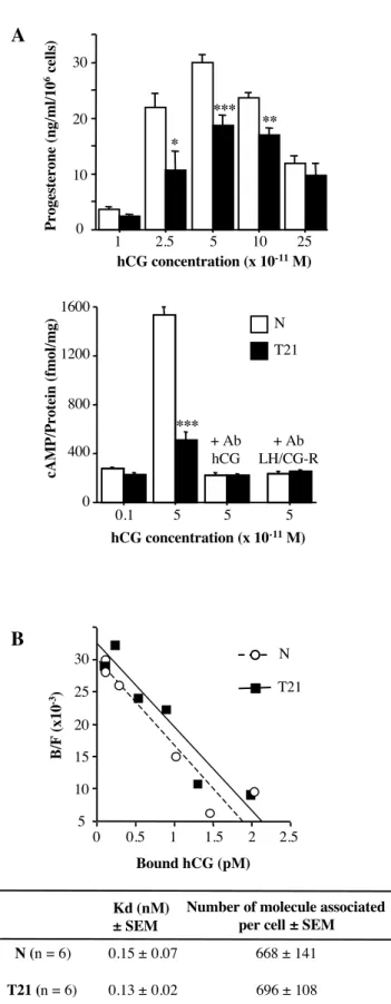
LH/CG receptors of T21-affected trophoblasts bind endogenous weakly bioactive hCG We have shown that T21-affected CT bear a reduced number of LH/CG-R. We then analyzed the bioactivity of the endogenous hCG ligand by using the well established test of hCG function on MA-10 Leydig cells (38). HCG secreted into the culture medium at 72h by normal (n=3) and T21-affected trophoblasts (n=3) were used to stimulate steroid production by Leydig cells, which constitutively express LH/CG receptors. At equivalent hCG concentrations in the culture medium (from 2.5x10⁻¹¹M to 10-10M), the ability of hCG secreted by T21-affected trophoblasts to
stimulate Leydig cell progesterone secretion was significantly decreased (Fig.5A, upper panel). To emphasize this result, we quantified the production of intracellular cAMP by Leydig cells after stimulation with hCG from normal (n=3) and T21 (n=3) culture medium. In view of previous results, we used two hCG concentrations to stimulate Leydig cells: 0.1x10 -11M, which does not elicit progesterone secretion, and 5x10-11M, which leads to maximal progesterone secretion. The histogram in Fig.5A (lower panel) shows that stimulation with hCG secreted at 72h of culture by T21-affected trophoblasts was associated with significantly lower (at least 3-fold) cAMP production than was hCG secreted by normal trophoblasts (510±64 versus 1535±61 fmol/mg of protein; p<0.0001). Intracellular cAMP accumulation occurred after hCG stimulation, as no cAMP production was detectable when the culture media were preincubated with anti-hCG (Ab-hCG) or anti-LH/CG-R (Ab-LH/CG-R) blocking antibodies before hCG stimulation. We obtained similar results when we used hCG secreted at 24h by normal and T21-affected trophoblasts (data not shown).
This reduction in progesterone secretion and cAMP production after stimulation with hCG secreted by T21-affected trophoblasts was not due to lesser binding of this hormone to its receptor. Scatchard analysis (Fig.5B) showed that hCG secreted by T21-affected trophoblasts bound to LH/CG receptors expressed by Leydig cells with the same affinity (Kd=0.13±0.01nM) as hCG secreted by normal trophoblasts (Kd=0.15±0.07nM).
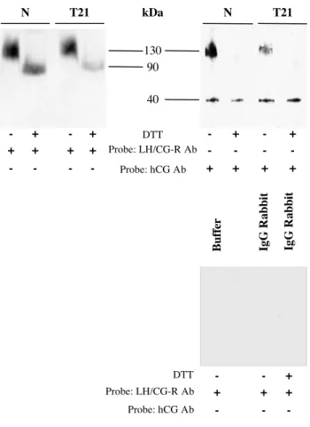
Our results clearly show that hCG secreted by T21-affected trophoblasts is less bioactive than normal hCG, and that this is not due to deficient binding to the LH/CG receptor, as expressed on Leydig cells. We then studied the interaction of the LH/CG-R expressed by T21-affected CT with endogenous hCG secreted by the same cells. For this purpose we cultured T21-affected CT and cross-linked the endogenous hCG secreted into the culture medium to its receptor to form hCG-LH/CG-R complexes. These complexes were immunoprecipitated (Fig.6) by using an anti-LH/CG-R polyclonal antibody (LHR-H50) and were then incubated in presence or absence of DTT. We used the DTSSP cross-linker agent, which is cleavable in the presence of DTT (used as a reducing agent). After immunoprecipitation
the complexes were probed with an anti-LH/CG-R antibody (Fig.6, left). Without the reducing agent (-DTT), we observed a band at approximately 130-kDa, corresponding to hCG cross-linked to LH/CG-R. Presence of DTT (+DTT) disrupted the hormone-receptor complex and the band at 130-kDa disappeared; a band corresponding to the receptor alone then appeared at 90-kDa. To ensure that the 130-kDa band corresponded to hCG-LH/CG-R complexes, we used an anti-hCG polyclonal antibody to treat the previous immunoprecipitates (Fig.6, right). In non denaturing conditions, a 130-kDa band corresponding to hCG bound to the LH/CG-R and a smaller band (40-kDa), corresponding to total hCG, was observed. In reducing conditions only the 40-kDa band was observed. No 130-kDa band was found as hCG-LH/CG-R complexes were disrupted. The interactions and complexes observed in these experiments were specific, as no cross-reactions occurred in a range of control conditions (Fig.6, lower).
These results show that T21 trophoblastic cells produce an abnormal hCG that is weakly bioactive but that can nonetheless bind its receptor LH/CG.
Defective ST formation by T21
cytotrophoblastic cells is overcome by recombinant hCG treatment
Interestingly, as shown in figure 7A, addition of recombinant hCG (rhCG, 10-8M) to the culture medium of T21-affected CT induced syncytiotrophoblast formation. T21 CT cultured for 72h contained twice as many mononuclear cells in control conditions (76.2±0.6%) than when treated with rhCG (39.3±0.9%; p<0.0001). In other words, rhCG induced T21-affected CT differentiation and fusion, as more than 60% of the cells participated in syncytia formation (versus 23% of untreated cells) (Fig.7B). With T21 cells the percentage of syncytia containing 10 to 50 nuclei rose from 1.6±0.3% with untreated cells (controls) to 13.0±1.7% with treated cells (+rhCG) (p<0.003). In contrast, the proportion of mononuclear cells observed at 72 h of culture of normal CT was not affected by rhCG (+rhCG: 15.8±1.2%, -rhCG: 17.2±0.4%; Fig.7B). Indeed, rhCG promoted the fusion of already formed syncytia, producing larger syncytia (Fig.7B); the percentage of syncytia containing more than 50 nuclei was significantly
higher with treated cells than with control cells (p<0.0001).
These results show that the defective cell fusion and differentiation of T21-affected CT may be overcome. More importantly, they show the key role of hCG in ST formation, as the addition of recombinant hCG to the T21 CT culture medium led to CT fusion and ST formation.
Discussion
Placental development is severely abnormal in women bearing a T21 fetus, with defective syncytiotrophoblast formation and function leading to the secretion of an abnormal hyperglycosylated hCG (12, 13). We show here, for the first time, that treatment of T21-affected cytotrophoblasts with normal or recombinant hCG restores their capacity to form a syncytiotrophoblast. This reversal of the T21 phenotype indicates that T21 CT have all the equipment required to fuse and differentiate. The abnormal CT fusion and differentiation observed in T21-affected pregnancies may thus be due to impaired hCG signaling. This is supported by our observation that specific inhibition of LH/CG-R expression by siRNA in normal CT mimics the T21 phenotype (defective ST formation).
In this study, we clearly show that LH/CG-R expression, present at the cell surface of the cells, was far lower on T21-affected CT than on normal CT, as shown by using several complementary methods and a well-characterized model of human villous trophoblast differentiation in vitro. LH/CG-R mRNA and protein levels were lower in T21 cells, as also observed in situ by means of immunohistochemical studies (data not shown). These results are in line with those reported by K. Nicolaides, who demonstrated in total placental extracts that LH/CG-R expression was significantly lower in T21 pregnancies than in of controls (42), whereas CV. Rao described stronger expression of LH/CG-R in T21 placentas (43). This divergence may stem from the use of different approaches and tools. Rao quantified LH/CG-R immunostaining in total samples of placental villous tissue and may thus have over-estimated LH/CG-R expression, as the receptor is also expressed in Hofbauer and endothelial cells (44, 45) in villous stromal tissue and by intermediate trophoblasts (46). Moreover, experiments with radiolabeled probes, such as in
situ hybridization, use porcine cDNA that shares
only 88% of the human sequence. In this study, we designed specific probes for the human LH/CG receptor in normal and T21-affected villous CT. In addition, we quantified the mature form of the receptor expressed at the CT surface, whereas the other authors quantified all LH/CG-R isoforms.
Scatchard plots clearly showed that the maximum number of hCG molecules bound per cell was significantly lower on T21-affected CT than on normal CT. This lower cell-surface receptor expression by T21-affected CT was confirmed by the lower cAMP production observed after stimulation with recombinant hCG. This decrease in cAMP production was not due to a loss of receptor affinity for the recombinant hormone, as recombinant hCG was able to bind to LH/CG-R on both normal and T21-affected CT with the same apparent affinity (same Kd values). Apart from the smaller number of LH/CG-R molecules expressed at the surface of T21-affected CT, the LH/CG-R seems to be normal, as sequencing revealed no mutations or deletions. Moreover, the receptor was functional and able to signal after stimulation with recombinant hCG. Indeed, replacing the abnormal endogenous hCG by the recombinant hormone in T21-affected CT cultures enhanced ST formation. This also implied that a functional hormone is necessary for ST formation. In other words, the secretion of abnormal hCG by T21-affected cells might be responsible for the defective CT differentiation. We have previously shown that hCG is hyperglycosylated in T21 pregnancies by using different lectins (13). Indeed, mRNA levels of two enzymes involved in the glycosylation pathway, sialyl-transferase-1 (which adds a sialyl group to antennary structures) and fucosyl-transferase-1 (which adds a fucose to the first N-acetyl-glucosamine of glycoproteins) were significantly higher in cultured trophoblasts isolated from trisomy 21 placenta {Frendo, 2004 #285}. We show here that it is biologically less functional on cytototrophoblast differentiation. We also demonstrate here that this abnormal hormone is able to bind its receptor.
One particularly interesting result is the differential effect of recombinant hCG on normal and T21-affected mononuclear CT (Fig. 1C). Recombinant hCG did not reduce the percentage of normal unfused mononuclear CT but rather
induced the fusion of already formed syncytia with one another, leading to huge ST containing more than 100 nuclei. In contrast, recombinant hCG induced the differentiation of T21 CT into an ST. At 24h of culture, the percentage of unfused CT was higher than in normal placenta, suggesting that their maturation or differentiation is delayed. Recombinant hCG enhanced CT fusion and differentiation into ST, possibly through the induction of LH/CG-R expression at the CT surface. A similar form of regulation has been described for EGF, which upregulates EGF-receptor mRNA and protein expression in human prostate cancer (47). Another possible explanation is that, in T21, despite the lower LH/CG-R expression, the level of expression is still sufficient (above a critical threshold of receptor density required to induce differentiation) and the defective differentiation is due to the abnormal secreted hCG molecules. By removing the latter from the culture medium and replacing them with recombinant hCG, we restored the CT fusion and differentiation process. However, even after stimulation with recombinant hCG the rate of fusion was never as high as that observed with normal cells, whether or not they were treated with recombinant hCG. This difference may be due to the lower number of LH/CG-R molecules expressed at the surface of T21 CT. Indeed, our results show that LH/CG-R is directly involved in human trophoblast cell fusion and differentiation, as its inhibition by specific siRNA reduce trophoblast cell fusion. It appears that the hCG-LH/CG-R system acts as a positive feedback system: if hCG signaling is intact, then ST formation is increased and hCG production as well, resulting in increased ST formation.
Screening strategies used to identify women at an increased risk of bearing a T21 fetus are partly based on maternal serum markers such as hCG. The hCG level in maternal serum is elevated during T21 pregnancies, for reasons that remain largely unknown. We demonstrate that, despite this increase, the autocrine/paracrine effects of hCG on the placenta are severely impaired owing to a loss of hormone function and reduced expression of the mature form of the LH/CG receptor at the cell surface. The conjunction of these two phenomena results in inadequate receptor-mediated signaling, leading to hCG accumulation in maternal serum. Abnormal receptor expression leading to
hormone accumulation has already been described in various systems (48-50).
The main clinical relevance of this report is that it shows the significance of hCG in establishing and maintaining placental and fetal development during human pregnancy. We clearly demonstrate that, in pregnancies associated with a T21 fetus, the placenta secretes an abnormal and weakly bioactive hCG molecule that cannot correctly stimulate CT differentiation. In addition, the subnormal expression of functional LH/CG-R protein in the placenta of T21 pregnancies may have far-reaching consequences. For instance, the rate of spontaneous abortion is high in T21-affected pregnancies. Aneuploidy might alter the fetal cells’ ability to differentiate properly. The morphological, phenotypic and functional differences among T21-affected trophoblastic cells may explain why a significant number of pregnancies end in spontaneous miscarriage.
Acknowledgments
We thank Dr Fanny Lewin for her support and the staff of the Saint Vincent de Paul Obstetrics Department for providing us with placentas. This work was supported by la Caisse d'Assurance Maladie des Professions Libérales Province. Guillaume Pidoux was supported by a fellowship from Conseil Regional d'Ile-de-France and Jean-Louis Frendo by a grant from INSERM (Projet Avenir).
References:
1. Red-Horse K, Zhou Y, Genbacev O, Prakobphol A, Foulk R, McMaster M, Fisher SJ
2004 Trophoblast differentiation during embryo implantation and formation of the maternal-fetal interface. J Clin Invest 114:744-754
2. Bo M, Boime I 1992 Identification of the transcriptionally active genes of the chorionic
gonadotropin beta gene cluster in vivo. J Biol Chem 267:3179-3184
3. Malassiné A, Frendo J-L, Evain-Brion D 2003 A comparison of placental development and
endocrine functions between the human and mouse model. Hum Reprod Update 9:531-539
4. Cronier L, Bastide B, Hervé J-C, Delèze J, Malassiné A 1994 Gap junctional
communication during human trophoblast differentiation: influence of human chorionic gonadotropin. Endocrinology 135:402-408
5. Shi Q, Lei Z, Rao CV, Lin J 1993 Novel role of human chorionic gonadotropin in
differentiation of human cytotrophoblasts. Endocrinology 132:1387-1395
6. Frendo J-L, Thérond P, Guibourdenche J, Bidart J-M, Vidaud M, Evain-Brion D 2000
Modulation of copper/zinc Superoxide Dismutase expression and activity with in vitro differentiation of human villous cytotrophoblast. Placenta 21:773-781
7. Frendo J-L, Thérond P, Bird T, Massin N, Muller F, Guibourdenche J, Luton D, Vidaud
M, Anderson W, Evain-Brion D 2001 Overexpression of copper zinc superoxide dismutase impairs human trophoblast cell fusion and differentiation. Endocrinology 142:3638-3648
8. Getsios S, MacCalman C 2003 Cadherin-11 modulates the terminal differentiation and
fusion of human trophoblastic cells in vitro. Dev Biol 257:41-54
9. Frendo J-L, Cronier L, Bertin G, Guibourdenche J, Vidaud M, Evain-Brion D,
Malassine A 2003 Involvement of connexin 43 in human trophoblast cell fusion and differentiation. J Cell Sci 116:3413-3421
10. Frendo J-L, Olivier D, Cheynet V, Blond J-L, Vidaud M, Rabreau M, Evain-Brion D,
Mallet F 2003 Direct involvement of HERV-W Env glycoprotein in human trophoblast cell fusion and differentiation. Mol Cell Biol 23:3566-3574
11. Rossant J, Cross J 2001 Placental development: lessons from mouse mutants. Nat Rev Genet
2:538-548
12. Frendo J-L, Vidaud M, Guibourdenche J, Luton D, Muller F, Bellet D, Giovangrandi Y,
Tarrade A, Porquet D, Blot P, Evain-Brion D 2000 Defect of villous cytotrophoblast differentiation into syncytiotrophoblast in Down syndrome. J Clin Endocrinol Metab 85:3700-3707
13. Frendo J-L, Guibourdenche J, Pidoux G, Vidaud M, Luton D, Giovangrandi Y, Porquet
D, Muller F, Evain-Brion D 2004 Trophoblast Production of a Weakly Bioactive Human Chorionic Gonadotropin in Trisomy 21-Affected Pregnancy. J Clin Endocrinol Metab 89:727-732
14. Wright A, Zhou Y, Weier JF, Caceres E, Kapidzic M, Tabata T, Kahn M, Nash C,
Fisher SJ 2004 Trisomy 21 is associated with variable defects in cytotrophoblast differentiation along the invasive pathway. Am J Med Genet A 130:354-364
15. Bogart M, Pandian M, Jones O 1987 Abnormal maternal serum chorionic gonadotropin
levels in pregnancies with fetal chromosome abnormalities. Prenat Diagn 7:623-630
16. Pierce J, Parsons T 1981 Glycoprotein hormones: structure and function. Annu Rev Biochem
50:465-495
17. O'Connor J, Birken S, Lustbader J, Krichevsky A, Chen Y, Canfield R 1994 Recent
advances in the chemistry and immunochemestry of human chorionic gonadotropin: impact on clinical measurements. Endocr Rev 15:650-683
18. Yang M, Lei Z, Rao C 2003 The central role of human chorionic gonadotropin in the
formation of human placental syncytium. Endocrinology 144:1108-1120.
19. Gudermann T, Birnbaumer M, Birnbaumer L 1992 Evidence for dual coupling of the
murine luteinizing hormone receptor to adenylyl cyclase and phophoinositide breakdown and Ca2+ mobilization. Studies with the cloned murine luteinizing hormone receptor expressed in L cells. J Biol Chem 267:4479-4488
20. Hipkin R, Sanchez-Yague J, Ascoli M 1992 Identification and characterization of a luteinizing hormone/chorionic (LH/CG) receptor precursor in a human kidney cell line stably transfected with the rat luteal LH/CG receptor complementary DNA. Mol Endocrinol 6:2210-2218
21. McFarland K, Sprengel R, Phillips H, Kohler M, Rosemblit N, Nikolics K, Segaloff D,
Seeburg P 1989 Lutropin-choriogonadotropin receptor: an unusual member of the G protein-coupled receptor family. Science 245:494-499
22. Rousseau-Merck M, Misrahi M, Atger M, Loosfelt H, Milgrom E, Berger R 1990
Localization of the human luteinizing hormone/choriogonadotropin receptor gene (LHCGR) to chromosome 2p21. Cytogenet Cell Genet 54:77-79
23. Loosfelt H, Misrahi M, Atger M, Salesse R, Vu Hai-Luu Thi M, Jolivet A,
Guiochon-Mantel A, Sar S, Jallal B, Garnier J 1989 Cloning and sequencing of porcine LH-hCG receptor cDNA: variants lacking transmembrane domain. Science 245:525-528
24. Minegishi T, Nakamura K, Takakura Y, Miyamoto K, Hasegawa Y, Ibuki Y, Igarashi M
1990 Cloning and sequencing of human LH/hCG receptor cDNA. Biochem Biophys Res Commun 172:1049-1054
25. Rocha A, Gomez A, Zanuy S, Cerda-Reverter JM, Carrillo M 2007 Molecular
characterization of two sea bass gonadotropin receptors: cDNA cloning, expression analysis, and functional activity. Mol Cell Endocrinol
26. Maugars G, Schmitz M 2006 Molecular cloning and characterization of FSH and LH
receptors in Atlantic salmon (Salmo salar L.). Gen Comp Endocrinol 149:108-117
27. Kumar RS, Ijiri S, Trant JM 2001 Molecular biology of channel catfish gonadotropin
receptors: 1. Cloning of a functional luteinizing hormone receptor and preovulatory induction of gene expression. Biol Reprod 64:1010-1018
28. Kwok HF, So WK, Wang Y, Ge W 2005 Zebrafish gonadotropins and their receptors: I.
Cloning and characterization of zebrafish follicle-stimulating hormone and luteinizing hormone receptors--evidence for their distinct functions in follicle development. Biol Reprod 72:1370-1381
29. Zhang F, Rannikko A, Manna P, Fraser H, Huhtaniemi I 1997 Cloning and functional
expression of the luteinizing hormone receptor complementary deoxyribonucleic acid from the marmoset monket testis: absence of sequences encoding exon 10 in other species. Endocrinology 138:2481-2490
30. Howell-Skalla L, Bunick D, Bleck G, Nelson RA, Bahr JM 2000 Cloning and sequence
analysis of the extracellular region of the polar bear (Ursus maritimus) luteinizing hormone receptor (LHr), follicle stimulating hormone receptor (FSHr), and prolactin receptor (PRLr) genes and their expression in the testis of the black bear (Ursus americanus). Mol Reprod Dev 55:136-45
31. Alsat E, Cedar L 1974 Demonstration of a specific fixation of radio-iodinated human
chorionic gonadotropin (HCG I-125) in fragments of human placentas. C r hebd seances Acad Sci ser D, Sci nat 178:2665-2668
32. Reshef E, Lei Z, Rao C, Pridham D, Chegini N, Luborsky J 1990 The presence of
gonadotropin receptors in nonpregnant human uterus, human placental, fetal membranes, and decidua. J Clin Endocrinol Metab 70:421-430
33. Lei Z, Rao C, Ackerman D, Day T 1992 The expression of human chorionic
gonadotropin/human luteinizing hormone receptors in human gestational trophoblastic neoplasms. J Clin Endocrinol Metab 74:1236-1241
34. Pidoux G, Gerbaud P, Tsatsaris V, Marpeau O, Ferreira F, Meduri G, Guibourdenche J,
Badet J, Evain-Brion D, Frendo J-L 2007 Biochemical characterization and modulation of LH/CG-receptor during human trophoblast differentiation. J Cell Physiol 212:26-35
35. Alsat E, Haziza J, Evain-Brion D 1993 Increase in epidermal growth factor receptor and its
messenger ribonucleic acid levels with differentiation of human trophoblast cells in culture. J Cell Physiol 154:122-128
36. Schmon B, Hartmann M, Jones CJ, Desoye G 1991 Insulin and glucose do not affect the
glycogen content in isolated and cultured trophoblast cells of human term placenta. J Clin Endocrinol Metab 73:888-893
37. Cronier L, Defamie N, Dupays L, Théveniau-Ruissy M, Goffin F, Pointis F, Malassiné A 2002 Connexin expression and gap junctional intercellular communication in human first trimester trophoblast. Mol Hum Reprod 8:1005-1013
38. Ascoli M 1981 Characterization of sevral clonal lines of cultured Leydig tumor cells:
gonadotropin receptors and steroidogenic responses. Endocrinology 108:88-95
39. Munson P, Rodbard D 1980 A versatile computerized approach for charatcterization of
ligand-binding systems. Anal Biochem 107:220-239
40. Hunter W, Greenwood F 1962 Preparation of iodine-131 labelled human growth hormone of
high specific activity. Nature 194:495-496
41. Ascoli M, Fanelli F, Segaloff D 2002 The lutropin/choriogonadotropin receptor, a 2002
perspective. Endocr Rev 23:141-174
42. Banerjee S, Smallwood A, Chambers A, Papageorghiou A, Loosfelt H, Spencer K,
Campbell S, Nicolaides K 2005 A link between high serum levels of human chorionic gonadotrophin and chorionic expression of its mature functional receptor (LHCGR) in Down's syndrome pregnancies. Reprod Biol Endocrinol 3:25-39
43. Jauniaux E, Bao S, Eblen A, Li X, Lei Z, Meuris S, Rao C 2000 HCG concentration and
receptor gene expression in placental tissue from trisomy 18 and 21. Mol Hum Reprod 6:5-10
44. Sonoda N, Katabuchi H, Tashiro H, Ohba T, Nishimura R, Minegishi T, Okamura H
2005 Expression of variant luteinizing hormone/chorionic gonadotropin receptors and degradation of chorionic gonadotropin in human chorionic villous macrophages. Placenta 26:298-307
45. Toth P, Lukacs H, Gimes G, Sebestyen A, Pasztor N, Paulin F, Rao C 2001 Clinical
importance of vascular LH/hCG receptors--a review. Biol Reprod 1:5-11
46. Tao Y, Lei Z, Hofmann G, Rao C 1995 Human intermediate trophoblasts express chorionic
gonadotropin/luteinizing hormone receptor gene. Biol Reprod 53:899-904
47. Seth D, Shaw K, Jazayeri J, Leedman P 1998 Complex post-trancriptional regulation of
EGF-receptor expression by EGF and TGF-alpha in human prostate cancer cells. B J C 80:657-669
48. Ying H, Furuya F, Zhao L, Araki O, West BL, Hanover JA, Willingham MC, Cheng SY
2006 Aberrant accumulation of PTTG1 induced by a mutated thyroid hormone beta receptor inhibits mitotic progression. J Clin Invest 116:2972-2984
49. Dorman SE, Holland SM 1998 Mutation in the signal-transducing chain of the
interferon-gamma receptor and susceptibility to mycobacterial infection. J Clin Invest 101:2364-9
50. de Roux N, Milgrom E 2001 Inherited disorders of GnRH and gonadotropin receptors. Mol
Cell Endocrinol 179:83-87
Figure legends
Figure 1: Defective ST formation by T21 cytotrophoblastic cells
(A): Differentiation of CT into ST, at 24h and 72h of culture, with normal (N) and T21 cells. The cells were visualized by phase contrast microscopy (upper panel) and by immunostaining (lower panel) of the cellular membrane (cm) with an anti-desmoplakin monoclonal antibody. Nuclei (nu) were counterstained by DAPI. At 24h of culture, normal and T21-affected CT had aggregated. At 72h, normal CT had fused, as immunofluorescence staining of the cell boundaries disappeared, owing to the formation of a large syncytium (ST) containing many nuclei. T21 cytotrophoblasts were still aggregated and had not fused. (B): hCG secretion into the culture medium at the indicated times, in normal (N) and T21-affected cell cultures. Results are means ± SEM of three culture dishes. This figure illustrates one experiment representative of three.
Figure 2: Defective LH/CG-R mRNA expression in T21 cytrophoblastic cells
(A): Schematic representation of the LH/CG receptor, and location of primers used for this study. (B): Ethidium bromide-staining gel after RT-PCR. Amplified products were separated on 1.8% agarose gel and analyzed by densitometry. Primers P1, P2 and P3 generate 800-bp, 660-bp and 647-bp amplified fragments, respectively. Hybridization with 32P-labelled specific probes confirmed that the amplified fragments were part of the LH/CG-R. RT-PCR was done with total mRNA extracted from cytotrophoblasts obtained from 3 normal (N) and 3 trisomic (T21) placentas. (C): The histograms represent LH/CG-R mRNA normalized to actin mRNA after RT-PCR with primers P1, P2 and P3. The data are means ± SEM of four independent experiments similar to the one shown in (B). bp: base pairs. *: p<0.05.
Figure 3: Defective LH/CG-R protein expression in T21 cytrophoblastic cells
(A): western-blot analyses were performed with normal (N) and T21-affected trophoblast cell extracts. The polyclonal antibody LHR-H50 raised against the human LH/CG receptor yielded two major bands on SDS-PAGE: a 65-75 kDa band corresponding to the LH/CG-R precursor (p) and a 85-95 kDa band corresponding to the mature LH/CG-R (m) expressed at the cell surface. The table shows mature LH/CG-R protein expression (m) normalized to actin expression (43-kDa). The results are the mean ± SEM of five culture dishes. Fig.3A illustrates one experiment representative of five. (B): Scatchard analyses of 125I-hCG binding in vitro to normal and T21-affected trophoblasts. Binding was allowed to proceed for 30 minutes at room temperature, on normal () and T21-affected cells (), after 24 hours of culture. Apparent dissociation constants (Kd) and the maximum number of molecules bound per cell were calculated with the LIGAND program (table). Results are means ± SEM of five experiments. (C): Intracellular cAMP production by normal () and T21-affected cells () after stimulation with recombinant hCG (10-12M to 10-6M) or with a positive control reagent (epinephrin 10-3M), compared to non stimulated cells (0). To show the specificity of stimulation by hCG, we used an anti-hCG (Ab-hCG) blocking antibody. Stimulation of normal trophoblasts was performed with 10-10M and 10-8M hCG in the presence or absence of the blocking hCG antibody. Figure 3C illustrates one experiment representative of three. **: p<0.01.
Figure 4: Specific inhibition of LH/CG-R expression by siRNA reduces syncytium formation and hCG secretion by normal cytotrophoblastic cells
(A): Normal cytotrophoblasts were transfected with scrambled or LH/CG-R-specific siRNA. The extent and number of syncytia were assessed by desmoplakin immunostaining and by determining the number of DAPI-stained nuclei. After 72h of culture mononuclear cells were counted and the fusion index was determined as (N–S)/T, where N is the number of nuclei in the syncytia; S the number of syncytia; and T the total number of nuclei counted. Results are expressed as percentages of the control fusion index. Larger syncytia were observed with cells treated with scrambled siRNA than with cells treated with LH/CG-R siRNA. (B): Western blot analysis of LH/CG-R expression in lysates of untransfected cells (control) and cells transfected with scrambled siRNA or LH/CG-R siRNA. LH/CG-R was detected with polyclonal antibody LHR-H50 raised against the human LH/CG receptor and standardized with an anti-actin polyclonal antibody. The histogram shows LH/CG-R protein expression normalized to actin expression. (C): Levels of hCG secreted into the culture medium at 72h
by untransfected cells (control) and by cells transfected with scrambled or LH/CG-R-specific siRNA. Results are expressed as the mean ± SEM, *: p<0.05; **: p<0.01; ***: p<0.001. Figure 4 illustrates one experiment representative among five.
Figure 5: HCG secreted by T21-affected trophoblast binds LH/CG-R but is weakly bioactive
(A, upper panel) MA-10 Leydig cells were stimulated with hCG secreted in the culture media of normal (N) and T21-affected cells. Various volumes of these media, corresponding to the indicated concentrations of hCG, were used to stimulate MA-10 cells. Progesterone was assayed in MA-10 culture medium 3h later. To confirm the lesser bioactivity of T21 hCG, we determined the intracellular cAMP accumulation in MA-10 cells after stimulation with medium conditioned by normal (N) and T21-affected cells (A, lower panel). The specificity of stimulation by hCG was determined with anti-hCG (Ab-anti-hCG) and anti-LH/CG-R (Ab-LH/CG-R) blocking antibodies. *: p<0.05; **: p<0.01; ***: p<0.001. (B) Scatchard analysis of 125I-hCG binding to LH/CG receptors expressed by Leydig cells in the presence of hCG secreted by normal () and T21-affected cells (). The apparent dissociation constants (Kd) and maximum number of molecules bound per cell (table) were calculated with the LIGAND program.
Figure 6: LH/CG-R of T21-affected trophoblasts binds endogenous hCG
Endogenous hCG from normal (N) and trisomic (T21) cultures was cross-linked to its receptor by using DTSSP, a reversible and cleavable (in reducing conditions) cross-linker. Cellular extracts were purified with immobilized LH/CG-R antibody (LHR H50) on protein G Plus-agarose. LH/CG-R–hCG complexes were analyzed by SDS-PAGE in reducing conditions in the presence or absence of DTT and probed with anti-LH/CG-R antibody (+) (left panel) or anti-hCG antibody (+) (right panel). When anti-LH/CG-R was used as probe (left panel), in non reducing conditions (-DTT), extracts of normal (N) and T21-affected cells contained a 130-kDa band corresponding to the hormone/receptor complex. In reducing conditions (+DTT), the hormone/receptor complexes were disrupted and a 90-kDa band corresponding to LH/CG-R was observed. When anti-hCG was used to probe the immunoblot (right panel), in non reducing conditions (-DTT), hCG in the hormone/receptor complex (130-kDa) and free total hCG (40-kDa) were detected in extracts from normal (N) and T21-affected cells. In reducing conditions (+DTT), the hormone/receptor complexes were disrupted and only free total hCG (40-kDa) was observed. The observed interactions and complexes were specific, as no cross-reactions were seen between the buffer used for cell extract preparation and protein G-agarose beads alone or between cell extracts and protein G-agarose beads coated with rabbit IgG, in either reducing or non reducing conditions (lower panel).
Figure 7: ST formation in T21 cytotrophoblastic cells is induced by recombinant hCG
(A): Normal (N) and trisomic (T21) cytotrophoblasts were cultured for 72h in the presence (+rhCG) or absence (control) of 10-8 M recombinant hCG. After 72h of culture the cells were immunostained with an anti-desmoplakin monoclonal antibody and the nuclei were counterstained with DAPI. (B): Mononuclear cells were counted and the distribution of nuclei was evaluated as follows: 100 syncytia were scored and the nuclei were counted in each syncytium. Data (from one representative experiment among five) are expressed as the distribution of the number of nuclei per syncytium. Results are expressed as the mean ± SEM. **: p<0.01; ***: p<0.001; ND: non detectable.
Figure 1
A
24h 72h CT CT CT ST cm N T21 N T21 CT ST CT CT cm nuB
72h 1089 ± 61 366 ± 28 ***, p < 0.0001 Time of culture N (n = 3) T21 (n = 3) 24h 48h 7.4 ± 2.3 334 ± 28 16.8 ± 9.5 85 ± 23 p = 0.37 ***, p < 0.0001hCG secretion in mUI/ml/106 cells
Figure 2
A
P3(+) 199 845 ATG 2099b P2(-) 1055 P 1 (+ ) P1(-) P2(+) 386 TAA 1 P3(-) 1300C
B
0 0.4 0.8 1.2 1.6 N T21 * A rb it ra ry u n it s P1/Actin Actin P1 P1 Actin P1 800pb N T21 N T21 P2/Actin 0 0.4 0.8 1.2 1.6 N T21 A rb it ra ry u n it s * P2 660pb N T21 N T21 Actin P2 P2 Actin A rb it ra ry u n it s P3/Actin 0 0.4 0.8 1.2 1.6 N T21 * P3 647pb N T21 N T21 Actin P3 P3 ActinFigure 3
A
B
C
B /F ( x1 0 -4) Bound hCG (pM) 0 5 10 15 20 0.5 1 1.5 2 T21 N LH/CG-R Actin m p 43 N T21 kDa 150 100 75 50 N (n = 5) T21 (n = 5) 9.2 ± 0.7 2.9 ± 1.4 p < 0.004 Normalization of LH/CG-R by Actin ± SEM N (n = 5) T21 (n = 5) Kd (nM) ± SEM Number of molecules associated per cell ± SEM0.50 ± 0.23 0.40 ± 0.16 3511 ± 693 1124 ± 350 p < 0.04 cA M P /P ro te in ( fm ol /m g) rhCG concentration (log M) 0 50 100 150 200 250 300 350 0 -10 -8 ** + Ab-hCG N T21 N 1000 3000 5000 -3 Epinephrin concentration (log M) 40 80 120 160 200 240 rhCG concentration(log M) cA M P /P ro te in ( fm ol /m g) 0 -12 -10 -8 -6 **
Scrambled siRNA LH/CG-R siRNA P er ce n ta ge Fusion index Mononuclear cytotrophoblasts 0 10 20 30 40 50 60 70 * ** LH/CG-R siRNA Scrambled siRNA
A
h C G m U I/ m l/ 10 6 c el ls 0 200 400 600 800 1000 1200 1400 Control Scrambled siRNA LH/CG-R siRNA ***C
B
LHCG-R S cr am b le d s iR N A Actin 90 kDA 43 kDA L H /C G -R s iR N A C on tr ol 0 LHCG-R/Actin A rb it ra ry u n it s Cont rol Scra mbl ed siRN A LH/C G-R siRN A 2 4 6 8 10 12 ** Figure 4Figure 5
A
P ro ge st er on e (n g/ m l/ 10 6 c el ls ) hCG concentration (x 10-11 M) 0 10 20 30 1 2.5 5 10 25 * *** ** T21 N hCG concentration (x 10-11 M) cA M P /P ro te in ( fm ol /m g) 0 400 800 1200 1600 0.1 5 5 5 *** + Ab hCG + Ab LH/CG-R N T21 B /F ( x1 0 -3) Bound hCG (pM) 0 5 10 15 20 25 30 0.5 1 1.5 2 2.5B
N (n = 6) T21 (n = 6) Kd (nM) ± SEMNumber of molecule associated per cell ± SEM
0.15 ± 0.07 0.13 ± 0.02
668 ± 141 696 ± 108
Figure 6 130 90 kDa 40 N T21 N T21 Probe: hCG Ab DTT Probe: LH/CG-R Ab - + - + + + + + - - - -- + - + - - - -+ + + + Ig G R ab b it Ig G R ab b it B u ff er Probe: hCG Ab DTT Probe: LH/CG-R Ab - - + + + + - -
Figure 7 + rhCG (10-8M) N T21 Control