. . . .
. . . .
Endothelial overexpression of LOX-1
increases plaque formation and promotes
atherosclerosis in vivo
Alexander Akhmedov
1,2†, Izabela Rozenberg
1,2†, Francesco Paneni
1,2,
Giovanni G. Camici
1,2, Yi Shi
1,2, Carola Doerries
1,2, Anna Sledzinska
2,3,
Pavani Mocharla
1,2, Alexander Breitenstein
1,2, Christine Lohmann
1,2, Sokrates Stein
1,2,
Tobias von Lukowicz
1,2, Michael O. Kurrer
4, Jan Bore´n
5, Burkhard Becher
2,3,
Felix C. Tanner
1,2, Ulf Landmesser
1,2, Christian M. Matter
1,2, and Thomas F. Lu¨scher
1,2*
1
Cardiovascular Research, Institute of Physiology, University of Zurich; and Cardiovascular Center, Cardiology, University Hospital Zurich, 8091 Zurich, Switzerland;2
Zurich Center of Integrative Human Physiology, University of Zurich, Zurich, Switzerland;3
Neuroimmunology Unit, Institute of Experimental Immunology, University of Zurich, Zurich, Switzerland; 4
Division of Pathology, University Hospital Zurich, Zurich, Switzerland; and5
Sahlgrenska Center for Cardiovascular and Metabolic Research, University of Go¨teborg, Gothenburg, Sweden
Received 2 July 2013; revised 8 November 2013; accepted 21 November 2013; online publish-ahead-of-print 12 January 2014
This paper was handled by GUEST EDITOR Filippo Crea (Universita Catolica del Santo Cuore; [email protected])
Aims Lectin-like oxLDL receptor-1 (LOX-1) mediates the uptake of oxidized low-density lipoprotein (oxLDL) in endothelial
cells and macrophages. However, the different atherogenic potential of LOX-1-mediated endothelial and macrophage oxLDL uptake remains unclear. The present study was designed to investigate the in vivo role of endothelial LOX-1 in atherogenesis.
Methods and results
Endothelial-specific LOX-1 transgenic mice were generated using the Tie2 promoter (LOX-1TG). Oxidized low-density lipoprotein uptake was enhanced in cultured endothelial cells, but not in macrophages of LOX-1TG mice. Six-week-old male LOX-1TG and wild-type (WT) mice were fed a high-cholesterol diet (HCD) for 30 weeks. Increased reactive oxygen species production, impaired endothelial nitric oxide synthase activity and endothelial dysfunction were observed in LOX-1TG mice as compared with WT littermates. LOX-1 overexpression led to p38 phosphorylation, increased nuclear factor kB activity and subsequent up-regulation of vascular cell adhesion molecule-1, thereby favouring
macro-phage accumulation and aortic fatty streaks. Consistently, HCD-fed double-mutant LOX-1TG/ApoE2/2displayed
oxida-tive stress and vascular inflammation with higher aortic plaques than ApoE2/2 controls. Finally, bone marrow
transplantation experiments showed that endothelial LOX-1 was sufficient for atherosclerosis development in vivo.
Conclusions Endothelial-specific LOX-1 overexpression enhanced aortic oxLDL levels, thereby favouring endothelial dysfunction, vascular inflammation and plaque formation. Thus, LOX-1 may serve as a novel therapeutic target for atherosclerosis.
-Keywords Endothelium † Vascular inflammation † Atherosclerosis
Introduction
Oxidized low-density lipoprotein (oxLDL) is internalized by en-dothelial cells and macrophages and its accumulation in the
subendo-thelial space is a key event preceding plaque formation.1,2Plasma
levels of oxLDL are increased in both experimental and human
atherosclerosis. While in macrophages, oxLDL is internalized by several receptors such as scavenger receptor A (SR-A), CD36, and
CD68,3in endothelial cells, its uptake depends solely on LOX-1.4
LOX-1 is a type II membrane glycoprotein that has a C-terminal extracellular C-type lectin-like domain, which is essential for binding
to oxLDL.5
†These authors contributed equally to this work.
*Corresponding author. Tel:+41 44 255 21 21, Fax: +41 44 255 42 51, Email:[email protected]
Endothelial dysfunction induced by oxLDL has been implicated in
atherogenesis.6Moreover, LOX-1 expression is increased in
athero-sclerotic plaques and its genetic deletion is associated with reduced
plaque formation in hypercholesterolemic mice.7–9In endothelial
cells, oxLDL-induced activation of LOX-1 triggers the expression of inflammatory genes involved in endothelial dysfunction and atherogenesis such as monocyte chemotactic protein-1 (MCP-1), intercellular adhesion molecule-1 (ICAM-1), and vascular cell
adhe-sion molecule-1 (VCAM-1).10,11 Moreover, LOX-1 activation in
vitro reduces nitric oxide (NO) availability via increased reactive
oxygen species (ROS) generation.12Although these studies provided
interesting insights on the putative role of LOX-1 in atherogenesis, the in vivo effects of endothelial LOX-1 activation remain to be elucidated.
To specifically explore the role of LOX-1 in the vascular endothe-lium, endothelial-specific LOX-1 transgenic mice were generated using the Tie2 promoter (LOX-1TG). We found that endothelial-specific overexpression of LOX-1 is associated with atherosclerotic features resulting from increased oxidative stress and NFkB-driven inflammation. Our results highlight opportunities for selective inhib-ition of LOX-1 in condinhib-itions of hypercholesterolemia.
Methods
Generation of LOX-1 transgenic mice
To obtain mice overexpressing LOX-1 in endothelial cells, a murine tyro-sine kinase receptor Tie2 promoter was used (Figure1A). Targeted LOX-1 gene expression in endothelial cells was achieved using the coding se-quence for the murine LOX-1 gene inserted into the expression vector pSP14/15, which contains the murine 2 kb Tie2 promoter together with 10 kb Tie2 enhancer originated from intron 1 of the endogenous murine Tie2 gene (the pSP14/15 vector was a kind gift of Thomas N. Sato, MD, PhD, University of Texas, TX, USA) (see Supplementary material online, Methods).
To obtain LOX-1/ApoE2/2double-mutant mice, hemizygous LOX-1 transgenic mice from the line, carrying maximal number of copies of the transgene, were cross-bred with homozygous ApoE knockout mice on a C57BL/6 background. Offsprings carrying the LOX-1 transgene and being heterozygous for the ApoE locus were used to generate LOX-1TG/ApoE2/2mice (see Supplementary material online, Methods).
All animal experiments were performed on male mice. All procedures were in accordance with institutional guidelines and approved by the local animal committee.
Bone marrow transplantation
Bone marrow transplantation experiments were performed as described13(for details, see Supplementary material online, Methods).
Diet, tissue harvesting and processing
See Supplementary material online, Methods.
Plaque quantifications
Serial cross sections of the aortic root (8 mm thickness) from LOX-1TG and corresponding C57BL/6 wild-type (WT) mice were cut and thaw-mounted on glass slides for oil red O staining. For quantification of ath-erosclerotic plaques in LOX-1TG/ApoE2/2and ApoE2/2mice, en face ana-lysis of thoraco-abdominal aortas was performed as described.14Plaque area was visualized by fat staining (oil red O), photographed with a digital camera (Olympus DP70, 12.5 megapixels) that was mounted on
binocular microscope (Olympus Schweiz AG), and quantified (Analysis 5; SoftImaging System).
Plasma lipids
Plasma cholesterol and triglycerides levels were determined using InfinityTMCholesterol, InfinityTMTriglycerides (Thermo Electron
Cor-poration Standard) and MC Cal (Abbott) (see Supplementary material online, Methods).
Oxidized low-density lipoprotein
measurements
To determine mouse oxLDL concentrations in mouse serum and aorta homogenates isolated from 12- to 14-week-old LOX-1TG and corre-sponding WT male mice, commercially available Mouse Oxidized Low Density Lipoprotein ELISA Kit (Cusabio Biotech, Wuhan, PR China) was used according to the manufacturer’s instructions.
Endothelium-dependent relaxation of
intact aorta
The thoracic aortas of ApoE2/2and LOX-1TG/ApoE2/2mice were iso-lated after 20 weeks of HCD. Aortas were dissected, excised and placed into cold modified Krebs– Ringer solution of the following com-position (mM): NaCl 118, KCl 4.7, CaCl22.5, MgSO41.2, NaHCO3
25.0, KH2PO41.18 and calcium disodium EDTA 0.026, glucose 11.1
(control solution). The aortic rings (2 mm in length) were suspended in organ chambers containing control solution (378C) aerated with 95% O2and 5% of CO2. They were connected to a force transductor
(Power-lab Model ML785 and ML119). Changes in isometric tension were recorded. The rings were stretched progressively to their optimal resting tension (0.75 g) and were allowed to equilibrate for 90 min. Con-centration – response curves were obtained in a cumulative way. To study endothelium-dependent relaxations to acetylcholine, the preparations were exposed to U 46619 [in order to obtain 50 – 70% of response to KCl (60 mM)]. Sodium nitroprusside (SNP) was applied to study endothelium-independent relaxation.
Measurement of total reactive oxygen species
production by electron spin resonance
spectroscopy
Total levels of ROS were determined in murine descending aorta sec-tions by electron spin resonance (ESR) spectroscopy using the spin probe 1-hydroxy-3-methoxycarbonyl-2,2,5,5-tetramethyl-pyrrolidine and an e-scan ESR spectrometer (Bruker). The intensity of ESR spectra was quantified after calibration of ESR signals with the free radical 3-carboxy-2,2,5,5-tetramethyl-1-pyrrolidinyloxy. The intensity values were divided by the dry weight of aorta sections.
Isolation of murine endothelial cells
Murine aortic endothelial cells were isolated as described15(for details, see Supplementary material online, Methods).
Lipid uptake
Endothelial cells or macrophages were stimulated with 10 mg/mL Dil-oxLDL (Intracel).16,17After 6 h cells were harvested and stained with anti-mouse CD105 antibody (Pharmingen), followed by incubation with FITC-labelled anti-rat secondary antibody (Molecular Probes). Mean fluorescence was monitored with FACS (BD, Canto II) and analysis of the data was performed using FlowJo software.
A. Akhmedov et al.
2840
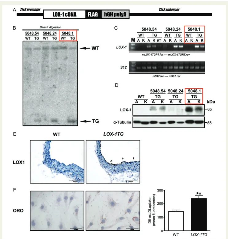
Figure 1 Generation and characterization of endothelial-specific LOX-1TG mice. (A) Scheme of endothelial-specific LOX-1 transgenic construct. (B) Southern blot analysis of F1 generation. Genomic DNA isolated from tail biopsies was digested with BamHI, and hybridized with LOX-1-specific radioactively labelled probe. The presence of additional BamHI restriction site located within LOX-1 cDNA introduced into murine genome upon integration of the transgenic construct allowed to detect the corresponding band in transgenic lanes with the specific probe used. This band is absent in wild-type lanes. (C ) real-time polymerase chain reaction analysis. Total RNA isolated from either aorta (A) or kidney (K) of three different trans-genic lines 5048.54, 5048.24, and 5048.1 are transformed into cDNA and amplified with LOX-1-specific primers. As a loading control, murine S12-specific primers are used. Real-time-negative control without reverse transcriptase; M, 100 bp DNA molecular weight marker. (D) Western blot of aortic lysates using anti-murine LOX-1 antibody. Anti-murine a-tubulin antibody is used as a loading control. Transgenic line 5048.1 (red rectangle) is used for all subsequent experiments. (E) Aortic cross sections from C57BL/6 wild type (left) and LOX-1TG (right) are stained with anti-murine LOX-1 antibody. Bar ¼ 100 mm. (F ) Murine endothelial cells isolated from LOX-1TG mice and stimulated with DiI-oxidized low-density lipo-protein for 6 h, reveal increased oxidized low-density lipolipo-protein uptake compared with endothelial cells from wild-type mice as shown by oil red O staining and monitored by FACS with an anti-mouse CD105 antibody. Quantification of FACS measurements is given as mean fluorescence of the CD105-positive cells; n ¼ 4 for each group, **P , 0.01.
RT – PCR analysis, immunohistochemistry
and western blotting
See Supplementary material online, Methods.
Statistical analysis
Data are presented as mean + SEM. Statistical significance was calculated using either ANOVA with post hoc Tukey’s test or Student’s unpaired t-test. Significance at the level of two-tailed P , 0.05 was accepted for the corresponding statistical analysis.
Results
Generation and molecular
characterization of LOX-1 transgenic mice
To obtain mice overexpressing LOX-1 in endothelial cells, a murine
tyrosine kinase receptor Tie2 promoter was used (Figure1A). Mice
overexpressing LOX-1 in endothelial cells carried a different copy
number of LOX-1 transgene (Figure 1B). The line 5048.1, which
showed the highest transgene expression, was selected for further
experiments (Figure 1B through D). LOX-1 expression was the
highest in the aorta, while lower expression levels were observed
in the kidney (Figure1C). Increased aortic expression of LOX-1 in
transgenic mice was confirmed by Western blot (Figure1D). In
con-trast, LOX-1 was not detectable in WT littermates (Figure1D).
Immu-nohistochemistry confirmed that overexpression of LOX-1 was
confined to the vascular endothelium (Figure 1E). Moreover,
oxLDL uptake was increased only in endothelial cells isolated from
LOX-1TG but not WT mice (Figure 1F). Importantly, in cultured
macrophages oxLDL uptake did not differ between LOX-1TG and WT mice (Supplementary material online, Figure S1). Plasma lipopro-tein levels remained unchanged in transgenic mice compared with
their WT littermates (Table1). In contrast, vascular oxLDL levels
were increased in LOX-1TG mice as compared with WT (Supplemen-tary material online, Table S1) suggesting a key role of endothelial LOX-1 for oxLDL uptake in vivo.
Endothelial LOX-1 overexpression
promotes fatty streak formation
To examine the effects of endothelial LOX-1 overexpression on the initial stages of atherogenesis, we fed LOX-1TG and WT mice a
high-cholesterol diet (HCD) for 30 weeks (Figure2A). In LOX-1TG mice,
aortic fatty streak formation was significantly increased (Figure2B).
Upon initiation of plaque formation, oxLDL activates inflam-matory pathways in vascular cells. In line with this concept,
immunohistochemical stainings of aortas showed increased endothe-lial expression of VCAM-1 in LOX-1TG mice compared with WT
(Figure2C). Adhesion molecules are important for recruiting
inflam-matory cells to the activated endothelium. Indeed, the number of activated macrophages, as assessed by the number of CD68-positive
cells, was increased in aortas of LOX-1TG mice (Figure2D).
Endothelial LOX-1 overexpression on
ApoE background increases
atherosclerosis
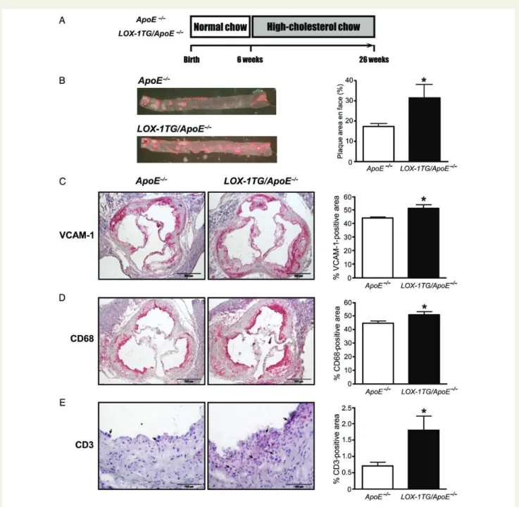
To investigate whether LOX-1 overexpression plays a role in plaque formation, LOX-1TG mice were crossed with apolipoprotein E
knock-out (ApoE2/2, C57BL/6 background) mice, a widely used mouse
model for studying atherosclerosis.18 The resulting LOX-1TG/
ApoE2/2 and ApoE2/2 male littermates were fed a HCD for
20 weeks (Figure3A). Interestingly, en face plaque area was almost
two-fold increased in LOX-1TG/ApoE2/2as compared with ApoE2/2
littermates (Figure3B). To determine the effects of endothelial-specific
LOX-1 overexpression on vascular inflammation, immunohistochem-ical staining for VCAM-1 and P-selectin was performed on cross sec-tions of aortic roots. Morphometric analyses revealed increased
expression levels of VCAM-1 and P-selectin in LOX-1TG/ApoE2/2
(Figure3C and Supplementary material online, Figure S2). mRNA
ana-lysis for VCAM-1 confirmed these results (data not shown). In addition,
the number of plaque macrophages (CD68-positive area; Figure3D) and
T-cells (CD3-positive area; Figure3E) was increased in LOX-1-1TG/
ApoE2/2mice.
To understand whether endothelial LOX-1 is required for atherosclerosis development in vivo, bone marrow transplantation experiments were performed. Bone marrow cells isolated from
ApoE2/2 or LOX-1-1TG/ApoE2/2 donor mice were transplanted
into irradiated 5-week-old recipient ApoE2/2 and LOX-1-1TG/
ApoE2/2mice (Supplementary material online, Figure S3A). Chimeric
LOX-1-1TG/ApoE2/ 2recipient mice receiving either ApoE2/ 2or
LOX-1-1TG/ApoE2/2bone marrow cells still showed more
atheroscler-otic plaques than those recipient ApoE2/2mice receiving either bone
marrow (Supplementary material online, Figure S3B). In addition,
immunostaining of aortic cross section from ApoE2/ 2 and
LOX-1-1TG/ApoE2/2mice kept 20 weeks on HCD with LOX-1 and
CD68 showed no LOX-1 expression in activated macrophages (Supplementary material online, Figure S3C). Finally, FACS analysis of blood and spleen samples from transplanted mice revealed no dif-ference in frequencies of monocytes, T-cell subtypes or macro-phages (Supplementary material online, Figure S4).
. . . . Table 1 Plasma lipid profiles in wild-type, LOX-1TG, ApoE2/2, and LOX-1TG/ApoE2/2mice
WT (n 5 5) LOX-1TG (n 5 6) ApoE2/2(n 5 8) LOX-1TG/ApoE2/2(n 5 10)
Total cholesterol (mmol/L) 5.64 + 1.20 6.27 + 0.94 19.97 + 2.98** 16.98 + 2.83**
Triglycerides (mmol/L) 0.77 + 0.07 0.75 + 0.03 0.84 + 0.18 1.13 + 0.28
Free fatty acids (mmol/L) 0.30 + 0.06 0.28 + 0.08 0.36 + 0.06 0.44 + 0.07
Statistical significance was calculated using analysis of variance. **P , 0.005 compared with wild-type and LOX-1TG, mean + SEM.
A. Akhmedov et al.
2842
LOX-1 overexpression increases reactive
oxygen species production and
inflammation
As endothelial dysfunction precedes plaque formation, we assessed endothelium-dependent vasorelaxation to acetylcholine in isolated
aortic rings obtained from WT, LOX-1TG, ApoE2/2 and LOX-1TG/
ApoE2/2 mice. Whereas no difference was seen in
endothelium-dependent vasorelaxation to acetylcholine between LOX-1TG and
WT mice, in LOX-1TG/ApoE2/2 mice, acetylcholine-induced
en-dothelium-dependent vasorelaxation was impaired (pEC50 (2log
M ): 6.34 + 0.12); Emax(% of contraction) 6.52 + 3.05% compared
with that of ApoE2/2mice (6.67 + 0.40, P , 0.05; Emax(% of
con-traction) 22.18 + 6.68%, P , 0.05) (Figures4A and5A, respectively).
In contrast, endothelium-independent relaxation to SNP was similar
(data not shown). Next, we tested whether impaired endothelial function would have any impact on endothelial nitric oxide synthase (eNOS) activity. Indeed, eNOS activation through phosphorylation
was markedly reduced in both LOX-1TG and LOX-1TG/ApoE2/2
mice (Figures 4B and 5B, respectively; Supplementary material
online, Figure S5A). Since eNOS is known to be redox sensitive and thus can be regulated by ROS, we measured ROS in aortas isolated
from LOX-1TG and LOX-1TG/ApoE2/2. Aortic levels of ROS in both
transgenic groups were markedly increased compared with corre-sponding littermate controls, but they decreased when aortas from
both LOX-1TG and LOX-1TG/ApoE2/2were pre-incubated with
super-oxide dismutase, a known scavenger of supersuper-oxide (Figures4C and5C,
respectively).
LOX-1 receptor activation leads to accumulation of ROS and in turn activates a cascade of redox-sensitive events including MAPK Figure 2 Increased aortic fatty streak formation, expression of VCAM-1, and CD68 in LOX-1TG mice. (A) Scheme of the diet set up. Six-week-old male LOX-1TG and wild-type control mice kept on normal chow diet were then fed a high-cholesterol diet for additional 30 weeks. (B) Immunostain-ing of aortic roots from LOX-1TG mice 30 weeks after high-cholesterol diet showed increased lipid accumulation as assessed by oil red O stainImmunostain-ing with corresponding quantification. Immunohistochemistry of aortic roots from LOX-1TG mice for (C ) VCAM-1 and (D) CD68 showed increased inflam-mation compared with wild-type mice; n ¼ 10 for wild-type and n ¼ 8 for LOX-1TG, *P , 0.05. Values are expressed as mean + SEM. Bar ¼ 100 mm.
pathways.12,19–22To test this in our setting, both protein expression and phosphorylation of p38 MAPK were assessed in LOX-1TG aortic lysates and were both increased compared with non-transgenic
controls (Figure4D and Supplementary material online, Figure S5B).
Further, activation of the redox-sensitive transcription factor nuclear factor kB (NFkB) was increased in LOX-1TG mice due to
increased binding of the p65 subunit of NFkB to DNA (Figure4E).
Finally, we assessed the expression of VCAM-1 and E-selectin, both
known to be regulated by ROS through NFkB.23Indeed, mRNA
expression of VCAM-1 and E-selectin was higher in aortic lysates
obtained from transgenic mice (Figure4F and Supplementary material
online, Figure S5C).
Figure 3 Increased aortic plaque formation and inflammation in LOX-1TG/ApoE2/2mice. (A) Twenty-six-week-old mice were sacrificed for tissue
isolation. Six-week-old male LOX-1TG/ApoE2/2and ApoE2/2mice kept on normal chow diet were fed a high-cholesterol diet for 20 weeks (lower panel). Twenty-six-week-old mice were euthanized for tissue harvesting. (B) En face plaque staining with corresponding quantification of thoraco-abdominal aortas from LOX-1TG/ApoE2/2 mice after 20 weeks of high-cholesterol diet showed increased lipid accumulation; n ¼ 12 for ApoE2/2 and n ¼ 9 for LOX-1TG/ApoE2/2, *P , 0.05. (C ) Immunostaining of aortic roots from LOX-1TG/ApoE2/2 and ApoE2/2 mice with
VCAM-1 antibody with corresponding quantification; n ¼ 7; *P , 0.05. (D, E) Cross sections of aortic roots from LOX-1TG/ApoE2/2 mice stained with (D) antibody against CD68, or (E) antibody against CD3 (arrows indicate positive staining) showed increased macrophage and T cell accumulation compared with ApoE2/2mice; n ¼ 7 for each group, *P , 0.05. Bar ¼ 500 mm (C, D) and 100 mm (E). Values are expressed as mean + SEM. Statistical significance was calculated using unpaired Student’s t-test.
A. Akhmedov et al.
2844
Discussion
In the present study, we sought to elucidate whether and through which mechanisms endothelial LOX-1 accelerates fatty streak formation and atherosclerosis development. To this end, we gener-ated Tie2-LOX-1TG mice with endothelium-targeted LOX-1 overex-pression and compared the development of atherosclerosis in these
mice and their WT littermates. Our results suggest that endo-thelial LOX-1 is critically involved in the development of endoendo-thelial dysfunction, vascular inflammation and atherosclerotic lesions. Several lines of evidence support our conclusions: (i) endothelial-specific LOX-1 overexpression accelerates fatty streak formation, induces adhe-sion molecule expresadhe-sion, and increases macrophage recruitment;
(ii) LOX-1 overexpression in ApoE2/2 mice causes endothelial
Figure 4 Increased fatty streaks are associated with decreased endothelial nitric oxide synthase phosphorylation, increased ROS levels, p38 phos-phorylation and NFkB activation in LOX-1TG mice. (A) Relaxation to acetylcholine in aortic rings from WT (blue line) and LOX-1TG (black line). Values were normalized to preconstruction with U46619 and shown as mean + SEM. n ¼ 6 per genotype. (B) Western blot reveals decreased endothelial nitric oxide synthase activation through phosphorylation of Ser1177 residue in aortic lysates from LOX-1TG mice. Quantification of protein expression is given as ratio to total endothelial nitric oxide synthase expression levels; n ¼ 4 for each group, *P , 0.05. Statistical significance was calculated with unpaired Student’s t-test. (C ) Measurements of total ROS production by ESR spectroscopy in aortas show increased ROS levels in LOX-1TG mice com-pared with WT controls. Pre-treatment of aortas from LOX-1TG mice with 150 U/mL superoxide dismutase (SOD) significantly reduces ROS accumu-lation. Quantification is given as a percentage of average control value; n ¼ 6 for each group, *P , 0.05. (D) Western blot shows increased p38 phosphorylation in aortic lysates from LOX-1TG mice. Quantification of protein expression is given as ratio to total p38 expression levels; n ¼ 4 for each group, *P , 0.05. (E) Increased p38 phosphorylation is further associated with increased nuclear translocation and binding capacity of NFkB as shown by DNA-binding assay for RelA/p65 subunit of NFkB in aortic lysates. Quantification of binding activity is given as mean absorbance and normal-ized to the average value of unstimulated LOX-1TG endothelial cells; n ¼ 6 for each group, *P , 0.05. (F) Increased NFkB further activates expression of inflammation molecule VCAM-1. The VCAM-1 mRNA was analysed by quantitative real time PCR. RNA was normalized to the internal control S12 (n ¼ 6 in each group). Data are shown as percentage of WT average. Values are expressed as the mean + standard error of mean (SEM). *P , 0.05 vs. control wild-type (WT) mice.
dysfunction and accelerates plaque formation; (iii) endothelium-targeted LOX-1 overexpression increases aortic ROS production and activates redox-sensitive pathways such as MAPK p38 and NFkB, ensuing in endothelial inflammation.
Uptake of oxLDL by endothelial cells and macrophages is a key event preceding plaque formation (i) and (ii). However, it is not clear whether endothelial LOX-1 activation translates into an atherolsclero-tic phenotype in the vasculature. In the present study, we have demon-strated that endothelial overexpression of LOX-1 accelerates
endothelial dysfunction in atherosclerosis-prone ApoE2/2mice. This
finding is in line with the notion that LOX-1 antagonism improves
NO availability in the coronary circulation.24Indeed, we have found
that in LOX-1TG mice LOX-1 overexpression induces oxidative stress and reduces eNOS activation without affecting endothelial
func-tion. In contrast, using LOX-TG1/ApoE2/2 mice, we have observed
impaired vasorelaxation to acetylcholine. We do speculate that in LOX-1TG mice some compensatory mechanisms may still be operating in the absence of a severe atherosclerotic phenotype. Another possi-bility is that increased ROS production observed in LOX-1TG mice was not sufficient to affect endothelium-dependent relaxation by such a strong agonist as acetylcholine. Impaired vasorelaxation in LOX-1TG/
ApoE2/2mice were explained by reduced eNOS expression and
acti-vation as well as increased aortic ROS generation. Indeed,
eNOS-activating phosphorylation was blunted in LOX-1TG/ApoE2/2
mice, thus contributing to endothelial dysfunction observed in these mice. Moreover, LOX-1-dependent oxidative stress led to phosphor-ylation of MAPK p38 and subsequent activation of NFkB signalling. These molecular events were associated with up-regulation of the in-flammatory molecules VCAM-1, E- and P-selectin, favouring mono-cytes recruitment. Moreover, phosphorylation of p38 is a key initiator of the apoptotic process, thus precipitating vascular pathology
in vessels with LOX-1 activation.25,26This first set of findings strongly
suggests that LOX-1 overexpression in the vascular endothelium is key event preceeding the atherogenic process. Indeed, LOX1-TG mice
without ApoE2/2background already showed clear signs of vascular
inflammation together with an increased oxidative burden. The strength of our work is the in vivo characterization of endothelial LOX-1 in an unanticipated transgenic mouse model. The design of this specific transgene allowed us to understand the relevance of endo-thelial LOX-1, regardless of its role in macrophages. Notably, we could show that selective activation of LOX-1 in the endothelium is sufficient to induce early precursors of atherosclerotic disease such as Figure 5 Increased plaque formation is associated with endothelial dysfunction, decreased endothelial nitric oxide synthase phosphorylation and increased ROS levels in LOX-1TG/ApoE2/2mice. (A) Relaxation to increasing concentrations of acetylcholine in aortic rings from ApoE2/2and LOX-1TG/ApoE2/2mice. Values were normalized to preconstruction with U46619 and shown as mean + SEM. ApoE2/2(blue line), LOX-1TG/ ApoE2/2(black line). n ¼ 8/genotype, *P , 0.05. (B) Western blot reveals decreased endothelial nitric oxide synthase activation through phosphor-ylation of Ser1177 residue in aortic lysates from LOX-1TG/ApoE2/2mice. Quantification of protein expression is given as ratio to total endothelial nitric oxide synthase expression levels; n ¼ 4 for each group, *P , 0.05. Statistical significance was calculated with unpaired Student’s t-test. (C ) Measurements of total reactive oxygen species production by electron spin resonance spectroscopy in aortas show increased reactive oxygen species levels in LOX-1TG/ApoE2/2mice compared with ApoE2/2controls. Pre-treatment of aortas from LOX-1TG/ApoE2/2mice with 150 U/ mL SOD significantly reduces reactive oxygen species accumulation. Quantification is given as a percentage of average control value; n ¼ 6 for each group, *P , 0.05.
A. Akhmedov et al.
2846
endothelial dysfunction and NFkB-driven inflammation. Indeed, macrophages did not show any up-regulation or functional activity of LOX-1 in our genetic model, further strengthening the importance of endothelial LOX-1 in atherogenesis. Although under certain
condi-tions the Tie2 promoter may also be active in macrophages,27,28it
appears that other scavenger receptors play a more dominant role for the oxLDL uptake in these cells. Indeed, our initial characterization of the LOX-1TG mice revealed high levels of LOX-1 transgene mRNA in aortic endothelial cells, and lower levels in peritoneal macrophages. However, functional assay of oxLDL uptake showed no difference between transgenic and non-transgenic macrophages ruling out a con-tribution of other scavenger receptors such as SR-A and SR-B under our experimental conditions. Moreover, bone marrow transplantation experiments together with immunostaining of atherosclerotic aortas demonstrated that bone marrow-derived cells, in particular macro-phages, do not account for the increase in atherosclerosis observed in our LOX-1 transgenic mouse model. Thus, we were able to specif-ically overexpress LOX-1 in the endothelium of mice in vivo and to demonstrate distinct functional changes of endothelial cells, i.e. a marked increase in oxLDL uptake as a crucial step in the atheroscler-osis process.
Since plasma lipid levels were similar in LOX-1TG and WT mice, the difference in fatty streak formation observed in transgenic mice must be related to an increased activity of the LOX-1 receptor as demon-strated in transgenic endothelial cells in culture. Indeed, aortic levels of lipoproteins were augmented in LOX-1TG as compared with WT littermates. Using a preproendothelin-1 promoter and a bovine LOX-1 transgene, others found no phenotype of the transgene in WT C57BL/6 mice, but an inflammatory intramyocardial
vasculopa-thy on the ApoE background.7This discrepancy may be related to the
fact that the preproendothelin-1 promoter, unlike the Tie2 promoter used in our study, drives LOX-1 transgene expression predominantly in microvessels rather than in conduit arteries that are prone to ath-erosclerotic plaque formation. Furthermore, species differences between bovine and murine LOX-1 gene may have contributed.
In summary, our data indicate that endothelial-specific overex-pression of LOX-1 enhances lipid deposition and inflammation in the aorta and leads to endothelial dysfunction and atherosclerotic plaque formation. At the molecular level, LOX-1 activates p38-NFkB pathway resulting in increased VCAM-1, E- and P-selectin tran-scription, and vascular inflammation. As LOX-1 is also overexpressed in human plaques, endothelial-specific inhibition of LOX-1 may represent a new therapeutic target for the prevention and treatment of atherosclerosis.
Study limitations
There are still some limitations applied for the present study which need to be considered and addressed in the future. First of all, ex-trapolation of data obtained in genetically modified mice to human is always a difficult issue. Therefore, in order to support our conclu-sion regarding clinical applications, further experiments on human samples should be considered in the future. Second, the use of trans-genic mice does not completely exclude the existence of alternative LOX-1-dependent molecular pathways. Finally, smooth muscle cells may also contribute to LOX-1-mediated atherogenesis, since the basal expression of LOX-1 and the activity of Tie2 promoter have been previously reported in these cells.
Supplementary material
Supplementary material is available at European Heart Journal online.
Acknowledgements
We thank Thomas N. Sato for providing us with the pSP14/15 vector, Elin Bjo¨rk for technical assistance, Nicola Schaefer for providing us with the methodology for the cultivation of murine endothelial cells.
Funding
This study was supported by Swiss National Science Foundation (Grant No. 3100-068118.02/1 to T.F.L.); the Swiss Heart Foundation, Bern, Switzerland; the Hartmann Mu¨ller Foundation, Zu¨rich, Switzerland; MERCATOR Foundation Switzerland as well as by a strategic alliance with Pfizer, Inc., New York, USA.
Conflict of interest: none declared.
References
1. Witztum JL, Steinberg D. Role of oxidized low density lipoprotein in atherogenesis. J Clin Invest 1991;88:1785 – 1792.
2. Zingg JM, Ricciarelli R, Azzi A. Scavenger receptor regulation and atherosclerosis. Biofactors 2000;11:189 – 200.
3. Mehta JL, Chen J, Hermonat PL, Romeo F, Novelli G. Lectin-like, oxidized low-density lipoprotein receptor-1 (LOX-1): a critical player in the development of ath-erosclerosis and related disorders. Cardiovasc Res 2006;69:36 – 45.
4. Pluddemann A, Neyen C, Gordon S. Macrophage scavenger receptors and host-derived ligands. Methods 2007;43:207 – 217.
5. Chen M, Narumiya S, Masaki T, Sawamura T. Conserved C-terminal residues within the lectin-like domain of LOX-1 are essential for oxidized low-density-lipoprotein binding. Biochem J 2001;355:289 – 296.
6. Ross R. Atherosclerosis – an inflammatory disease. N Engl J Med 1999;340:115 – 126. 7. Inoue K, Arai Y, Kurihara H, Kita T, Sawamura T. Overexpression of lectin-like oxi-dized low-density lipoprotein receptor-1 induces intramyocardial vasculopathy in apolipoprotein E-null mice. Circ Res 2005;97:176 – 184.
8. Mehta JL, Sanada N, Hu CP, Chen J, Dandapat A, Sugawara F, Satoh H, Inoue K, Kawase Y, Jishage K, Suzuki H, Takeya M, Schnackenberg L, Beger R, Hermonat PL, Thomas M, Sawamura T. Deletion of LOX-1 reduces atherogenesis in LDLR knockout mice fed high cholesterol diet. Circ Res 2007;100:1634 – 1642. 9. Kataoka H, Kume N, Miyamoto S, Minami M, Moriwaki H, Murase T, Sawamura T,
Masaki T, Hashimoto N, Kita T. Expression of lectinlike oxidized low-density lipopro-tein receptor-1 in human atherosclerotic lesions. Circulation 1999;99:3110 –3117. 10. Li D, Mehta JL. Antisense to LOX-1 inhibits oxidized LDL-mediated upregulation of
monocyte chemoattractant protein-1 and monocyte adhesion to human coronary artery endothelial cells. Circulation 2000;101:2889 – 2895.
11. Li D, Chen H, Romeo F, Sawamura T, Saldeen T, Mehta JL. Statins modulate oxidized low-density lipoprotein-mediated adhesion molecule expression in human coron-ary artery endothelial cells: role of LOX-1. J Pharmacol Exp Ther 2002;302:601 – 605. 12. Cominacini L, Rigoni A, Pasini AF, Garbin U, Davoli A, Campagnola M, Pastorino AM, Lo Cascio V, Sawamura T. The binding of oxidized low density lipoprotein (ox-LDL) to ox-LDL receptor-1 reduces the intracellular concentration of nitric oxide in endothelial cells through an increased production of superoxide. J Biol Chem 2001; 276:13750 – 13755.
13. Becher B, Durell BG, Miga AV, Hickey WF, Noelle RJ. The clinical course of experi-mental autoimmune encephalomyelitis and inflammation is controlled by the ex-pression of CD40 within the central nervous system. J Exp Med 2001;193:967 – 974. 14. von Lukowicz T, Hassa PO, Lohmann C, Boren J, Braunersreuther V, Mach F, Odermatt B, Gersbach M, Camici GG, Stahli BE, Tanner FC, Hottiger MO, Luscher TF, Matter CM. PARP1 is required for adhesion molecule expression in atherogenesis. Cardiovasc Res 2008;78:158 – 166.
15. Kobayashi M, Inoue K, Warabi E, Minami T, Kodama T. A simple method of isolating mouse aortic endothelial cells. J Atheroscler Thromb 2005;12:138 – 142.
16. Biocca S, Falconi M, Filesi I, Baldini F, Vecchione L, Mango R, Romeo F, Federici G, Desideri A, Novelli G. Functional analysis and molecular dynamics simulation of LOX-1 K167N polymorphism reveal alteration of receptor activity. PLoS ONE 2009;4:e4648.
17. Madonna R, Salerni S, Schiavone D, Glatz JF, Geng YJ, De Caterina R. Omega-3 fatty acids attenuate constitutive and insulin-induced CD36 expression through a sup-pression of PPAR alpha/gamma activity in microvascular endothelial cells. Thromb Haemost 2011;106:500 – 510.
18. Plump AS, Smith JD, Hayek T, Aalto-Setala K, Walsh A, Verstuyft JG, Rubin EM, Breslow JL. Severe hypercholesterolemia and atherosclerosis in apolipoprotein E- de-ficient mice created by homologous recombination in ES cells. Cell 1992;71:343–353. 19. Imanishi T, Hano T, Sawamura T, Takarada S, Nishio I. Oxidized low density lipopro-tein potentiation of Fas-induced apoptosis through lectin-like oxidized-low density lipoprotein receptor-1 in human umbilical vascular endothelial cells. Circ J 2002;66: 1060 – 1064.
20. Chen J, Mehta JL, Haider N, Zhang X, Narula J, Li D. Role of caspases in Ox-LDL-induced apoptotic cascade in human coronary artery endothelial cells. Circ Res 2004;94:370 – 376.
21. Li D, Liu L, Chen H, Sawamura T, Ranganathan S, Mehta JL. LOX-1 mediates oxidized low-density lipoprotein-induced expression of matrix metalloproteinases in human coronary artery endothelial cells. Circulation 2003;107:612 – 617.
22. Mehta JL, Chen J, Yu F, Li DY. Aspirin inhibits ox-LDL-mediated LOX-1 expression and metalloproteinase-1 in human coronary endothelial cells. Cardiovasc Res 2004; 64:243 – 249.
23. Weber C, Erl W, Pietsch A, Strobel M, Ziegler-Heitbrock HW, Weber PC. Antiox-idants inhibit monocyte adhesion by suppressing nuclear factor-kappa B mobilization
and induction of vascular cell adhesion molecule-1 in endothelial cells stimulated to generate radicals. Arterioscler Thromb 1994;14:1665 – 1673.
24. Xu X, Gao X, Potter BJ, Cao JM, Zhang C. Anti-LOX-1 rescues endothelial function in coronary arterioles in atherosclerotic ApoE knockout mice. Arterioscler Thromb Vasc Biol 2007;27:871 – 877.
25. Bao MH, Zhang YW, Zhou HH. Paeonol suppresses oxidized low-density lipopro-tein induced endothelial cell apoptosis via activation of LOX-1/p38MAPK/ NF-kappaB pathway. J Ethnopharmacol 2013;146:543 – 551.
26. Ogura S, Kakino A, Sato Y, Fujita Y, Iwamoto S, Otsui K, Yoshimoto R, Sawamura T. Lox-1: the multifunctional receptor underlying cardiovascular dysfunction. Circ J 2009;73:1993 – 1999.
27. De Palma M, Venneri MA, Galli R, Sergi Sergi L, Politi LS, Sampaolesi M, Naldini L. Tie2 identifies a hematopoietic lineage of proangiogenic monocytes required for tumor vessel formation and a mesenchymal population of pericyte progenitors. Cancer Cell 2005;8:211 – 226.
28. De Palma M, Venneri MA, Roca C, Naldini L. Targeting exogenous genes to tumor angiogenesis by transplantation of genetically modified hematopoietic stem cells. Nat Med 2003;9:789 – 795.
CARDIOVASCULAR FLASHLIGHT
. . . .
doi:10.1093/eurheartj/eht521
Online publish-ahead-of-print 13 December 2013
Surgical banding of the inferior vena cava for the facilitation of transcatheter
valve implantation in a patient with severe secondary tricuspid regurgitation
Georg Daniel Duerr1*†, Matthias Endlich1†, Jan-Malte Sinning2, Bahman Esmailzadeh1,
Nikos Werner2, and Fritz Mellert1
1
Department of Cardiac Surgery, University Clinical Center of Bonn, Bonn, Germany; and2
Department of Cardiology, Pneumology and Angiology, Medizinische Klinik II, University Clinical Center of Bonn, Bonn, Germany
*Corresponding author. E-mail:[email protected]
†These authors contributed equally to this work.
Pathological tricuspid regurgitation (TR) is more often secondary due to annular dilatation and increased tricuspid leaflet tethering. Although ring annuloplasty is key to surgery for TR, surgical treat-ment of TR in high-risk patients is associated with increased mortality. Percutaneous single or dual caval transcatheter heart valve-(THV)-prosthesis implantation seems feasible, but not realizable in many patients due to increased caval vein diameter. In an 85-year-old woman with severe TR (Panels A and B) associated with advanced right-heart failure, ascites, and portal hypertension (Panel C) at prohibitive risk for open-heart surgery (Euro-SCORE 21.7), transesophageal echocardiography (TEE) and multi-slice computed tomography (MSCT) revealed inferior vena cava (IVC) dilatation
(Panel D; 34× 43 mm).
To downsize the IVC to a mean diameter ,30 mm, surgical banding was performed via right-lateral mini-thoracotomy using a longitudinally-opened goretex-prosthesis, which was ‘wrapped’ around the IVC below the diaphragm just after the confluence of the hepatic veins, while a 30 mm Z-MED II valvuloplasty balloon was inflated in that position. Thereafter, a balloon-expandable
stent (AndraStent-XXL, 35 mm, Andramed) was deployed within the banded IVC-segment, which was tightened with 5-0 Prolene-suture. Finally, an Edwards-SAPIEN 29 mm was implanted into the stent (Panels E and F ).
Although pacemaker leads in the superior vena cava (SVC) prohibited implantation of an upper caval valve, TR declined significantly (Panels G – I), and general condition had significantly improved at discharge after 2 weeks (decreased ascites and peripheral oedema; 9 kg weight loss). TEE/MSCT showed trace-leakage with decrease of RV and RA volumes and hepatic vein diameters.
We conclude that in high-risk patients with severe TR and enlarged IVC, downsizing of IVC is feasible to enable THV implantation.
Published on behalf of the European Society of Cardiology. All rights reserved.&The Author 2013. For permissions please email: [email protected]
A. Akhmedov et al.