Klaus Strobel Beata Bode Didier Lardinois Ulrich Exner Received: 13 February 2006 Revised: 11 April 2006 # ISS 2006
PET-positive fibrous dysplasia - a potentially
misleading incidental finding in a patient
with intimal sarcoma of the pulmonary artery
Abstract Benign bone tumors can show an increased FDG uptake in FDG-PET/CT investigations. In the presented case, an incidentally de-tected PET-positive asymptomatic fibrous dysplasia was initially misinterpreted as a metastasis in a patient with intimal sarcoma of the pulmonary artery.
Keywords PET/CT . Fibrous dysplasia . Sarcoma
Introduction
Fibrous dysplasia (FD) is a developmental disorder in which normal bone marrow is replaced with fibro-osseous tissue. Patients are often asymptomatic, and FD is often detected incidentally on radiographs, computed tomogra-phy (CT) or bone scans. FD can lead to complications like deformity, pathologic fractures and—rarely—malignant transformation. FD can be monostotic or polyostotic. Polyostotic FD may be a component of the McCune-Albright syndrome [1–4]. 18-fluoro-deoxyglucose positron emission tomography (FDG-PET/(CT) is increasingly used for the staging of different malignant diseases [5,6]. FDG is not a tumor-specific agent, and an increase in FDG uptake can be shown by many benign or physiologic “disorders” , such as infections and inflammations [7,8], brown fatty tissue [9], traumatic lesions [10], ovaries [11] and benign tumors like Warthin`s tumor of the parotid
gland [12]. If PET/(CT) is performed to stage malignan-cies, an FDG uptake in benign lesions can lead to their misinterpretation as metastases. To our knowledge, there is no report in the literature of a biopsy-proven PET/CT-positive fibrous dysplasia.
Case report
A 40-year-old male non-smoker was presented who had had a cough for 3 months and suffered from weight loss, night sweat and recurrent haemoptysis. A chest X-ray and a contrast-enhanced chest CT scan showed a mass at the left hilus, most likely arising from the left pulmonary artery. The differential diagnosis of a tumor of the intima of the pulmonary artery, a bronchial carcinoma, tuberculosis or other tumor was established. A bronchoscopy was not conclusive. The patient was referred to us with suspicion of
K. Strobel (*)
Division of Nuclear Medicine, Department of Medical Radiology, University Hospital, Zurich, Switzerland e-mail: klaus.strobel@usz.ch Tel.: +41-44-2552850 Fax: +41-44-2554414 B. Bode
Institute of Surgical Pathology, University Hospital,
Zurich, Switzerland
D. Lardinois
Department of Thoracic Surgery, University Hospital,
Zurich, Switzerland U. Exner
Department of Orthopaedic Tumor Surgery, Orthopedic University Hospital Balgrist,
Zurich, Switzerland Accepted: 24 April 2006
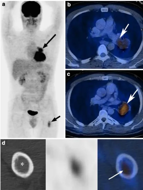
a pulmonary artery intimal sarcoma, and 18F-FDG-PET/CT (combined PET/CT in-line system; Discovery ST, GE Health Systems, Milwaukee, WI, USA) was performed for staging. The central lung lesion at the left hilus showed an increased FDG uptake (SUV max. 4.1) (Fig. 1a–c). Another asymptomatic FDG-active lesion was found in the proximal diaphysis of the left femur (SUV max. 2.9; Fig.1d). Because of the increased FDG uptake, this lesion was initially interpreted as suspicious for a metastasis of the lung tumor.
However, due to the recurrent character of the hemop-tysis, and due to the absence of effective induction therapy for intimal sarcomas, a lung resection was performed with a palliative intention. The patient underwent extended left pneumonectomy. The histology confirmed the presumed diagnosis of a high-grade intimal sarcoma of the left pulmonary artery (Fig.2).
Fig. 1 MIP image (a) demon-strating an increased FDG up-take (SUV max. 4.1) centrally in the left lung (long arrow) and in the proximal left femur di-aphysis (short arrow). Axial fused PET/CT images show that the tumor has a close relation-ship to the left pulmonary artery (b, c; arrow). On axial images (d) of the lesion in the femur, “ground-glass” appearance (asterisk) of the lesion is visible with increased FDG-uptake (SUV max. 2.9)
Fig. 2 Hematoxilin and eosin staining (original magnification 400×) of the centrally located, intraluminal tumor of the pulmonary artery, in a pneumonectomy specimen showing highly pleomorphic and mitotically active malignant tumor tissue corresponding to a high-grade spindle-cell intimal sarcoma
The bone lesion was further evaluated by means of conventional X-rays (Fig.3) and MRI (Fig.4). The X-ray demonstrated a homogeneous grayish lesion with a “ground-glass” pattern in the femur. There were no visible periosteal reactions or destructions, and no deformity could be detected. MR images presented a T1-hypointense lesion in the diaphysis with sharp borders and homogenous contrast enhancement on fat-suppressed T1-weighted im-ages. All images were reviewed by an experienced musculoskeletal radiologist together with a nuclear physi-cian. Because of the typical pattern, the most likely diagnosis of incidentally detected FD was established. The lesion was biopsied under fluoroscopic guidance, and the histology confirmed the diagnosis of FD (Fig.5).
Discussion
This case demonstrates that an increased FDG uptake in a clinically occult fibrous dysplasia can be misleading in the evaluation of a patient with a malignant primary tumor. The literature on PET and fibrous dysplasia is very limited. Aoki et al. evaluated primary benign and malignant bone tumors, and found an increased FDG uptake (SUV max. >2.0) in three of six patients with fibrous dysplasia. There was no statistically significant difference in SUV between FD (n=6; SUV max. 2.05±0.98) and osteosarcoma (n=6; SUV max. 3.07±0.96) or chrondrosarcoma (n=7; SUV max. 2.23±0.74) [13]. With a cut-off SUV max. of 2.0,
only 64% of the 52 cases were classified correctly as benign or malignant. They found out that histiocytic and giant cell-containing benign bone lesions in particular can show a high FDG uptake. In another publication, the authors described a case of a female patient with breast cancer, where the bone scan showed multiple focal uptakes due to polyostotic FD. All of the lesions were PET-negative [14]. Toba et al. described a case of a patient with FD in the craniofacial bone without an increased FDG uptake [15]. It is generally accepted that FD is a developmental failure in the remodeling of primitive bone to mature lamellar bone. One explanation for the different degrees of FDG
metab-Fig. 3 X-ray of the bone lesion (arrows) showing a slightly expansile subtrochanteric lesion in the left femur without periostal reactions or destruction of the cortex indicating a benign tumor. Homogeneously increased density of the lesion compared with surrounding normal bone structure
Fig. 4 Coronal contrast-enhanced, fat-suppressed MR image showing a homogeneous contrast enhancement of the well-defined lesion (arrows)
Fig. 5 Elastica and van Gieson staining (original magnification 50×) of the fluoroscopically guided biopsy of the left femur lesion: bland spindle-cell proliferation with bone trabeculae without osteoblastic rimming and directly ingrowing collagen bundles (arrow) characteristic of fibrous dysplasia
olism of FD may be that the turnover of the remodeling process may have various velocities in different patients in different stages of the disease. Fibroblasts are the predominant proliferating cells in FD lesions, and the difference in SUV among FD may be due to the difference in the amount of proliferating fibroblasts or their metabolic turnover. Furthermore, a bone reaction may be influenced by the mechanical stress on the lesion, which may be higher in weight-bearing bones like the proximal femur than in the face. Experience with FDG-PET and other fibrous lesions, such as cortical desmoid, desmoplastic fibroma, nonossifying fibroma and osteofibrous dysplasia, is also very limited.
A malignant transformation of FD is rare, with reported prevalences ranging from 0.4% to 4% [16–18]. The most common malignant tumors reported are osteosarcoma, fibrosarcoma and chondrosarcoma. The FDG uptake correlates with the grade of bone sarcomas, and it is possible that an increased FDG uptake is a sign of the transformation of a benign tumor into a malignant one, but this hypothesis has not been proven yet [19, 20]. Unfortunately, because the differentiation, for example, between enchondroma and low-grade chondrosarcoma is sometimes impossible even for the pathologist, one can not expect FDG-PET or PET/CT studies to provide a solution to this problem.
Other benign conditions with an increased uptake in the bone are fractures, which can be misleading in tumor patients. The amount of the FDG uptake of fractures depends on the time interval between trauma and PET imaging. In most cases, the different pattern of uptake and clinical correlation allows an accurate differentiation of a fracture from malignant tumors [10,21,22].
This case also shows that thorough interpretation of the CT information from the PET/CT study with the help of an experienced musculoskeletal radiologist is essential to making a correct diagnosis. CT is still the best technique for demonstrating the typical radiographic findings of FD: the “ground-glass” pattern of the bone, the lesion being characteristically surrounded by a distinct rim of reactive bone, the endosteal scalloping. With CT, the extent of the lesion becomes clearly visible.
There are reports suggesting that FD accumulates other PET tracers: Tsuyuguchi et al. described two cases of FD of the skull base with mild accumulation (SUV values were about 2.0) of 11C-methyl-L-methionine on PET [23]. 11C Met PET is clinically used for the evaluation of brain tumors [24]. Tsuyuguchi and his colleagues speculated that “the mechanism of 11
C Met uptake in FD is due to increased metabolism and active transport of this amino acid because of the increased density of spindle cells”.
We found no publications on FD and18F-fluoride-PET. First studies indicate that 18F-fluoride is more sensitive than a conventional bone scan for detecting skeletal metastases in patients with prostate, lung or thyroid cancer [25]. Because 18F- fluoride-PET is also cost-effective, some authors expect that “classic” bone scans will be replaced with18F- fluoride PET completely in the next few years [26,27].18F-fluoride is a very potential non-specific bone tracer. Because18F-fluoride is a bone agent with an accumulation mechanism that is similar to methylene diphosphonate (MDP), one might expect that FD would show a significant uptake, even higher than in FDG-PET examinations.
The findings of whole-body intergrated PET/CT imag-ing can clearly affect therapeutic management. Lardinois and colleagues found extrapulmonary lesions in 110 out of 250 NSCLC patients with PET/CT. In 72 patients, solitary lesions were present. 32 (46%) of these lesions were unrelated to the lung primary. In 26 patients, the extrapul-monary lesion was caused by a benign tumor or an inflammatory lesion [12]. An extrathoracic solitary FDG accumulation in a patient with a lung tumor requires a histopathologic diagnosis, not only to exclude a false positive result or to confirm an occult metastasis, but also to diagnose lesions that are not related to the primary.
In conclusion, PET-CT readers should be aware of FDG-positive benign bone tumors, and should be familiar with the typical CT appearance of these lesions.
Acknowledgement The authors thank Dr. Paul Skandera for his support in preparing the manuscript.
References
1. Fitzpatrick KA, Taljanovic MS, Speer DP, et al. Imaging findings of fibrous dysplasia with histopatho-logic and intraoperative correlation. AJR Am J Roentgenol 2004;182:1389– 1398
2. Kransdorf MJ, Moser RP Jr, Gilkey FW. Fibrous dysplasia. Radiographics 1990;10:519–537
3. Sathekge MM, Clauss RP. Criteria and quantification of fibrous dysplasia on MDP scanning. Nuklearmedizin 1995;34:229–231
4. Parekh SG, Donthineni-Rao R, Ricchetti E, Lackman RD. Fibrous dysplasia. J Am Acad Orthop Surg 2004;12:305–313
5. Macapinlac HA. FDG PET and PET/ CT imaging in lymphoma and mela-noma. Cancer J 2004;10:262–270 6. Lardinois D, Weder W, Hany TF, et al.
Staging of non-small-cell lung cancer with integrated positron-emission tomography and computed tomography. N Engl J Med 2003;348:2500–2507
7. Blockmans D, Knockaert D, Maes A, et al. Clinical value of [(18)F]fluoro-de-oxyglucose positron emission tomog-raphy for patients with fever of unknown origin. Clin Infect Dis 2001;32:191–196
8. Stumpe KD, Dazzi H, Schaffner A, von Schulthess GK. Infection imaging using whole-body FDG-PET. Eur J Nucl Med 2000;27:822–832
9. Cohade C, Osman M, Pannu HK, Wahl RL. Uptake in supraclavicular area fat (“USA-Fat”): description on 18F-FDG PET/CT. J Nucl Med
2003;44:170–176
10. Zhuang H, Sam JW, Chacko TK, et al. Rapid normalization of osseous FDG uptake following traumatic or surgical fractures. Eur J Nucl Med Mol Imaging 2003;30:1096–1103
11. Lerman H, Metser U, Grisaru D, Fishman A, Lievshitz G, Even-Sapir E. Normal and abnormal 18F-FDG endo-metrial and ovarian uptake in pre- and postmenopausal patients: assessment by PET/CT. J Nucl Med 2004;45:266–271 12. Lardinois D, Weder W, Roudas M, et
al. Etiology of solitary extrapulmonary positron emission tomography and computed tomography findings in patients with lung cancer. J Clin Oncol 2005;23:6846–6853
13. Aoki J, Watanabe H, Shinozaki T, et al. FDG PET of primary benign and ma-lignant bone tumors: standardized up-take value in 52 lesions. Radiology 2001;219:774–777
14. Shigesawa T, Sugawara Y, Shinohara I, Fujii T, Mochizuki T, Morishige I. Bone metastasis detected by FDG PET in a patient with breast cancer and fibrous dysplasia. Clin Nucl Med 2005;30:571–573
15. Toba M, Hayashida K, Imakita S, et al. Increased bone mineral turnover with-out increased glucose utilization in sclerotic and hyperplastic change in fibrous dysplasia. Ann Nucl Med 1998;12:153–155
16. Huvos AG, Higinbotham NL, Miller TR. Bone sarcomas arising in fibrous dysplasia. J Bone Joint Surg Am 1972;54:1047–1056
17. Harris WH, Dudley HR Jr, Barry RJ. The natural history of fibrous dysplasia. An orthopaedic, pathological, and roentgenographic study. J Bone Joint Surg Am 1962;44:207–233
18. Present D, Bertoni F, Enneking WF. Osteosarcoma of the mandible arising in fibrous dysplasia. A case report. Clin Orthop 1986;204:238–244
19. Folpe AL, Lyles RH, Sprouse JT, Conrad EU 3rd, Eary JF. (F-18) fluorodeoxyglucose positron emission tomography as a predictor of pathologic grade and other prognostic variables in bone and soft tissue sarcoma. Clin Cancer Res 2000;6:1279–1287 20. Eary JF, Conrad EU, Bruckner JD,
et al. Quantitative [F-18]fluorodeoxy-glucose positron emission tomography in pretreatment and grading of sarcoma. Clin Cancer Res 1998;4:1215–1220
21. Fayad LM, Cohade C, Wahl RL, Fishman EK. Sacral fractures: a poten-tial pitfall of FDG positron emission tomography. AJR Am J Roentgenol 2003;181:1239–1243
22. Shon IH, Fogelman I. F-18 FDG positron emission tomography and benign frac-tures. Clin Nucl Med 2003;28:171–175 23. Tsuyuguchi N, Ohata K, Morino M,
et al. Magnetic resonance imaging and [11C]methyl-L-methionine positron emission tomography of fibrous dysplasia-two case reports. Neurol Med Chir (Tokyo) 2002;42:341–345 24. Ogawa T, Shishido F, Kanno I, et al.
Cerebral glioma: evaluation with methionine PET. Radiology 1993;186:45–53
25. Schirrmeister H, Guhlmann A, Elsner K, et al. Sensitivity in detecting osseous lesions depends on anatomic
localization: planar bone scintigraphy versus 18F PET. J Nucl Med 1999;40:1623–1629
26. Hetzel M, Arslandemir C, Konig HH, et al. F-18 NaF PET for detection of bone metastases in lung cancer: accuracy, cost-effectiveness, and im-pact on patient management. J Bone Miner Res 2003;18:2206–2214 27. Langsteger W, Heinisch M, Fogelman
I. The Role of Fluorodeoxyglucose, (18)F-Dihydroxyphenylalanine, (18)F-Choline, and (18)F-Fluoride in Bone Imaging with Emphasis on Prostate and Breast. Semin Nucl Med 2006;36:73–92