Hypernatraemia and polyuria in a patient with acute myeloid leukaemia and allogeneic bone marrow transplant
3
0
0
Texte intégral
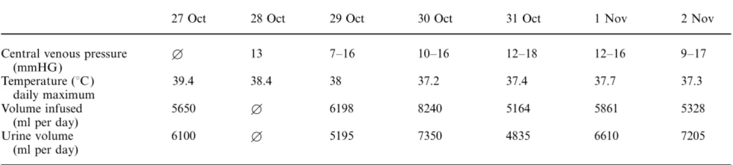
(2) 2688. M. J. Dickenmann & F. P. Brunner. Table 2. Clinical findings during the development of hypernatraemia. Central venous pressure (mmHG) Temperature (°C ) daily maximum Volume infused (ml per day) Urine volume (ml per day). 27 Oct. 28 Oct. 29 Oct. 30 Oct. 31 Oct. 1 Nov. 2 Nov. B. 13. 7–16. 10–16. 12–18. 12–16. 9–17. 39.4. 38.4. 38. 37.2. 37.4. 37.7. 37.3. 5650. B. 6198. 8240. 5164. 5861. 5328. 6100. B. 5195. 7350. 4835. 6610. 7205. B Not measured.. (137–141 mmol/l ). The increase in plasma osmolality, induced by a rising plasma sodium concentration results in water movement out of the cells into the extracellular fluid and the resultant cellular dehydration of brain cells is responsible for the dominant neurological symptoms ( lethargy, seizures, coma). The decrease in brain volume can cause rupture of cerebral veins, resulting in intracerebral or subarachnoid haemorrhage [1,2]. This did occur in our patient and may have been precipitated by thrombocytopenia after the bone marrow transplantation. The severity of hypernatraemic neurological symptoms is related primarily to the rate of rise in plasma osmolality rather than to the absolute degree of hypernatraemia. Thus patients with chronic stable hypernatraemia can be asymptomatic [3]. Hypernatraemia is induced either by negative water balance or by sodium overloading. Normally, whenever plasma sodium concentration increases, thirst develops and antidiuretic hormone is secreted. If the patient is conscious and has free access to drinking water, hypernatraemia will not develop. However, thirst and ingestion of water may not be provoked in severely ill patients because of altered mental state, weakness or mechanical ventilation. Table 3 summarizes the different aetiologies of hypernatraemia. Table 3. Aetiology of hypernatraemia [7] Water loss 1. Insensible loss sweating, fever burns respiratory infections 2. Gastrointestinal loss Osmotic diarrhoea 3. Renal loss Central diabetes insipidus Acquired disorder of urine concentration (‘renal diabetes insipidus’) Osmotic diuresis 4. Hypothalamic disorders 5. Water loss into cells (by increase of intracellular osmolality) Seizures Lactic acidosis Rhabdomyolysis Sodium retention Administration of hypertonic NaCl or NaHCO 3 Oral salt overloading. Aetiology of hypernatraemia in the above case No hypertonic NaCl or NaHCO infusions were given 3 and accidental Na+ overload could thus be excluded. Plasma lactate and creatine kinase levels repeatedly determined during the course of the illness were within the normal range, which excluded water loss into the intracellular space as a result of lactic acidosis or rhabdomyolysis. Gastrointestinal water loss by diarrhoea was not observed. In spite of hypernatraemia heart rate, blood pressure and central venous pressure remained within normal limits. Thus hypovolaemia due to dehydration was not readily apparent. However, the rising plasma creatinine concentration (maximal plasma creatinine 148 mmol/l ) pointed to a decreasing glomerular filtration rate, which was presumably provoked by borderline hypovolaemia despite generous fluid replacement with isotonic saline and glucose infusions (see Table 2). In spite of rising plasma Na+ levels polyuria persisted (Tables 1, 2). Thus hypernatraemia had to be caused by renal water loss in the face of either undisturbed or defective urinary concentrating ability (Table 4). Polyuria and hypernatraemia induced by central or renal diabetes insipidus can be distinguished from the consequences of solute diuresis with intact urinary concentration by measuring the urine osmolality and by calculating free water clearance. Osmotic diuresis is likely when daily solute excretion exceeds 1400 mosmol (or >20 mosmol/kg body weight/day) [4], as observed in our patient (e.g. on 1.11.96, 2829 mosmol/day). A hypTable 4. Aetiology of hypernatraemia combined with polyuria (adapted from [4]) Water diuresis (impaired urine concentration): Urine osmolality <250 mosmol/kg Central diabetes insipidus Renal disturbance of urine concentration (‘renal diabetes insipidus’) Solute diuresis: Urine osmolality >300 mosmol/kg Saline loading Hyperglycaemia Mannitol therapy Urea-induced osmotic diuresis Na+ wasting nephropathy.
(3) Continuing Nephrological Education. 2689. Table 5. Urea excretion and calculated protein catabolism. Plasma sodium (mmol/l ) Urea excretion/24 h (mmol/24 h) Protein catabolism (g/24 h)*. 21 Oct. 25 Oct. 2 Nov. 133 365 63.8. 136 709 124.0. 162 2174 380.4. *Protein catabolism/24 h (g)=urea (mmol/24 h)×0.175.. erosmotic urine as measured repeatedly in our patient (Table 1) is indicative of solute diuresis. Furthermore central diabetes insipidus was unlikely because desmopressin failed to raise urine osmolality. Diminished urine concentrating ability would lead to free-water excretion or an appreciably positive free-water clearance (C ) H2O [4]. Free-water clearance in our patient, however, was negative, indicating free-water reabsorption by a concentrating kidney (data obtained on 31.10.96): C. C. D. urine osm =urine vol× 1− H2O plasma osm 387 =4835× 1− =652 ml/24 h 341. C. D. This amount of free water reabsorbed, though relatively small, is well within the normal urinary concentrating capacity in view of reduced renal perfusion with decreased glomerular filtration rate due to dehydration. The most common causes of polyuria due to solute diuresis are uncontrolled diabetes mellitus with glucosuria and infusions with hypertonic mannitol, which both were absent in our patient. Another possibility is urea induced osmotic diuresis as observed following high-protein tube feedings [5], or urea administration to correct hyponatraemia in the syndrome of inappropriate ADH secretion [6 ]. In fact our patient’s blood urea concentration rose quite excessively from 10 mmol/l (24.10.96) to 36 mmol/l (2.11.96) and so did urea excretion, which peaked at over 2000 mmol/24 h on 2.11.96 ( Table 5). Where did all this urea come from? From the case history we know that parenteral nutrition was discontinued for unclear reasons when the plasma sodium concentration started to rise. Massive catabolism of body proteins due to severe illness and inadequate caloric supply must have led to the endogenous production and urinary excretion of large quantities of urea which occurred simultaneously with the development of hypernatraemia. The amount of free water needed to excrete this tremendous urea load can be estimated indirectly by calculating the electrolyte free water reabsorption Te [4]. Our patient’s calculated Te on CH2O CH2O 01.11.96:. C. D. U +U K−1 Te =urine vol× NA CH2O P NA 35+41 =6661× −1 =−3415 ml/24 h 156. C. D. The negative value indicates that on this day 3.4 litres of normally reabsorbed free water were lost by ‘nonelectrolyte’ solute diuresis.The cause of hypernatraemia becomes apparent: free-water loss by urea osmotic diuresis. The excretion of large amounts of urea induced by severe metabolic stress with high protein turnover required large urine volumes. The free-water loss led to hypernatraemia. Because of mechanical ventilation and sedation the patient was incapable of correcting his water deficit by drinking.. Diagnosis 1. Life-threatening hypernatraemia induced by urea osmotic diuresis; 2. Hypernatraemia-induced subarachnoid haemorrhage.. Conclusion Differential diagnosis of hypernatraemia in severely ill patients with metabolic stress should include osmotic urea diuresis. Treatment requires administration of adequate amounts of electrolyte-free glucose solution to supply sufficient water and calories.. Follow up After correction of free-water depletion with isotonic glucose infusion and starting adequate parenteral nutrition, plasma sodium concentration normalized (see Table 2). The patient left the hospital in good condition without any neurological deficit despite the subarachnoid haemorrhage.. References 1. Arieff AI, Guisado R. Effects on the central nervous system of hypernatremic and hyponatremic states. Kidney Int 1976; 10: 104–116 2. Finberg L, Luttrell E, Red H. Pathogenesis of lesions in the nervous system in hypernatremic states. Pediatrics 1959; 23: 46–53 3. Kastin AJ, Lipsett MB, Ommaya AK, Moser JM Jr. Asymptomatic hypernatremia. Am J Med 1965; 38: 306–315 4. Robertson GL. Differential diagnosis of polyuria. Annu Rev Med 1988; 39: 425–442 5. Gault MH, Dixon ME, Doyle M, Cohen WM. Hypernatremia, azotemia, and dehydratation due to high-protein tube feeding. Ann Intern Med 1968; 68: 778–791 6. Decaux G, Genette F. Urea for long term treatment of syndrome of inappropriate secretion of antidiuretic hormone. Br J Med 1981; 283:1081–1083 7. Rose BD. Hyperosmolal states-hypernatremia. In: Rose BD ed. Clinical Physiology of Acid–Base and Electrolyte Disorders. McGraw-Hill, New York, 1994; 695–736.
(4)
Figure

Documents relatifs
According to the China Banking Regulatory Committee, the banking institutions include 10 categories which are policy banks, state-owned commercial banks (SOCBs), joint
The oxygen permeability of Fe and Fe–Ni alloys was measured at 1150 and 1100 °C in dry and wet conditions using the internal oxidation technique. In this material, high permeability
To access the nutrigenomic effect of an acute intake of PS juice in PBMCs, we performed a pangenomic gene expression analysis 3 h after PS juice and PB drink consumption for the
For example, the model predicts that a higher volatility increases informed participation in dark pools, but can reduce dark-pool market share; and that the addition of a dark
Je souhaite aborder les domaines dans lesquels se dessine un renforcement des actions et des projets entre nos deux pays, régulièrement examinés par le Conseil consultatif
Deux ensembles de recherches, que nous passerons en revue, nous permettent cependant de dégager des recommandations thérapeutiques : les travaux consacrés au traitement des
S’enroulant vers le centre on devra reculer Accepter de souffrir de ces adversités En retrouvant la voie le but réapparait Le défi de la vie au milieu est caché J’avance et je
La toile exprime toute l'horreur et la colère ressenties par Picasso à la suite du bombardement de Guernica dont il veut dénoncer la violence et la barbarie.. « La peinture n'est
