Printed in the United States of America
Evolving concepts of sleep cycle
generation: From brain centers to
neuronal populations
J. A. Hobson, R. Lydic, and H. A. Baghdoyan
Laboratory of Neurophysiology, Harvard Medical School, Boston, Mass. 02115
Abstract: As neurophysiological investigations of sleep cycle control have provided an increasingly detailed picture of events at the cellular level, the concept that the sleep cycle is generated by the interaction of multiple, anatomically distributed sets of neurons has gradually replaced the hypothesis that sleep is generated by a single, highly localized neuronal oscillator.
Cell groups that discharge during rapid-eye-movement (REM) sleep (REM-on) and neurons that slow or cease firing during REM sleep (REM-off) have long been thought to comprise at least two neurochemically distinct populations. The fact that putatively cholinoceptive and/or cholinergic (REM-on) and putatively aminergic (REM-off) cell populations discharge reciprocally over the sleep cycle suggests a causal interdependence.
In some brain stem areas these cell groups are not anatomically segregated and may instead be neurochemically mixed (interpenetrated). This finding raises important theoretical and practical issues not anticipated in the original reciprocal-interaction model. The electrophysiological evidence concerning the REM-on and REM-off cell groups suggests a gradient of sleep-dependent membrane excitability changes that may be a function of the connectivity strength within an anatomically distributed neuronal network. The connectivity strength may be influenced by the degree of neurochemical interpenetration between the REM-on and REM-offcells. Recognition of these complexities forces us to revise the reciprocal-interaction model and to seek new methods to test its tenets.
Cholinergic microinjection experiments indicate that some populations of REM-on cells can execute specific portions of the REM sleep syndrome or block the generation of REM sleep. This observation suggests that the order of activation within the anatomically distributed generator populations may be critical in determining behavioral outcome. Support for the cholinergic tenets of the reciprocal-interaction model has been reinforced by observations from sleep-disorders medicine.
Specific predictions of the reciprocal-interaction model and suggestions for testing these predictions are enumerated for future experimental programs that aim to understand the cellular and molecular basis of the mammalian sleep cycle.
Keywords: brain stem; cholinergic systems; monoamines; neuronal populations; oscillators; sleep; sleep disorders; ultradian rhythms
I. From brain centers to neuronal populations: A scientific paradigm shift?
Mammalian neurobiology seeks to identify the neurons that mediate behavioral acts and states and to specify the mechanisms by which the critical neuronal ensembles are activated and inactivated. Most studies of the neural control of behavior have focused upon specifiable motor acts (e.g., limb flexion or extension) and have ignored the sets of background conditions or behavioral states (e.g., sleep, wakefulness, or anesthesia) out of which the acts arise. The investigation of motor acts can offer the relative advantage of the reflex paradigm. For example, cellular studies of segmental reflex control can often be localized to restricted neuronal circuitry. By contrast, physiologi-cal studies of behavioral states such as sleep must contend with endogenous changes occurring simultaneously in a multiplicity of diffuse neuronal circuits. Hobson (1978) has discussed the evolution of the state concept and defined a behavioral state operationally as a set of numer-ical values that its component variables may be assigned at a given point in time.
Only since the 1950s has it been recognized that sleep is a heterogeneous behavioral state. Mammalian sleep is a bistable process consisting of continuous alterations be-tween two different states. Information about the nomen-clature (Czeisler, Borbely, Hume, Kobayashi, Kronaure, Schulz, Weitzman, Zimmerman & Zulley 1980; Roffwarg 1979; Ursin & Sterman 1981) and the temporal organiza-tion of sleep states has been presented in detail elsewhere (Kraemer, Hole & Anders 1984; Lavie & Kripke 1981; Schulz, Dirlich, Balteskonis & Zulley 1980). Briefly, the two distinct sleep states have been given a variety of names, which are usually descriptors of bioelectric re-cordings used to characterize these two behavioral states. For example, one state of sleep is characterized by rapid eye movements (REM) but is also referred to as de-synchronized, paradoxical, or active sleep. The other state of sleep is characterized by a synchronized elec-troencephalogram and is referred to as synchronized, slow-wave, quiet, or non-REM (NREM) sleep. The REM-and-NREM nomenclature will be used throughout this paper.
perspective, is that REM sleep occurs as an endogenously generated rhythm in all terrestrial, placental mammals, including man (Tobler 1984). The REM-and-NREM sleep cycle exhibits species-specific period lengths that are always in the ultradian (much less than 24 hrs) range (Campbell & Tobler 1984). Thus, these cyclic alterations in behavioral state represent a fundamental biological rhythm.
Until recently, the states of waking and sleep were studied within the paradigm of research on neural cen-ters. The neural-centers concept is a special case of the cerebral-localizationist trend that dominated the physio-logical study of behavior in the 1930s and 1940s. Early exponents of the neural-centers concept were Hess (1931), who postulated waking and sleep centers in the diencephalon, and Bremer (1935), who first suggested that waking and sleep were regulated intrinsically, per-haps by the brain stem. The subsequent studies of arousal by Moruzzi and Magoun (1949), of NREM sleep by Moruzzi (1960), and of REM sleep by Jouvet (1962) all postulated specific brain regions or even particular nuclei involved in behavioral-state control. These hypotheses were heuristically useful because they focused attention on the physiological mechanisms underlying behavioral states. Such hypotheses were practically limited, howev-er, since the lesion and electrical-stimulation techniques were incapable of determining the precise cellular and molecular mechanisms involved in triggering and main-taining behavioral states.
Gradually replacing the concept of discrete regulatory centers controlling sleep and wakefulness is the emerging concept of dynamic interaction between anatomically distributed neuronal populations. The notion of func-tionally distinct neuronal populations was foreshadowed by Hess's (1931) concept of trophotrophic (cholinergic) and ergotrophic (aminergic) centers in the hypothalamus. Applied to behavioral-state control, this idea was further concretized and extended in Jouvet's (1962) work on the pontine mechanisms of REM sleep, which first impli-cated a cholinergic trigger mechanism. Jouvet later incor-porated the histofluorescent anatomical data of the Swed-ish school (Dahlstrom & Fuxe 1965) when he suggested that each specific state was controlled by a specific aminergic cell group (Jouvet 1969). Since many of the chemically specific neurons were found in extended sets of brain stem nuclei (Morgane & Stern 1974), the idea of an anatomically distributed system was thus clearly im-plied even in Jouvet's highly specified formulations of sleep cycle control.
The distributed-neuronal-population concept has also been reinforced by more recent evidence obtained when the electrical activity of individual neurons was recorded from behaving animals. The contrast between the brain-centers concept and the neuronal-populations concept becomes particularly sharp when distinctive single-cell firing patterns are displayed by neurons concentrated in certain brain regions or localized in scattered, but chem-ically distinct, brain nuclei. Groups of cells may attract attention because of their unique behavioral-state-specific discharge patterns. This is what has been found in recent research on the brain stem mechanisms control-ling the behavioral states of waking and sleep. For exam-ple, during REM sleep some neurons discharge more
(REM-on cells), and some neurons cease discharging (REM-oflf cells). (See Figure 2 caption for a quantitative definition of REM-on and REM-off cells.) The REM-on and REM-off discharge patterns will be discussed in detail to illustrate the interpretative and methodological problems of sleep neurobiology. Throughout this discus-sion we will distinguish between generator and modu-lator populations. Generator populations include neu-rons that may act as initiators or effectors of a state or the physiological or behavioral components of the state. Modulator neurons alter the postsynaptic response of a cell to some parallel presynaptic input. In so doing, they may alter the mode of neuronal operation, including the output of generator populations, thereby determining state properties.
It is the purpose of this paper to trace the conceptual and theoretical evolution of sleep neurophysiology in its transition from the centers to the neuronal-populations paradigm. In doing so, the present account will focus on evidence regarding the first cellular and mathematical model of mammalian behavioral-state con-trol, that of reciprocal interaction between putatively cholinergic and aminergic brain stem neurons, which appeared 10 years ago (Hobson, McCarley & Wyzinski 1975; McCarley & Hobson 1975b). In the subsequent decade, the initial finding and concepts have stimulated discussion and controversy. The central goal of the pre-sent paper is to move beyond the debate about the significance of correlations (between cellular discharge patterns and sleep states) and to stimulate a new era of causal hypothesis testing by reformulating the reciprocal-interaction hypothesis in terms more amenable to unam-biguous empirical evaluation.
A. General features of the reciprocal-interaction model of sleep cycle control
We believe it essential to preface our consideration of the relationship between empirical data and conceptual mod-els with some discussion of the nature and utility of models in general and of the reciprocal-interaction con-cept in particular. Models are useful and perhaps even essential in the conceptual organization of data at critical points in the growth of science. With the growth in its data base, neurobiology has developed models of increas-ing complexity. For example, the neuron doctrine of Ram6n y Cajal might be described as the fundamental first-order or structural model of modern neurobiology. In his sketches of linear neuronal interactions, Cajal anticipated Sherrington's second-order or dynamic-reflex model. The reflex concept made possible the detailed analysis of individual sensorimotor systems. Sherring-ton's coworker, Graham-Brown, added the neuronal-oscillator concept to the dynamic-reflex model. This ex-tension was based on Graham-Brown's (1914) observation that neuronal activity was not only responsive to external stimuli but also spontaneous, dynamic, and intrinsically periodic (Gallistel 1980).
The reciprocal-interaction model of sleep cycle control tries to account for neuronal discharge activity, which is characteristically spontaneous> continuous, and periodic. These formal properties of neuronal activity are precisely those which are likely to be of more fundamental
rele-372 THE BEHAVIORAL AND BRAIN SCIENCES (1986) 9:3
https://doi.org/10.1017/S0140525X00046318
vance to the still-higher-order concept of behavioral state control. One way to make the conceptual transition from the reflex paradigm to the behavioral-state paradigm is to view a given behavioral state (e.g., REM sleep) as a constellation of temporally coordinated physiological traits, each of which manifests the reflexivity of individual sensorimotor systems.
While model building is essential for integrating facts, making predictions, and testing hypotheses, we also recognize two pitfalls in the use of any model. One is reification, which occurs when the abstract model (a hypothetical construct) is confused with the reality of the physiological system being modeled. The other pitfall is the untestability of a model so flexible that it can be bent to suit any datum. In offering this revision of the re-ciprocal-interaction model for open peer commentary, we appeal to our colleagues for help in avoiding either of these pitfalls.
In constructing this revision we have tried to clarify the present nature, value, and limitations of the reciprocal-interaction model. Because this model superficially re-sembles the Sherringtonian reflex model it has some-times mistakenly been construed as positing a mere variant of simple reciprocal innervation (as in the crossed-extension reflex). It is important to note that such a reading involves both a lexical mistake and a misunder-standing since the reciprocal-interaction model assumes - and begins to account for - the temporal organization of spontaneous fluctuations in the excitability levels of neu-rons that may be organized reflexively, or nonreflexively, throughout the brain. In contrast to the reflex concept, the reciprocal-interaction model of behavioral-state con-trol is thus neither restricted to local circuits nor input-dependent. A patient and careful evaluation of these revised concepts can productively contribute to the con-tinued development of sleep research. We therefore appeal to our readers - especially those serving as com-mentators - for a detailed assessment of the weaknesses as well as the strengths of these concepts. We believe that only through such a properly balanced evaluation can our dialogue contribute fresh perspectives and avoid sterile debate.
B. Heuristic value of the reciprocal-interaction model: When should a model be altered and when should it be abandoned?
When a model can be altered by a simple modification of its assumptions (without violating its essential or funda-mental structure) it should be so altered and retained. When a model is demonstrated to be fundamentally wrong, or when it fails to generate testable hypotheses, it should be abandoned. As we hope to make clear, the testing of hypotheses derived from the reciprocal-interac-tion model has only begun, and the initial results strongly encourage further work.
A critical question about the usefulness of the model is: Can the reciprocal-interaction model be proved wrong? In other words, is it, in Popper's (1962; 1974) terms, a genuinely scientific hypothesis? The answer to this ques-tion is yes, as will be shown in Part V of this paper, where testable predictions are enumerated that could further confirm or unequivocally refute the model.
C. The problem of correlation versus causation One problem fundamental to behavioral neurobiology, and indeed to all studies of regulatory physiology, is the distinction between correlation and causation. We be-lieve that this conceptual issue is at the root of the interpretative differences that have divided investigators in the field of sleep neurophysiology.
The problem concerns the adequacy of evidence from single-cell recording or central-nervous-system (CNS) lesions as a basis for accepting or rejecting the hypothesis that a given brain region plays a causal role in generating a behavioral or physiological event. Data on behavioral state control will be discussed in detail using the concepts and interpretive logic that follow.
For cellular discharge data:
1. A strong positive or negative correlation between single-cell firing rate and a behavioral state is compatible with a hypothesis of cause but cannot be taken as more than suggestive evidence that the cellular activity causes the behavioral state. An example would be the high discharge rate of pyramidal tract motoneurons during REM sleep (Evarts 1964), a finding that, in and of itself, cannot be distinguished from similar results at the level of the brain stem (Steriade & Hobson 1976). Which is cause, which is effect, and which a mere correlate?
2. A corollary of the first principle concerns the fact that most brain regions and their component nuclear groups have multiple regulatory roles. An example would be the putative role of serotonergic neurons in pain modulation, temperature control, and sleep regulation. It would thus be inappropriate to conclude that because such neurons mediate pain and temperature control they cannot medi-ate sleep. For example, serotonergic neurons cease firing during REM sleep (McGinty & Harper 1976), tem-perature control is lost in REM sleep (Parmeggiani 1981), and pain sensation is rare in dream reports (McCarley & Hoffman 1981). For any neurophysiological theory of sleep to be adequate it should aim to integrate observa-tions from multiple physiological systems (Figure 1; Table 1).
3. Since neuronal populations commonly subserve many regulatory functions, and since the CNS exhibits considerable functional plasticity after experimental le-sions, the relationships between neuronal discharge and a given behavioral state may not be immutable. After surgical or pharmacological manipulations, neuronal fir-ing patterns may even be dissociated from a given behav-ioral state. An example of altered phase dependence would be the state-independent pontogeniculooccipital (PGO) waves that are produced after brain amine deple-tion with parachlorophenylalanine (Jouvet 1972) or with localized administration of cholinergic agonists (Vivaldi, McCarley & Hobson 1980).
For data obtained from experimental lesions: 1. The loss of a behavioral state after a CNS lesion is compatible with the idea that the brain region may be involved in generating that behavioral state, but such evidence is no more than suggestive and may even be quite misleading. Numerous examples are discussed in Section A, Part II; the complexity of this point is also illustrated by the cessation of respiration or loss of sleep seen after midline knife cuts in the brain stem (Mancia
1969). Should Mancia's finding be attributed to destruc-tion of cell bodies (e.g., raphe neurons) or to destrucdestruc-tion of decussating fibers? And would such fibers be of local or remote origin?
2. Persistence of a behavioral state following a lesion indicates only that the destroyed CNS structure is not necessary for the expression of the behavioral state; it does not prove that the brain region in question normally plays no physiological role in the generation of that behavioral state. The redundancy that is typical of most CNS control systems may explain, for example, the fact that animals with visual cortex ablations can still "see" with their colliculi (Sprague 1966) or that monkeys can still move after destruction of the pyramidal tract (Bucy, Ladpli & Erlich 1966). One would not conclude from these data that the cortex has no role in vision or that the pyramidal tract has no role in movement (see also Campion, Latto & Smith 1983).
The general conclusion emerging from single-cell re-cording and experimental lesion studies is that neither method, either alone or in combination with the other, provides evidence adequate for accepting or rejecting any hypothesis of causality with complete confidence. Given these severe methodological limitations, it is important to be both cautious in interpreting data and imaginative in developing additional ways to test hypotheses that a neuronal population causes a particular stage of sleep. The difficulties in obtaining definitive evidence for or against such causal hypotheses in the case of sleep cycle control will be discussed to illustrate (1) the strengths and problems of the neuronal-population approach and (2) the need for developing specific criteria with which to evalu-ate the hypothesis that a cellular population causes a behavioral state.
II. A neuronal-population concept of
behavioral-state control: Evolution of the reciprocal-interaction model
Over the past decade, the initial formulation of the reciprocal-interaction model of mammalian sleep cycle control (Hobson et al. 1975; McCarley & Hobson 1975b) has been revised in accordance with new evidence from single-unit recording and chemical microinjection ex-periments in animals and from human clinical studies (Hobson 1984; McCarley 1978; 1980; McCarley & Massa-quoi 1985). In its original form, the reciprocal-interaction model postulated that the behavioral states constituting the mammalian sleep cycle were a function of the out-of-phase discharge profiles displayed by two brain stem neuronal populations. The REM-on cells were first found in the gigantocellular tegmental field (FTG) of the pon-tine reticular formation and were postulated to play a cholinergically mediated role in generating REM sleep. The REM-off cells were first found in the locus coeruleus (LC) and were postulated to play a noradrenergic, per-missive role by disinhibiting the REM-on cells. The model, which aimed to be synaptically explicit and math-ematically precise in its attempt to account for the cycling of NREM and REM sleep, could also be extended to account for waking state features in terms of high levels of aminergic neuronal activity.
Replacing these original postulates of a REM sleep
generator zone highly localized to the pontine FTG is the present concept of an anatomically distributed population of neurons (REM-on cells) in the brain stem whose activation is postulated to play a key role in generating REM sleep. Figure 1 schematizes the anatomically dis-tributed nature of some of the neuronal populations studied in relation to behavioral-state control.
A note regarding the anatomical nomenclature in Fig-ure 1 and throughout the text: In constructing this revi-sion of the reciprocal-interaction model, we have con-tinued to use the terminology of Berman (1968). Ber-man's stereotaxic atlas is a standard reference found in virtually every neurophysiology laboratory where the domestic cat is the animal studied. We appreciate the cytological heterogeneity of the brain stem reticular for-mation, and when we refer to the FTG of Berman (1968), unless otherwise stated, we are emphasizing that portion of the FTG defined by Brodal (1969) as the medial pontine reticular formation. The general features of the figure include:
1. The putatively cholinergic/cholinoceptive REM-on cell network is widespread, and its component neurons at the cortical, brain stem, and spinal cord levels are postu-lated to interact with motor-control systems during wakefulness, as well as during REM sleep.
2. The excitatory interconnections of the REM-on cell network are such that the REM-on activation process could theoretically start from several places within the network if the inhibitory aminergic input were removed uniformly. Once the REM-on discharge is started it is postulated to increase exponentially to some maximum level, reinforced by the excitatory synaptic interconnec-tions.
3. Figure 1 distinguishes between two critically selec-tive neuronal populations: (a) the aminergic (REM-off) cells, which are postulated to exert an inhibitory re-strain during waking so as to release the REM-on neu-rons progressively to the point of autoactivation during REM sleep, and (b) the inhibitory pontomedullary neu-rons, which are postulated to actively block the somatic motor output during REM sleep.
4. The interaction between the REM-off (putative modulatory) and the REM-on (putative generator) net-works is also anatomically widespread throughout the CNS (for details see Table IB). Based on this diffuse anatomical distribution, any localization of a putative REM sleep generator even to the brain stem could, theoretically, be an illusion. Our reasoning in making this suggestion is the following:
(a) The concentration in the brain stem of aminergic cell bodies (see again Figure 1; and for details, Table IB) would indicate that the aminergic portion of the putative .REM sleep oscillator is localizable to the brain stem. The widespread distribution of the REM sleep generator population (Figure 1) suggests that the other, putatively cholinoceptive/cholinergic side of the proposed oscillator is much less likely to be so localized. The experimental evidence supporting these ideas is discussed in detail in Part IV and further illustrated in Figures 2 and 4.
(b) The putative effectors of some REM sleep phe-nomena (Figure 1) are localizable to the brain stem (e.g., those neurons generating rapid eye movements, PGO waves, and facial muscle twitches), but other effectors of REM sleep are not in the brain stem (e.g., the effector 374 THE BEHAVIORAL AND BRAIN SCIENCES (1986) 9:3
https://doi.org/10.1017/S0140525X00046318
BRAIN LEVEL Cortex
Thalamus
Mid bra in
NEURONAL SYSTEM REM SLEEP PHENOMENON
Spinal Cord
I Inhibitory OEicitatory
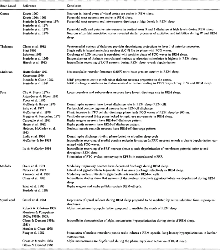
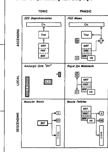
Figure 1. Schematic representation of the REM sleep genera-tion process. A distributed network involves cells at many brain levels (left). The network is represented as comprising three neuronal systems (center) that mediate REM sleep electro-graphic phenomena (right). Postulated inhibitory connections are solid circles; postulated excitatory connections are open circles. In this diagram, no distinction is made between neurotransmission and neuromodulatory functions of the de-picted neurons. Table 1A gives a selective outline of the physio-logical evidence supporting this schematization, and the ana-tomical evidence on which the synaptic connections are based is outlined in Table IB. We caution the reader that the precise synaptic signs of many of the aminergic and reticular pathways remain to be demonstrated. This is particularly true with re-spect to the possible state dependency of the postulated synap-tic signs. In many cases, the neuronal architecture is also known to be far more complex than we have indicated (e.g., the thalamus and cortex).
There are postulated to be two additive effects of the marked diminution in firing by aminergic neurons at REM sleep onset: disinhibition (through removal of negative restraint) and facilita-tion (through positive feedback). The net result is strong tonic and phasic activation of reticular and sensorimotor neurons.
REM sleep phenomena are postulated to be mediated as follows:
EEG desynchronization results from a net tonic increase in reticular, thalamocortical, and cortical neuronal firing.
PGO waves are the result of tonic disinhibition and phasic excitation of burst cells in the lateral pontomesencephalic teg-mentum. (Note that no excitatory input is shown because none has yet been demonstrated.)
The rapid eye movements are the consequence of phasic firing by reticular and vestibular cells; the latter (not shown) directly excite the oculomotor neurons.
Muscular atonia is the consequence of tonic postsynaptic inhibition of the anterior horn cells by the pontomedullary reticular formation.
Muscle twitches occur when excitation by reticular and pyra-midal tract motoneurons phasically overcomes the tonic inhibi-tion of the anterior horn cells.
Anatomical abbreviations: RN = raphe nuclei; LC = locus coeruleus; P = peribrachial region; FTG = gigantocellular tegmental field; FTC = central tegmental field; FTP = par-vocellular tegmental field; FTM = magnocellular tegmental field; TC = thalamocortical; CT = cortical; PTcell = pyramidal
neurons of EEG desynchronization and alpha moto-neurons effecting somatic muscular atonia).
(c) According to this concept, one could anticipate that extensive destruction of REM-on cells in the brain stem might be without effect on some of the tonic manifesta-tions of REM sleep which are derived from extra-brain-stem effectors.
(d) A key remaining question concerns the degree of dissociation of the physiological signs of REM sleep that could be effected by neuronal destruction. Part IV of this paper describes the dissociations of the physiological signs of REM sleep that can be systematically produced using chemical microinjection techniques.
The most essential neurophysiological difference be-tween the waking and the RE M sleep state now appears to be the level of discharge activity in the subpopulation of putatively modulatory REM-off cells located in several aminergic nuclei throughout the pontomesencephalic brain stem. These aminergic nuclei are widespread in the pontine tegmentum, display progressive decreases in firing rate during REM sleep, and may disinhibit the anatomically distributed REM-on neurons (Table 1A). Like the REM-on populations, these level-setting or modulatory REM-off populations are more widely dis-tributed than was first supposed. They are currently known to include at least two chemically distinct groups: the noradrenergic and the serotonergic subpopulations; and they.are found to be concentrated in at least three brain stem regions: the raphe nuclei, the locus coeruleus, and the peribrachial region. Many brain stem nuclei contain cells of both the REM-on and the REM-off type, thus indicating that the REM sleep oscillator network is not only more widely distributed than previously be-lieved but also neurochemically interpenetrated.
A. The putative generator population
1. Identity and characteristics of the REM-on cells.
During REM sleep, most of the neurons recorded throughout the brain have been shown to be activated to levels at least as high as those seen in the waking state (Steriade & Hobson 1976). The most conspicuous excep-tion is the populaexcep-tion of REM-off cells that will be discussed below (Part II, Section B) with respect to their proposed modulatory or level-setting interaction with the REM-on population.
Since so many neurons are of the REM-on type, it is logical to propose that a vast ensemble of cells constitutes the population that finally acts as effectors of the physio-logical signs of REM sleep, such as cortical desynchroni-zation, muscular atonia, or the rapid eye movements themselves. A putative neuronal trigger zone that initi-ates the recruitment process leading to the coordinated physiological manifestations of REM sleep remains to be unequivocally identified.
The neuronal mechanisms to be accounted for in REM sleep generation ultimately include tonic and phasic excitation of sensory and motor neurons at many levels of the nervous system during sleep. From the tentative cell; III = oculomotor; IV = trochlear; V = tirgeminal motor nuclei; AHC = anterior horn cell.
The time calibration for the four simultaneously recorded polygraphic records is 5 seconds.
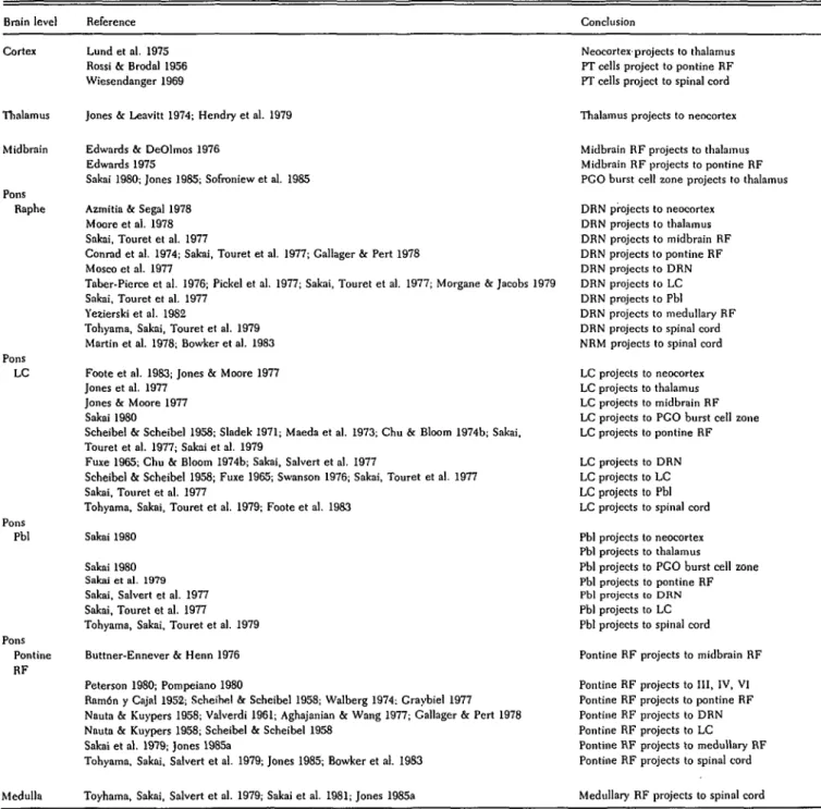
Table 1A. Electrophysiological studies of the REM sleep generation process
Brain Level Reference Conclusion Cortex Evarts 1960
Evarts 1964; 196S Steriade & Deschenes 1974 Steriade et al. 1974 Steriade et al. 1978 Steriade et al. 1979
Thalamus Clenn et al. 1982 Bizzi 1966 Sakakura 1968 Steriade et al. 1969 Hirsch et al. 1983
Neurons in lateral gyrus of visual cortex are active in REM sleep. Pyramidal tract neurons are active in REM sleep.
Pyramidal tract neurons and interneurons discharge at high levels in REM
sleep-Pyramidal cells and putative interneurons in cortical areas 5 and 7 discharge at high levels during REM sleep. Neurons of parietal association cortex revealed similar processes of excitation and inhibition during W and REM sleep.
Ventromedial nucleus of thalamus provides depolarizing projections to layer ] of anterior neocortex. Single cells in lateral geniculate nucleus (LGN) fire in phase with PGO waves.
Discharge of LGN neurons is correlated with positive phase of PGO waves in REM sleep. Responsiveness of thalamic ventrolateral nucleus to electrical stimulation is highest in REM sleep. Intracellular recording of LGN neurons during REM sleep reveals depolarization.
Midbrain Huttenlocher 1961 Kasamatsu 1970 Steriade & Clenn 1982 Steriode et al. 1982
Mesencephalic reticular formation (MRF) units have greatest activity in REM sleep. MRF projections excite intralaminar thalamic neurons projecting to the cortex.
MRF discharge contributes to thalamocortical activation leading to EEC desynchrony in W and REM sleep. Pons Chu & Bloom 1974a
Aston-Jones & Bloom 1981 Foote et al. 1983 McGinty & Harper 1976 Saito et al. 1977 McCarley et al. 1978 Mergner & Pompeiano 1978 Cespuglio et al. 1981 Heym et al. 1982 Hobson, McCarley et al. 1983
Lydic et al. 1984 McCarley & Ito 1983 Ito & McCarley 1984
Locus coeruleus and subcoeruleus neurons have lowest discharge rate in REM sleep.
Dorsal raphe neurons have lowest discharge rate in REM sleep (REM-ofi). Peribrachial pontine tegmental neurons have REM-off discharge.
Phasic increase in FTG cellular discharge phase leads PGO waves of REM sleep by 900 ms. Vestibular neuronal firing phase locked to rapid eye movements in REM sleep.
Raphe magnus neurons have REM-off discharge pattern. Raphe pontis neurons have REM-off discharge pattern.
Nucleus linearis centralis neurons have REM-off discharge pattern. Dorsal raphe discharge rhythm phase-locked to ultradian sleep cycle.
Intracellular recording of medial pontine reticular formation (mPRF) neurons reveals a phasic depolarization cor-related wjth PGO waves.
Intracellular recording of mPRF neurons shows a tonic depolarization of membrane potential prior to and throughout REM sleep.
Stimulation of FTG evokes monosynaptic EPSPs in contralateral mPRF. Medulla Orem et al. 1974
Netick et al. 1977 Kanamori et al. 1980 Chase et al. 1981 Sakai et al. 1983 Steriade et al. 1984
Medullary respiratory neurons have decreased discharge during REM sleep.
Lateral and gigantocellular tegmental field neurons discharge selectively in REM sleep. Medullary nucleus reticularis gigantocellularis contains REM-on cells.
Intracellular studies show that neurons of the nucleus reticularis gigantocellularis are depolarized during REM sleep.
Raphe magnus and raphe pallidus contain REM-off cells.
Spinal cord Cassel et al. 1964 Kubota & Kidokoro 1965 Morrison & Pompeiano 1965a; 1965b; 1965c Glenn & Dement 1981a; 1981b
Morales & Chase 1978 Fung et al. 1982 Chase & Morales 1983 Clenn & Dement 1985
Depression of spinal reflexes during REM sleep proposed to be mediated by active inhibition from supraspinal structures.
Alpha motoneuron hypopolarization proposed to mediate the atonia of REM sleep.
Intracellular demonstration of alpha motoneuron hyperpolarization during atonia of REM sleep.
Stimulation of nucleus reticularis pontis oralis induces a REM-specific, long-latency hyperpolarization in lumbar motoneurons.
Alpha motoneurons are depolarized during the phasic myoclonic activation of REM sleep.
Note: This table provides selected references supporting the functional role of the schematized neuronal connections outlined in Figure 1.
scheme drawn in Figure 1 and documented in Tables 1A and IB, it can be seen that the components of this vast network are mutually enhancing; it is therefore difficult to assert with certainty that one or another part of the system plays the role of trigger or prime mover for the excitation process initiating a REM sleep episode. The initial defense of the idea that FTG neurons were the
executors of REM sleep helped to perpetuate a contro-versy which one hopes can be superseded by the more supple account of sleep cycle generation developed here. Questions concerning the appropriateness of hier-archically arranged control models are not limited to REM-sleep-generating mechanisms. For example, since cortical, rubral, and pontine neurons may all contribute
376 THE BEHAVIORAL AND BRAIN SCIENCES (1986) 9:3
https://doi.org/10.1017/S0140525X00046318
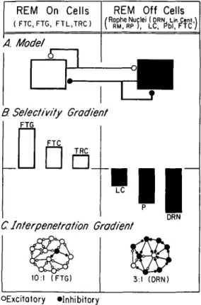
Table IB. Anatomical substrates of REM sleep generation
Brain level Reference Conclusion
Cortex Thalan Midbrain Pons Raphe Pons LC Pons Pbl Pons Pontine RF Medulla Lund et al. 1975 Rossi & Brodal 1956 Wiesendanger 1969
Jones & Leavitt 1974; Hendry et al. 1979
Edwards & DeOlmos 1976 Edwards 1975
Sakai 1980; Jones 1985; Sofroniew et al. 1985 Azmitia & Segal 1978
Moore et al. 1978 Sakai, Touret et al. 1977
Conrad et al. 1974; Sakai, Touret et al. 1977; Gallager & Pert 1978 Mosco et al. 1977
Taber-Pierce et al. 1976; Pickel et al. 1977; Sakai, Touret et al. 1977; Morgane & Jacobs 1979 Sakai, Touret et al. 1977
Yezierski et al. 1982
Tohyama, Sakai, Touret et al. 1979 Martin et al. 1978; Bowker et al. 1983 Foote et al. 1983; Jones & Moore 1977 Jones et al. 1977
Jones & Moore 1977 Sakai 1980
Scheibel & Scheibel 1958; Sladek 1971; Maeda et al. 1973; Chu & Bloom 1974b; Sakai, Touret et al. 1977; Sakai et al. 1979
Fuxe 1965; Chu & Bloom 1974b; Sakai, Salvert et al. 1977
Scheibel & Scheibel 1958; Fuxe 1965; Swanson 1976; Sakai, Touret et al. 1977 Sakai, Touret et al. 1977
Tohyama, Sakai, Touret et al. 1979; Foote et al. 1983 Sakai 1980
Sakai 1980 Sakai et al. 1979 Sakai, Salvert et al. 1977 Sakai, Touret et al. 1977 Tohyama, Sakai, Touret et al. 1979 Buttner-Ennever & Henn 1976 Peterson 1980; Pompeiano 1980
Ramon y Cajal 1952; Scheihel & Scheibel 1958; Walberg 1974; Craybiel 1977 Nauta & Kuypers 1958; Valverdi 1961; Aghajanian & Wang 1977; Gallager & Pert 1978 Nauta & Kuypers 1958; Scheibel & Scheibel 1958
Sakai et al. 1979; Jones 1985a
Tohyama, Sakai, Salvert et al. 1979; Jones 1985; Bowker et al. 1983
Toyhama, Sakai, Salvert et al. 1979; Sakai et al. 1981; Jones 1985a
Neocortex projects to thalamus PT cells project to pontine RF PT cells project to spinal cord
Thalamus projects to neocortex
Midbrain RF projects to thalamus Midbrain RF projects to pontine RF PGO burst cell zone projects to thalamus DRN projects to neocortex DRN projects to thalamus DRN projects to midbrain RF DRN projects to pontine RF DRN projects to DRN DRN projects to LC DRN projects to Pbl DRN projects to medullary RF DRN projects to spinal cord NRM projects to spinal cord LC projects to neocortex LC projects to thalamus LC projects to midbrain RF LC projects to PGO burst cell zone LC projects to pontine RF LC projects to DRN LC projects to LC LC projects to Pbl LC projects to spinal cord Pbl projects to neocortex Pbl projects to thalamus
Pbl projects to PGO burst cell zone Pbl projects to pontine RF Pbl projects to DRN Pbl projects to LC Pbl projects to spinal cord Pontine RF projects to midbrain RF Pontine RF projects to III, IV, VI Pontine RF projects to pontine RF Pontine RF projects to DRN Pontine RF projects to LC Pontine RF projects to medullary RF Pontine RF projects to spinal cord
Medullary RF projects to spinal cord
Note: This table provides selected references supporting the structural basis of the schematic model outlined in Figure 1.
to the phasic excitation of spinal motoneurons, it would be difficult to assign a principal role to any one of them in generating movement; the ensemble works as a whole, and motor action may be initiated at several points in the system.
a. The FTG generator hypothesis. Historically, the initial studies of cellular discharge during REM sleep suggested that the FTG might constitute a trigger zone for the initiation of REM sleep (Hobson, McCarley, Pivik & Freedman 1974; Hobson, McCarley, Freedman & Pivik 1974; McCarley & Hobson 1975a). The hypothesis that the FTG constitutes the REM sleep generator is at the center of the discussion and debate stimulated by the reciprocal-interaction model.
It is now clear that the process of REM sleep generation is far too complex to be accounted for solely by the FTG generator hypothesis. First, it has been shown that the neuronal discharge selectivity of FTG neurons recorded from head-restrained animals is not limited to REM sleep. In freely moving animals these pontine cells, like cells elsewhere throughout the CNS, discharged as in-tensely in association with particular movements in wak-ing as they did durwak-ing REM sleep (Siegel & McGinty 1977; Siegel, McGinty & Breedlove 1977; Vertes 1977). It has also been shown that kainic acid lesions in the pontine reticular formation that destroy FTG cells do not prevent REM sleep (Drucker-Colin & Bernal-Pedraza 1983; Sastre, Sakai & Jouvet 1981). This finding reinforces the
notion that the REM-on population is anatomically dis-tributed and that only large lesions of what still appears to be the most critical portion for REM sleep, the pontine reticular formation, can eliminate all manifestations of REM sleep (Jones 1979).
The discovery that REM sleep discharge selectivity characterized FTG cells only in head-restrained animals overthrew the original assumption that the FTG popula-tion was the sole executor or even an essential generator of REM sleep. This finding also supported the idea that the FTG was only one of a set of upper motor neuronal systems to be activated in REM sleep. The relationship between FTG discharge and certain movements does not refute the more cautious suggestion that activation of the FTG and other motor neuronal systems during REM sleep could nonetheless be involved in the generation of many REM sleep events (e.g., the rapid eye movements themselves, the muscle twitches, and even the PGO waves).
Since all results agree that FTG cells are dramatically activated in REM sleep, numerous studies have indi-cated the importance of understanding how this activa-tion is produced. Unfortunately, the cellular mecha-nisms of FTG activation during REM sleep are in no way explained by establishing a correlation between specific movements and waking-state FTG discharge. Hence, it is clearly premature to conclude that FTG activation either is entirely movement-related or has no signifi-cance in the normal triggering of REM sleep. Neither of these alternative hypotheses has been supported by de-tailed intracellular studies. Recent intracellular analyses of the membrane characteristics of FTG neurons have demonstrated that FTG cells are depolarized before and throughout REM sleep (Ito & McCarley 1984; McCarley & Ito 1983). McCarley (1978) has emphasized this point and coined the term "selectivity of modulation" to con-vey the idea that depolarization of the entire FTG cell population, not just subpopulations subserving particu-lar movements, is a mechanistically critical and dis-tinctive REM sleep phenomenon.
Although the kainic acid lesions clearly show that the FTG cells of the pontine reticular formation are neither necessary nor sufficient for initiating REM sleep, the results are at variance with the consistent finding that the rostral FTG, when pharmacologically stimulated, ap-pears to function as a critical zone for triggering the electrographic signs of REM sleep (Baghdoyan, Rodrigo-Angulo, McCarley & Hobson 1984; Drucker-Colin & Bernal-Pedraza 1983). Furthermore, the lesion data do not rule out the possibility that the FTG might function as a REM sleep trigger zone under physiological conditions. There are numerous examples where lesions produce complex interpretative problems rather than supporting or refuting the hypothesis of a causal, regulatory role for a putative pattern generator. For example, selective le-sions of the medullary dorsal and ventral respiratory nuclei do not eliminate breathing (Speck & Feldman 1982), and lesions of the hypothalamic suprachiasmatic nuclei do not disrupt all circadian rhythms (Albers, Lydic, Gander & Moore-Ede 1984; Fuller, Lydic, Sulz-man, Albers, Tepper & Moore-Ede 1981; Prosser, Kit-trell & Satinoff 1984; Szafarczyk, Ixart, Malaval, Nou-guier-Soule & Assenmacher 1979). In addition, surgical
removal of well-defined epileptic foci does not always eliminate seizures in humans (Elazar & Hobson 1985). There are two important reasons for keeping these caveats in mind. One challenge to the interpretation that REM sleep is in part caused by FTG discharge was based on the finding that FTG neurons fire during waking movements and are thus not selectively active during REM sleep (Siegel & McGinty 1977; Vertes 1977). A key observation, and one that tended to become lost in the discharge selectivity debate, is that the change in firing of FTG neurons precedes by several minutes the onset of REM sleep and all of its phasic phenomena. This long phase lead in FTG discharge prior to REM onset is difficult to explain as a mere correlate of phasic motor activity, since there is normally little or no motor activity during the early transition into REM sleep. No similar changes in FTG discharge have been reported during the transition from sleep to waking. On the contrary, after reaching an early peak the firing level of pontine reticular neurons falls progressively during the last half of each REM sleep episode and then decreases greatly as REM sleep gives way to wakefulness. Such a long tonic latency suggests that it would be useful to design experiments to explore the possibility of a change in FTG neuronal excitability level preceding REM onset (Ito & McCarley 1984).
The second reason for continuing to consider the hy-pothesis that the rostral FTG is one of the regions from which REM sleep can be generated is that the anterodor-sal pontine tegmentum is the only brain stem reticular site from which a behavioral state resembling REM sleep has been induced pharmacologically (Baghdoyan, Rod-rigo-Angulo, McCarley & Hobson 1982; Baghdoyan, Rodrigo-Angulo, McCarley & Hobson 1986). These important data will be discussed in detail in Part IV of this paper, which explores the nature of the REM sleep activation process.
b. The cholinergic generator hypothesis. The postulated cholinergic identity of neurons has been confirmed in numerous brain stem areas from which REM-on cells have been recorded (Table 3D). In no case to date, however, has an electrophysiologically identified REM-on cell also been cREM-onfirmed to be cholinergic. Although it is not yet possible to demonstrate the presence of acetyl-choline (ACh) in neurons, immunohistochemical identifi-cation of the synthetic enzyme choline acetyltransferase (ChAT) has been shown to be a reliable marker for cholinergic cells (Cuello & Sofroniew 1984). The pro-posed cholinergic identity of pontine FTG cells has been supported (Kimura, McGeer, Peng & McGeer 1981). This study showed that large cells in the rostral FTG stained heavily for ChAT, and Kimura et al. (1981) sug-gested that these cells are both cholinoceptive and cho-linergic. However, the cholinergic nature of the pontine FTG cells remains to be confirmed using a monoclonal antibody for ChAT.
The density of ChAT-positive cells in the FTG and FTM has been found to be greater in the medullary than in the pontine extent of these reticular fields (FTG, FTL, FTM; Kimura et al. 1981). Sakai (1985c) has also demon-trated that reticular cells in the ventromedial medulla (FTM) are ChAT immunopositive. These medullary cells (magnocellular or FTM of Berman) have recently been
378 THE BEHAVIORAL AND BRAIN SCIENCES (1986) 9:3
https://doi.org/10.1017/S0140525X00046318
implicated in the generation of EEG desynchrony during REM sleep (Steriade, Sakai & Jouvet 1984). Neurons in the FTM, interacting with pontine peri-LC alpha cells, have also been shown to play a role in generating the somatic muscular atonia of REM sleep (Sakai 1985c; Sakai, Sastre, Kanamori & Jouvet 1981). Both the medul-lary FTM cells and the pontine peri-LC alpha neurons have been shown to be ChAT-immunopositive (Jones 1985; Sakai 1985c).
Of all the physiological signs of REM sleep, PGO waves have most convincingly been shown to be generated by cholinergic neurons. As discussed below, the cellular mechanisms responsible for generating PGO waves have been localized to the region surrounding the brachium conjunctivum near the junction of the pons and midbrain. Nuclei in this region consistently have been shown to contain cells that stain heavily for ChAT (Jones 1985; Mesulam, Mufson, Levey & Wainer 1984; Sakai 1985c; Sofroniew, Priestley, Consolazione, Eckenstein & Cuello 1985).
Finally, cholinergic neurons are known to play a role in generating the rapid eye movements of REM sleep. The ocular motor nuclei of the brain stem contain ChAT-positive neurons (Cuello & Sofroniew 1984; Kimura et al. 1981; Mesulam et al. 1984).
It thus seems justified to conclude that detailed physio-logical, pharmacophysio-logical, and anatomical studies have substantiated many parts of this hypothetical picture of how REM sleep may be generated (Figures 1 and 3; Tables 1A and IB; Table 3). These results are consistent with the inference from the early transection studies (Jouvet 1962) that the pons plays a critical role during REM sleep in coordinating the activity of neurons in such widely separated sites as the cortex and the spinal cord.
2. Neurons involved in generating the tonic and phasic events of REM sleep. Superimposed upon the tonic
(greater than 1 sec) background of a desynchronized electroencephalogram and postural atonia are the REM sleep phasic (less than 1 sec) events: the rapid eye movements themselves, the muscle twitches, and PGO waves recorded in the pontine reticular formation, the lateral geniculate bodies, and in the occipital cortices.
a. The tonic events of REM sleep
i. EEG desynchrony The original "paradox" of REM
sleep was recorded by Jouvet and Michel (1959), who noted in a laboratory notebook: "The cat appears to be asleep but the EEG shows it to be awake!" Subsequent lesion, stimulation, and single-cell recording studies have indicated that this EEG desynchronization, which is characteristic of REM sleep but phenomenologically identical to that of wakefulness, is due to the following processes:
(1) Rostrally projecting neurons of the midbrain re-ticular formation are activated (Huttenlocher 1961; Kasamatsu 1970; Steriade 1981).
(2) These reticular neurons convey excitatory signals to the medial thalamus (Steriade, Ropert, Kitsikis & Oakson 1980; Steriade & Deschenes 1984) where
(3) the resulting depolarization of thalamic cells is relayed to cortical targets (Steriade 1981), including
(4) a wide variety of pyramidal and nonpyramidal cor-tical interneurons (Steriade 1978) and,
(5) putatively excitatory and inhibitory neurons of the cortex (Steriade 1984).
Thus, at the level of extracellular recording, there is no visible difference in the mechanisms of cortical activation observed during REM sleep and waking. These findings vindicate and elegantly extend to the cellular level the concept of the ascending reticular activating system (Moruzzi & Magoun 1949). The reciprocal-interaction model postulates that during REM sleep the reticulotha-lamocortical system is activated via the disinhibition associated with arrest of firing in the diffusely projecting (Azmitia & Segal 1978) aminergic neurons, whereas dur-ing wakdur-ing the cellular generators of EEG de-synchronization must overcome aminergically mediated inhibitory restraint.
It has been shown that when parts of the midbrain reticular formation are microinjected with cholinergic agonists, the cortical EEG is desynchronized and the occurrence of the waking state is enhanced at the expense of time spent in REM sleep (Baghdoyan, Rodrigo-Angulo, McCarley & Hobson 1984). It would thus be of particular interest to know how the aminergic neurons respond to cholinergic stimulation of the midbrain. The reciprocal-interaction model predicts no change or an increased aminergic cell discharge in association with midbrain cholinergic stimulation. Finding a significant reduction or a cessation of aminergic cell firing in such an experiment would weigh heavily against the reciprocal-interaction model.
it. Muscular atonia During REM sleep in intact ani-mals there is a powerful postsynaptic inhibition of the brain stem and final-common-path motoneurons that restrains all but the oculomotor movements, respiration, and phasic twitches of the skeletal musculature (Pom-peiano 1967). Intracellular recordings from spinal moto-neurons (Chandler, Chase & Nakamura 1980; Glenn & Dement 1981a; 1981b; Morales & Chase 1978) have revealed tonic hyperpolarizations of up to 10 mV, as strong as the simultaneous depolarization of brain stem premotor effector neurons (Ito & McCarley 1984). A likely source of such inhibition is the medullary reticular formation (Chase & Morales 1984; Fung, Boxer, Morales & Chase 1982), elements of which show the strongest tonic state-specific discharge yet observed (maximal dis-charge ratios for REM/waking of about 700/1; Netick, Orem & Dement 1977). It has been proposed that these bulbar inhibitory neurons may be activated by the gener-alized disinhibition attributed to putatively aminergic REM-off cells and/or by specific excitatory projections from the anterodorsal pons (Sakai 1980). Compounding the obvious difficulty of performing long-term intra-cellular recordings in behaving animals, a major obstacle to obtaining critical evidence about the hypothesis of disinhibition is the difficulty of ascertaining distant effects as originating from the anatomically interpenetrated REM-on or REM-off populations (see Figures 1 and 2).
b. The phasic events of REM sleep
i. PGO waves The PGO waves of REM sleep are
associated with sequential phasic activation of cells in the medulla, pons, midbrain, thalamus, and cortex (Nelson, McCarley & Hobson 1983; Orem 1980; Steriade & Hob-son 1976). The original descriptions and experimental analyses of PGO waves (Bizzi & Brooks 1963; Brooks &
Bizzi 1963) suggested a strict temporal and spatial order of activation that has been substantiated by recent studies. For example, when the eyes move to the right during REM sleep, bursts of PGO-related cellular discharge occur in the right (and not the left) midbrain (Nelson et al. 1983), and the PGO waves of the right geniculate body and the right posterolateral cortex are larger in amplitude than those recorded from the left side of the brain (Monaco, Baghdoyan, Nelson & Hobson 1984). Latency studies have revealed that the midbrain cellular dis-charge always precedes the ipsilateral geniculate wave onset by 11.0 (±2.45) milliseconds (Nelson et al. 1983). The spatial and temporal specificity of PGO waves during REM sleep suggests that these patterns of elec-trical activity may represent an internal information sys-tem (Bowker 1985; Morrison 1979; Nelson et al. 1983) whose excitability is markedly enhanced during REM sleep. These findings are relevant to the efferent-copy hypothesis of von Hoist and Mittelstaedt (1950) and of Sperry (1950). Both postulate an internal signal system by which the consequences of motor acts could be antici-pated by sensorimotor systems whose inputs would nec-essarily be perturbed by the motor acts. [See also Berkin-blit et al: "Adaptability of Innate Motor Patterns and Motor Control Mechanisms" 9(4) 1986.] 1985.)
it. Eye movements The eye movements of REM sleep appear to be generated by the premotor neurons of the reticular formation (Henn, Buttner-Ennever & Hepp 1982; Henn 1980; Henn, Hepp & Buttner-Ennever 1982). These include pontine giant cells which have been shown to project remotely to the spinal cord and locally to oculomotor and vestibular neurons (Peterson 1980; Pom-peiano 1980). These cells display the same range of firing patterns in REM sleep as in waking (i. e., short lead burst, long lead burst, and tonic discharge). However, this similar activation pattern does not guarantee identical mechanisms of activation or identical functional signifi-cance of these cells during REM sleep and wakefulness. With respect to mechanisms activating the rapid eye movements, intracellular recording of pontine giant cells has revealed a powerful, tonic influence, with 10-mV depolarizations in resting membrane potential reflecting excitation and/or disinhibition of these cells during REM sleep (Ito & McCarley 1984). While the depolarization mechanism remains to be elucidated, disinhibition of the pontine giant cells by aminergic neurons is one pos-sibility. This notion fits well with the state-specific cessa-tion of midline aminergic neuronal activity during REM sleep (McGinty & Harper 1976). This cessation of firing by aminergic neurons may provide a mechanism for REM-on cell activation that is analogous to the funda-mental mechanism by which saccadic eye movements are generated in waking: in part through the cessation of midline omnipause neuron discharge (Keller 1974; Nelson et al. 1983).
in. Muscle twitches The somatic musculature during REM sleep exhibits phasic twitches synchronized with bursts of eye movements. In the cat and in the human infant (Emde & Metcalf 1970; Aaronson, Rashed, Biber & Hobson 1982) the facial and digital limb and trunk mus-cles show this phasic activation to be in part patterned (in smiling, grimacing, and frowning) and in part sporadic (in clonic flexion and extension movements of whole body
parts punctuating the REM sleep epoch). Recent investi-gations of the mechanisms that mediate these muscle twitches (Chase & Morales 1983; Glenn & Dement 1985) have focused on clarifying the relationship between de-scending excitatory input (Pompeiano 1967) and the hy-perpolarization of alpha motoneurons, discussed above as a tonic event occurring throughout REM sleep. The intracellular studies suggest that during REM sleep alpha motoneurons are coactivated by both excitatory and in-hibitor drives. Myoclonic muscle twitches during REM sleep are currently believed to result from brief episodes of synaptic excitation rather than the withdrawal of tonic inhibition.
The concept that central motor pattern generators are being activated during REM sleep (Pompeiano 1967) is supported by two further findings. One is the phasic firing of pyramidal tract, rubral, and brain stem moto-neurons correlated with movement (Steriade & Hobson 1976). The other finding is the dramatic expression of stereotyped and patterned motor acts by cats during a REM-sleep-like state that appears after bilateral lesions of the anterodorsal pontine tegmentum (Henley & Mor-rison 1974; Jouvet & Delorme 1965). During this REM-sleep-like state, these lesioned cats walk and demonstrate sequences of stereotypic behavior similar to the defensive posturing and predatory stalking normally observed dur-ing wakefulness (Hendricks, Morrison & Mann 1982; Morrison 1983).
In summary, the problems inherent in efforts to under-stand the cellular and molecular substrates underlying the tonic and phasic events of REM sleep (Vertes 1984) are not unique to neurophysiological investigations of behavioral-state control. For example, studies of respira-tory rhythm generation show such complex interpenetra-tion and overlapping of inspiratory and expiratory respi-ratory neurons that the discretely-defined-respirespi-ratory- discretely-defined-respiratory-centers concept has long been abandoned (Cohen 1979). In addition, recent reviews of the circadian-control sys-tem (Moore 1982; Groos 1984) report no data derived from single-unit recordings of the suprachiasmatic nu-cleus using intact, behaving animals. Thus, there is pres-ently no empirical base to support cellular-level models of the otherwise extensively studied circadian system. For sleep neurophysiology, despite the foregoing limitations as to detail, the emerging picture is impressively co-herent in suggesting that the sleep cycle is a function of dramatic and highly organized shifts in discharge activity of a widespread and complex neuronal network (Figures 1 and 5).
B. The putative level-setting or modulatory population
1. Identity and characteristics of the REM-off cells. In its
original form the reciprocal-interaction model identified those cells of the LC with lowest activity levels in REM sleep as the highly localized, permissive component of the putative sleep cycle oscillator (Hobson et al. 1975; McCarley & Hobson 1975b). These REM-off cells of the LC were postulated to exert a tonic restraint on the REM-on cell populatiREM-on in the FTG by releasing the inhibitory neurotransmitter norepinephrine. Current revisions of
380 THE BEHAVIORAL AND BRAIN SCIENCES (1986) 9:3
https://doi.org/10.1017/S0140525X00046318
the reciprocal-interaction hypothesis take account of new data indicating that (1) the REM-off cell populations are far more extensive than originally supposed (Figures 1 and 5; Table IB); (2) at least one other aminergic neuro-transmitter, serotonin, could jointly mediate the postu-lated modulatory action of the REM-off cells; and (3) the overlapping and interpenetration (Figure 2) of REM-on and REM-off cells seems to occur in many brain stem regions. The extent of the REM-on and REM-off inter-penetration (Figures 1 and 2) may, therefore, serve to determine the discharge selectivity of a neuronal popula-tion during a given behavioral state.
REM-off cells were described in the dorsal raphe nu-cleus (DRN) (McGinty & Harper 1976) at about the same time that they were discovered in the LC (Hobson, McCarley, Wyzinski & Pivik 1973; Chu & Bloom 1974a; 1974b). Since then REM-off cells have been found in numerous other brain regions. These include the puta-tively serotonergic nucleus linearis centralis and centralis inferior (Hobson, McCarley & Nelson 1983), raphe pontis (Heyme, Steinfels & Jacobs 1982), raphe pallidus (Sakai, Vanni-Mercier & Jouvet 1983), raphe magnus (Ce-spuglio, Faradzi, Gomez & Jouvet 1981), and the putatively noradrenergic peribrachial zone of the dor-solateral pontine tegmentum (Saito, Sakai & Jouvet 1977; Sakai 1980). There is also a smattering of REM-off cells in the hypothalamus (Glotzbach & Heller, personal commu-nication, 1985), in the medullary reticular formation (Sakai et al. 1983) and in the central tegmental field of the pontine reticular formation (Hobson, McCarley & Nelson 1983b), where the chemical identity of the REM-off cells is less certain than in those brain stem areas listed above. The evidence concerning the aminergic identity of REM-off cells, though strong, is not yet absolutely con-clusive. The assumption of aminergic identity is based on nuclear membership (Dahlstrom & Fuxe 1965), dis-tinctive spike train characteristics (McGinty & Harper 1976; Aston-Jones & Bloom 1981), proximity of REM-off cell recording sites to histochemically identified nor-adrenergic cells in the LC (Chu & Bloom 1974a; 1974b), and arrest of firing after the administration of phar-macological agents that enhance serotonergic autoinhibi-tion in the raphe (Gallager & Aghajanian 1976). None of these points constitutes the certain, positive evidence of aminergic transmission that combined intracellular re-cording, dye staining, and/or immunocytochemistry could provide. Perhaps the most important piece of evidence in support of the noradrenergic nature of REM-off cells comes from the finding that the cells of the rat LC, an exclusively noradrenergic population, exhibit the REM-off firing pattern (Foote, Bloom & Aston-Jones 1983).
The idea that the slowing and cessation of REM-off cell discharge before and during REM sleep may serve to release REM-on neurons from aminergic restraint is supported by evidence suggesting that norepinephrine and serotonin are inhibitory neurotransmitters (Foote et al. 1983; Gallager & Aghajanian 1976). However, the axonal projection patterns of many REM-off neurons do not indicate particularly dense inriervation of the re-ticular regions in which the most highly selective REM-on cells are found (Sakai 1980). In additiREM-on, as is well known, aminergic neurons project to regions remote
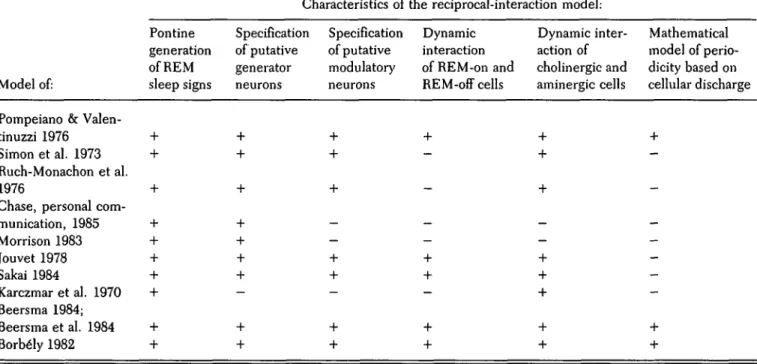
REM On Cells
( FTC, FTG, FTLJRC)
A Model
REM Off Cells
/Rophe Nuclei ( DRN. Lin.Cem., \ RM.RP ), LC, F*bl, FTC B. Selectivity Gradient FTG
I I
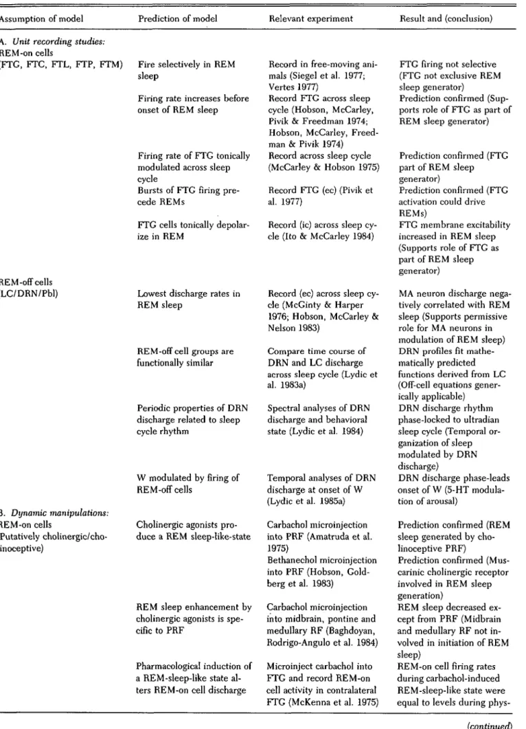
TRC C. Interpenetration Gradient DRN 10:1 (FTG) 3:1 (DRN) OExcitatory •InhibitoryFigure 2. Relationship between interpenetration and selec-tivity. Schematic illustration of the hypothesized relationship between physiological selectivity (B) and anatomical inter-penetration (C) of REM-on (left column) and REM-off (right column) cells whose reciprocal interconnections are modeled in A. REM-on cells are those neurons whose discharge rate in REM sleep is at least twice that of waking and NREM sleep. REM-off cells are those neurons whose discharge rate is at most one-half that of waking and NREM sleep. Inhibitory neurons are represented by filled circles; excitatory neurons are repre-sented by open circles. The same caveats regarding connectivity and synaptic function advanced in Figure 1 apply to this figure. The model (A) shows the REM-on cells as having feedforward and feedback (auto) excitation while the REM-off cells have feedforward and feedback (auto) inhibition. The neurotransmit-ters postulated to mediate these effects are shown in Figure 5C. Selectivity (B) is defined as the ratio of the mean firing rate of a given population in REM sleep relative to the firing rate during waking without movement. The proportional strengths of the values (not given) are accurately represented. Note that the significantly positive selectivity values of the REM-on popula-tions and the significantly negative values of the REM-off populations form a continuous gradient.
The interpenetration (C) of excitatory and inhibitory cells (or synapses) in a given nucleus may determine the selectivity gradient shown in B since the proportions of on and REM-off cells in a nucleus correlates with the selectivity ratios. In the examples shown, REM-on cells exceed REM-off cells by 10 to 1 in the area with the greatest positive selectivity (e.g., the FTG) while REM-off cells conversely exceed REM-on cells by 3 to 1 in the nucleus with the greatest negative selectivity (e.g., the DRN).
Anatomical nomenclature following Berman (1968):
REM-on nuclei: FTG = gigantocellular tegmental field; FTC = central tegmental field; FTL = lateral tegmental field; TRC = tegmental reticular nucleus.
REM-off nuclei: DRN = dorsal raphe nucleus; Lin Cent = linearis centralis; RM = raphe magnus; RP = raphe pontis; LC = locus coeruleus; P = peribrachial region.
from the brain stem, especially the cortex and spinal cord (Azmitia & Segal 1978; Tohyama, Sakai, Salvert, Touret & Jouvet 1979; Tohyama, Sakai, Touret, Salvert & Jouvet 1979). The fact that REM-off cell projections are not exclusive to the REM-on cell zones indicates that some aspects of the postulated reciprocal interaction are ana-tomically distributed, not only at the level of the brain stem but also throughout the brain. The possibility of anatomically distributed (Figure 1) and histochemically interpenetrated (see Figure 2C) REM-off and REM-on cell populations gives the reciprocal-interaction concept an even more distributed field of action (Figure 2A) than the original discussion of the REM-on cell data had implied (Hobson etal. 1975; McCarley & Hobson 1975b). This leads logically to the consideration that sleep cycle control may involve a system with both localized and remote cellular elements, both of which may play signifi-cant regulatory roles under physiological conditions.
One example will suffice to illustrate the functional implications of a reciprocal interaction between the ana-tomically distributed systems thought to be involved in sleep cycle control. As noted above, the eye movements of REM sleep are generated by premotor neurons in the brain stem reticular formation and by final-common-path oculomotor neurons whose own excitability may also be increased by disinhibition (Henn, Baloh & Hepp 1984). However, the increase in firing that causes this system to generate bursts of saccades during REM sleep may derive from (1) remote excitatory inputs to the cortex and superi-or colliculi, (2) excitation from recurrent collaterals, and (3) local vestibular feedback. Thus, a variety of neurons related both locally and remotely to the saccade-generat-ing cells may be rendered more excitable by the disin-hibition postulated to result from the REM-off firing pattern of aminergic neurons.
The hypothesis that recruitment of the distributed REM-on population is centripetal (e.g., via corticopon-tine fibers) as well as centrifugal (e.g., via pontogenicu-late and pontospinal pathways) may account for the important finding that the eye movements of REM sleep are less intense after lesions of the colliculi or visual and motor cortices Qeannerod, Monet & Jouvet 1965) and that electrical stimulation of the lateral geniculate body can trigger PGO waves and REM sleep if stimulation is delivered during the transition period from NREM to REM sleep (Nelson, McCarley & Hobson 1978). In addition, it has been shown that REM latency can be decreased and REM duration can be increased by audito-ry stimulation, which increases PGO waves (Drucker-Colin, Bernal-Pedraza, Fernandez-Cancino & Morrison 1983).
The relative ratio of interpenetration between REM-on and REM-off cells in the brain stem of the cat (Figure 2C) may also be associated with the degree to which both of these populations reveal discharge patterns selective for a given behavioral state (Figure 2B). For example, the most impressive REM-off selectivity is seen in the DRN, where the proportion of cells that cease firing in REM sleep (rather than merely slow their discharge) is highest and REM-on cells have not been reported (Figure 2B, right). Conversely, REM-on selectivity in head-restrained animals is greatest in the FTG, where REM-off cells have not been recorded (Figure 2B, left). Finally, in
regions where there is an admixture of REM-on and REM-off cells (such as the FTC), both classes of neuron show a relatively weak tendency to discharge selectively in a given behavioral state.
A parsimonious explanation of this apparent gradient of discharge selectivities would be a parallel gradient in density of net excitatory versus inhibitor synaptic con-tacts (Figure 2C). According to this concept, the func-tional homogeneity of cell type (i.e., on or REM-off) in a nucleus should enhance the tendency of that nucleus to discharge with either a REM-on or REM-off selectivity pattern (Figure 2B). Thus, on and REM-off discharge selectivity in a brain stem region appears to be, in part, a function of local homogeneity. In other words, this local homogeneity may take the form of uniformly signed (i.e., excitatory or inhibitory) local pro-jections that would tend to bias the balance of competing excitatory and inhibitory inputs of remote origin. 2. Deactivation of REM-off cells: Inferences derived from correlative data. According to the reciprocal-interaction concept, arrest of firing by aminergic REM-off cells disinhibits REM-on cells and this process is critical in determining the REM sleep state. Our continued tests of the foregoing hypotheses have recently focused upon the pontine DRN. The DRN represents an ideal site for testing these hypotheses since it has been shown (1) to contain the highest concentration of serotonin in the brain (Dahlstrom & Fuxe 1965), (2) to be the source of most of the forebrain serotonin (Azmitia & Segal 1978), and (3) to be a component of an extensive collection of monoaminergic nuclei that extend from the medulla to the mesencephalon. Furthermore, DRN neurons show a regular discharge during wakefulness, a slowing in NREM sleep, and a near cessation of firing during REM sleep.
So robust is the negative correlation between DRN discharge and REM sleep that the cessation of raphe discharge has consistently been observed in a variety of experimental preparations. For example, extracellular recordings of DRN discharge reveal a cessation of firing during the REM-sleep-like state induced by cooling the locus coeruleus (Cespuglio et al. 1981). A slowing of DRN firing was observed during the REM-sleep-like state without atonia produced by lesions of the pontine teg-mentum (Trulson, Jacobs & Morrison 1981). Phar-macological studies have revealed a REM-off discharge in the DRN during the REM-sleep-like state produced by pontine injections of carbachol (Steinfels, Heym, Streck-er & Jacobs 1983). Finally, the DRN has been shown to exhibit a REM-off firing pattern in recordings from one-day-old kittens, before the DRN has completed its bio-chemical and anatomical development (Adrien & Lan-fumey 1984).
Similarly, if one reviews the cellular studies of DRN discharge during physiologically occurring REM sleep, a slowing and cessation of DRN discharge has always been reported (Heym et al. 1982; Hobson, McCarley & Nelson 1983; Lydic, McCarley & Hobson 1984; McGinty & Harper 1976; Trulson & Jacobs 1979). There have been no exceptions to the observation of a REM-off discharge in studies using intact, undrugged adult animals. This decremental discharge pattern, therefore, appears to
382 THE BEHAVIORAL AND BRAIN SCIENCES (1986) 9:3
https://doi.org/10.1017/S0140525X00046318