HAL Id: hal-00925042
https://hal.archives-ouvertes.fr/hal-00925042
Submitted on 7 Jan 2014
HAL is a multi-disciplinary open access
archive for the deposit and dissemination of
sci-entific research documents, whether they are
pub-lished or not. The documents may come from
teaching and research institutions in France or
abroad, or from public or private research centers.
L’archive ouverte pluridisciplinaire HAL, est
destinée au dépôt et à la diffusion de documents
scientifiques de niveau recherche, publiés ou non,
émanant des établissements d’enseignement et de
recherche français ou étrangers, des laboratoires
publics ou privés.
Programmable multimetallic linear nanoassemblies of
ruthenium-DNA conjugates
Joris Irvoas, Arielle Noirot, Nadia Chouini-Lalanne, Olivier Reynes,
Jean-Christophe Garrigues, Valérie Sartor
To cite this version:
Joris Irvoas, Arielle Noirot, Nadia Chouini-Lalanne, Olivier Reynes, Jean-Christophe Garrigues, et al..
Programmable multimetallic linear nanoassemblies of ruthenium-DNA conjugates. RSC Advances,
Royal Society of Chemistry, 2012, vol. 2, pp. 9538-9542. �10.1039/c2ra21645k�. �hal-00925042�
To link to this article
: DOI:10.1039/c2ra21645k
URL : http://dx.doi.org/10.1039/c2ra21645k
This is an author-deposited version published in:
http://oatao.univ-toulouse.fr/
Eprints ID: 9990
To cite this version:
Irvoas, Joris and Noirot, Arielle and Chouini-Lalanne, Nadia and Reynes,
Olivier and Garrigues, Jean-Christophe and Sartor, Valerie Programmable
multimetallic linear nanoassemblies of ruthenium–DNA conjugates.
(2012) RSC
Advances, vol. 2 (n° 25). pp. 9538-9542. ISSN 2046-2069
O
pen
A
rchive
T
oulouse
A
rchive
O
uverte (
OATAO
)
OATAO is an open access repository that collects the work of Toulouse researchers
and makes it freely available over the web where possible.
Any correspondence concerning this service should be sent to the repository
administrator:
[email protected]
!Programmable multimetallic linear nanoassemblies of ruthenium–DNA
conjugates
Joris Irvoas,
abArielle Noirot,
abNadia Chouini-Lalanne,
abOlivier Reynes,
cdJean-Christophe Garrigues
aband
Valerie Sartor*
abDOI: 10.1039/c2ra21645k
A new ruthenium–DNA conjugates family was synthesized, made up of a ruthenium complex bound to one or two identical DNA strands of 14–58 nucleotides. The formation of controlled linear nanoassemblies containing one to seven ruthenium complexes is described.
Introduction
Metals are largely used by chemists to make original and varied nanostructures by complexation with organic backbones.1,2 More recently, inorganic complexes were employed to stabilize, to study and to form DNA assemblies.3–5 The nature of the metals used is as diverse as the nature of the assemblies formed. Ruthenium is one of them due to the remarkable photophysical and photochemical properties of ruthenium complexes, which can be easily modulated by the nature of the ligands employed. This allowed researchers to develop a wide range of applications concerning these components, for example, dye-sensitized solar cells,6cancer treatments and medical diagnosis7,8and catalysis.9 Despite the potential of these complexes in various fields of scientific research, only few examples of DNA assemblies involving ruthenium compounds are described.10–15
It was previously shown that the insertion of DNA backbones on terpyridine ruthenium complexes permitted the formation of linear arrays.11,12 The formation of cyclic structures has also been described. The control of the hybridization process allowed the formation of such constructions bearing one or two ruthenium molecules.13 Another original work reported the
assembly of cyclic DNA architectures by using the tris(2,29-bi-pyridine) ruthenium complex as a molecular template not being part of the structure, to control the duplex assembly.14 Furthermore, a star building block for future nanoassemblies was achieved. Indeed, a ruthenium tris(bipyridine) centre with
six identical oligonucleotide arms was synthesized and the formation of six DNA double strands surrounding the complex was shown.15
Considering the potential of ruthenium complexes on varied applications and the development of DNA hybridization strategies to construct nanodevices, we developed a very simple synthesis pathway to obtain ruthenium–DNA conjugates and a flexible hybridization strategy to control the distance and the position of the functionalized building blocks. The synthetic pathway is only based on two reaction steps and on commercially available modified oligonucleotides and chemical products. This synthetic strategy relies on the use of the carboxylic acid moiety. As it is a commonly used functionalization, it allows us to easily modulate the nature of the molecules inserted into DNA, therefore broad-ening the range of possible applications. In this paper, we describe the work made with Ru(II)(2,29-bipyridine)2
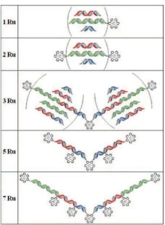
(4,49-dicarboxy-2,29-bipyridine) complex.16 We developed a new family of ruthenium complexes bearing one or two DNA single strands with lengths varying between 14 and 58 nucleic bases. The modular hybridization strategy is based on the choice of the oligonucleotide sequences. They have been wisely designed to self-assemble into linear nanoarrays in a modular fashion to lead to original one-dimensional constructions with one to seven ruthenium complexes at different controlled positions and distances (Fig. 1).
Results and discussion
Synthesis of ruthenium–DNA conjugates
This new series of ruthenium–DNA hybrids were synthesized by coupling oligonucleotides substituted by an amino hexyl linker in the 59 position with a Ru(II)(2,29-bipyridine)2
(4,49-dicarboxy-2,29-bipyridine) complex.16The amide link has been performed
with the DNA strand kept on a solid support. The reaction took place using 4-(4,6-dimethoxy-1,3,5-triazin-2-yl)-4-methylmor-pholinium chloride (DMT-MM) in 0.8 M 3-morpholinopro-pane-1-sulfonic acid (MOPS) buffer (pH = 7) in DMSO/H2O
solvents with a large excess of ruthenium complex.17 After
deprotection and cleavage of crude DNA products from the solid
aUniversite´ de Toulouse; Universite´ Paul Sabatier; Laboratoire IMRCP; Bat. II R1, 118 route de Narbonne, F-31062, Toulouse cedex 09, France. E-mail: [email protected]; Fax: +33 (0)5 61 55 81 55;
Tel: +33 (0)5 61 55 25 62 74 b
CNRS; Laboratoire IMRCP UMR 5623, F-31062, Toulouse cedex 09, France. E-mail: [email protected]; Fax: +33 (0)5 61 55 81 55; Tel: +33 (0)5 61 55 62 74
cUniversite´ de Toulouse; Universite´ Paul Sabatier; Laboratoire de Genie Chimique; Bat. II R1, 118 route de Narbonne, F-31062, Toulouse cedex 09, France
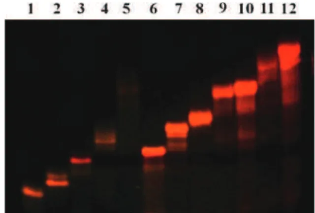
support, analysis and purification were realized using denaturing polyacrylamide gel electrophoresis (PAGE). The polyacrylamide gels were directly observed under UV light at two wavelengths (254 and 365 nm), which were convenient to discriminate DNA and Ru–DNA conjugates. Indeed, at l = 365 nm or on a Safe ImagerTM 2.0 Blue-light transilluminator, the presence of a Ru centre induces a characteristic orange luminescence. The latter does not appear at l = 254 nm, but DNA is still visible. Two molecules per reaction were isolated and characterized both carrying a ruthenium complex. The fast-moving orange band in the gel corresponds to the incorporation of one DNA sequence on the ruthenium complex. The slowest moving orange band in the gel was identified as the bis-DNA-functionalized ruthenium complex centre. With this experimental protocol, we obtained a family of ruthenium–DNA hybrids constituted of one 14 (1m and 2m), two 14 (2b), one 20 (3m and 4m), two 20 (4b), one 24 (5m and 6m), two 24 (6b), two 34 (7b) or two 58 (8b) nucleotide arms (Fig. 2).
The modified oligonucleotides were characterized by PAGE, UV-Vis and fluorescence spectrophotometries and mass spectro-scopy.
The polyacrylamide gel electrophoresis of this new family of ruthenium–DNA conjugates was visualized directly on a Safe ImagerTM2.0 Blue-light transilluminator (Fig. 3). The increase of the sequence lengths and the insertion of two DNA single strands on one ruthenium complex consequently showed a decreased migration length of the molecules in the gel.
The UV-Vis absorption spectra of these conjugates show at l = 260 nm the characteristic band of DNA oligomers and at l =
280 nm and l = 458 nm, the bipyridine p–p* and the complex MLCT (metal-to-ligand charge-transfer) transitions, respec-tively. The fluorescence spectra are all identical and character-istic of the ruthenium complex, namely having an emission peak at l = 650 nm with an excitation wavelength at 458 nm. An example of UV-Vis and fluorescence spectra of a mono DNA-functionalized ruthenium complex is shown in Fig. 4.
High resolution electrospray mass spectra of 1m, 2m, 2b, 3m, 4m, 4b and 5m confirm the structure of these ruthenium–DNA conjugates (see in ESI{). The compounds 6m, 6b, 7b and 8b did not provide HRMS. However, the migration of their bands in PAGE compared to the fully characterized compounds and the presence of a ruthenium signal in UV-Vis and fluorescence spectrophotometries provided convincing evidence and charac-terizations of theirs structures.
Multimetallic linear nanoassemblies
The selective and specific hybridization of these ruthenium– DNA conjugates afforded a variety of linear assemblies. They provided DNA double helices with a length of 14 to twice 58
Fig. 2 Synthesized mono-DNA-functionalized ruthenium complexes (Xm) and bis-DNA-functionalized ruthenium complexes (Xb) with their corresponding sequences.
Fig. 3 Denaturing PAGE analysis of ruthenium–DNA conjugates on a Safe ImagerTM2.0 Blue-light transilluminator. Lane 1: 1m; lane 2: 2m; lane 3: 2b; lane 4: 3m; lane 5: 4m; lane 6: 4b; lane 7: 5m; lane 8: 6m; lane 9: 6b; lane 10: 7b; lane 11: 8b.
Fig. 1 Multimetallic linear nanoassemblies bearing one to seven ruthenium complexes and DNA sequence lengths of 14–58 nucleotides.
base pairs and made up of one to seven ruthenium polypyridine complexes (Fig. 1). They were highlighted by native PAGE (Fig. 5 and 6 and in ESI{).
Hybridization of mono DNA-functionalized ruthenium com-plex 1m with 2 or 2m yields a 14-base double strand with one (Fig. 5, lane 3) or two ruthenium moieties (Fig. 5, lane 4), respectively. Similar assemblies were obtained based on the mono 20 (Fig. 5, lanes 6 and 7) and 24 (Fig. 5, lanes 9 and 10) nucleotide-long DNA-substituted ruthenium conjugates. Three ruthenium complex–DNA duplex structures were built by hybridization of the bis(DNA)-ruthenium hybrids with two equivalents of the sequence complementary mono(DNA)-ruthe-nium conjugates (Fig. 6). In these three-ruthemono(DNA)-ruthe-nium assemblies,
the distances between two ruthenium complexes are modulated by the duplexes lengths, which are 14 (lane 6), 20 (lane 7), 24 (lane 8), 34 (lane 9) or 58 (lane 11) base pairs long.
To easily and efficiently modulate the number of ruthenium motifs and their positions in the linear assemblies, the 34 and 58 nucleotide-long DNA oligomers 7 and 8 were designed to be successively composed with the 14- and 20-base sequences (1 and 2) and 14-, 20- and 24-base sequences (1, 2 and 3), respectively. Consequently, longer three-ruthenium linear assemblies were generated by hybridization of the bis(34 bases oligonucleotide)– ruthenium hybrid 7b with two complementary mono(20 bases oligonucleotide)–Ru conjugates 3m and two 14 nucleotide-long single strands 1 (Fig. 6 lane 9). Hybridization of the bis(58 bases oligonucleotide)–ruthenium hybrid 8b with two complementary mono(24 bases oligonucleotide)–Ru conjugates 5m and two 14 and 20 nucleotide-long single strands 1 and 3 led to an even longer three-ruthenium linear structure (Fig. 6 lane 11). This approach allowed us to easily adjust and increase the number of ruthenium moieties in the architecture. Indeed, it was achieved by using Ru–DNA conjugates instead of DNA single strands in the previously formed structures. With this methodology, hybridization of 7b with 2 eq. of 1m and 3m (Fig. 6, lane 10), and 8b with 2 eq. of 1m, 3m and 5m (Fig. 6, lane 12) provided linear DNA assemblies bearing five and seven ruthenium complexes, respectively.
All of these linear assemblies were studied by UV-thermal denaturation experiments and circular dichroism (CD) spectro-scopy.
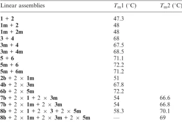
The melting temperature (Tm) analysis shows no significant
difference in Tmvalues for duplexes in presence of no, one, two
or three ruthenium complexes (Table 1). Nevertheless, the Tm
curves are broadened (Tmcurves available in ESI{). The metallic
motifs seem to affect the thermal stability of the double helices. It can be watched on the polyacrylamide gel in Fig. 5. The bands for one- and two-ruthenium–DNA conjugate duplexes (lanes 3, 4, 6, 7, 9 and 10) are thicker and more diffuse than duplexes without a ruthenium complex (lanes 2, 5 and 8). For the 34 and 58 base pair duplexes formed with two and three distinct double strands, respectively, two Tm were observed, the values
Fig. 5 Non-denaturing PAGE analysis of linear assemblies with 0, 1 and 2 ruthenium complexes with l = 254 nm (A) and on Safe ImagerTM (B). Lanes 1 and 11: two products for migration control, upper: xylene cyanol, lower: bromophenol blue; lane 2: 1 + 2; lane 3: 1m + 2; lane 4: 1m + 2m; lane 5: 3 + 4; lane 6: 3m + 4; lane 7: 3m + 4m; lane 8: 5 + 6; lane 9: 5m + 6; lane 10: 5m + 6m.
Fig. 6 Non-denaturing PAGE analysis of multimetallic linear assem-blies on Safe ImagerTM. Lane 1: 2b; lane 2: 4b; lane 3: 6b; lane 4: 7b; lane 5: 8b; lane 6: 2b + 2 6 1m; lane 7: 4b + 2 6 3m; lane 8: 6b + 2 6 5m; lane 9: 7b + 2 6 1 + 2 6 3m; lane 10: 7b + 2 6 1m + 2 6 3m; lane 11: 8b + 2 61 + 2 6 3 + 2 6 5m; lane 12: 8b + 2 6 1m + 2 6 3m + 2 6 5m. Fig. 4 UV-Vis (solid line) and fluorescence emission (dash line) spectra
corresponding to an average of the hybridization temperatures of the separate double strands.
Circular dichroism (CD) was used to check Ru complex effects on the duplex integrity. The CD spectra are identical for the whole duplex family. They present a characteristic spectrum of DNA B form with a positive band at 282 nm and a negative band at 252 nm (Fig. 7). No CD signal next to the MLCT band of the ruthenium complex was observed. The presence of internal and/ or terminal metallic centres did not affect the CD signals and thus the shape of the duplexes.
Conclusions
Herein, we have shown the synthesis and the characterization of a new family composed of eleven mono- and bis-DNA-functionalized ruthenium complexes. A simple and efficient route was described to assemble ruthenium complexes in a linear and programmable fashion using the DNA self-assembly property. This strategy allowed us to selectively choose the number and the position of metallic complexes introduced in a DNA backbone. The extension of this methodology could afford symmetrical nanowires of various lengths with other metals.
Experimental
Materials and methods
59-C6 amino-modified oligonucleotides were purchased from Eurogentech. 4,49-dicarboxy-2,29-bipyridine, cis-bis(2,29-bipyridine) dichlororuthenium(II) and 3-morpholinopropane-1-sulfonic acid (MOPS) was purchased at Alfa-Aesar France. 4-(4,6-dimethoxy-1,3,5-triazin-2-yl)-4-methylmorpholinium chloride (DMT-MM) was purchased at Sigma Aldrich France. [Ru2+(2,29-bipyridine)2
(4,49-dicarboxy-2,29-bipyridine](PF62)2 was synthesized as already
reported.16
UV-Vis spectra were obtained using Hewlett-Packard 8452A equipment. Fluorescence spectra were acquired on a Photon Technology International modular setup. Circular dichroism was achieved on a Jasco J-815 CD spectrometer. Polyacrylamide gel electrophoresis (PAGE) analysis was carried out on a Hoefer SE40-15-1.5 unit and the gels finally visualized on a Safe ImagerTM2.0 blue light transilluminator, under UV light at l =
254 nm or at l = 365 nm. HRMS analyses were realized on a Qtof Ultima API (Waters) mass spectrometer, see ESI{. The capillary, cone and RF Lens tensions were, respectively, 3.5 kV, 130 V and 40 V. The source and desolvation temperatures were 60 uC and 80 uC. The collision energy (Ar) was fixed at 8 eV in Tof ms mode, with nitrogen for the nebulization gas. The solutions were analyzed by infusion (10mL min21), with an acquisition time of 5 min, realized with the Masslynx 4.1 software (Waters).
General procedure for ruthenium–DNA hybrids synthesis A 2 mL aqueous buffer solution (pH = 7) of 3-morpholino-propane-1-sulfonic acid (MOPS) (1,046 g, 0.8 mol L21) and
4-(4,6-dimethoxy-1,3,5-triazin-2-yl)-4-methylmorpholinium chlor-ide (DMT-MM) (138,4 mg, 0.08 mol L21) was prepared. 4 mL DMSO solution of [Ru2+
(2,29-bipyridine)2(4,49-dicarboxy-2,
29-bipyridine)](PF62)2) (4,7 mg, 5 mmol, 25 eq.) was added,
followed by the introduction of 59-C6 amino-modified oligo-nucleotide kept on a solid support (200 nmol, 1 eq.). The solution was stirred for 48 h at 25 uC and then filtered. The resulting orange beads were washed once with DMSO and 3 times with water. The beads were put in a concentrated ammonia solution (32%) at 55 uC for 20 h and then filtered. The filtrate was evaporated and redissolved in an aqueous solution. Ruthenium–DNA conjugates were purified by PAGE on 20% denaturing polyacrylamide gels. The bands were cut, crushed in water and incubated at 55 uC. The mixture was filtered to remove the gel. The filtrate was concentrated and washed twice with water on a Amicon1 Ultra centrifugal filter 3 K (Millipore Ireland Ltd).
Hybridization conditions
A solution with the appropriated number of equivalents of each DNA strand was prepared for a final 50 mM (duplex) concentration in a buffer solution (50 mM Tris, 100 mM NaCl and 10 mM MgCl2at pH = 7.2). Hybridization was achieved by
heating this solution to 90 uC for 3 min and then leaving its temperature to decrease slowly to room temperature. The solution was finally incubated for 12 h at 4 uC.
Table 1 Melting temperatures of linear assemblies
Linear assemblies Tm1 (uC) Tm2 (uC)
1 + 2 47.3 1m + 2 48 1m + 2m 48 3 + 4 68 3m + 4 67.5 3m + 4m 68.5 5 + 6 71.1 5m + 6 72.2 5m + 6m 71.2 2b + 2 6 1m 51 4b + 2 6 3m 67.8 6b + 2 6 5m 72.2 7b + 2 6 1 + 2 6 3m 54 66.6 7b + 2 6 1m + 2 6 3m 54 66.8 8b + 2 6 1 + 2 6 3 + 2 6 5m 58.3 70.1 8b + 2 6 1m + 2 6 3m + 2 6 5m — 69
Fig. 7 Circular dichroism spectra of 2b + 2 6 1m (dash grey line); 4b + 2 6 3m (grey line); 6b + 2 6 5m (dash dot black line); 7b + 2 6 1 + 2 6 3m (dot grey line); 7b + 2 6 1m + 2 6 3m (dash black line); 8b + 2 6 1 + 2 6 3 + 2 6 5m (black line); 8b + 2 6 1m + 2 6 3m + 2 6 5m (dot black line).
Acknowledgements
Financial support was provided by the French National Agency of Research (ANR) (ANR-08-JCJC-0141).
References
1 Y. E. Alexeev, B. I. Kharisov, T. C. H. Garcia and A. D. Garnovskii, Coord. Chem. Rev., 2010, 254, 794–831.
2 R. Chakrabarty, P. S. Mukherjee and P. J. Stang, Chem. Rev., 2011, 111, 6810–6918.
3 H. Yang, K. L. Metera and H. F. Sleiman, Coord. Chem. Rev., 2010, 254, 2403–2415.
4 S. Ghosh and E. Defrancq, Chem.–Eur. J., 2010, 16, 12780–12787. 5 J. Mu¨ller, Eur. J. Inorg. Chem., 2008, 2008, 3749–3763.
6 G. C. Vougioukalakis, A. I. Philippopoulos, T. Stergiopoulos and P. Falaras, Coord. Chem. Rev., 2011, 255, 2602–2621.
7 O. Lentzen, C. Moucheron and A. K.-D. Mesmaekerin Metallotherapeutic Drugs and Metal-Based Diagnostic Agents, John Wiley & Sons, Ltd, 2005, pp. 359-378.
8 W. H. Ang, A. Casini, G. Sava and P. J. Dyson, J. Organomet. Chem., 2011, 696, 989–998.
9 A. L. Noffke, A. Habtemariam, A. M. Pizarro and P. J. Sadler, Chem. Commun., 2012, 48, 5219–5246.
10 K. Wiederholt and L. W. McLaughlin, Nucleic Acids Res., 1999, 27, 2487–2493.
11 K. M. Stewart and L. W. McLaughlin, Chem. Commun., 2003, 2934–2935.
12 S. Ghosh, I. Pignot-Paintrand, P. Dumy and E. Defrancq, Org. Biomol. Chem., 2009, 7, 2729–2737.
13 D. Mitra, N. Di Cesare and H. F. Sleiman, Angew. Chem., Int. Ed., 2004, 43, 5804–5808.
14 F. A. Aldaye and H. F. Sleiman, J. Am. Chem. Soc., 2007, 129, 10070–10071.
15 K. M. Stewart, J. Rojo and L. W. McLaughlin, Angew. Chem., Int. Ed., 2004, 43, 5808–5811.
16 E. Terpetschnig, H. Szmacinski, H. Malak and J. R. Lakowicz, Biophys. J., 1995, 68, 342–350.
17 X. Li, J. Gartner, B. N. Tse and D. R. Liu, J. Am. Chem. Soc., 2004, 126, 5090–5092.