HAL Id: hal-02612552
https://hal.archives-ouvertes.fr/hal-02612552v2
Submitted on 11 Jun 2020
HAL is a multi-disciplinary open access
archive for the deposit and dissemination of
sci-entific research documents, whether they are
pub-lished or not. The documents may come from
teaching and research institutions in France or
abroad, or from public or private research centers.
L’archive ouverte pluridisciplinaire HAL, est
destinée au dépôt et à la diffusion de documents
scientifiques de niveau recherche, publiés ou non,
émanant des établissements d’enseignement et de
recherche français ou étrangers, des laboratoires
publics ou privés.
Bidimensional lamellar assembly by coordination of
peptidic homopolymers to platinum nanoparticles
Ghada Manai, Hend Houimel, Mathilde Rigoulet, Angélique Gillet,
Pier-Francesco Fazzini, Alfonso Ibarra, Stéphanie Balor, Pierre Roblin,
Jérôme Esvan, Yannick Coppel, et al.
To cite this version:
Ghada Manai, Hend Houimel, Mathilde Rigoulet, Angélique Gillet, Pier-Francesco Fazzini, et al..
Bidimensional lamellar assembly by coordination of peptidic homopolymers to platinum nanoparticles.
Nature Communications, Nature Publishing Group, 2020, 11, pp.2051. �10.1038/s41467-020-15810-y�.
�hal-02612552v2�
ARTICLE
Bidimensional lamellar assembly by coordination of
peptidic homopolymers to platinum nanoparticles
Ghada Manai
1,2,8
, Hend Houimel
1,8
, Mathilde Rigoulet
1
, Angélique Gillet
1
, Pier-Francesco Fazzini
1
,
Alfonso Ibarra
3
, Stéphanie Balor
4
, Pierre Roblin
5
, Jérôme Esvan
6
, Yannick Coppel
2
, Bruno Chaudret
1
,
Colin Bonduelle
2,7
✉
& Simon Tricard
1
✉
A key challenge for designing hybrid materials is the development of chemical tools to control
the organization of inorganic nanoobjects at low scales, from mesoscopic (~µm) to
nano-metric (~nm). So far, the most ef
ficient strategy to align assemblies of nanoparticles consists
in a bottom-up approach by decorating block copolymer lamellae with nanoobjects. This well
accomplished procedure is nonetheless limited by the thermodynamic constraints that
govern copolymer assembly, the entropy of mixing as described by the Flory
–Huggins
solution theory supplemented by the critical in
fluence of the volume fraction of the block
components. Here we show that a completely different approach can lead to tunable 2D
lamellar organization of nanoparticles with homopolymers only, on condition that few
ele-mentary rules are respected: 1) the polymer spontaneously allows a structural
preorganiza-tion, 2) the polymer owns functional groups that interact with the nanoparticle surface, 3) the
nanoparticles show a surface accessible for coordination.
https://doi.org/10.1038/s41467-020-15810-y
OPEN
1Laboratoire de Physique et Chimie des Nano-Objets, INSA, CNRS, Université de Toulouse, Toulouse, France.2Laboratoire de Chimie de Coordination, CNRS,
Université de Toulouse, Toulouse, France.3Instituto de Nanociencia de Aragón, Universidad de Zaragoza, Zaragoza, Spain.4Plateforme de Microscopie Électronique Intégrative, Centre de Biologie Intégrative, CNRS, Université de Toulouse, Toulouse, France.5Laboratoire de Génie Chimique, Fédération Fermat,
INPT, CNRS, Université de Toulouse, Toulouse, France.6Institut Carnot– Centre Inter-universitaire de Recherche et d’Ingénierie des Matériaux,
INP-ENSIACET, CNRS, Université de Toulouse, Toulouse, France.7Laboratoire de Chimie des Polymères Organiques, Université de Bordeaux, CNRS, Bordeaux
INP, Pessac, France.8These authors contributed equally: Ghada Manai, Hend Houimel. ✉email:colin.bonduelle@enscbp.fr;tricard@insa-toulouse.fr NATURE COMMUNICATIONS| (2020) 11:2051 | https://doi.org/10.1038/s41467-020-15810-y | www.nature.com/naturecommunications 1
123456789
S
tructuring hybrid materials combining metallic
nano-particles (NPs) and polymers has stimulated a large effort to
make new physical properties emerging: optical, electronic,
or magnetic
1–3. Such composites have the potential of improving
the functionality of devices ranging from memory storage to
sensors or microelectronic systems
4,5. In addition, the structural
properties of the polymer matrix can give added value to the
hybrid materials, such as stimuli-responsiveness
6or chirality
7.
Among a variety of possible structuring, disposing NPs in lines on
substrates paves a way toward anisotropic ordering of matter at
the nanoscale, which can be observed, manipulated, and
con-nected. To reach such a supraparticular organization, the most
efficient strategy developed so far consists in decorating diblock
copolymer lamellar assemblies by NPs, tuning the strength and
the nature of weak interactions between the two components
(essentially Van de Waals interactions)
5–11. Diblock copolymer
templating is a robust and versatile approach that has been
extended to thermal evaporation
12, atomic layer deposition
13, or
for structuring molecules such as polyoxometallates
14. The
obtained morphologies are driven by (1) the entropy of mixing of
the two blocks, described by the Flory–Huggins solution
theory
15,16, and (2) their corresponding volume fractions
17,
tak-ing into account that, in presence of NPs, there is a significant
decrease in the copolymers’ conformational entropy coming from
particle sequestration
10. Although very promising in many
situations, the diblock copolymer approach shows important
limitations: (1) a constrained size ratio between the two blocks to
access lamellar assemblies (generally comprised between 0.4 and
0.6 volume fraction with random coil polymers), (2) in presence
of NPs, an imprecise control of their localization (as it is governed
by entropy) and (3) in solution, a strong dependence to
experi-mental conditions (concentration, temperature, presence of
co-solvent, etc.)
10,15,16. Here, we present an alternative strategy to
dispose NPs in lines by simply mixing metallic ultra-small NPs
with structured peptidic homopolymers. The coordination
bonding between the polymer functional groups and the NP
surface indeed affords lamellar organization, where the patterning
distances are linearly controlled by the molecular weight of the
polymer.
Self-assembly is a widely applied strategy for preparing various
materials based on metallic NPs, which lead to e.g., innovative
photonic materials, new microelectronic devices, or structured
templates for nanolithography
1. NPs are generally obtained by a
chemical approach that consists in constraining the growth of the
crystal by the presence of a limiting agent (ligand, polymer, etc.).
An elegant approach resides in decomposing organometallic
precursors in mild conditions to obtain clean NPs, i.e. having a
surface composition perfectly described and controllable. In this
work, we choose, as a standard, ultra-small (<2 nm) platinum
NPs, as their sizes are in the same range as the one of a monomer
moiety and their surfaces are only stabilized by carbon monoxide
(CO) and labile tetrahydrofuran (THF)
18. The absence of strong
organic ligands gives the opportunity to fully describe the
coordination between the additional component, i.e. the
poly-peptide polymer, and the NP surface. So far, most approaches
involving NPs consider their assemblies as a packing of hard
spheres, but the ligand itself has emerged as a chemical leverage,
which enables exquisite control to promote supraparticular
chemistry
19,20. Polypeptide polymers are simple macromolecules
in which an amino-acid moiety is repeated many times. They
adopt ordered secondary conformations such as
α-helices or
β-sheets, which offer a way to guide structuration at the nanoscale
through intermolecular and/or intramolecular interactions
21,22.
In this work, we choose poly(γ-benzyl-L-glutamate) (PBLG), a
synthetic polypeptide that gives rise to a rigid rod-like
α-helical
conformation in organic solvents and that has often been
employed as a model system to drive lamellar morphologies,
when included in block copolymer structures
23.
Results
Description of the nanostructuration. The NPs were
synthe-sized by decomposition of Pt
2(dba)
3(dba
=
dibenzylideneace-tone) under a CO atmosphere in THF, followed by complete
elimination of the organic dba residue by washing with pentane
18.
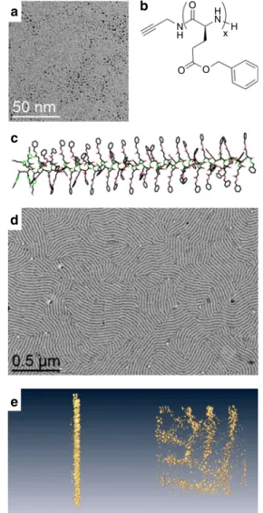
TEM pictures showed well-dispersed NPs, with diameters of
1.2 ± 0.3 nm (Fig.
1
a). On another hand, PBLG was synthesized
by initiating the ring-opening polymerization of
γ-benzyl-L-glu-tamate-N-carboxyanhydride with propargylamine in
dimethyl-formamide (Supplementary Fig. 1)
24. A library of PBLGs spanning
a wide range of molecular weights were obtained (Fig.
1
b), with
five degrees of polymerization (Dp), as measured by
1H NMR and
SEC chromatography (Supplementary Table 1): PBLG1 presented
a Dp of 28, PBLG2 of 69, PBLG3 of 120, PBLG 4 of 217, and
PBLG 5
of 481. Their
α-helix secondary structures were confirmed
by circular dichroism measurements in THF (Fig.
1
c and
O
a
c
d
e
b
O O H H x N N HFig. 1 Building blocks and self-assembly. a TEM image of pristine ultra-small platinum nanoparticles of 1.2 ± 0.3 nm.b Chemical structure of PBLG —in the present study, x = 28 (PBLG1), 69 (PBLG2), 120 (PBLG3), 217 (PBLG4), 481 (PBLG5). c Geometrical model of theα-helix conformation of PBLG (example forx = 60). Lamellar structuration of an assembly of platinum nanoparticles withPBLG4 at 0.5 eq.: d Low-magnification TEM image;e, Tomographv 3D reconstruction at two viewing directions: each yellow dot corresponds to an individual nanoparticle.
Supplementary Fig. 2). The assembly of NPs and peptidic
poly-mers was carried out in THF: solutions of platinum NPs and of
PBLG were mixed and stirred for 2 h at different equivalent
numbers (eq.
– defined as the ratio between the monomer unit
and the platinum atom quantities). Transmission electron
microscopy (TEM) on PBLG4 at 0.5 eq. showed unexpected
lamellar assemblies of the hybrid materials (Fig.
1
d), alternating
zones containing (dark), or excluding (white) NPs. Such an
organization has been observed on both silicon substrates and
TEM grids by different microscopy techniques: atomic force
microscopy and scanning electron microscopy, in addition to
TEM (Supplementary Fig. 3a–c). Tomography imaging showed
that the NPs constitute cylindrical lamellae, without any
pre-ferential arrangement within each lamella (Fig.
1
e and
Supple-mentary Fig. 3d, e). As we did not notice any effect of deposition
substrate (hydrophobic carbon vs. hydrophilic silicon), nor of
deposition process (drop-casting vs. spin-coating), we
hypothe-sized that these structured lamellae were present in solution and
did not form during the solvent evaporation. The existence of the
structured lamellae in solution was then confirmed by cryo-TEM
imaging, as a same morphology was observed after a fast freezing
of the assembly solution (Supplementary Fig. 4). Besides, in some
cases, we noticed the presence Moiré patterns, resulting from the
superimposition of up to four lamellae (Supplementary Fig. 5). As
the lamellae structures are preformed in solution, they can
deposit on top of each other during the drop casting of the TEM
grid preparation. Such observations gave insight that the system
organized in a lamellae-within-lamellae hierarchical assembly
25.
Information on the polymer structure before and after assembly
was
first given by
13C HR-MAS NMR at slow speed
(Supple-mentary Fig. 6). After NP addition, an increase of the spinning
sideband intensities, due to stronger chemical shift anisotropies,
highlighted a stiffening of the PBLG in the assembly, which also
led to greater conformational homogeneity, as evidenced by the
resonance sharpening. Second, SAXS measurements showed a
broad peak centered at 0.25 Å
−1(Supplementary Fig. 7),
mean-ing that NPs were separated from each other by an average
cor-relation distance equal to 2.5 nm. The importance of the ratio
between the monomer unit and the platinum atom quantities was
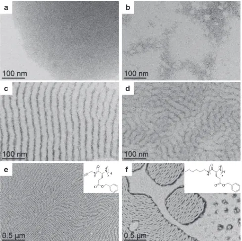
then studied by varying the eq. number from 0.05 up to 5 eq. with
PBLG4. Without any polymer, the NPs simply aggregated
because of the capillary forces generated by THF evaporation
(Fig.
2
a), without showing any specific average distance by SAXS
analysis (Supplementary Fig. 7). As soon as PBLG was added to
the reaction mixture, depletion zones without NPs formed
(Fig.
2
b–d), attributed to the presence of the polymer. Very clean
patterns were obtained at 0.5 eq, with alternation of regular
lamellae. At higher ratios, this NP organization became looser
(1 eq.—Fig.
2
d), and totally disappeared at 5 eq. (Supplementary
Fig. 8). A window of two orders of magnitude in eq. number
(from 0.05 to 5 eq.) was thus accessible to tune the pattern
structure of the lamellar assemblies.
a
c
b
d
x H H N N H O O Oe
f
xH H N N H O O OFig. 2 Effect of the relative quantity of polymer vs. nanoparticle and effect of the terminal group. TEM micrographs of the assemblies after 2 h of reaction between platinum nanoparticles and:PBLG4 at a 0 eq. (nanoparticles alone); b 0.05 eq.; c 0.5 eq.; d 1 eq. (an equivalent eq. refers to the number of introduced monomers per platinum atom). The assembly process occurs at an optimum relative ratio of polymer vs. nanoparticle equal to 0.5 eq. TEM micrograph of the assemblies between platinum nanoparticles and:e PBLG4; f PBLG-H, at 0.5 eq., after 2 h of reaction. Insets represent the chemical structures of the polymers. The nature of the terminal group of PBLG influences the global cohesion of the hybrid system, as alkyne groups lead to better structuration than hexyl groups.
NATURE COMMUNICATIONS | https://doi.org/10.1038/s41467-020-15810-y
ARTICLE
Origin of the patterning. To determine key parameters at the
origin of the patterning, coordination of the polymer to the NP
surface was studied. First, a peptidic dimer DBLG was specifically
prepared and mixed with NPs to confirm the possible
coordi-nation of the alkyne moiety at the NP surface.
13C solid-state
MAS NMR showed the disappearance of the peaks at 72 and
80 ppm attributable to the alkyne moiety upon NP addition,
whereas the other peaks of the molecule remained unchanged
(Supplementary Fig. 9). This disappearance strongly supported
the occurrence of similar alkyne coordination to the NP surface
with PBLG4. A polymeric analogue without alkyne was then
synthesized from hexylamine, PBLG-H. After mixing with NPs,
TEM imaging showed a significantly less structured system, with
free NPs and more discontinuous NP arrangement within the
lamellae (Fig.
2
e, f), thus confirming the importance of the alkyne
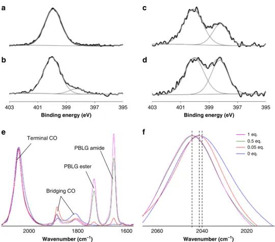
coordination. Second, another coordination bonding between the
NP surface and the polymer was identified by X-ray
photoelec-tron spectroscopy (XPS) at the N1s edge, which showed the
appearance of a new peak at 398 eV in addition to the neutral
component at 400 eV of the free polymer. The relative intensity of
this peak significantly increased when the eq. number decreased
(Fig.
3
a–d and Supplementary Fig. 10), confirming a rise of the
electronic density on the PBLG amide nitrogen upon NP
addi-tion. Simultaneously, both the relative increase of surface
plati-num oxidation measured by XPS at the Pt4f edge (Supplementary
Fig. 11), and the progressive shift of the adsorbed CO peak in
infrared spectroscopy from 2040 cm
−1at 0 eq. to 2041 cm
−1at
0.05 eq. and to 2045 cm
−1at 0.5 and 1 eq. confirmed a decrease of
the electronic density at the NP surface upon PBLG addition
(Fig.
3
e, f and Supplementary Fig. 12)
18. Overall, both XPS and
infrared measurements reflected an electronic transfer from the
NP surface to reduce the PBLG nitrogen in the composite
materials, as already observed for amides in platinum molecular
complexes
26,27. The present study thus indicates that the polymer
interacts with the NP surface by both coordinating the terminal
alkyne group of the polypeptide and its peptide linkages.
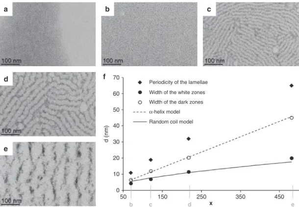
Influence of the degree of polymerization. We further explored
the Dp influence on the lamellar assemblies. No arrangement was
observed with PBLG1, but image analysis showed that the width
of the lamellae average periodicity regularly increased from
PBLG2
to PBLG5 (Fig.
4
, Supplementary Fig. 13, and
Supple-mentary Table 2). Such a tendency was confirmed by SAXS
measurements (Supplementary Fig. 14 and Supplementary
Table 2). In addition, the average width of the white zones
con-taining the polymer was equal at any Dp to the length of the
PBLG model in
α-helices conformation. Similarly, the evolution
of the average width of the dark zones containing the NPs as a
function of Dp (x) could be
fitted by the diameter of gyration of a
random coil model R
g= R
0x
ν. The
ν value was fixed to 0.6, as
predicted by theory and confirmed by experiments for
excluded-volume chains
28. Extensive study on chemically unfolded proteins
found a R
0value equal to 1.33
28, whereas the
fit of our
experi-mental data gave a R
0value equal to 0.22, thus divided by six.
Such a result can be interpreted by the presence of an average of
6–7 bridges within the polymer
29, which is coherent considering
the PBLG polymer in a coil disordered state, internally connected
Wavenumber (cm–1) Wavenumber (cm–1) Terminal CO Bridging CO PBLG amide PBLG ester 1 eq. 0.5 eq. 0.05 eq. 0 eq.
a
e
c
f
b
403 2000 1800 1600 2060 2040 2020 401 399 397 395 403 401 399 397 395d
Binding energy (eV) Binding energy (eV)
Fig. 3 Spectroscopic signature of coordination of the peptidic polymer to the nanoparticle surface. a XPS spectrum at the N1s edge of PBLG4 alone. XPS spectra at the N1s edge of assemblies between platinum nanoparticles and PBLG4 at: b 1 eq. (12% of component at 398 eV); c 0.5 eq. (33% of component at 398 eV);d 0.05 eq. (45% of component at 398 eV). e Infrared spectra of CO coordinated at the nanoparticle surface and of peptide bond of the polymer within the assemblies (at 0 eq., 0.05 eq., 0.5 eq., and 1 eq.). The spectra are normalized to the signal of the CO vibration around 2040 cm−1.f Zoom on the infrared spectra of Fig.2e at the terminal CO region. The assembly process is characterized by a specific signature both in XPS and infrared spectroscopies.
by NP bridges. Analysis of tomography reconstruction confirmed
that the thickness of the NP containing zones was comparable to
their width (Supplementary Fig. 15). Interpretation of TEM
imaging is thus in line with geometrical models where aligned
polymers in
α-helices alternate with alignments of coiled
poly-mers interacting with NPs. Similar dependence with Dp was
obtained if the ratio of polymer number over NP was kept
con-stant (Supplementary Fig. 16, here the eq. number varied from
0.06 to 0.81), confirming the robustness of the patterning to
variations of the polymer to NP ratio. Our assembly approach is
thus a robust and simple alternative to diblock copolymer
tem-plating for structuring metallic NPs. It allows a large patterning
period window comprised between 10 and 100 nm, depending on
the polypeptide Dp, and can be easily described by simple
geo-metrical models of polymers (α-helices and excluded-volume
chains).
First steps of the assembly. In order to give insight on the
lamellae formation mechanism, a time dependent study was
performed with PBLG3 and PBLG4 (Supplementary Fig. 17).
Although the standard assembly time was set to 2 h to be sure to
reach a steady state (Supplementary Fig. 17a, b), we noticed that
the lamellar structure was already present after
five seconds, but
significantly less advanced (Supplementary Fig. 17c, d). Some free
NPs were indeed not assembled and aggregated around the
structured zones, and the NPs lamellae were discontinuous. In
order to probe effects of a fraction of second of mixing, we
first
deposited the PBLG on the TEM grid and we added the NP in a
second time (Supplementary Fig. 17e, f). We confirmed a
pre-structuration of the polymer materials, illustrated by depletion
zones without NP, and a beginning of decoration of such zones by
the NP, which will then interact with and coordinate to the
functional groups of the peptidic polymer.
Discussion
The results of the present study show it is possible (1) to generate
anisotropic ordering of platinum NPs in lamellar assemblies by
mixing with peptidic homopolymers and (2) to easily tune the
dimension of these lamellar assemblies by simply varying the
molar mass of the homopolymer. Generally, NP-homopolymer
systems do not give extended nanostructured materials but
rosaries of NPs that decorate the polymer
30. Our results can be
explained by the fact that PBLG adopt ordered secondary
con-formation,
α-helices, which could spontaneously interact and
align with each other following a nematic liquid crystal
beha-vior
23. The addition of NPs does not result in a simple decoration
of the liquid crystal
31,32, and we hypothesized that a destructuring
of some of the
α-helices occurred thanks to coordination of the
peptide linkages with the NP surface (a scheme of the
self-assembly process is presented on Supplementary Fig. 18). This
NP/polymer association would then drive a small amount of
PBLG to adopt a coil-disordered state, where the NPs bridge
some part of the polymers. The demixing of the two resulting
phases (the NP/coil polymer hybrid on one hand and the aligned
α-helices on the other hand) was facilitated by the coordination of
the terminal alkyne groups on the NP surface, and led to an
anisotropic ordering of NPs. In the future, to ensure optimal
performances, rational design of lamellar NP assembly including
polymer should consider functional and structured polymers such
as peptidic polymers and a
fine-tuning of the chemical interaction
between the anchoring moieties of the polymers and the NP
surface.
Methods
Starting materials. All chemicals were purchased from Sigma-Aldrich and used as received.γ-benzyl-L-glutamate N-carboxyanhydride (γ-BLG NCA) was purchased from Isochem. Propargylamine and hexadecylamine were distilled before use. DMF and THF were obtained from a Solvent Purification System and freshly used.
a
e
d
b
c
b c d e
Periodicity of the lamellae Width of the white zones Width of the dark zones α-helix model Random coil model
f
60 50 40 d (nm) 30 20 10 0 50 150 250 x 350 450 70Fig. 4 Effect of the degree of polymerization. TEM micrographs of assemblies between platinum nanoparticles and: a PBLG1; b PBLG2; c PBLG3; d PBLG4; and e PBLG5. f, Evolution of average characteristics as a function of the degree of polymerizationx (the α-helix model curve represents the length of the polymer as shown in Fig.1c forx = 60; the random coil model curve represents the diameter of gyration evolution of excluded volume random coil model withR0= 0.22 and ν = 0.6). Positions corresponding to the micrographs of Fig.4b, c, d, and e are made explicit on thex-axis.
NATURE COMMUNICATIONS | https://doi.org/10.1038/s41467-020-15810-y
ARTICLE
General procedure for the synthesis of PBLGs. The NCA monomer of γ-benzyl-L-glutamate (BLG-NCA, 2 g, 7.6 mmol) was weighed in a glovebox under pure argon, introduced in aflame-dried schlenk, and dissolved with 4 mL of anhydrous DMF. The solution was stirred for 10 min, and propargylamine (for instance for PBLG4, 2μL, 0.03 mmol) was added with an argon purged syringe. The solution was stirred for 3 days at room temperature under argon. The polymer was then recovered by precipitation in diethylether and dried under high vacuum, analyzed by1H NMR (CDCl3+ 15% trifluoroacetic acid). Yield: 81–92%. Molar masses were
first determined by1H NMR using the intensity of methylene protons of the
initiator at 3.9 ppm and the intensity of methylene protons of the PBLG at 5.1 ppm. Representative1H-NMR of the polypeptide backbone (400 MHz,δ, ppm): 2.13 (m,
2H,CH2), 2.59 (t, 2H, CH2, J= 7.09 Hz), 4.37 (t, 1H, CH, J = 6.56 Hz), 5.13 (s, 2H,
CH2O), 6.75 (s, 1H, NH), 7.35 (m, 5H, ArH)24. PBLG1 presented a Dp of 25,
PBLG 2a Dp of 59, PBLG3 a Dp of 92, PBLG 4 a Dp of 171, and PBLG 5 a Dp of 373 (Supplementary Table 1). PBLG-H was synthesized following the same pro-cedure but was initiated by hexylamine instead. Polymer molar masses were determined by SEC using dimethyformamide (DMF+ LiBr 1 g L−1) as the eluent. Measurements were performed on an Ultimate 3000 system from Thermoscientific equipped with diode array detector DAD. The system also includes a multi-angles light scattering detector MALS and differential refractive index detector dRI from Wyatt technology. Polymers were separated on three Shodex Asahipack gel col-umns [GF-1G 7B (7.5 × 8 mm), GF 310 (7.5 × 300 mm), GF510 (7.5 × 300), exclusion limits from 500–300 000 Da] at a flowrate of 0.5 mL min−1. Easivial kit of
Polystyrene from Agilent was used as a standard (Mn from 162 to 364,000 Da). Individual offline batch-mode measurements were performed to determine the homopolymers accurate refractive index increments (dn/dc) at 50 °C. All the samples (5 mg mL−1) were dissolved in DMF and were run at aflow rate of 0.5 mL min−1at 55 °C: PBLG1 presented a Dp of 28, PBLG2 a Dp of 69, PBLG3 a Dp of 120, PBLG 4 a Dp of 217, and PBLG 5 a Dp of 481.
The secondary structure of the PBLG blocks were studied by CD spectroscopy in THF using the following procedure: thefinal concentration (the concentration in the cuvette used for the CD analyses) was 180μM in monomer units. The pathlength used was 0.01 mm to decrease the THF UV absorbance and to access correct CD signal down to 200 nm. In these conditions, the CD monitoring was performed in high resolution mode. The molar ellipticity also called the mean residue ellipticity has been calculated as follow: [ϕ] = (10 qobs)/(l × c). [ϕ] is
expressed in deg cm2dmol−1. qobswas the observed ellipticity in degrees (deg), l is
the path length in dm, and c is the polypeptide concentration in mol L−1. The range from 190 to 250 nm corresponds to the peptide bond absorption. The CD shape of all PBLGs presented two minima at about 208 and 222 nm that were attributable toα-helical structuring (Supplementary Fig. 1 for PBLG4 for which the CD signature of the helix displayed a slightly smaller 208 nm minimum as compared with a 222 nm minimum: [ϕ] value of −11.52 mdeg cm2dmol−1at 208 nm and−12.59 mdeg cm2dmol−1at 222 nm)22.
Synthesis of DBLG. The NCA monomer ofγ-benzyl-L-glutamate (BLG-NCA, 2 g, 7.6 mmol) was weighed in a glovebox under pure argon, introduced in a flame-dried schlenk, and dissolved with 6 mL of anhydrous DMF. The solution was stirred for 10 min. at 0 °C, and 1 mL of a DMF solution containing propargylamine (243μL, 3.8 mmol) was added with an argon purged syringe. The solution was stirred for 3 h at 0 °C under argon. Upon lyophilization, the crude residue was purified by chromatography on silica gel using CH2Cl2/MeOH as an eluent. The
dimer was isolated as a white solid (54%, 0.9 g).
Synthesis of platinum nanoparticles. The PtNPs have been synthesized as follows18,33: all operations were carried out using Fischer–Porter bottle techniques
under argon. A solution of Pt2(dba)3(90 mg; 0.165 mmol of Pt) in 20 mL of freshly
distilled and deoxygenated THF was pressurized in a Fischer–Porter bottle with 1 bar of CO during 30 min at room temperature under vigorous stirring. During this time, the solution color changed from violet to brown (attesting the formation of the NPs). The mixture was evaporated and washed with pentane to eliminate the dba (3 × 20 mL), and to obtain native NPs. The colloid was then redissolved in 20 mL of THF. The size of the NPs was found to be equal to1.2 ± 0.3 nm. For each series of measurements, the sizes were determined by TEM imaging.
Self-assembly. 1 mL of a solution of PBLG polymer in THF (2 mg in 1 mL for 0.50 eq.) was added to 4 mL of the native nanoparticle mixture under vigorous mixing. The precursor concentrations were adapted to obtain the desired equiva-lent of PBLG monomer per introduced Pt. The brown solution was agitated for 2 h. Drops of the crude solution were deposited on specific substrates for each char-acterization (see below).
Spectroscopy for PBLG polymers.1H NMR spectra were recorded on a Bruker
AC 400 spectrometer.
For circular dichroism in THF: CD spectra were recorded on a JASCO J-815 Spectropolarimeter between 205 and 260 nm (far-UV), by using a quartz cell of 0.1 cm path length, at 20 °C. The measure parameters were optimized as follows: high sensitivity, between 5 and 20 mdeg, 0.01 mdeg resolution, 8 s response time (digital integration time), 1 nm bandwidth and 5 nm min−1scanning rate.
Microscopy. Samples for TEM were prepared by deposition of one drop of the crude solution on a carbon covered holey copper grid. TEM analyses were per-formed at the centre de microcaractérisation Raimond Castaing using a JEOL JEM 1400 electron microscope operating at 120 kV. The mean size of the particles and the mean widths of the white and dark zones of the lamellae were determined by image analysis on a large number of objects (~300) using the ImageJ software. The auto-correlation analysis, for determining the average periodicity of the lamellae (shown on Supplementary Fig. 13), has been perform with Gatan DigitalMicro-graph software. Low resolution electron tomoDigitalMicro-graphy has been performed on a JEOL JEM 1400 microscope operated at 120 kV installed in the METI platform in Toulouse. Angles between−60° and 60° with a 2° interval where used for the acquisition. The 3D volume reconstruction has been obtained using the weighted back projection algorithm in IMOD. High resolution STEM HAADF tomography was performed at the Advanced Microscopy Laboratory (LMA), Instituto Uni-versitario de Nanociencia de Aragon (INA), Zaragoza, Spain, by a FEI Tecnaifield emission gun operated at 300 kV. 3D reconstruction was carried out with FEI tomography acquisition software, Inspect 3D and Amira 3D reconstruction soft-ware after the acquisition of 140 images.
Cryo-TEM has been performed on a JEOL 2100 microscope, equipped with a LaB6 cathode, and operated at 200 kV under low dose conditions. To prepare the samples, 3 µL of sample were deposited onto glow-discharged lacey carbon grids and placed in the thermostatic chamber of a Leica EM-GP automatic plunge freezer, set at 20 °C and low humidity. Excess solution was removed by blotting with Whatman n°1filter paper for 0.5 s, and the grids were immediately flash frozen in liquid nitrogen. The frozen specimens were placed in a Gatan 626 cryo-holder for imaging. Images were acquired with SerialEM software, with defocus of 1–2 μm, on a Gatan US4000 CCD camera. This device was placed at the end of a GIF Quantum energyfilter (Gatan, Inc.), operated in zero-energy-loss mode, with a slit width of 25 eV. Images were recorded at a nominal magnification of 4000 corresponding to calibrated pixel sizes of 1.71 Å.
AFM images were performed with an AIST-NT SmartSPM 1000 microscope. We used silicon tips (Mikromash HQNSC15/ALBS). SEM images were acquired. For both AFM and SEM experiments, the samples were prepared by drop casting of one drop of the crude solution on silicon wafers.
FT-IR spectra were recorded on a Thermo Scientific Nicolet 6700 FT-IR spectrometer in the range 4000–700 cm−1, using a Smart Orbit ATR platform. The sample deposition was performed by drop casting of the crude solution on the germanium crystal of the platform; the measurement was acquired after evaporation of the THF solvent.
Diffraction measurement. X-ray diffraction patterns were recorded on a PANa-lytical Empyrean diffractometer using the Co Ka radiation. Small angle measure-ments were performed on a microscopy glass, on which the crude solution was drop-casted. An advantage of working with particles smaller than 2 nm is that the inter-particle distance is sufficiently small to observe correlation distances between two particles with a regular XRD diffractometer without the need of any dedicated SAXS facilities.
Regular Small Angle X-Ray Scattering (SAXS) measurements were performed on a XEUSS 2.0 laboratory source equipped with a pixel detector PILATUS 1 M (DECTRIS) and an X-ray source provided by GeniX3D with afixed wavelength based on Cu Kα radiation (λ = 1.54 Å). The sample to detector distance was fixed at 1216.5 mm giving a q range starting from 0.005 to 0.5 Å−1assuming that q is the scattering vector equal to 4π/λ sin θ with 2θ the scattering angle. The distance was calibrated in the small angle region using silver behenate (d001= 58.34 Å). Measurements were performed on samples in solution in capillaries. Concentration of the sample was necessary to observe a signal, so that measurements have been performed on a system that started to precipitate. The capillaries were sealed to prevent solvent evaporation and traces of water, and placed on motorized sample holder. To remove scattering and absorption from air, a primary vacuum has been applied to the entire instrument. Acquisition time per sample was set to 1 h and all scattering curves were corrected for the solvent and capillary contributions, divided by the transmission factor, acquisition time and optical path in order to obtain SAXS curves in absolute units (cm−1).
Spectroscopy. X-Ray Photoelectron Spectroscopy (XPS) analyses were performed at CIRIMAT Laboratory (Toulouse) using a Thermoelectron Kalpha device. The photoelectron emission spectra were recorded using Al-Kα radiation (hν = 1486.6 eV) from a monochromatized source. The analyzed area was about 0.15 mm2. The pass energy wasfixed at 40 eV. The spectrometer energy calibration
was made using the C1s (284.5 ± 0.1 eV) photoelectron lines. XPS spectra were recorded in direct mode N(Ec). The background signal was removed using the Shirley method. The atomic concentrations were determined from photoelectron peak areas using the atomic sensitivity factors reported by Scofield, taking into account the transmission function of the analyzer. The photoelectron peaks were analyzed by Gaussian/Lorentzian (G/L= 50) peak fitting.
Solid-state NMR experiments were recorded on a Bruker Avance III HD 400 spectrometer equipped with a 4 mm probehead. Samples were wetted with 20μl of THF-d8and spun between 1 and 5 kHz at 293 K.1H MAS was performed
with DEPTH pulse sequence and a relaxation delay of 3 s. For13C MAS, single
pulse experiments were performed with a recycle delay of 2 s. All chemical shifts for13C and1H are relative to TMS.
Data availability
Data are provided in the article or in Supplementary information. Original data are available from the corresponding authors upon reasonable request.
Received: 7 November 2019; Accepted: 25 March 2020;
References
1. Kao, J., Thorkelsson, K., Bai, P., Rancatore, B. J. & Xu, T. Toward functional nanocomposites: taking the best of nanoparticles, polymers, and small molecules. Chem. Soc. Rev. 42, 2654–2678 (2013).
2. Mei, S., Staub, M. & Li, C. Y. Directed nanoparticle assembly through polymer crystallization. Chem. Eur. J. 26, 349–361 (2020).
3. Yi, C., Yang, Y., Liu, B., He, J. & Nie, Z. Polymer-guided assembly of inorganic nanoparticles. Chem. Soc. Rev. 49, 465–508 (2020).
4. Huynh, W. U., Dittmer, J. J. & Alivisatos, A. P. Hybrid nanorod-polymer solar cells. Science 295, 2425–2427 (2002).
5. Balazs, A. C., Emrick, T. & Russell, T. P. Nanoparticle polymer composites: where two small worlds meet. Science 314, 1107–1110 (2006).
6. Zhao, Y. et al. Small-molecule-directed nanoparticle assembly towards stimuli-responsive nanocomposites. Nat. Mater. 8, 979–985 (2009).
7. Sanwaria, S. et al. Helical packing of nanoparticles confined in cylindrical domains of a self-assembled block copolymer structure. Angew. Chem. Int. Ed. 53, 9090–9093 (2014).
8. Lin, Y. et al. Self-directed self-assembly of nanoparticle/copolymer mixtures. Nature 434, 55–59 (2005).
9. Warren, S. C. et al. Ordered mesoporous materials from metal nanoparticle–block copolymer self-assembly. Science 320, 1748–1752 (2008).
10. Haryono, A. & Binder, W. H. Controlled arrangement of nanoparticle arrays in block-copolymer domains. Small 2, 600–611 (2006).
11. Leffler, V. B. et al. Controlled assembly of block copolymer coated nanoparticles in 2D arrays. Angew. Chem. Int. Ed. 58, 8541–8545 (2019). 12. Lopes, W. A. & Jaeger, H. M. Hierarchical self-assembly of metal
nanostructures on diblock copolymer scaffolds. Nature 414, 735–738 (2001). 13. Peng, Q., Tseng, Y.-C., Darling, S. B. & Elam, J. W. Nanoscopic patterned
materials with tunable dimensions via atomic layer deposition on block copolymers. Adv. Mater. 22, 5129–5133 (2010).
14. Lunkenbein, T. et al. Direct synthesis of inverse hexagonally ordered diblock copolymer/polyoxometalate nanocompositefilms. J. Am. Chem. Soc. 134, 12685–12692 (2012).
15. Darling, S. B. Directing the self-assembly of block copolymers. Prog. Polym. Sci. 32, 1152–1204 (2007).
16. Mai, Y. & Eisenberg, A. Self-assembly of block copolymers. Chem. Soc. Rev. 41, 5969–5985 (2012).
17. Jain, S. & Bates, F. S. On the origins of morphological complexity in block copolymer surfactants. Science 300, 460–464 (2003).
18. Tricard, S. et al. Chemical tuning of Coulomb blockade at room-temperature in ultra-small platinum nanoparticle self-assemblies. Mater. Horiz. 4, 487–492 (2017).
19. Nie, Z., Petukhova, A. & Kumacheva, E. Properties and emerging applications of self-assembled structures made from inorganic nanoparticles. Nat. Nanotechnol. 5, 15–25 (2010).
20. Wei, J., Schaeffer, N. & Pileni, M.-P. Ligand exchange governs the crystal structures in binary nanocrystal superlattices. J. Am. Chem. Soc. 137, 14773–14784 (2015).
21. Bellomo, E. G., Wyrsta, M. D., Pakstis, L., Pochan, D. J. & Deming, T. J. Stimuli-responsive polypeptide vesicles by conformation-specific assembly. Nat. Mater. 3, 244–248 (2004).
22. Bonduelle, C. Secondary structures of synthetic polypeptide polymers. Polym. Chem. 9, 1517–1529 (2018).
23. Klok, H.-A., Langenwalter, J. F. & Lecommandoux, S. Self-assembly of peptide-based diblock oligomers. Macromolecules 33, 7819–7826 (2000). 24. Schatz, C., Louguet, S., LeMeins, J. F. & Lecommandoux, S.
Polysaccharide-block -polypeptide copolymer vesicles: towards synthetic viral capsids. Angew. Chem. Int. Ed. 48, 2572–2575 (2009).
25. Ruokolainen, J. et al. Supramolecular routes to hierarchical structures: comb-coil diblock copolymers organized with two length scales. Macromolecules 32, 1152–1158 (1999).
26. Liang, L.-C., Lin, J.-M. & Lee, W.-Y. Benzene C–H activation by platinum(ii) complexes of bis(2-diphenylphosphinophenyl)amide. Chem. Commun. 19, 2462–2464 (2005).
27. Unger, Y. & Strassner, T. Platinum(II) complexes with amide-functionalized NHC ligands. J. Organomet. Chem. 713, 203–208 (2012).
28. Kohn, J. E. et al. Random-coil behavior and the dimensions of chemically unfolded proteins. Proc. Natl Acad. Sci. USA 101, 12491–12496 (2004). 29. Zhu, L., Wang, X., Li, J. & Wang, Y. Radius of gyration, mean span, and
geometric shrinking factors of bridged polycyclic ring polymers. Macromol. Theory Simul. 25, 482–496 (2016).
30. Akcora, P. et al. Anisotropic self-assembly of spherical polymer-grafted nanoparticles. Nat. Mater. 8, 354–359 (2009).
31. Mitov, M., Portet, C., Bourgerette, C., Snoeck, E. & Verelst, M. Long-range structuring of nanoparticles by mimicry of a cholesteric liquid crystal. Nat. Mater. 1, 229–231 (2002).
32. Gupta, M., Mohapatra, S. S., Dhara, S. & Pal, S. K. Supramolecular self-assembly of thiol functionalized pentaalkynylbenzene-decorated gold nanoparticles exhibiting a room temperature discotic nematic liquid crystal phase. J. Mater. Chem. C 6, 2303–2310 (2018).
33. Dassenoy, F. et al. Platinum nanoparticles stabilized by CO and octanethiol ligands or polymers: FT-IR, NMR, HREM and WAXS studies. N. J. Chem. 22, 703–712 (1998).
Acknowledgements
We thank Abdellaziz Jouaiti and Rodrigo Fernandez-Pacheco for insightful discussions, and Vanessa Soldan for assistance for cryo-TEM imaging. Financial support from Université de Toulouse (NaSAPeP grant APR), from CNRS (MITI interdisciplinary programs, COCOPIE project) and from Agence Nationale de la Recherche (PhoCatSA grant ANR-10-LABX-0037-NEXT, and MOSC grant ANR-18-CE09-0007) is acknowl-edged. This study has been partially supported through the EUR grant NanoX n° ANR-17-EURE-0009 in the framework of the Programme des Investissements d’Avenir.
Author contributions
S.T., C.B., G.M., H.H., M.R., and A.G. performed the experimental syntheses and characterizations; P-F.F. and A.I. performed tomography imaging by TEM; S.B. per-formed cryo-TEM imaging; P.R. perper-formed SAXS measurements; J.E. perper-formed XPS measurements; Y.C. performed HRMAS NMR measurements; S.T., C.B., and B.C. supervised the project; S.T. and C.B. conceived the project and wrote the manuscript. All authors discussed the results and commented on the manuscript.
Competing interests
The authors declare no competing interests.
Additional information
Supplementary informationis available for this paper at https://doi.org/10.1038/s41467-020-15810-y.
Correspondenceand requests for materials should be addressed to C.B. or S.T.
Peer review informationNature Communications thanks Zhihong Nie, Andreas Thünemann and the other, anonymous, reviewer(s) for their contribution to the peer review of this work. Peer reviewer reports are available.
Reprints and permission informationis available athttp://www.nature.com/reprints
Publisher’s note Springer Nature remains neutral with regard to jurisdictional claims in published maps and institutional affiliations.
Open Access This article is licensed under a Creative Commons Attribution 4.0 International License, which permits use, sharing, adaptation, distribution and reproduction in any medium or format, as long as you give appropriate credit to the original author(s) and the source, provide a link to the Creative Commons license, and indicate if changes were made. The images or other third party material in this article are included in the article’s Creative Commons license, unless indicated otherwise in a credit line to the material. If material is not included in the article’s Creative Commons license and your intended use is not permitted by statutory regulation or exceeds the permitted use, you will need to obtain permission directly from the copyright holder. To view a copy of this license, visithttp://creativecommons.org/ licenses/by/4.0/.
© The Author(s) 2020
NATURE COMMUNICATIONS | https://doi.org/10.1038/s41467-020-15810-y
ARTICLE
Supplementary information
Bidimensional lamellar assembly by coordination of peptidic polymers to
platinum nanoparticles
Ghada Manai,
a,b,ǂHend Houimel,
a,ǂMathilde Rigoulet,
aAngélique Gillet,
aPier-Francesco Fazzini,
aAlfonso Ibarra,
cStéphanie Balor,
dPierre Roblin,
eJérôme Esvan,
fYannick Coppel,
bBruno Chaudret,
aColin Bonduelle,
b,g,*and Simon Tricard
a,*a
Laboratoire de Physique et Chimie des Nano-Objets, INSA, CNRS, Université de Toulouse,
Toulouse, France
b
Laboratoire de Chimie de Coordination, CNRS, Université de Toulouse, Toulouse, France
cInstituto de Nanociencia de Aragón, Universidad de Zaragoza, Zaragoza, Spain
d
Plateforme de Microscopie Électronique Intégrative, Centre de Biologie Intégrative, CNRS,
Université de Toulouse
, Toulouse, France
e
Laboratoire de Génie Chimique, Fédération Fermat, INPT, CNRS,
Université de Toulouse
,
Toulouse, France
f
Institut Carnot – Centre Inter-universitaire de Recherche et d’Ingénierie des Matériaux,
INP-ENSIACET, CNRS, Université de Toulouse, Toulouse, France
g
Laboratoire de Chimie des Polymères Organiques, Université de Bordeaux, CNRS, Bordeaux INP,
Pessac, France
*